Article
0103 - 5053 $6.00+0.00
*e-mail: [email protected], [email protected]
Binuclear Cu
IIComplexes as Catalysts for Hydrocarbon and Catechol Oxidation
Reactions with Hydrogen Peroxide and Molecular Oxygen
Luciana R. Martins,a Elizabeth T. Souza,a Tatiana L. Fernandez,a Bernardo de Souza,b
Sílvio Rachinski,c Carlos B. Pinheiro,d Roberto B. Faria,a Annelise Casellato,a
Sérgio P. Machado,a Antonio S. Mangrich*,c and Marciela Scarpellini*,a
aDepartamento de Química Inorgânica, Instituto de Química, Universidade Federal do Rio de Janeiro,
21945-970 Rio de Janeiro-RJ, Brazil
bDepartamento de Química, Universidade Federal de Santa Catarina, 88040-900 Florianópolis-SC, Brazil
cDepartamento de Química, Universidade Federal do Paraná, 81531-970 Curitiba-PR, Brazil
dDepartamento de Física, Instituto de Ciências Exatas, Universidade Federal de Minas Gerais,
31270-901 Belo Horizonte-MG, Brazil
Dois ligantes tridentados, HL1, [(2-hidroxibenzil)(2-(imidazol-2-il)etil)]amina, e HL2,
[(2-hidroxibenzil)(2-(piridil-2-il)etil]amina, foram usados na síntese dos complexos binucleares [Cu2(L1)2]Cl2•2H
2O, complexo 1, e [Cu2(L2)2]
(ClO4)2•1.5H
2O, complexo 2,para serem empregados como catalisadores em processos de oxidação. Os complexos foram
caracterizados por análise elementar e espectroscopias na região do infravermelho, ultravioleta-visível e ressonância paramagnética eletrônica. Foram também estudados por voltametria cíclica e titulação potenciométrica, a im de caracterizar
seus comportamentos em solução. A resolução da estrutura cristalina do complexo 1 mostrou um cátion binuclear contendo
dois grupos fenóxido em ponte. Este arranjo possui uma distância Cu…Cu de 3.043(10) Å, similar à observada na catecol
oxidase (2.90 Å). Os comportamentos catalíticos destes complexos foram investigados nas oxidações de cicloexano e de catecol. Na oxidação de cicloexano observou-se baixa atividade para ambos os complexos, que pode ser atribuída ao estereoimpedimento produzido pela falta de coplanaridade entre os anéis aromáticos do arcabouço dos ligantes, sugerindo que a aproximação entre o substrato e o centro ativo binuclear é uma etapa determinante no mecanismo da reação. Com
relação à atividade de catecolase, foram observadas altas eiciências, com o complexo 2 sendo mais ativo que o complexo
1. Isto indica que o ligante piridina é capaz de estabilizar melhor o intermediário proposto para esse processo, que contém
o centro CuICuI. Isto é corroborado pela grande participação da piridina no LUMO (lowest unoccupied molecular orbital)
do complexo 2, que pode ajudar a acomodar a carga negativa adicional quando o complexo é reduzido da forma CuIICuII
para CuICuI.
The tridentate ligands HL1, [(2-hydroxybenzyl)(2-(imidazol-2-yl)ethyl)]amine, and HL2, [(2-hydroxybenzyl)
(2-(pyridil-2-yl)ethyl]amine, were used to synthesize binuclear CuII complexes, [Cu
2(L1)2]Cl2•2H2O, complex 1,
and [Cu2(L2)2](ClO4)2•1.5H
2O, complex 2, in order to obtain catalysts for oxidative processes. Both complexes were
characterized by elemental analysis, IR, UV-Vis and EPR spectroscopies. In addition, they were studied by cyclic voltammetry and potentiometric titration in order to investigate their behavior in solution. The crystal structure of complex
1 revealed a binuclear cation where the metal centers are bridged by two phenoxo groups. This arrangement provides a
Cu…Cu distance of 3.043(10) Å, which is similar to the observed for catechol oxidase (2.90 Å). The catalytic reactivities
of both complexes were investigated for hydrocarbon and catechol oxidations. Complexes 1 and 2 led to low overall
hydrocarbon oxidation conversion values of 6.34 % and 7.15 %, respectively. However, for complex 1, only cyclohexanol
(Cy-OH) and cyclohexanone (Cy=O) were isolated as reaction products, with selectivities of 68.1% for Cy-OH. This low overall conversion is tentatively attributed to steric hindrance effects produced by the non-coplanar aromatic rings of the ligand scaffolds, which suggest that the access of the hydrocarbon molecule to the binuclear active center is a determinant
step in the reaction mechanism. Investigation of catecholase activities has shown high eficiencies, with complex 2 being
more active than complex 1. It indicates that the pyridine-containing ligand is able to stabilize the intermediate CuICuI
center which is proposed to be formed in this process. This is corroborated by the strong participation of pyridine in the
LUMO (lowest unoccupied molecular orbital) of complex 2, which can help to accommodate the additional negative
charge when the complex is reduced from CuIICuII to CuICuI.
Introduction
Bleaching processes are among the most outstanding oxidative processes worldwide. They are largely employed in household, hospitals, commercial installations and in a variety of industries, such as textile, pulp and paper,
and detergents.1 Actually, chlorine-based bleaching
systems including chlorine gas, chlorites (Ca(ClO2)2
and NaClO2), hypochlorites (NaOCl, Ca(OCl)2) and
chlorine dioxide (ClO2) are largely employed.2 However,
environmental concerns have led to restrict their use
mainly because of the resulting chlorine-based residuals.3
This restriction focuses basically on their chlorinated organic subproducts that are potentially hazardous, present low biodegradability, are recalcitrant, and then considered not environmentally friendly. In order to overcome the disadvantages of chlorine-containing products, totally chlorine free (TCF) systems emerged as an alternative, including molecular oxygen, ozone, hydrogen peroxide, perborates, percarbonates, enzymes and others.4 Under the green chemistry concept, some
strategies are being spread out, as the use of enzymes able to activate oxygen, or hydrogen peroxide, to perform oxidation reactions.5,6 This class of enzymes is called
oxidoreductases and comprises several copper-containing metalloenzymes such as laccases,7 tyrosinase,8,9 catechol
oxidase10 and Cu-containing quercetinase.11,12 Laccase is
probably the most employed in bleaching studies. It is a multi-copper containing oxidoreductase (EC 1.10.3.2) with four copper centers (one type 1, one type 2 and two type 3 Cu atoms), which catalyzes the oxidation of a variety of phenols to quinones through the activation of
molecular oxygen.13 Laccase has been studied in several
organic transformations such as the preparation of phenol-derivatives, aldehydes, ketones, lactones, oxidations of azo-dyes, hormones, polymerization reactions, and others.13
In addition to copper-containing laccase, tyrosinase and catechol oxidase have also received special attention related to their use as green catalysts because of their catecholase activity, i.e., their ability to convert 1,2-dihydroxyphenols
(o-diphenol or catechol) to the respective o-quinones.
However, besides this feature, tyrosinase is also able to
promote the o-hydroxylation of phenols (cresolase activity)
and may contribute to bioremediation of phenol-containing industrial residues.7 Unlike laccase, both tyrosinase and
catechol oxidase are binuclear (type 3) copper enzymes. Despite tyrosinase crystal structure has not been solved, several spectroscopic studies indicate a close similarity with catechol oxidase. In the met form, catechol oxidase presents
two CuII ions coordinated to three histidine residues each,
bridged by a hydroxo group 2.87 Å apart.14
Considering the high academic and industrial interest on these enzyme systems, and that their use is sometimes limited mainly because of the optimal pH and temperature ranges,15 model complexes are considered as an alternative.
Because of this, several compounds have been synthesized, characterized and tested as catalysts for oxidative processes
involving molecular oxygen and hydrogen peroxide.16 It is
a matter of great concern to understand how small changes on the ligand scaffold can interfere in the reactivity of copper-containing coordination complexes, in order to
improve their eficiency.17
In view of our previous interest in copper model complexes and in polipodal ligands,18-22 we present here
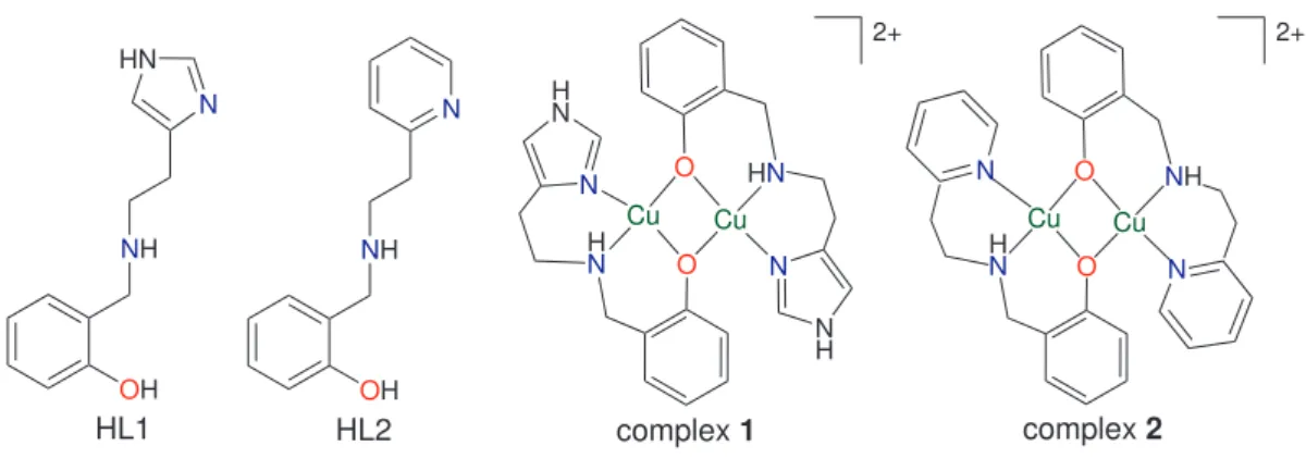
two new binuclear CuII complexes (1 and 2, Figure 1)
employing the tridentate N,O-donor ligands HL1,
[(2-hydroxybenzyl)(2-(imidazol-2-yl)ethyl)]amine, and HL2, [(2-hydroxybenzyl)(2-(pyridil-2-yl)ethyl]amine. These complexes were designed intending to develop new catalysts for oxidative processes, and their reactivity towards cyclohexane and catechol oxidation reactions were investigated using hydrogen peroxide or molecular oxygen as oxidants.
Cu N H N O H N Cu N HN O N H Cu H N O Cu NH O 2+ NH OH N HN HL1 N NH OH HL2 N N
complex 1 complex 2
2+
Figure 1. Schematic views of HL1, HL2 and complexes 1 and 2.
Cu N H N O H N Cu N HN O N H Cu H N O Cu NH O 2+ NH OH N HN HL1 N NH OH HL2 N N
complex 1 complex 2
Experimental
Materials and measurements
HL1and HL2 were synthesized and purified as
previously described.23,24 All chemicals for syntheses
and analyses were of analytical grade and used without further puriication. Infrared spectra (CsI pellet or ilm) were recorded using a NICOLET, MAGNA-IR 760 (4000
to 200 cm-1) spectrophotometer. Elemental analyses were
performed using a Perkin-Elmer 2400 analyzer coupled to an AD-4 Perkin-Elmer microbalance. Electronic absorption spectra were recorded using a Perkin-Elmer Lambda-19 spectrophotometer. Cyclic voltammetry studies were performed with a Princeton Applied Research (PAR) 273 potentiostat, at room temperature (25 ± 1 °C), in acetonitrile solutions kept under argon atmosphere. The standard three-electrode cell was composed by the following electrodes: a glassy carbon working, a platinum auxiliary and a Ag/AgCl pseudo-reference. Hexaluorophosphate
of tetrabutylammonium (tba(PF6), 0.1 mol L-1) was used
as supporting electrolyte and the ferrocenium-ferrocene
couple25 was employed to monitor the reference electrode
potential. Molar conductivities were measured on a Digimed CD-21 conductivity meter in methanol solutions.
Syntheses
[Cu2(L1)2]Cl2•2H 2O, 1
Complex 1 was obtained by the slow addition of
CuCl2•2H
2O (0.17 g, 1 mmol in 20 mL of methanol) to a
methanolic solution of HL1 (0.217 g, 1 mmol in 20 mL). The reaction mixture was heated under stirring for ca.
15 min, iltered and left to crystallize on bench at room temperature (near 25 °C). After a few days, dark green single crystals suitable for X-ray crystallographic analysis were collected from the mother liquor. Decomposition point: 195 °C. FTIR (CsI, cm-1): ν(NH
sec) 3134; ν(CHar/CHalif)
3037-2909; ν(C=N/C=C) 1596-1454; ν(C-O) 1275 and
δ(CHar) 771. Elemental analysis calculated (found) for C24H28Cl2Cu2N6O2•2H
2O: C 43.25 (43.11); H 4.84 (4.61);
N 12.61 (12.87)%. ΛM = 151 Ω-1 mol-1 cm2 (electrolyte 2:1
in methanol).26
[Cu2(L2)2](ClO4)2•1.5H 2O, 2
Complex 2 was synthesized by the same procedure
described for complex 1, but with the addition of excess of sodium perchlorate at the end of reaction. Dark green single crystals were also obtained from the mother liquor, but none of them presented a satisfactory diffraction pattern. Recrystallizations in other media failed. Decomposition
point: 229 °C. FTIR (CsI, cm-1): ν(NH
sec) 3240; ν(CHar/CHalif)
3072-2941; ν(C=N/C=C) 1610-1423; ν(C-O) 1259; ν(Cl-O)
1130-1028 and δ(CHar) 775. Elemental analysis calculated
(found) for C28H30Cl2Cu2N4O10•1.5H
2O: C 41.64 (41.61);
H 4.12 (3.79); N 6.94 (6.54)%. ΛM = 162 Ω-1 mol-1 cm2
(electrolyte 2:1 in methanol).26
Warning: perchlorate salts are potentially explosive and must be handled very carefully and only in small quantities.
Potentiometric titration
Potentiometric studies were carried out in CH3CN/H2O
(1:1 v/v) solution using a Micronal B375 pHmeter itted with blue-glass and calomel reference electrodes calibrated to read –log[H+] directly, designated as pH. Bidistilled
water in the presence of KMnO4 was used to prepare the CH3CN/H2O (1:1 v/v) solutions. The electrode was calibrated using the data obtained from a potentiometric
titration of a known volume of a standard 0.0100 mol L-1
HCl solution (0.1 mol L-1 in KCl) with a standard CO
2
-free 0.100 mol L-1 KOH solution. The measurements were
carried out in a thermostatized cell containing a solution of the complex (0.05 mol/50 mL) with ionic strength adjusted
to 0.100 mol L-1 by addition of KCl, at 25.00 ± 0.05 °C.
The experiments were performed under argon low to
eliminate the presence of atmospheric CO2. The samples
were titrated by addition of ixed volumes of a standard
CO2-free KOH solution (0.100 mol L-1). Computations were
carried out with the BEST program, and species diagrams
were obtained with SPE and SPEPLOT programs.27
Crystal structure
X-ray diffraction data collection for compound 1
was performed on a Bruker-Kappa-CCD diffractometer
(LDRX) using graphite-monochromatized Mo-Kα
radiation (λ = 0.71069 Å) at room temperature. Final unit
cell parameters were based on the itting of all relections
positions.28 Data integration and scaling of the relections
were performed with the EVALCCD suite.29 Empirical
multiscan absorption corrections using equivalent
relections were performed with the program SADABS.30
The structure was solved by direct methods using the
SHELXS31 program. The positions of all atoms could be
unambiguously assigned on consecutive difference Fourier
maps. Refinements were performed using SHELXL31
based on F2 through full-matrix least square routine.
in the structure reinements. All attempts to localize the hydrogen atoms around the water molecules failed. Thus, the hydrogen bonds of the N–H group of the imidazole towards the water molecules could not be properly assigned. Crystal structure and reinement data for complex 1 are summarized in Table 1. Selected bond distances and angles are indicated in Table 2. Atomic coordinates and complete crystal structure results are given as supplementary information (see SI).
Reactivity
Cyclohexane oxidation
The catalytic activities of complexes 1 and 2 towards cyclohexane oxidation were determined following
previously published methods.33 The reaction was performed
in acetonitrile at room temperature and inert atmosphere, using H2O2 as oxidant. The catalyst:substrate:oxidant reaction ratio was 1:1000:1000, with a catalyst concentration of 7×10⁻⁴ mol L-1. In a typical experiment, the catalytic
reaction was triggered with the addition of cyclohexane to a degassed acetonitrile solution containing the catalyst and the oxidant. The reaction was quenched after 24 h with the addition of an aqueous solution of Na2SO4 (0.4 mol L-1).
The products were extracted with diethyl ether; the organic
fractions were dried over anhydrous Na2SO4 and analyzed
by gas chromatography. The results are expressed as relative yields.33
Oxidation of 3,5-di-tert-butyl-catechol
The cathecolase activities of complexes 1 and 2 were measured by the oxidation of 3,5-di-tert-butylcatechol
(3,5-dtbc) to the respective quinone at 25 °C. The experiments
were performed with an Agilent 8453 spectrophotometer, and the reactions were followed at 400 nm, which is the characteristic absorption band of the oxidation product
(3,5-di-tert-butylquinone, 3,5-dtbq). The pH effect on the
reaction rate was determined over the pH range 5.5-10.0. A typical experiment was performed using the following conditions: 100 µL of freshly prepared aqueous buffer solution, [buffer]final = 3×10⁻³ mol L-1 (buffers: MES
(2-(N-morpholino)ethanesulfonic acid), pH 5.5, 6.0 and 6.5;
HEPES (4-(2-hydroxyethyl)piperazine-1-ethanesulfonic acid), pH 7.0 and 7.5; and CHES (2-(cyclohexylamino) ethanesulfonic acid), 8.0, 8.5 , 9.0, 9.5 and 10.0), 100 µL
of a methanolic solution of the complex ([complex]inal =
2.4×10⁻⁵ mol L-1) and 3.0 mL of O
2-saturated methanol. A
1-cm-path-length quartz cell was used as a reactor. The kinetic experiments under conditions of excess of substrate
were performed using 100 µL of freshly prepared aqueous CHES buffer, 100 µL of a methanolic solution of the
complex ([complex]inal = 2.4×10⁻⁵ mol L-1) and
oxygen-saturated methanol, at pH 8.5 ([buffer]inal = 3×10⁻³ mol L-1).
The reaction was initiated by the addition of volumes varying from 60 µL to 240 µL of a 3,5-dtbc solution ([3,5-dtbc]inal 3×10⁻³-12×10-3 mol L-1) and monitored for 20 min.
Correction for the spontaneous oxidation of the 3,5-dtbc was carried out by subtracting the initial rate for the blank experiment (without adding the catalyst). The initial rate at each substrate concentration was obtained by itting a second degree polynomial to the absorbance vs. time
plot,34 using ε(400 nm) = 1900 mol L-1 cm-1 for 3,5-di-tert
-butylquinone.35 A Michaelis-Menten approach was applied
using a non-linear it to obtain Vmax and KM parameters.36
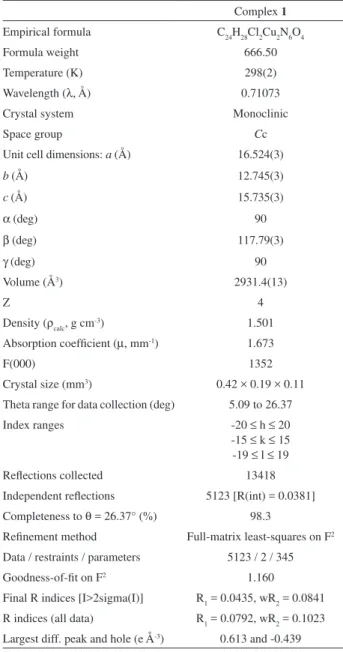
Table 1. Crystal structure data collection and reinement for compound 1
Complex 1 Empirical formula C24H28Cl2Cu2N6O4
Formula weight 666.50
Temperature (K) 298(2)
Wavelength (λ, Å) 0.71073
Crystal system Monoclinic
Space group Cc
Unit cell dimensions: a (Å) b (Å)
c (Å)
α (deg)
β (deg)
γ (deg)
16.524(3) 12.745(3) 15.735(3)
90 117.79(3)
90
Volume (Å3) 2931.4(13)
Z 4
Density (ρcalc, g cm-3) 1.501 Absorption coeficient (µ, mm-1) 1.673
F(000) 1352
Crystal size (mm3) 0.42 × 0.19 × 0.11 Theta range for data collection (deg) 5.09 to 26.37
Index ranges -20 ≤ h ≤ 20
-15 ≤ k ≤ 15 -19 ≤ l ≤ 19
Relections collected 13418
Independent relections 5123 [R(int) = 0.0381] Completeness to θ = 26.37° (%) 98.3
Reinement method Full-matrix least-squares on F2 Data / restraints / parameters 5123 / 2 / 345
Goodness-of-it on F2 1.160
Theoretical calculations
Geometry optimizations were performed as described
before,using Gaussian 03 package with the B3LYP hybrid
density functional theory combined with the 6-31G* basis
sets and LANL2DZ for the Cu atom.37
Results and Discussion
Synthesis and initial characterization
L1 and L2 were chosen in order to provide insights about the inluence of the substitution of pyridine by imidazole rings on the electronic structure and reactivity
of the complexes. Both ligands are able to stabilize CuII
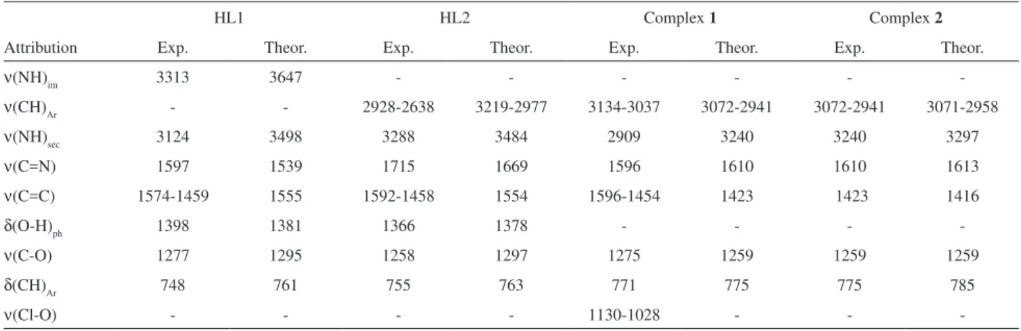
complexes. Infrared spectra of complexes 1 and 2 have shown characteristic bands of the ligands scaffold, and are shifted when compared to free HL1 and HL2, which is clear evidence of complexation. In order to help band attribution, theoretical studies were carried out using DFT (Figure S2). Considering the similarities of ligands and complexes spectra, the attribution data are summarized on Table S1, and only the main differences are discussed. In both spectra of complexes 1 and 2, the out-of-the-plane OH angular deformation band of the phenol group is absent, indicating the presence of phenolate groups coordinated to the metal centers. For complex 2, the presence of free perchlorate anions is evidenced by a broad band from 1130 to 1028 cm-1 assigned to the Cl-O stretchings of this anion
as a counter ion.38 For both complexes, elemental analyses
indicate the 1:1 ligand:metal ratio, and the presence of chloride or perchlorate anions. The binuclear arrangement for both complexes is irstly suggested by conductivity data obtained in freshly prepared methanol solutions (complex 1: ΛM = 151 Ω-1 mol-1 cm2, and complex 2:
ΛM = 162 Ω-1 mol-1 cm2), which are in the typical range of
2:1 electrolytes.26
X-ray crystal structure
Crystallographic data and bond parameters for complex 1 are presented in Tables 1 and 2, respectively. An ORTEP39
drawing of the cation complex is shown in Figure 2. The binuclear structure in the solid state is conirmed, where
each CuII atom is coordinated to a deprotonated L1, and
bridged by the two phenoxo groups provided by the ligand. In close proximity, there are two chloride anions and two water molecules (Figure 2). In addition to the two
Figure 2. ORTEP39 representation of complex 1, showing the coordination sphere atoms labeling and 50% probability ellipsoids.
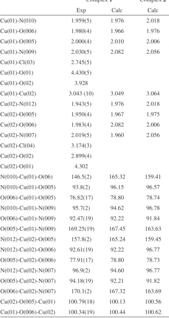
Table 2. Experimental (X-ray) and calculated (DFT) main bond distances (Å) and angles (o) for complexes 1 and 2
Complex 1 Complex 2
Exp Calc Calc
Cu(01)-N(010) 1.959(5) 1.976 2.018
Cu(01)-O(006) 1.980(4) 1.966 1.976
Cu(01)-O(005) 2.000(4) 2.010 2.006
Cu(01)-N(009) 2.030(5) 2.082 2.056
Cu(01)-Cl(03) 2.745(5)
Cu(01)-O(01) 4.430(5)
Cu(01)-O(02) 3.928
Cu(01)-Cu(02) 3.043 (10) 3.049 3.064
Cu(02)-N(012) 1.943(5) 1.976 2.018
Cu(02)-O(005) 1.950(4) 1.967 1.975
Cu(02)-O(006) 1.983(4) 2.082 2.006
Cu(02)-N(007) 2.019(5) 1.960 2.056
Cu(02)-Cl(04) 3.174(3)
Cu(02)-O(02) 2.899(4)
Cu(02)-O(01) 4.302
oxygen atoms from the phenoxo bridges, each CuII atom is
coordinated to one imidazole and the amine nitrogen atoms. Thus, the CuII atoms can be considered as tetracoordinated,
since their distances to the chloride anions and the water molecules are longer than 2.745(5) Å (Table 2). In fact, these Cu-Cl bond distances are longer than those reported for coordinated chloride anions.19,40,41 The geometries
adopted by the CuII centers are both distorted square-planar,
with angles varying from 76.8(2) Å to 95.7(2) Å, for Cu(01), and from 77.9(2) Å to 96.9(2) Å, for Cu(02).
In both CuII centers, the shorter bond distances are
Cu-Nimidazole [Cu(01)-N(10) = 1.959(5) and Cu(02)-N(12) =
1.943(5) Å], which are in the range observed for other CuII
complexes with imidazole-containing ligands.18-21,65 The
longer ones are Cu-Namine [Cu(01)-N(09) = 2.030(5) and Cu(02)-N(07) = 2.019(5) Å], which also agree with this type of bond distances in CuII complexes.18-21,42,43 Also in the expected
range are the distances Cu-Ophenoxo [Cu(01)-O(006) = 1.980(4), Cu(01)-O(005) = 2.000(4), Cu(02)-O(005) = 1.950(4) and Cu(02)-O(006) = 1.983(4) Å].24,44-46 Finally, the Cu…Cu
distance is 3.043(10) Å and is in the range observed for other binuclear CuII complexes bridged by two phenoxo groups.43,44
These distances are similar to that observed for the oxidized form of catechol oxidase (2.90 Å).10,17
Spectroscopic analyses
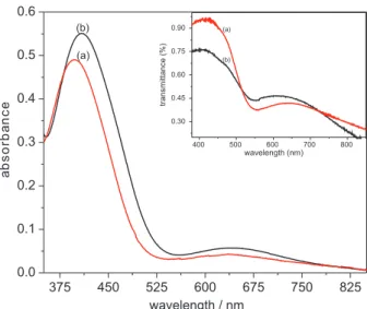
The electronic spectra of complexes 1 and 2 were
recorded in the solid state and acetonitrile solutions (Figure 3). In both cases, the two complexes present the same electronic features, indicating that their structures are maintained in solution. In acetonitrile, the spectra of both complexes are quite similar and show bands at 635 nm (ε = 212 L mol-1 cm-1) and 398 nm (ε = 3,340 L mol-1 cm-1)
for complex 1, and at 640 nm (ε = 280 L mol-1 cm-1) and
408 nm (ε = 2,620 L mol-1 cm-1) for complex 2. The low
molar extinction coeficients presented by the lower energy bands classify them as typical ligand ield transitions in the
CuII centers.47 The higher energy bands can be assigned
to ligand-to-metal charge transfer transitions (LMCT)
from the phenoxo bridges to the CuII ions, as proposed
for other phenolate-coordinated CuII complexes.24,40,46,48
A slight batochromic shift is observed for complex 2
when compared to complex 1. As observed before for
cobalt(III) complexes with these same ligands,49 this shift
is a consequence of substitution of an imidazole by a pyridine ring in the ligand scaffold. In fact, we have also observed this behavior in other systems and assigned it to the greater basicity of 1-methylimidazole group compared to pyridine.49,50 Finally, electronic spectroscopy has been
used to infer the geometry adopted by CuII complexes in
solution.48 Based on the similarities of the spectral data
presented by complexes 1 and 2 we can conclude that they might present the same kind of geometry. The very broad shape of the spectra in the range of the low energy band makes a precise geometry attribution dificult, but points
out to a distorted octahedron,47 which suggests interaction
of metal centers with solvent molecules.
EPR spectroscopy was used to give some insight on this issue. X-band spectra of complexes 1 and 2 were collected in frozen ethanol solutions at 77 K. Using the WINEPR
SimFonia51 program, the experimental data were simulated;
both experimental and simulated spectra are presented in Figure 4. The following parameters were determined: g⊥ = 2.068, g// = 2.268, A⊥ = 20×10-4 cm-1, A
// = 175×10-4 cm-1
and g// / A// = 129 cm (for complex 1), and g⊥ = 2.0590, g// = 2.261, A⊥ = 20×10-4 cm-1, A
// = 182×10-4 cm-1 and
g///A// = 125 cm (for complex 2). The frozen solution spectra of both complexes (Figure 4) are typically axial, presenting
four well-deined lines around g= 2, with g// > g⊥ > 2.0
and A// > A⊥. These features are characteristic of elongated
octahedral, square-pyramidal or square-planar geometries,52
and have been observed for other binuclear CuII complexes
with two phenoxide bridges.46,53 The empirical g
// / A// ratio
can be employed to estimate the geometry distortion around
CuII centers, and values in the range of 105 to 135 cm are
attributed to square-planar or octahedral (with tetragonal
distortion) geometries.54,55 Considering the electronic
in the metal-ligand bond.21,55 Addison and Sakaguchi55
reported that the replacement of “hard” by “soft” donor
atoms around CuII centers results in a decrease of g
//, and consequently a decrease in the g// / A// ratio. This fact is interpreted as an increase in the electron delocalization away from the metal center, or an increase in covalency. For complexes 1 and 2, the g// values are very similar, but an
increase in A// is observed when the softer base imidazole
(complex 1) is replaced by pyridine (complex 2), which
results in the decrease of the g// / A// ratio. This fact suggests a slight increase of the ligand ield strength around the metal center for complex 2 compared to complex 1. In fact, as imidazole is a better σ-donor than pyridine,19-21,23,56 it was
expected that HL1 presented higher ligand ield strength than HL2, which is exactly the opposite behavior of the observed here.
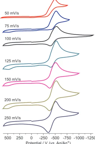
Cyclic voltammetry studies
The cyclic voltammograms for complex 1 in acetonitrile solution (Figure S1) do not undergo signiicant variation upon several scan rate measurements, presenting a broad
irreversible peak at -315 mV vs. NHE (normal hydrogen
electrode). Considering that the coordination environments around the CuII ions are identical, their reduction is expected
to occur at very similar potentials, which can explain the broadening of the peak and allow us to consider it as a CuIICuII→ CuICuI process.20 This irreversibility indicates
that HL1 might not be able to stabilize the reduced complex state leading it to break down (EC process). In contrast, complex 2 presents a redox activity dependent on the scan
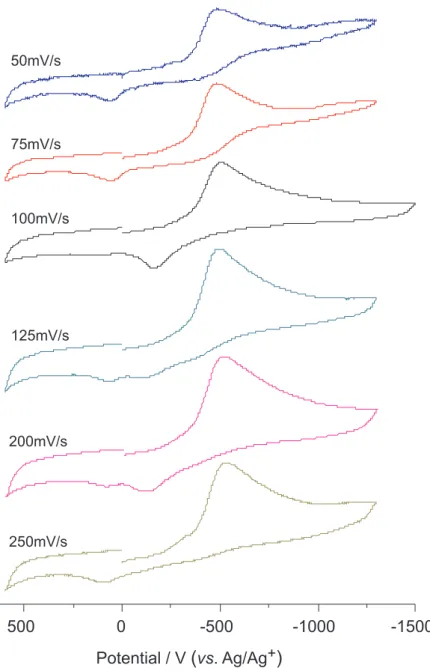
rate. As shown in Figure 5, from 50 and 100 mV s-1, an
irreversible wave is observed at –340 mV vs. NHE that is
also tentatively attributed to the CuIICuII→ CuICuI process.
The more negative value observed for complex 2 compared
to complex 1 suggests that HL2 acts as a better electron donor than HL1, despite the expected behavior based on the σ-donor ability of imidazole and pyridine groups.
As reported before,49 it is probably related to the LUMO
features of these complexes.
When cyclic voltammograms of complex 2 are recorded
at higher scan rates (125 to 250 mV s-1), an enhancement
of the system reversibility is observed (125 mV s-1:
E1/2 = –332 mV vs. NHE, ∆Ep = 125 mV; 200 mV s-1:
E1/2 = –328 mV vs. NHE, ∆Ep = 135 mV; 250 mV s-1:
E1/2 = –323 mV vs. NHE, ∆Ep = 155 mV). This behavior
suggests that, at least on the time scale of the measurements, the complex scaffold is maintained after reduction, and so a
quasi-reversible process is detected. This fact can tentatively
be rationalized in terms of the better π-acceptor properties of the pyridine ring which, by withdrawing electron density from the metal center after reduction, stabilizes the CuICuI
species under the time scale of the experiments.
Potentiometric titration
In order to examine the pKa of water molecules
coordinated to the metal centers, potentiometric studies
Figure 4. X-band experimental (——) and simulated (...) EPR spectra for complexes 1 (black) and 2 (blue/gray), in frozen ethanol solutions at 77 K.
were performed in acetonitrile/water mixture (1:1 v/v).
As can be seen in Figure 6, two pKa values were observed
for both complexes: for complex 1, pKa1 = 10.32 and
pKa2 = 11.36, and for complex 2, pKa1 = 10.25 and
pKa2 = 11.27.
Comparison of these results with those reported for other copper complexes in which the water molecules are considered completely coordinated to the metal center (pKaca. 8),19,36,40 reveals that these pK
a values are much
higher. This fact can be tentatively explained by the existence of a weak metal-water interaction, as observed in the crystal structure of complex 1, in which the water molecule is 2.899 Å away from the Cu(02) center. In fact, a strong Jahn-Teller effect associated with the donating
ability of the phenolate groups may weaken the Cu–Ow
bond, not permitting a signiicant polarization on the O–H bond of the closest water molecule and raising its pKa. It is
worth to mention that the phenolate protonation constants could not be determined, probably because these groups
are coordinated in a bridge mode and have very low pKa.
Theoretical calculations
As mentioned earlier, the EPR and the voltammetric data obtained for complexes 1 and 2 present an inverse behavior of that expected considering only the substitution of a pyridine by an imidazole group in the ligand scaffold.
As previously reported,49 this might be correlated to the
features of the complexes frontier orbitals. Since the crystal
structure of complex 1 revealed that the chloride anions
and the water molecules are only interacting with the metal center, the geometry optimizations were carried out without them. As shown in Table 2, a good agreement was achieved between the experimental (X-ray) and the theoretical results
for complex 1 and, then, the same approach was used to
predict the geometry of complex 2. Figures 7 and 8 show the optimized geometries and the graphical representations of the frontier orbitals for complexes 1 and 2, respectively. For complex 1, the HOMO (highest occupied molecular orbital) representation shows a small participation of the nitrogen atoms of the secondary amine and a strong contribution of both phenolate bridges. On the other hand, the LUMO has the participation of the nitrogen atoms of the secondary amine and the imidazole, the phenolate groups and the dx2-y2 orbital of the Cu center. For complex 2, the
HOMO presents high similarity with that of complex 1, and
shows a small participation of the amine nitrogen atoms and a large participation of the phenolate groups. However, the LUMO of complex 2 has strong contribution of the two pyridine rings. As the HOMO of both complexes are quite similar, the behavior observed for complexes 1 and 2 might be related to the different atomic contributions to the LUMO. While the LUMO of complex 1 is composed by the contribution of several atoms and is delocalized all over Figure 6. Diagram of species distribution for complexes 1 (up) and 2
(down).
Figure 7. (a) Geometry optimization, (b) HOMO and (c) LUMO for complex 1.
the molecule, in complex 2 it has mainly the involvement of the pyridine rings and is localized over these groups. This fact can explain the more negative potential observed for complex 2 compared to complex 1, since the increase of electron density due to the reduction process might be more easily transferred to the LUMO of complex 1 than to the LUMO of complex 2.
Reactivity
Cyclohexane oxidation
Since our intention is to develop new catalysts for oxidative processes, and alkane oxidations have great industrial interest,58,59 the cyclohexane oxidation was
investigated using hydrogen peroxide at room temperature. Complexes 1 and 2 lead to low overall conversion values
of 6.34 and 7.15%, respectively. For complex 1, only
cyclohexanol (Cy–OH) and cyclohexanone (Cy=O) were isolated as reaction products, with selectivities of 68.1% for Cy–OH and 31.9% for Cy=O, and turnover
number of 70. For complex 2, the formation of 31.9%
of Cy–OH, 29% of Cy=O and 32% of Cy–OOH (cyclohexylhydroperoxide) was observed, and turnover
number of 78. Comparing these values with other CuII
complexes presenting vacant positions on the metal coordination sphere, the overall yields are fairly lower.60,61
This fact may be explained by the non-coplanarity of the central square-plane core and the ligand aromatic rings, which can make dificult the substrate approach, even
though both complexes (1 and 2) present two vacant
positions per CuII center.
Catechol oxidation
The most widely employed substrate to investigate the catecholase activity of model complexes is
3,5-di-tert-butylcatechol (3,5-dtbc). It is due to its low redox
potential that facilitates the oxidation to quinone, and the presence of bulky substituents that prevents further oxidation reactions.62
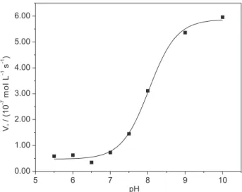
The pH dependence of the catecholase activities promoted by complexes 1 and 2 was investigated in a range (5.5 to 10) that includes the optimal pH activity range observed for the enzyme.15 The plots of v
0 (initial rate)
vs. pH (Figure 9 for complex 1, and S2 for complex 2)
show sigmoid-shape proiles and reveal pKa values of 9.1
for 1 and 8.0 for complex 2, which are the kinetic pKa.
The difference between these values and those obtained by potentiometric titration can be explained by the fact that the kinetic pKa represents the water molecules
deprotonation in the presence of substrate. In addition, the potentiometric pKa was determined in CH3CN/H2O
(1:1 v/v) solution and the reactivity experiments were carried out in methanol. Based on the pKa values, the
active species for 1 and 2 can be assigned to the
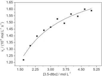
aqua-hydroxo complex.14,62-64 The substrate dependence was
carried out at pH 9, and a Michaelis-Menten proile was obtained (Figure 10 for complex 1, and Figure S3 for complex 2). The parameters obtained by a non-linear it are presented in Table 3. The values for complexes 1 and 2 indicate that they are very eficient compared to other complexes in the literature.35,64,65 The kinetic eficiency
(kcat/KM) shows that complex 2 is approximately 10 times
more active than complex 1. Usually, the catecholase
activity of model complexes is largely attributed to the distance Cu…Cu, but other effects such as redox potentials,
neighboring groups, and the individual coordination spheres can play important roles.66 For complexes 1 and 2, the
Cu…Cu distances are very similar (Table 2) and may not
be the reason for their different activities. Since the irst step in the proposed mechanism is the oxidation of one substrate molecule coupled to a two-electron reduction of Figure 9. pH dependence for the oxidation of 3,5-dtbc catalyzed by complex 1, in methanol/water (30:1 v/v) solution. Experimental conditions: [1]final 2.4×10⁻⁵ mol L-1, [3,5-dtbc]
final 5×10⁻³ mol L-1, [buffer]inal 3×10⁻³ mol L-1, at 25 ºC.
Table 3. Kinetic parameters for complexes 1 and 2
Complex Kcat/KM (mol L-1 s-1) V
max (mol L-1 s-1) KM (mol-1L) Kcat (s-1) Kass
1 10 1.84×10⁻⁶ 7.72×10⁻³ 7.69×10⁻² 129
the active site,67 it was expected that complex 1 presented
higher activity than complex 2, which has a more negative reduction potential, but this kind of correlation has been questioned.66
The non-reversibility shown in the voltammograms of complex 1 indicates that, after reduction, the complex integrity is not maintained in solution and this can be a reason of its lower reactivity. On the other hand, the reversibility shown by complex 2 at high scan rates shows that this complex does not decompose as fast as complex 1 after reduction, leading to its higher reactivity.
Conclusions
The CuII binuclear complexes 1 and 2 are reactive in
the cyclohexane oxidation, with selectivity close to 70% for the formation of cyclohexanol (for complex 1), but with conversion values low for both complexes. This low hydrocarbon oxidation activity can be attributed to steric hindrance effects produced by the non-coplanar aromatic rings of the ligand scaffold. It seems that the access of the hydrocarbon molecule to the binuclear active center is a determinant step in the mechanism of this process. As mimetic for catecholase activity, both complexes were very eficient, with complex 2 being approximately 10 times
more effective than complex 1. These high eficiencies
indicate that the pyridine ligand is capable to stabilize an intermediate CuICuI center, which is proposed to be
formed in this process, avoiding the breaking down of the complex. This is corroborated by the strong participation of pyridine in the LUMO of complex 2, which can help to accommodate the additional negative charge when the complex is reduced from CuIICuII to CuICuI.
Supplementary Information
Crystallographic data have been deposited with the Cambridge Crystallographic Data Centre (deposition number CCDC 749493). Copies of the data can be obtained, free of charge, via www.ccdc.cam.ac.uk/conts/retrieving.html, or by request to CCDC, 12 Union Road, Cambridge CB2 1EZ, UK (fax 44-1223-336033 or e-mail: deposit@ccdc. cam.ac.uk). Tables with torsion angles and hydrogen bond distances for complex 1, Tables S1 and S4, and Figures S1 to S3 are presented as supplementary information, available free of charge at http://jbcs.sbq.org.br, as PDF ile.
Acknowledgments
Authors are grateful for the facilities of the LDRX Laboratory (Laboratório de Difração de Raios X at UFF), LABEPR (Laboratório Regional Sul de EPR at UFPR), Prof. Octavio A. C. Antunes (in memorian), and grants
from CAPES, CNPq and FAPERJ.
References
1. Julémon, M. In Handbook of Detergents, Part A: Properties; Broze, G. ed., Marcel Dekker, Inc: New York, 1999, p. 631. 2. Emmanuel, J.; Non-IncinerationMedical Waste Treatment
Technologies, Health Care without Harm: Washington, 2001, p. 61. 3. Tarchitzky, J.; Chen, Y. In Handbook of Detergents, Part B:
Environmental Impact; Zoller, U. ed., Marcel Dekker: New York, 2004, p. 645.
4. Sippola, V. O.; Krause, A. O. I.; Catal. Today 2005, 100, 237. 5. Torres, E.; Bustos-Jaimes, I.; Le Borgne, S.; Appl. Cat., B 2003,
46, 1.
6. Durán, N.; Esposito, E.; Appl. Cat., B 2000, 28, 83.
7. Chiacchierini, E.; Restuccia, D.; Vini, G.; Food Sci. Technol. Int. 2004, 10, 373.
8. Ensuncho, L.; Alvarez-Cuenca, M.; Legge, R. L.; Bioprocess Biosyst. Eng. 2005, 27, 185.
9. Seetharam, G. B.; Saville, B. A.; Water Res. 2003, 37, 436. 10. Gerdemann, C.; Eicken, C.; Krebs, B.; Acc. Chem. Res. 2002,
35, 183.
11. Fusetti, F.; Schröter, K. H.; Steiner, R. A.; van Noort, P. I.; Pijning, T.; Rozeboom, H. J.; Kalk, K. H.; Egmond, M. R.; Dijkstra, B. W.; Structure 2002, 10, 259.
12. Dunwell, J. M.; Purvis, A.; Khuri, S.; Phytochemistry 2004, 65, 7. 13. Witayakran, S.; Ragauskas, A. J.; Adv. Synth. Catal. 2009, 351,
1187.
14. Klabunde, T.; Eicken, C.; Sacchettini, J. C.; Krebs, B.; Nat. Struct. Biol. 1998, 5, 1084.
15. Hildén, K.; Hakala, T. K.; Lundell, T.; Biotechnol. Lett. 2009, 31, 1117.
16. Alamsetti, S. K.; Mannam, S.; Mutupandi, P.; Sekar, G.; Chem. Eur. J. 2009, 15, 1086.
17. Korpi, H.; Lahtinen, P.; Sippola, V.; Krause, O.; Leskelä, M.; Repo, T.; Appl. Cat., A 2004, 268, 199.
18. Scarpellini, M.; Neves, A.; Bortoluzzi, A. J.; Joussef, A. C.; Acta Crystallogr., Sect. C: Cryst. Struct. Commun. 2001, C57, 356.
19. Scarpellini, M.; Neves, A.; Bortoluzzi, A. J.; Szpoganicz, B.; Zucco, C.; Drago, V.; Mangrich, A. S.; Ortiz, W. A.; Passos, W. A. C.; Hörner, R.; Terenzi, H.; Silva, R. A. N.; Oliveira, M. C. B.; Inorg. Chem. 2003, 42, 8353.
20. Scarpellini, M.; Neves, A.; Castellano, E. E.; Franco, D. W.; J. Mol. Struct. 2004, 694, 193.
21. Scarpellini, M.; Neves, A.; Castellano, E. E.; Almeida, E. F.; Franco, D. W.; Polyhedron 2004, 23, 511.
22. Oliveira, M. C. B.; Mazera, D.; Scarpellini, M.; Severino, P. C.; Neves, A.; Hernán. T.; Inorg. Chem. 2009, 48, 2711.
23. Scarpellini, M.; Neves, A.; Bortoluzzi, A. J.; Vencato, I.; Drago, V.; Ortiz, W. A.; Zucco, C.; J. Chem. Soc., Dalton Trans. 2001, 2616.
24. Yajima, T.; Shimazaki, Y.; Ishigami, N.; Odani, A.; Yamauchi, O.; Inorg. Chim. Acta 2002, 337, 193.
25. Gagné, R.; Koval, C.; Licenski, G.; Inorg. Chem.1980, 19, 2854.
26. Geary, W. J.; Coord. Chem. Rev. 1971, 7, 81.
27. Martell, A. E.; Motekaitis. R. J.; Determination of Stability Constants, 2nd ed.; VHC Publishers: Weinheim, Germany, 1992.
28. Duisenberg, A. J. M.; J. Appl. Cryst. 1992, 25, 92.
29. Duisenberg, A. J. M.; Kroon-Batenburg, L. M. J.; Schreurs, A. M. M.; J. Appl. Cryst. 2003, 36, 220.
30. SADABS Bruker Analytical X-ray Systems, Inc., Madison WI, 1997.
31. Sheldrick, G. M.; Schneider, T. R.; Methods Enzymol. 1997, 277, 319.
32. Johnson, C. K. In Crystallographic Computing; Ahmed, F. R. ed., Copenhagen: Munksgaard, 1970, pp. 207-219.
33. Fernández, T. L.; Souza, E. T.; Visentin, L. C.; Santos, J. V.; Mangrich, A. S.; Faria, R. B.; Antunes, O. A. C.; Scarpellini, M.; J. Inorg. Biochem. 2009, 103, 474.
34. Côrtes, C. E. S; Faria, R. B.; Inorg. Chem. 2004, 43, 1395. 35. Peralta, R. A.; Neves A.; Bortoluzzi, A. J.; Anjos, A.; Xavier, F.
R.; Szpoganicz, B.; Terenzi, H.; Oliveira M. C. B.; Castellano, E.E.; Friedermann, G. R.; Mangrich, A. S.; Novak, M. A.; J. Inorg. Biochem. 2006, 100, 992.
36. Signorella, S.; Rompel, A.; Bült-Karentzopoulos, K.; Krebs, B.; Pecoraro, V. L. Tuchagues, J.–P.; Inorg. Chem. 2007, 46, 10864.
37. Frisch, M. J.; Trucks, G. W.; Schlegel, H. B.; Scuseria, G. E.; Robb, M. A.; Cheeseman, J. R.; Montgomery, Jr., J. A.; Vreven, T.; Kudin, K. N.; Burant, J. C.; Millam, J. M.; Iyengar, S. S.;
Tomasi, J.; Barone, V.; Mennucci, B.; Cossi, M.; Scalmani, G.; Rega, N.; Petersson, G. A.; Nakatsuji, H.; Hada, M.; Ehara, M.; Toyota, K.; Fukuda, R.; Hasegawa, J.; Ishida, M.; Nakajima, T.; Honda, Y.; Kitao, O.; Nakai, H.; Klene, M.; Li, X.; Knox, J. E.; Hratchian, H. P.; Cross, J. B.; Bakken, V.; Adamo, C.; Jaramillo, J.; Gomperts, R.; Stratmann, R. E.; Yazyev, O.; Austin, A. J.; Cammi, R.; Pomelli, C.; Ochterski, J. W.; Ayala, P. Y.; Morokuma, K.; Voth, G. A.; Salvador, P.; Dannenberg, J. J.; Zakrzewski, V. G.; Dapprich, S.; Daniels, A. D.; Strain, M. C.; Farkas, O.; Malick, D. K.; Rabuck, A. D.; Raghavachari, K.; Foresman, J. B.; Ortiz, J. V.; Cui, Q.; Baboul, A. G.; Clifford, S.; Cioslowski, J.; Stefanov, B. B.; Liu, G.; Liashenko, A.; Piskorz, P.; Komaromi, I.; Martin, R. L.; Fox, D. J.; Keith, T.; Al-Laham, M. A.; Peng, C. Y.; Nanayakkara, A.; Challacombe, M.; Gill, P. M. W.; Johnson, B.; Chen, W.; Wong, M. W.; Gonzalez, C.; Pople, J. A.; Gaussian 03, Revision C.02, Gaussian, Inc., Wallingford CT, 2004.
38. Nakamoto, K.; Infrared and Raman Spectra of Inorganic and Coordination Compounds, 3rd ed., John Wiley & Sons, Inc: New York, 1977, part III, pp. 231-232, 242.
39. Farrugia, L. J.; J. Appl. Crystallogr. 1997, 30, 565.
40. Oliveira, M. C. B.; Scarpellini, M.; Neves, A.; Terenzi, H.; Bortoluzzi, A. J.; Szpoganicz, B.; Mangrich, A. S.; Inorg. Chem. 2005, 44, 921.
41. Albada, G.A.; Smeets, W.J.J.; Spelk, A.L.; Reedjik, J.; Inorg. Chim. Acta 1999, 288, 220.
42. Belle, C.; Beguin, C.; Gautier-Luneau, I.; Hamman, S.; Philouze, C.; Pierre, J. L.; Thomas F.; Torelli, S.; Inorg. Chem. 2002, 41, 479.
43. Xie, Y.; Jiang, H.; Chan, A.S.; Liu, Q.; Xu, X.; Du, C.; Zhu, Y.; Inorg. Chim. Acta 2002, 333, 138.
44. Wang, X.; Ding, J.; Ranford, J. D.; Vital, J. J.; J. App. Phys. 2003, 98, 10.
45. Kong, D.; Mao, J.; Martell, A. E.; Cleariel, A.; Inorg. Chim. Acta 2003, 342, 260.
46. Taki, M.; Kumei, H.; Nagatomo, S.; Kitagawa, T.; Itoh, S.; Fukuzumi, S.; Inorg. Chim. Acta 2000, 300-302, 622. 47. Lever, A. B. P.; Inorganic Electronic Spectroscopy, 2nd ed.,
Elsevier Science Publishers B. V.: Amsterdam, 1984, pp. 553-572.
48. Karlin, K.D.; Gultneh, Y.; Nicholson, T.; Zubieta, J.; Inorg. Chem. 1985, 24, 3725.
49. Souza, E. T.; Castro, L. C.; Castro, F. A. V.; Visentin, L. C.; Pinheiro, C. B.; Pereira, M. D.; Machado, S. P.; Scarpellini, M.; J. Inorg. Biochem. 2009, 103, 1355.
50. Scarpellini, M.; Casellato, A.; Bortoluzzi, A. J.; Vencato, I.; Mangrich, A. S.; Neves, A.; Machado, S. P.; J. Braz. Chem. Soc. 2006, 17, 1617.
52. Hathaway, B. J.; Hodgson, P. G.; J. Inorg. Nucl. Chem. 1973, 35, 4071.
53. Kruse, T.; Weyhermüller, T.; Wieghardt, K.; Inorg. Chim. Acta 2002, 331, 81.
54. Silva, L. A.; Andrade, J. B.; Mangrich, A. S.; J. Braz. Chem. Soc. 2007, 18, 607.
55. Sakaguchi, U.; Addison, A. W.; J. Chem. Soc., Dalton Trans. 1979, 600.
56. Scarpellini, M.; Toledo Júnior, J. C.; Neves, A.; Ellena, J.; Castellano, E. E.; Franco, D. W.; Inorg. Chim. Acta 2004, 357, 707.
57. Greatti, A.; Scarpellini, M.; Peralta, R. A.; Casellato, A.; Bortoluzzi, A. J.; Xavier, F. R.; Jovito, R.; Brito, M. A.; Szpoganicz, B.; Tomkowicz, Z.; Rams, M.; Haase, W.; Neves, A.; Inorg. Chem. 2008, 47, 1107.
58. Schuchardt, U.; Cardoso, D.; Sercheli, R.; Pereira, R.; Cruz, R. S.; Guerreiro, M. C.; Mandelli, D.; Spinacé, E. V.; Pires, E. L.; Appl. Catal., A 2001, 211, 1.
59. Punniyamurthy, T.; Rout, L.; Coord. Chem. Rev. 2008, 252, 134.
60. Silva, A. C.; Fernández, T. L.; Carvalho, N. M. F.; Herbst, M. H.; Bordinhão, J.; Horn Jr, A.; Wardell, J. L.; Oestreicher, E. G.; Antunes, O. A. C.; Appl. Catal., A 2007, 317, 154 and references therein.
61. Canhota, F. P.; Salomão, G. C.; Carvalho, N. M. F.; Antunes, O. A. C.; Catal. Comm. 2008, 9, 182 and references therein. 62. Rompel, A.; Fischer, H.; Meiwes, D.; Buldt-Karentzopoulos,
K.; Magrini, A.; Eicken, C.; Gerdemann, C.; Krebs, B.; FEBS Lett. 1999, 103, 445.
63. Gerdmann, C.; Eicken, C.; Krebs, B.; Acc. Chem. Res. 2002, 35, 183.
64. Rey, N. A.; Neves, A.; Bortoluzzi, A.; Pich, C. T.; Terenzi, H.; Inorg. Chem. 2007, 46, 348.
65. Neves, A; Rossi, L. M.; Bortoluzzi, A.J.; Szpoganicz, B.; Wiezbicki, C.; Schwingel E.; Haase, W.; Ostrovsky, S.; Inorg. Chem. 2002, 41, 1788.
66. Relm, J.; Krebs, B.; Dalton Trans. 1997, 3793.
67. Ackermann, J., Meyer, F., Kaifer, E., Pritzkow, H.; Chem. Eur. J. 2002, 8, 247.
Supplementary Information
0103 - 5053 $6.00+0.00
*e-mail: [email protected], [email protected]
Binuclear Cu
IIComplexes as Catalysts for Hydrocarbon and Catechol Oxidation
Reactions with Hydrogen Peroxide and Molecular Oxygen
Luciana R. Martins,a Elizabeth T. Souza,a Tatiana L. Fernandez,a Bernardo de Souza,b
Sílvio Rachinski,c Carlos B. Pinheiro,d Roberto B. Faria,a Annelise Casellato,a
Sérgio P. Machado,a Antonio S. Mangrich*,c and Marciela Scarpellini*,a
aDepartamento de Química Inorgânica, Instituto de Química, Universidade Federal do Rio de Janeiro,
21945-970 Rio de Janeiro - RJ, Brazil
bDepartamento de Química, Universidade Federal de Santa Catarina, 88040-900 Florianópolis - SC, Brazil
cDepartamento de Química, Universidade Federal do Paraná, 81531-970 Curitiba - PR, Brazil
dDepartamento de Física, Instituto de Ciências Exatas, Universidade Federal de Minas Gerais,
31270-901 Belo Horizonte - MG, Brazil
Table S1. Experimental and theoretical infrared data (cm-1) for HL1, HL2 and complexes 1 and 2
HL1 HL2 Complex 1 Complex 2
Attribution Exp. Theor. Exp. Theor. Exp. Theor. Exp. Theor. ν(NH)
im 3313 3647 - - -
-ν(CH)
Ar - - 2928-2638 3219-2977 3134-3037 3072-2941 3072-2941 3071-2958
ν(NH)
sec 3124 3498 3288 3484 2909 3240 3240 3297
ν(C=N) 1597 1539 1715 1669 1596 1610 1610 1613
ν(C=C) 1574-1459 1555 1592-1458 1554 1596-1454 1423 1423 1416
δ(O-H)
ph 1398 1381 1366 1378 - - -
-ν(C-O) 1277 1295 1258 1297 1275 1259 1259 1259
δ(CH)
Ar 748 761 755 763 771 775 775 785
-N(10)-Cu(1)-Cu(2)-N(12) 102.0(2) O(6)-Cu(1)-Cu(2)-N(12) −44.7(3) O(5)-Cu(1)-Cu(2)-N(12) 159.4(3) N(9)-Cu(1)-Cu(2)-N(12) −25.5(3) Cl(3)-Cu(1)-Cu(2)-N(12) −123.59(19) N(10)-Cu(1)-Cu(2)-O(5) −57.3(3) O(6)-Cu(1)-Cu(2)-O(5) 156.0(3) N(9)-Cu(1)-Cu(2)-O(5) 175.1(3) Cl(3)-Cu(1)-Cu(2)-O(5) 77.0(2) N(10)-Cu(1)-Cu(2)-O(6) 146.7(3) O(5)-Cu(1)-Cu(2)-O(6) −156.0(3) N(9)-Cu(1)-Cu(2)-O(6) 19.2(3) Cl(3)-Cu(1)-Cu(2)-O(6) −78.9(2) N(10)-Cu(1)-Cu(2)-N(7) −44.3(3) O(6)-Cu(1)-Cu(2)-N(7) 169.0(3) O(5)-Cu(1)-Cu(2)-N(7) 13.0(3) N(9)-Cu(1)-Cu(2)-N(7) −171.9(3) Cl(3)-Cu(1)-Cu(2)-N(7) 90.0(2) N(12)-Cu(2)-O(5)-C(15) 138.5(6) O(6)-Cu(2)-O(5)-C(15) −155.1(5) N(7)-Cu(2)-O(5)-C(15) 18.8(5) Cu(1)-Cu(2)-O(5)-C(15) −170.5(6) N(12)-Cu(2)-O(5)-Cu(1) −50.9(6) O(6)-Cu(2)-O(5)-Cu(1) 15.5(2) N(7)-Cu(2)-O(5)-Cu(1) −170.6(2) N(10)-Cu(1)-O(5)-C(15) −57.8(5) O(6)-Cu(1)-O(5)-C(15) 154.7(5) N(9)-Cu(1)-O(5)-C(15) 150.2(10) Cl(3)-Cu(1)-O(5)-C(15) 54.5(5) Cu(2)-Cu(1)-O(5)-C(15) 170.3(6) N(10)-Cu(1)-O(5)-Cu(2) 131.9(2) O(6)-Cu(1)-O(5)-Cu(2) −15.6(2) N(9)-Cu(1)-O(5)-Cu(2) −20.1(13) Cl(3)-Cu(1)-O(5)-Cu(2) −115.75(17) N(10)-Cu(1)-O(6)-C(27) 137.4(5) O(5)-Cu(1)-O(6)-C(27) −145.9(5) N(9)-Cu(1)-O(6)-C(27) 33.3(5)
Cl(3)-Cu(1)-O(6)-C(27) −46.6(5) Cu(2)-Cu(1)-O(6)-C(27) −161.1(6) N(10)-Cu(1)-O(6)-Cu(2) −61.4(4) O(5)-Cu(1)-O(6)-Cu(2) 15.3(2) N(9)-Cu(1)-O(6)-Cu(2) −165.6(2) Cl(3)-Cu(1)-O(6)-Cu(2) 114.57(18) N(12)-Cu(2)-O(6)-C(27) −57.3(6) O(5)-Cu(2)-O(6)-C(27) 143.0(6) Cu(1)-Cu(2)-O(6)-C(27) 158.6(7) N(12)-Cu(2)-O(6)-Cu(1) 144.1(2) O(5)-Cu(2)-O(6)-Cu(1) −15.6(2) N(12)-Cu(2)-N(7)-C(20) −139.0(5) O(5)-Cu(2)-N(7)-C(20) 21.7(5) Cu(1)-Cu(2)-N(7)-C(20) 13.3(6) N(12)-Cu(2)-N(7)-C(25) −8.3(5) O(5)-Cu(2)-N(7)-C(25) 152.4(4) Cu(1)-Cu(2)-N(7)-C(25) 144.0(4) N(10)-Cu(1)-N(9)-C(29) −1.3(5) O(6)-Cu(1)-N(9)-C(29) 146.2(5) O(5)-Cu(1)-N(9)-C(29) 150.7(10) Cl(3)-Cu(1)-N(9)-C(29) −111.6(5) Cu(2)-Cu(1)-N(9)-C(29) 134.1(4) N(10)-Cu(1)-N(9)-C(17) −132.9(5) O(6)-Cu(1)-N(9)-C(17) 14.5(5) O(5)-Cu(1)-N(9)-C(17) 19.0(14) Cl(3)-Cu(1)-N(9)-C(17) 116.7(5) Cu(2)-Cu(1)-N(9)-C(17) 2.4(5) O(6)-Cu(1)-N(10)-C(19) 45.5(7) O(5)-Cu(1)-N(10)-C(19) −26.2(5) N(9)-Cu(1)-N(10)-C(19) 148.8(5) Cl(3)-Cu(1)-N(10)-C(19) −130.3(5) Cu(2)-Cu(1)-N(10)-C(19) 5.9(6) O(6)-Cu(1)-N(10)-C(30) −119.2(5) O(5)-Cu(1)-N(10)-C(30) 169.1(5) N(9)-Cu(1)-N(10)-C(30) −16.0(5) Cl(3)-Cu(1)-N(10)-C(30) 65.0(5) Cu(2)-Cu(1)-N(10)-C(30) −158.8(4)
O(5)-Cu(2)-N(12)-C(16) 34.1(8) O(6)-Cu(2)-N(12)-C(16) −29.6(5) N(7)-Cu(2)-N(12)-C(16) 153.4(5) Cu(1)-Cu(2)-N(12)-C(16) −2.8(6) O(5)-Cu(2)-N(12)-C(13) −130.9(6) O(6)-Cu(2)-N(12)-C(13) 165.3(5) N(7)-Cu(2)-N(12)-C(13) −11.7(5) Cu(1)-Cu(2)-N(12)-C(13) −167.9(4) C(16)-N(12)-C(13)-C(28) 0.4(7) Cu(2)-N(12)-C(13)-C(28) 167.9(4) C(16)-N(12)-C(13)-C(23) −176.5(5) Cu(2)-N(12)-C(13)-C(23) −9.0(8) C(22)-C(11)-C(15)-O(5) −178.4(6) C(22)-C(11)-C(15)-C(21) 2.2(9) Cu(2)-O(5)-C(15)-C(11) 152.7(5) Cu(1)-O(5)-C(15)-C(11) −15.1(8) Cu(2)-O(5)-C(15)-C(21) −27.8(8) Cu(1)-O(5)-C(15)-C(21) 164.4(4) C(13)-N(12)-C(16)-N(8) −0.1(7) Cu(2)-N(12)-C(16)-N(8) −167.4(4) C(28)-N(8)-C(16)-N(12) −0.2(7) C(29)-N(9)-C(17)-C(24) 167.6(6) Cu(1)-N(9)-C(17)-C(24) −55.7(7) C(30)-N(10)-C(19)-N(14) 0.1(7) Cu(1)-N(10)-C(19)-N(14) −166.8(4) C(31)-N(14)-C(19)-N(10) −0.1(7) C(25)-N(7)-C(20)-C(21) 170.3(6) Cu(2)-N(7)-C(20)-C(21) −55.6(7) C(11)-C(15)-C(21)-C(34) −4.1(9) O(5)-C(15)-C(21)-C(34) 176.4(5) C(11)-C(15)-C(21)-C(20) 171.5(6) O(5)-C(15)-C(21)-C(20) −7.9(9) N(7)-C(20)-C(21)-C(34) −131.1(6) N(7)-C(20)-C(21)-C(15) 53.3(9) C(15)-C(11)-C(22)-C(32) 1.0(10) C(28)-C(13)-C(23)-C(25) −123.8(7) N(12)-C(13)-C(23)-C(25) 52.3(8) N(9)-C(17)-C(24)-C(27) 58.1(8)
N(9)-C(17)-C(24)-C(36) −123.6(7) C(20)-N(7)-C(25)-C(23) 179.9(5) Cu(2)-N(7)-C(25)-C(23) 47.7(7) C(13)-C(23)-C(25)-N(7) −73.2(7) Cu(1)-O(6)-C(27)-C(18) 137.1(5) Cu(2)-O(6)-C(27)-C(18) −17.3(9) Cu(1)-O(6)-C(27)-C(24) −41.8(8) Cu(2)-O(6)-C(27)-C(24) 163.8(5) C(33)-C(18)-C(27)-O(6) −178.6(6) C(33)-C(18)-C(27)-C(24) 0.3(10) C(36)-C(24)-C(27)-O(6) 176.0(6) C(17)-C(24)-C(27)-O(6) −5.6(9) C(36)-C(24)-C(27)-C(18) −3.0(9) C(17)-C(24)-C(27)-C(18) 175.4(6) C(16)-N(8)-C(28)-C(13) 0.5(7) N(12)-C(13)-C(28)-N(8) −0.6(7) C(23)-C(13)-C(28)-N(8) 175.9(6) C(17)-N(9)-C(29)-C(26) 175.3(6) Cu(1)-N(9)-C(29)-C(26) 41.3(7) C(30)-C(26)-C(29)-N(9) −72.5(7) C(19)-N(10)-C(30)-C(31) −0.1(7) Cu(1)-N(10)-C(30)-C(31) 167.3(4) C(19)-N(10)-C(30)-C(26) −177.0(6) Cu(1)-N(10)-C(30)-C(26) −9.6(8) C(29)-C(26)-C(30)-C(31) −119.2(8) C(29)-C(26)-C(30)-N(10) 56.8(8) N(10)-C(30)-C(31)-N(14) 0.0(7) C(26)-C(30)-C(31)-N(14) 176.4(7) C(19)-N(14)-C(31)-C(30) 0.0(7) C(11)-C(22)-C(32)-C(34) −2.1(10) C(27)-C(18)-C(33)-C(35) 2.2(11) C(22)-C(32)-C(34)-C(21) 0.1(10) C(15)-C(21)-C(34)-C(32) 3.0(10) C(20)-C(21)-C(34)-C(32) −172.8(6) C(18)-C(33)-C(35)-C(36) −2.0(11) C(33)-C(35)-C(36)-C(24) −0.7(12) C(27)-C(24)-C(36)-C(35) 3.2(10) C(17)-C(24)-C(36)-C(35) −175.2(7)
Table S3. Hydrogen bond distances and angles for complex 1 (Å and °)
D-H…A d(D-H) d(H…A) d(D…A) <(DHA) N(8)-H(8)…Cl(3)#1 0.86 2.29 3.149(5) 173.9 N(14)-H(14)…Cl(4)#2 0.86 2.24 3.089(6) 172.2 Symmetry transformations used to generate equivalent atoms: #1 : x-1/2,y+1/2,z ; #2 : x-1/2,-y+3/2,z-1/2.
Table S4. Experimental (X-ray diffraction data for HL123) and calculated
(DFT) main bond distances (Å) and angles (o) for HL1 and HL2
HL1 Exp. Calc. HL2 Calc.
N(8)-C(9) 1.70(3) 1.465 N(8)-C(9) 1.462 N(8)-C(7) 1.476(3) 1.468 N(8)-C(7) 1.458 N(14)-C(13) 1.331(3) 1.365 C(14)-C(13) 1.396 N(14)-C(15) 1.359(3) 1.382 C(14)-C(15) 1.395 N(12)-C(11) 1.363(3) 1.385 N(12)-C(11) 1.344 N(12)-C(13) 1.326(3) 1.315 N(12)-C(13) 1.338 C(9)-N(8)-C(7) 113.34(19) 114.55 C(9)-N(8)-C(7) 115.53 C(13)-N(14)-C(15) 107.31(18) 107.21 C(13)-C(14)-C(15) 117.95 C(13)-N(12)-C(11) 105.61(19) 105.72 C(13)-N(12)-C(11) 118.09
Figure S2. pH dependence for the oxidation of 3,5-dtbc catalyzed by complex 2, in methanol/water (30:1 v/v) solution. Experimental conditions: [2]inal =
2.4 × 10⁻⁵ mol L-1; [3,5-dtbc]
inal = 5 × 10⁻³ mol L-1; [buffer]inal = 3 × 10⁻³ mol L-1; 25 ºC.
Figure S3. Dependence of the reaction rates on the 3,5-dtbc concentration for the oxidation reaction catalyzed by complex 2, in methanol/water (30:1 v/v)