Article
0103 - 5053 $6.00+0.00*e-mail: marcelog@iqsc.usp.br
Molecular Fluorescence in Silica Particles Doped with Quercetin-Al
3+Complexes
Rafael Frederice, Ana Paula Garcia Ferreira and Marcelo Henrique Gehlen*
Instituto de Química de São Carlos, Universidade de São Paulo, CP 780 13560-970 São Carlos-SP, Brazil
Partículas de sílica dopadas com o complexo quercetina-Al3+ foram preparadas através da
hidrólise e condensação do tetraetilortossilicato (TEOS) utilizando catálise ácida e básica. A catálise ácida resultou em partículas com diâmetro em torno de 200 nm. A catálise básica com TEOS foi executada sobre um sol de alumina dopado com quercetina utilizado como precursor caroço e com este procedimento foram obtidas partículas luorescentes de SiO2 na forma de dedos. Os decaimentos de luorescência do sistema quercetina-alumina-SiO2 são biexponenciais e este
caráter é atribuído aos dois tipos de complexos quercetina-Al3+ encapsulados no caroço do sistema.
Silica particles doped with quercetin-Al3+ complex were prepared by hydrolysis and
condensation of tetraethylorthosilicate (TEOS) using acid and basic catalysis. The acid catalysis resulted in particles with diameter about 200 nm. Basic catalysis with TEOS was performed over a quercetin doped alumina-sol used as a core precursor, and luorescent inger-shaped SiO2 particles were obtained. The quercetin-alumina-SiO2 luorescence decays are biexponential, and this character is ascribed to two types of quercetin-Al3+ complexes entrapped in the core of the system.
Keywords: silica particles, luorescence spectroscopy, probes, lavonoid
Introduction
Colloidal silica particles have many applications in the ield of adsorbents, drug delivers, and abrasive agents. On the other hand they are good support matrices for catalysts and probes used in chemical and biological systems, as well as in advanced materials of optics and microscopy.1-3 Silica nanoparticles can be prepared by Stöber’s method,4 which involves acid or basic catalyzed hydrolysis and condensation, for instance, of tetraethylorthosilicate (TEOS). Many studies have been reported about dye-doped silica nanospheres, and the increase of luorophore emission intensity and photo stability when the dye molecule is entrapped in the solid structure are effects that have been observed.5-7
The lavonoid quercetin forms complexes with Al3+ resulting in species with high luorescent quantum yield because excited state intramolecular proton transfer (ESIPT) process in quercetin is inhibited.8 These complexes have been studied in methanolic solution8,12 and they can be interesting systems to investigate the interaction of silica nanoparticles and to be used as probes for Al3+ determination because of their high emission intensity
upon complexation. The structures of quercetin and its Al3+ complexes are showed in Figure 1.8
In the present work, the quercetin-Al3+ complexes are studied in two types of colloidal silica matrices, using steady-state and time-resolved luorescence and UV-Vis absorption techniques. The complexes formed are determined by time-resolved luorescence, and scanning electron microscopy (SEM) is used to analyze the silica particle morphology.
Experimental
Reagents and techniques
T h e f l a v o n o i d q u e r c e t i n ( A l d r i c h , 9 9 % ) , Al(NO3)3•9H
2O (Fluka, 98%), TEOS (Fluka, 99%),
ammonium hydroxide (Chemis, 28%), NaOH (Synth, 97%), HCl (Synth, P.A.), ethanol (Synth, P.A.), methanol (J. T. Baker, HPLC) and 2-propanol (Mallinckrodt, HPLC) were used as received.
Scanning electron microscopy (SEM) images were obtained using a LEO-440 electronic microscope with an Oxford detector, operating with a 20 kV electronic beam. The silica samples were suspended in ethanol, sonicated and, after air-drying, covered with a sputtered gold layer in a Bal-Tec MED 020 metalizer.
The absorption measurements were performed on a Varian/Cary 5G spectrophotometer and the steady-state emission spectra were recorded on a Hitachi F-500 or on a CD-900 Edinburgh spectrofluorimeters. All the measurements were executed at 298K with 1 cm quartz cuvettes. All the absorption and emission measurements were performed with fresh methanolic suspensions (previously sonicated) of the powder particles.
Fluorescence decays were measured using the time-correlated single-photon counting technique in a spectrometer with a MCP-PMT photon detector (R3809U-50, Hamamatsu), cooled by Peltier system. The light pulse was provided by frequency doubling the 200 fs laser pulse of Mira 900 Ti-sapphire laser pumped by a Verdi 5 W Coherent. The laser pulsed frequency was reduced down to 800 kHz by using a Conoptics pulse picker system.9 The data were obtained with an acquisition Edinburgh TCC 900 counting board and the analysis was performed by the FAST program.
Synthesis of dye-doped silica nanoparticles
Stöber method in acid media (system QAl-1)
The silica nanoparticles were prepared using acid catalysis according to the Nassar10 modiied method with molar ratio 1 TEOS : 10 H2O : 10-3 HCl and ethanol as solvent. These reagents were added following the method with inclusion of 430 µL of a 1×10⁻² mol L-1 quercetin methanolic solution in the reaction system. The mixture was magnetically stirred during 30 min and maintained in rest for slow ethanol evaporation. The yellow solids obtained were washed many times with water, ethanol and methanol to eliminate the non reacting species and the weakly adsorbed quercetin over the silica surface. The elimination of all surface-bound quercetin was checked by UV-Vis absorption measurements of the washing solutions until indication of non detectable absorption traces at the maximum of quercetin at 370 nm. Finally, the product was dried at 70 ºC for 4 h, yielding a ine yellow powder.
Stöber method in alkaline media using an alumina sol (system QAl-2)
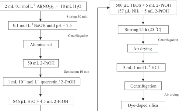
The alkaline catalysis was based on the Stöber method with a molar ratio of 1 TEOS : 21 H2O : 0.5 NH4OH. The procedure low diagram is illustrated in Figure 2. Firstly, an alumina-sol was prepared from base addition to an aqueous solution of Al(NO3)3. The alumina-sol was dispersed in 2-propanol and quercetin, water, TEOS and ammonium hydroxide were added in this order. The system
was magnetically stirred at 25 ºC during 24 h, centrifuged and sonicated with HCl 1 mol L-1 used to eliminate the uncoated alumina particles. The system was centrifuged again and the solid obtained was air-dried.
Results and Discussion
The system QAl-1 was submitted to SEM in order to analyze its morphology, and the samples were obtained from evaporation of a suspension of the nanoparticles in ethanol.
It is well known that silica nanospheres are preferentially obtained using basic catalysis. Acid catalysis can also provide globular silica particles, but very agglomerated and some irregular particle formation may occur.11 Because of the little amount of acid used in our preparation procedure, the dye-doped silica nanoparticles obtained were spherical, with a medium diameter about 200 nm, but little agglomeration was detected. Few irregular particles were also noted, in addition to close contact nucleation as indicated in Figure 3.
It is known that addition of Al3+ ions to quercetin solution results in bathochromic shift of the absorption spectrum due to the Al3+ ion complexation with the lavonoid (see structures on Figure 1).8,12 The quercetin in methanol solution has an absorbance maximum about 370 nm8,12 and its complex with Al3+ absorbs toward red region with maximum about 430 nm.8 The prepared quercetin doped silica nanoparticles QAl-1 dispersed in methanol have absorption maximum of the π→π* transition at 372 nm in absence of aluminum, but upon addition of an Al(NO3)3 solution, a new band towards the red region (maximum at 439 nm) appears. This result is a clear evidence of the interaction of the added aluminum cation with the silica colloid and formation of the complex quercetin-Al3+ inside the silica particle. The change in absorption spectrum is showed in Figure 4.
The quercetin-Al3+ complex formed in methanol solution has an emission maximum about 500 nm (Figure 5a), and it has been used as an emission standard. The emission spectrum of QAl-1 is similar to the emission spectrum of the standard quercetin-Al3+ solution in methanol, and
spectroscopic evidence of complex formation upon addition of Al3+ is shown in Figure 5b.
Considering the absorption and emission maxima of the Q-Al3+ complex in solution and those values found in the silica, the Stokes shift in solution corresponds to 70 nm while in the SiO2 matrix it hits only 57 nm indicating a less solvated or stabilized complex in excited state is formed due to the adsorption over the solid support.
The system QAl-2 was also submitted to a SEM analysis and photophysical measurements. The alumina-quercetin-SiO2 particles obtained are showed in Figure 6.
Finger-Figure 3. SEM image of the system QAl-1 acquired at 20 kV. Magniication: 20,000 times.
Figure 4. Electronic absorption spectra showing the spectral change upon titration of the system QAl-1 with Al3+ [Al(NO
3)3]= 0-71×10 -6 mol L-1.
Figure 5. Steady-state emission spectra of a methanolic Q-Al3+ standard
shaped particles with average size about 1 µm of diameter are observed. Some degree of agglomeration observed may be related to two effects. In the preparation condition used, with pH close to neutral, any isolated growing silica nanoparticles would nucleate with the alumina particles due to electrostatic interactions, 13 and this effect would be responsible for formation of large and non spherical particles as observed. On the other hand, the acid treatment used in the final step of preparation with the goal of elimination of non capped Al2O3 remaining sol particles may cause a secondary effect of further connection and nucleation of already formed particles.11
The QAl-2 particles have electronic absorption maximum at 450 nm, slightly red shifted when compared to QAl-1 nanoparticles, but nevertheless it conirms the presence of quercetin-Al3+ complex in the alumina core. Typical absorption spectrum is showed in Figure 7.
Emission profiles of the QAl-2 particles reveal a maximum at 532 nm (see Figure 8), which is red shifted in about 42 nm when compared to the emission of Quercetin Al3+ complex in methanol or that emission from QAl-1 nanoparticles with added aluminum cation. This change
in the maximum intensity to higher wavelength strongly suggests that both 1:1 and 1:2 quercetin:Al3+ complexes are present because 1:2 complex absorbs and emits at higher wavelengths.12
In order to elucidate the photophysical behavior of the complexes of Quercetin-Al3+ in alumina core-silica particles, time-resolved luorescence decay measurements were also performed. Samples suspended in methanol were excited at 400 nm and emission decays were recorded at two wavelengths, 490 nm and 530 nm. Decay proiles obtained are illustrated in Figure 9.
Fluorescence decay times obtained by reconvolution procedure with biexponential fitting function are summarized in Table 1.
The complex with 1:2 stoichiometry with red emission should contribute more for the decay recorded at 530 nm. The shorter decay time of 380 ps reported in Table 1 is thus ascribed to this species in a large aggregate that allows energy migration and fast deactivation of the excited state.8 The component with about 3.5 ns is related to the 1:1 quercetin:Al3+ complex.
Conclusions
Silica fluorescent particles due to the presence of entrapped quercetin-Al3+ emitting complex were obtained with success using both acid and basic catalysis with
Figure 6. SEM image of the system QAl-2 acquired at 20 kV. Magniication: 15,000 times. The arrow shows typical agglomerated particles.
Figure 7. Electronic absorption spectrum of the system QAl-2.
Figure 8. Steady-state emission spectra of the QAl-2 system. λexc = 430 nm.
Table 1. Time-resolved parameters of QAl-2 system obtained from reconvolution procedure. λexc = 400 nm
λem (nm) τ1 (ns) b1 a τ
2 (ns) b2
a χ2
490 1.28 ± 0.05 0.420 3.62 ± 0.03 0.580 1.020
530 0.38 ± 0.01 0.555 3.46 ± 0.01 0.445 1.301
Figure 9. Emission decays of the QAl-2 particles in methanol suspension. () Instrument Response Function, IRF, () decay, (—) biexponential itting. λexc = 400 nm, λem = 490 nm (A), λem = 530 nm (B).
formed complex with both 1:1 and 1:2 quercetin:Al3+ stoichiometry.
References
1. Maccraith, B. D.; McDonagh, C.; J. Fluoresc. 2002, 12, 333. 2. Rossi, L. M.; Shi, L.; Quina, F. H.; Rosenzweig, Z.; Langmuir
2005,21, 4277.
3. Enrichi, F.; Trave, E.; Bersani, M.; J. Fluoresc.2008, 18, 507. 4. Stöber, W.; Fink, A.; Bohn, E.; J. Colloid Interface Sci.1968,
26, 62.
5. Ow, H.; Larson, D. R.; Srivastava, M.; Baird, B. A.; Webb, W. W.; Wiesner U.; Nano Lett. 2005, 5, 113.
6. Larson, D. R.; Ow, H.; Vishwasrao, H. D.; Heikal, A. A.; Wiesner, U.; Webb, W. W.; Chem. Mater.2008, 20, 2677. 7. Rodembusch, F. S.; Campo, L. F.; Rigacci, A.; Stefani, V.;
J. Mat. Chem.2005, 15, 1537.
8. Gutierrez, A. C.; Gehlen, M. H.; Spectrochim. Acta, Part A
2002, 58, 83.
9. Pereira, R. V.; Gehlen, M. H.; J. Phys. Chem. A2006, 110, 7539. 10. Nassar, E. J.; Messaddeq, Y.; Ribeiro, S. J. L.;Quim. Nova2002,
25, 27.
11. Karmakar, B.; De, G.; Ganguli, D.; J. Non-Cryst. Solids 2000,
272, 119.
12. Cornard, J.P.; Merlin, J.C.; J. Inorg. Biochem.2002, 92, 19. 13. Osawa, C. C.; Bertran, C. A.; J. Braz. Chem. Soc.2005, 16,
251.
Received: September 13, 2009
Web Release Date: March 15, 2010
FAPESP helped in meeting the publication costs of this article.


![Figure 4. Electronic absorption spectra showing the spectral change upon titration of the system QAl-1 with Al 3+ [Al(NO 3 ) 3 ]= 0-71×10 -6 mol L -1 .](https://thumb-eu.123doks.com/thumbv2/123dok_br/18994131.461653/3.892.499.796.382.819/figure-electronic-absorption-spectra-showing-spectral-change-titration.webp)

