Ar
ti
cl
e
*e-mail: [email protected]
Determination of Cd in Blood by Microwave-Induced Combustion Coupled to
Flame Furnace Atomic Absorption Spectrometry
Lucélia Hoehne, Fabiane R. Bartz, Cezar A. Bizzi, José N. G. Paniz, Valderi L. Dressler and Érico M. de Moraes Flores*
Departamento de Química, Universidade Federal de Santa Maria, 97105-900 Santa Maria-RS, Brazil
Um sistema de combustão iniciada por microondas foi acoplado a um espectrômetro de absorção atômica com atomização em tubo metálico aquecido com chama (FF) do tipo ar-acetileno para a determinação de cádmio em sangue. Amostras de sangue lioilizado foram preparadas como comprimidos, posicionadas em um suporte de quartzo e introduzidas em uma câmara de vidro empregada para a etapa de combustão. A câmara foi acoplada ao tubo metálico usando tubos de politetraluoroetileno e de quartzo. A ignição foi iniciada por irradiação microondas usando solução de nitrato de amônio adicionada em papel previamente descontaminado. Oxigênio foi usado na etapa de combustão e para o transporte dos produtos gerados até o tubo aquecido. Graite em pó de alta pureza foi misturada com as amostras antes do preparo doscomprimidos. O volume da solução de NH4NO3, a vazão de oxigênio, a estequiometria da chama, o tipo de suporte e a quantidade de massa de amostra foram avaliados. A calibração foi possível usando solução de referência adicionada a comprimidos de graite de alta pureza, não sendo necessário o uso de material de referência certiicado (CRM). Foi possível a combustão de até 56 mg de amostra, permitindo a obtenção de limite de quantiicação de 0,018 µg g-1 e massa característica de 50 pg de Cd. Os valores dos brancos foram baixos (absorbância integrada menor que 0,01 s). Os resultados foram considerados satisfatórios com respeito à exatidão (concordância com valores de CRMs entre 95 e 99%) e à precisão (coeiciente de variação < 12%). Até 15 determinações podem ser feitas por hora incluindo a etapa de pesagem.
Microwave-induced combustion system was coupled to a metallic lame furnace (FF) atomic absorption spectrometer and applied for cadmium determination in blood. Lyophilized blood samples were prepared as pellets, positioned on a quartz holder and introduced into a glass chamber used for the combustion step. The glass chamber was coupled to the metallic lame furnace by using polytetraluoroethylene and quartz tubes. Ignition was performed by microwave radiation using ammonium nitrate solution added to a small piece of previously cleaned paper. Oxygen was used to assist the sample combustion and also to transport combustion products up to the FF heated by an air/acetylene lame. High purity graphite powder was mixed with samples before pellets preparation. The volume of NH4NO3 solution, oxygen low-rate, lame stoichiometry, sample holder type and the sample mass range were evaluated. Calibration step was feasible using adsorbed reference solution in pelletized high purity graphite avoiding the use of certiied reference material (CRM). Sample masses up to 56 mg could be used allowing quantiication limit of 0.018 µg g-1 and characteristic mass of 50 pg Cd. Low values for blanks were obtained (integrated absorbance lower than 0.01 s) and results were considered satisfactory regarding to both accuracy (agreement with results using CRMs between 95 to 99%), and precision (relative standard deviation < 12%). Up to 15 determinations can be performed per hour including the weighing step.
Keywords: lame furnace atomic absorption spectrometry, microwave induced combustion,
cadmium determination, solid sampling, blood analysis
Introduction
Analysis of solid samples offers a number of important advantages partially by avoiding the dissolution step: the
risk of contamination as well as the risk of analyte loss is considerably reduced, the limit of detection (LOD) is improved as sample is not diluted and the use of corrosive or hazardous reagents is not required, resulting in both economic and environmental benefits.1 Graphite furnace atomic
for solid sampling analysis2-4 allowing the achievement of
low LODs for many analytes.5,6 In spite of better suitability
of GF-AAS, lame atomic absorption spectrometry (FAAS) has been also used for solid sample introduction.7 In addition,
the simplicity, availability in many laboratories and relative low cost can make FAAS an attractive technique for this purpose. However, this technique presents relatively low sensitivity and no commercial device suitable for solid sampling (SoS) analysis by FAAS is available up to now. Several approaches have been described to improve the sensitivity of conventional FAAS. Special systems have been used in order to increase the density of atoms in the optical path of the spectrometer as slotted-tube atom trap,8
high-temperature nebulization,9 beam injection lame furnace
atomic absorption spectrometry,10,11 thermospray flame
furnace atomic absorption spectrometry,12-15 andpneumatic
nebulization lame furnace atomic absorption spectrometry.16
However, despite the good performance for liquids these procedures are not suitable for direct introduction of solid samples. In the case of solid introduction using FAAS another dificulty is related to the choice of a convenient analytical calibration procedure1 and some problems have
been reported if slurries are used for sample introduction using the pneumatic nebulizer.17 On the other hand, some
works have demonstrated the feasibility of using solid sampling lame atomic absorption spectrometry (SoS-FAAS) for trace analysis.7,18-21 In these works, a conventional air/
acetylene lame was used as atomizer and samples were weighed directly into a small polyethylene vial connected to a glass chamber. Air was used to transport ground samples as dry aerosol to a T-shaped quartz cell positioned above the burner in the optical path of spectrometer. In spite of good accuracy and relatively low LODs had been obtained with this system a limitation related to the small sample mass that could be introduced into the atomizer was reported.7,18-21 To
overcome this problem, solid samples were introduced as pellets into a quartz cell for analysis by FAAS.22 With this
system, samples up to 7 mg could be weighed into a small paper capsules and introduced into a quartz cell heated by an air/acetylene lame and LOD as low as 0.23 µg g-1 was
obtained for cadmium determination.
Some authors proposed to separate the sample vaporization step from atomization process. Infrared lamps were used to vaporize the analyte that was further transported to the lame or to a tube positioned above the burner of a conventional atomic absorption spectrometer. However, for the calibration step it was necessary the use of CRMs and the limitation of low masses still remained.23,24
Recently, a novel systemwas proposed using microwave induced combustion (MIC) to the combustion of organic samples in closed quartz vessels pressurized with oxygen
with the ignition step being performed by microwave radiation. Samples were pressed as pellets and positioned on a small piece of low-ash ilter paper that was previously placed on a quartz holder. About 50 µL of 6 mol L-1
ammonium nitrate solution is added as igniter, and the system is closed and pressurized with oxygen.25,26
The proposed mechanism is related to the heating of the NH4NO3 solution wetted-ilter paper by microwave radiation. The fast heating allows the creation of a small lame that, in presence of a convenient oxygen-pressure, causes a complete combustion of sample. In this system, sample combustion is performed in less than one minute with minimum acid consumption.27 Based in previous
works,25-27 some authors proposed the combination of
microwave induced combustion (MIC) system to a lame furnace (FF) for analysis of botanical samples by atomic absorption spectrometry (AAS).28 For the combustion
step, solid samples were introduced as pellets into a glass chamber using a quartz holder device. This chamber was coupled to a metallic FF by using polytetraluoroethylene and quartz tubes. The glass chamber was positioned inside a microwave oven where the sample was combusted. Ignition was performed by microwave radiation using a small piece of paper wetted with few microliters of NH4NO3 solution. Oxygen was used to assist the combustion and also to transport the combustion products up to a heated FF positioned above an air/acetylene type burner and calibration was feasible by using reference solutions. Cadmium and Pb were determined in botanical samples as examples to demonstrate the potential of the proposed procedure for trace analysis. Moreover, with microwave-induced combustion flame furnace atomic absorption spectrometry (MIC-FF-AAS) system the LOD was improved by a factor of 50 and 12, respectively for Cd and Pb in relation to the conventional FAAS technique.
In the present work, MIC-FF-AAS was applied to Cd determination in blood samples in order to increase the analyte amount introduced into the atomizer in comparison with conventional lame-AAS atomizers. Accuracy was evaluated by using CRMs. Operational conditions were evaluated and results were compared to those using other devices proposed in the literature for SoS using lame heated systems.
Experimental
Instrumentation
with deuterium lamp background correction system and a conventional burner (slit of 10 cm, air/acetylene lame). A cadmium hollow-cathode lamp was operated at 4 mA, wavelength was set at 228.8 nm and the spectral bandwidth was 0.5 nm. Integrated absorbance with an integration time of 30 s was used for signal evaluation.
A freeze dryer (Terroni Fauvel, Model LH 2000/3, São Carlos, Brazil) was used for blood lyophilization process. An ultra-micro balance (Sartorius, Model M2P, Goettingen, Germany) with a resolution of 1 µg and an electronic weighing range up to 2 g was used for sample weighing and a hydraulic press set at 1 ton (Specac, Model Hydraulic Press 15 ton, Orpington, England) was used to prepare the sample pellets.
For the combustion step, a microwave oven (Panasonic, Model NN-S52 B, Manaus, Brazil) with internal volume of 28 L and nominal maximum power of 900 W was used. Closed polytetraluoroethylene-perluoroalkoxy (PTFE-PFA) vessels heated by microwave radiation (microwave oven Model ETHOS 1, Milestone, Sorisole, Italy) were used for conventional sample digestion. Cadmium determination was also performed by inductively coupled plasma mass spectrometry (ICP-MS, PerkinElmer, Model Sciex-Elan DRC II, Thornhill, Canada), equipped with a concentric nebulizer (Meinhard Associates, Golden, USA), a cyclonic spray chamber (Glass Expansion, Inc., West Melbourne, Australia) and a quartz torch with a quartz injector tube (2 mm i.d.). Measurements were carried out according to the instructions of manufacturer.29
Reagents and samples
All glass or quartz apparatus were soaked in 10% (v/v) HNO3 solution for 24 h and washed with water before use. Water was distilled, deionized and puriied using a Milli-Q system (Millipore Corp., Bedford, USA). Nitric acid (Merck, Darmstadt, Germany) was doubly distilled in a Model duoPUR 2.01E sub-boiling system (Milestone, Bergamo, Italy). Analytical grade hydrogen peroxide (30%, Merck, Darmstadt, Germany) was used as aid for samples digestion.
Calibration reference solutions for Cd were prepared immediately before use by dilution of a Titrisol stock
solution containing 1000 mg L-1 in 2% (v/v) HNO 3
(Merck, Darmstadt, Germany). Ammonium nitrate solution (6 mol L-1) was used as ignition aid.
High purity graphite powder (RWA, nº 431815/00, series X/62/403) was obtained from SGL Carbon (Bonn, Germany). Filter paper (2.2 cm2, 25 mg) with low ash
content (Black Ribbon Ashless, Schleicher & Schuell GmbH, Dassel, Germany) was used to aid the combustion process. The ilter paper was cleaned with 10% (v/v) HNO3
in an ultrasonic bath (Odontobrás, 100W, Model 1440D, Ribeirão Preto, Brazil) for 20 min and dried in an oven (Nova Ética, 820 W, Model 400/2ND 300, São Paulo, Brazil) by 2 h at 60 ºC before use.
Human blood samples were obtained from a local hospital. Sodium citrate solution was added to the samples as anticoagulant. Samples were stored under freezing at −4 °C
before use. For the initial development and optimization of the procedure, a spike correspondent to 0.19 µg mL-1 Cd was
performed to a sample of blood before lyophilization process. Certified reference blood was not available and biological CRMs with different composition were used: dogish muscle and liver DOLT-2 (NRCC, National Research Council Canada), non defatted lobster hepatopancreas LUTS-1 (NRCC), skim milk powder BCR-151 (IRMM, Institute for Reference Materials and Measurement), oyster tissue 1566a (NIST, National Institute of Standards & Technology) and oyster tissue 1566b (NIST).
Sample preparation
Human blood samples were defrost and lyophilized for 12 h, ground using an agate mortar and pressed (1 ton) to prepare pellets of 5 mm diameter. Pellets of lyophilized blood and pellets of lyophilized blood mixed with high purity graphite were also evaluated.
Sample digestion for ICP-MS
Cadmium determination was also performed by ICP-MS after sample digestion using closed vessels with microwave heating. About 80 mg of lyophilized human blood were digested in PTFE-PFA vessels, using 8 mL of concentrated nitric acid and 2 mL of 30% hydrogen peroxide. After acid addition to samples the vessels were kept overnight. Then, vessels were closed and the decomposition was performed based on the manufacturer recommendation.30 Digested
samples were diluted with water to 25 mL in polypropylene vessels before analysis by ICP-MS.
Proposed MIC-FF-AAS procedure
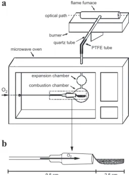
A domestic microwave oven was modiied in order to allow an oxygen low entrance in the combustion chamber and to aid the transport of the combustion products up to the FF (Figure 1a). Microwave power that was effectively transferred to the oven cavity was determined according to a procedure previously described.31 For the combustion
(3 mm i.d., 2 cm length) to a second glass chamber (expansion chamber). The expansion chamber was necessary to minimize eventual excessive pressure during the beginning of combustion. A PTFE tube (10 cm length, 3 mm i.d.) connected to a quartz tube (30 cm length and 3 mm i.d.) was used to the transport of combustion products inside the combustion chamber up to the FF. The end of this quartz tube was itted to the FF (10 cm length, 11 mm o.d. and 9 mm i.d.), that was positioned 4 mm above the burner.The FF was home-made and it was produced from a metallic bar obtained from Camacam, Inconel 600, Brazil (72% Ni, 15% Cr, 8% Fe, 0.5% Cu and 0.3% Tl).
A pellet of lyophilized blood mixed with graphite was placed on a ilter paper and it was wrapped with aid of polypropylene tweezers and positioned on the quartz holder. In order to have reproducible ignitions ammonium nitrate solution must be added to the extremity of ilter paper close to the oxygen supply. After holder introduction into the combustion chamber oxygen was passed through the device (1 to 2.5 L min-1) and microwave was irradiated
using the maximum power. Combustion time was typically between 8 and 15 s. After ignition, the microwave radiation was stopped and atomic signals were recorded. Integrated absorbance (peak area) of 30 s was used throughout for the proposed MIC-FF-AAS procedure. After combustion has been inished the quartz holder was removed for a subsequent cycle of analysis. Calibration was evaluated by using reference solution added directly on the ilter paper as described in previous work28 or using reference solution
added to high purity graphite powder pellets.
Results and Discussion
Optimization of operating conditions for MIC-FF-AAS
Initial experiments were performed by trying to introduce original blood samples (not lyophilized) directly into the metallic lame furnace. However, solid deposition in tube used for sample introduction was observed and results were not reproducible. On the other hand, preliminary tests using MIC-FF-AAS procedure for lyophilized samples were performed according to conditions described in previous work.28 Pellets of lyophilized blood were wrapped with
ilter paper and placed on the sample holder. Ammonium nitrate solution (40 µL) was added to the paper and the sample holder was introduced into the glass chamber. Then, microwave irradiation was applied using the maximum power up to starting sample combustion (about 10 s). The maximum real power of microwave was 615 W and this condition was always used for the experiments by MIC-FF-AAS. Combustion products were transported to the FF and atomic signal was recorded. However, black particles remained after combustion inside the glass chamber and in the PTFE tube. It occurred due to sample falling through the holes in the basis of sample holder during the combustion. The quartz sample holder was changed with holes narrower than the previous type (Figure 1b). Using this holder the combustion process always occurred with complete combustion.
The inluence of volume of 6 mol L-1 NH
4NO3 solution
was investigated from 10 to 50 µL. Volumes lower than 40 µL of ammonium nitrate solution were not suficient to start the combustion process. With 40 or 50 µL of NH4NO3 solution the combustion process always occurred. This result was in agreement with results previously reported.25
For further experiments, 40 µL of ammonium nitrate solution was selected.
Preliminary tests showed that characteristic mass of Cd was different for reference solution and solid sample. Addition of graphite has been proposed in direct solid sampling- graphite furnace atomic absorption spectrometry in order to allow sensitivities more close to those using reference solutions.32 In this work, tests were performed
by mixing samples with 30 mg of high purity graphite powder and pressed as pellets. With this condition, results for characteristic mass were more similar between samples and reference solution added to graphite pellets used for calibration and graphite addition was used for further oxygen low-rate, lame stoichiometry, calibration and sample mass evaluation.
The effect of oxygen low-rate on characteristics mass of Cd was evaluated (Figure 2). For oxygen low-rates
lower than 1.0 L min-1 (not shown in Figure 2) the ignition
was not reproducible and the Cd signal was lower due to the incomplete burning of sample. For oxygen low-rate higher than 2.0 L min-1 a decrease occurred in the analytical
signal. This result was probably caused by dilution of analyte in gaseous phase. Better sensitivity and relative standard deviation (RSD) were obtained with O2 low-rate of 1 L min-1
and this condition was used for subsequent studies.
Flame stoichiometry
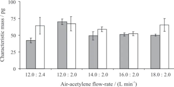
The inluence of lame composition on Cd characteristic mass was investigated and results are shown in Figure 3. The following air/acetylene low-rates were evaluated: 12.0/2.4, 12.0/2.0, 14.0/2.0, 16.0/2.0 and 18.0/2.0 L min-1.
Lower characteristic mass and standard deviation for Cd determination were obtained using air/acetylene low-rates of 16.0/2.0 L min-1. Background signals were always
lower than 0.035 in peak height scale for all investigated lame composition. These results were similar to previous work using MIC-FF-AAS.28 However, when compared
with results of the present work, the small difference of chosen lame composition was probably due to the graphite presence in the pellet. Then, an air/acetylene low-rate of 16.0/2.0 L min-1 was applied for subsequent tests.
Calibration
Similar results for sensitivity were obtained if pellets for reference solutions and samples were prepared with 30 mg of graphite powder. With graphite addition good agreement between signals for reference solution and solid samples were obtained. The correlation coeficient for calibration curve up to 60 ng Cd was 0.9996 with the use of reference solution adsorbed in graphite pellets. Figure 4 shows the
typical signal using MIC-FF-AAS for Cd determination in lyophilized blood samples using graphite pellets. It can be observed that Cd signal was completely integrated in 15 s and background was relatively low (< 0.03 in peak height). The low background signal was compensated by the deuterium corrector and a more powerful corrector (e.g., based on Zeeman effect) was not necessary.
Sample mass
In a similar way of a previous work,33 a study was
performed in order to evaluate the minimum and maximum limits feasible to apply the proposed procedure by MIC-FF-AAS (Figure 5). Pellets containing 2.7 to 90 mg of sample plus 30 mg of graphite were used and standard deviation was calculated using analysis of variance considering a conidence interval of 95% (Tukey-Kramer multiple comparisons test). Sample masses lower than 5 mg showed high dispersion of results probably due to heterogeneity of analyte in samples. With sample masses higher than 56 mg a black residue was observed in the glass chamber. This result was considered to be caused by incomplete sample combustion. In addition, memory effects were observed
Figure 2. Characteristic masses obtained for () reference solution adsorbed in 30 mg of high purity graphite pellet and () pellets of 10 mg of solid sample mixed with 30 mg of high purity graphite for Cd with different oxygen low-rate: air/acetylene, 16.0/2.0 L min-1. Cd mass in
reference solution added to graphite pellet was 5.0 ng and in solid sample was 6.2 ng (n = 5).
Figure 3. Characteristic masses obtained for () reference solution adsorbed in 30 mg of high purity graphite pellets and () pellets of 10 mg of solid sample mixed with 30 mg of high purity graphite for Cd with different lame stoichiometry. Oxygen low-rate: 1 L min-1. Cd mass in
reference solution added to graphite pellet was 5.0 ng and in solid sample was 6.2 ng (n = 5).
using this condition and a cleaning step must be applied after each run. However, up to 10 runs using 56 mg of sample mass could be carried out without changes in analytical signal. Therefore, sample masses between 5 to 56 mg could be used in the present procedure.
Determination of Cd in human blood and in certiied reference materials
Cadmium was determined in lyophilized human blood by MIC-FF-AAS and results were lower than 0.005 µg g-1
(correspondent to < 1.6 µg L-1 in blood samples). These
results were conirmed by ICP-MS analysis with previous sample digestion. For lyophilized blood sample with Cd spike the results by MIC-FF-AAS and ICP-MS after digestion presented an agreement better than 95%. Cadmium was also determined in different biological CRMs (Table 1) and an agreement to the mean value of certiied value between 95 to 99% was found for all certiied reference samples (no statistical difference based on t-test, conidence level of 95%).
Comparison of MIC-FF-AAS procedure
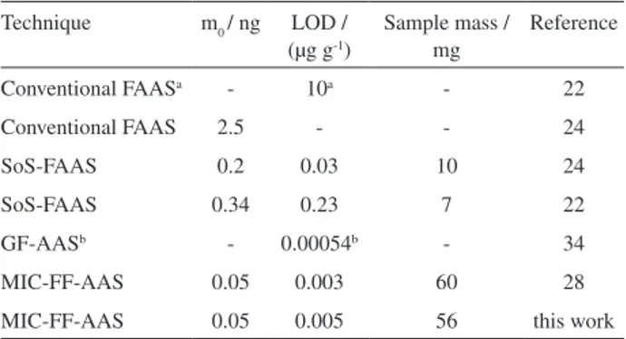
Results using MIC-FF-AAS were compared with those from different procedures for biological sample analysis using FAAS22,24,28 and conventional graphite furnace atomic
absorption spectrometry using diluted blood samples34
(Table 2). The characteristic mass of Cd (0.05 ng) obtained with proposed procedure was the same or better than by a factor up to 50 in comparison with other works using FAAS systems.22,24,28 For MIC-FF-AAS the LOD (0.005 µg g-1)
was similar when using a similar procedure (0.003 µg g-1) 28
but up to 40 times better than other procedures using FAAS systems.22,24 As expected, the LOD using GF-AAS34 was
lower (0.00054 µg mL-1). However, the obtained LOD using
the proposed MIC-FF-AAS system was considered suitable for Cd determination in biological samples, as blood.
On the other hand, maximum sample masses were similar by using MIC-FF-AAS procedure28 but higher
than those reported in other works using FAAS.22,24
Relative standard deviation was lower than 12% and it was considered suitable for the proposed procedure.
In this work, the limit of quantiication (10σ, n = 10)
was 0.018 µg g-1 and it was possible to perform up to
15 determinations in 1 h including the weighing step. Moreover, it was feasible the calibration using adsorbed reference solution in pelletized high purity graphite avoiding the use of solid CRMs.
Conclusions
Relatively low LOD and RSD were obtained using the proposed MIC-FF-AAS procedure for Cd determination in blood in view of the relatively high sample masses used (up to 56 mg). Calibration was carried out with reference solution added to graphite pellets which is an advantage in comparison with the use of CRMs for calibration. Moreover, the coupling of the MIC procedure to an inexpensive FF-AAS spectrometer makes this procedure
Figure 5. Inluence of sample mass for Cd determination by MIC-FF-AAS, using pellets of different sample mass mixed with 30 mg of high purity graphite. Flame stoichiometry: air/acetylene, 16.0/2.0 L min-1.
Oxygen low-rate: 1 L min-1. Horizontal lines represent the mean and the
standard deviation (sd) for the useful mass range.
Table 1. Results for Cd determination using MIC-FF-AAS (results, in µg g-1, represent the mean and respective standard deviation of results,
n = 5)
CRM Certiied value MIC-FF-AAS
IRMM BCR-151 0.101 ± 0.008 0.096 ± 0.010
NIST 1566a 4.15 ± 0.38 4.02 ± 0.32
NIST 1566b 2.48 ± 0.08 2.36 ± 0.22
NRCC DOLT-2 20.80 ± 0.50 20.64 ± 0.21
NRCC LUTS-1 14.2 ± 1.0 13.6 ± 1.6
Table 2. Characteristic mass, limit of detection and maximum sample mass used for different techniques for Cd determination
Technique m0 / ng LOD / (µg g-1)
Sample mass / mg
Reference
Conventional FAASa - 10a - 22
Conventional FAAS 2.5 - - 24
SoS-FAAS 0.2 0.03 10 24
SoS-FAAS 0.34 0.23 7 22
GF-AASb - 0.00054b - 34
MIC-FF-AAS 0.05 0.003 60 28
MIC-FF-AAS 0.05 0.005 56 this work
acalculated value considering the digestion of 100 mg of sample and
an alternative to more expensive techniques for direct solid sample analysis.
Acknowledgments
The authors thank to CAPES and CNPq for supporting this study.
References
1. Belarra, M. A.; Resano, M.; Vanhaecke, F.; Moens, L.; Trends Anal. Chem.2002, 21, 828.
2. Resano, M.; Vanhaecke, F.; de Loos-Vollebregt, M. T. C.; J. Anal. At. Spectrom. 2008, 23, 1450.
3. Welz, B.; Sperling, M.; Atomic Absorption Spectrometry, 3rd ed., Wiley-VCH: Weinheim, 1999.
4. Nomura, C. S.; Silva, C. S.; Nogueira, A. R. A.; Oliveira, P. V.; Spectrochim. Acta, Part B2005, 60, 673.
5. Mattos, J. C. P.; Flores, E. M. M.; Krivan, V.; J. Anal. At. Spectrom.2008, 23, 931.
6. Friese, K-C.; Krivan, V.; Schuierer, O.; Spectrochim. Acta, Part B1996, 51, 1223.
7. Flores, E. M. M.; Costa, A. B.; Barin, J. S.; Dressler, V. L.; Paniz, J. N. G.; Martins, A. F.; Spectrochim. Acta, Part B2001, 56, 1875.
8. Matusiewicz, H.; Spectrochim. Acta, Part B1997, 54, 1711. 9. Berndt, H.; Yáñez, J.; J. Anal. At. Spectrom. 1996, 11, 703. 10. Gáspár, A.; Berndt, H.; Anal. Chem.2000, 72, 240.
11. Aleixo, P. C.; Santos Júnior, D.; Tomazelli, A. C.; Ruini, I. A.; Berndt, H.; Krug, F. J.; Anal. Chim. Acta2004, 512, 329. 12. Gáspár, A.; Berndt, H.; Spectrochim. Acta, Part B2000, 55,
587.
13. Rosini, F.; Nascentes, C. C.; Neira, J. Y.; Nóbrega, J. A.; Talanta 2007, 73, 845.
14. Brancalion, M. L.; Sabadini, E.; Arruda, M. A. Z.; Spectrochim. Acta, Part B2009, 64, 89.
15. Nascentes, C. C.; Arruda, M. A. Z.; Nogueira, A. R. A.; Nóbrega, J. A.; Talanta2004, 64, 912.
16. Wu, P.; Liu, R.; Berndt, H.; Lv, Y.; Hou, X.; J. Anal. At. Spectrom. 2008, 23, 37.
17. Fuller, C. W.; Analyst1976, 101, 961.
18. Flores, E. M. M.; Paniz, J. N. G.; Martins, A. F.; Dressler, V. L.; Müller, E. I.; Costa, A. B.; Spectrochim. Acta, Part B2002, 57, 2187.
19. Flores, E. M. M.; Paniz, J. N. G.; Saidelles, A. P. F.; Müller, E. I.; Costa, A. B.; J. Anal. At. Spectrom.2003, 18, 769. 20. Flores, E. M. M.; Saidelles, A. P. F.; Flores, E. L. M.; Mesko,
M. F.; Pedroso, M. P.; Dressler, V. L.; Bittencourt, C. F.; Costa, A. B.; Microchem. J.2004, 77, 113.
21. Flores, E. M. M.; Paniz, J. N. G.; Saidelles, A. P. F.; Barin, J. S.; Dressler, V. L.; Müller, E. I.; Costa, A. B.; J. Braz. Chem. Soc.2004, 2, 199.
22. Costa, A. B.; Mattos, J. C. P.; Müller, E. I.; Paniz, J. N. G.; Dressler, V. L.; Flores, E. M. M.; Spectrochim. Acta, Part B 2005, 60, 583.
23. Berndt, H.; Spectrochim. Acta, Part B1984, 39, 1121. 24. Campos, R. C.; Curtius, A. J.; Berndt, H.; J. Braz. Chem. Soc.
1990, 1, 66.
25. Flores, E. M. M.; Barin, J. S.; Paniz, J. N. G.; Medeiros, J. A.; Knapp, G.; Anal. Chem.2004, 76, 3525.
26. Mesko, M. F.; Moraes, D. P.; Barin, J. S.; Dressler, V. L.; Knapp, G.; Flores, E. M. M.; Microchem. J.2006, 82, 183.
27. Flores, E. M. M.; Barin, J. S.; Mesko, M. F.; Knapp, G.; Spectrochim. Acta, Part B 2007, 62, 1051.
28. Barin, J. S.; Bartz, F. R.; Dressler, V. L.; Paniz, J. N. G.; Flores, E. M. M.; Anal. Chem.2008, 80, 9369.
29. PerkinElmer-SCIEX; ELAN Version 3.0 Software Guide, PerkinElmer Inc.: Waltham, 2003.
30. Milestone-ETHOS 1; Application Notes for Digestion, Milestone Inc.: Sorisole, 2006.
31. Flores, E. M. M.; Saidelles, A. P. F.; Barin, J. S.; Mortari, S. R.; Martins, A. F.; J. Anal. At. Spectrom. 2001, 16, 1419. 32. Krivan, V.; Janickova, P.; Anal. Bioanal. Chem. 2005, 382, 1949. 33. Rodrigues, L. F.; Mattos, J. C. P.; Dressler, V. L.; Pozebon, D.;
Flores, E. M. M.; Spectrochim. Acta, Part B2007, 62, 933. 34. Palmer, C. D.; Lewis Jr., M. E.; Geraghty, C. M.; Barbosa Jr.,
F.; Parsons, P. J.; Spectrochim. Acta, Part B2006, 61, 980.
Received: March 17, 2009