Dentoalveolar mandibular changes with self-ligating
versus
conventional bracket systems: A CBCT and dental cast study
Marcio Rodrigues de Almeida1, Cristina Futagami2, Ana Cláudia de Castro Ferreira Conti3, Paula Vanessa Pedron Oltramari-Navarro1, Ricardo de Lima Navarro4
How to cite this article: Almeida MR, Futagami C, Conti ACCF, Oltramari-Navarro PVP, Oltramari-Navarro RL. Dentoalveolar mandibular changes with self-ligating versus conventional bracket systems: A CBCT and dental cast study. Dental Press J Orthod. 2015 May-June;20(3):50-7.
DOI: http://dx.doi.org/10.1590/2176-9451.20.3.050-057.oar
Submitted: June 02, 2014 - Revised and accepted: September 26, 2014
Contact address: Marcio Rodrigues de Almeida Avenida Paris, 675 Jardim Piza, Londrina -PR - Brazil CEP: 86041-120 - E-mail: [email protected]
1 Full professor of Orthodontics, Universidade Norte do Paraná (UNOPAR),
Londrina, Paraná, Brazil.
2 MSc in Orthodontics, Universidade Norte do Paraná (UNOPAR), Londrina,
Paraná, Brazil.
3 Professor of Orthodontics, Universidade do Sagrado Coração (USC), Bauru,
São Paulo, Brazil.
4 PhD in Orthodontics, Universidade de São Paulo (USP), São Paulo, São Paulo,
Brazil.
» The authors report no commercial, proprietary or financial interest in the prod-ucts or companies described in this article.
Objective: The aim of the present study was to compare dentoalveolar changes in mandibular arch, regarding transversal measures and buccal bone thickness, in patients undergoing the initial phase of orthodontic treatment with self-ligating or conventional bracket systems. Methods: A sample of 25 patients requiring orthodontic treatment was assessed based on the bracket type. Group 1 comprised 13 patients bonded with 0.022-in self-ligating brackets (SLB). Group 2 included 12 patients bonded with 0.022-in conventional brackets (CLB). Cone-beam computed tomography (CBCT) scans and a 3D program (Dolphin) assessed changes in transversal width of buccal bone (TWBB) and buccal bone thickness (BBT) before (T1) and 7 months after treatment onset (T2). Measurements on dental casts were performed using a digital cali-per. Differences between and within groups were analyzed by Student’s t-test; Pearson correlation coefficient was also calculated. Results: Significant mandibular expansion was observed for both groups; however, no significant differences were found between groups. There was significant decrease in mandibular buccal bone thickness and transversal width of buccal bone in both groups. There was no significant correlation between buccal bone thickness and dental arch ex-pansion. Conclusions: There were no significant differences between self-ligating brackets and conventional brackets systems regarding mandibular arch expansion and changes in buccal bone thickness or transversal width of buccal bone.
Keywords:Orthodontic appliances. Corrective orthodontics. Orthodontic brackets.
DOI: http://dx.doi.org/10.1590/2176-9451.20.3.050-057.oar
Objetivo: o objetivo do presente estudo foi comparar as alterações dentoalveolares transversais e a espessura óssea da arcada inferior em pacientes submetidos ao tratamento ortodôntico utilizando sistemas de braquetes autoligáveis ou con-vencionais. Métodos: uma amostra de 25 pacientes requerendo tratamento ortodôntico foi recrutada com base no tipo de braquete. No Grupo 1, 13 pacientes foram tratados com braquetes autoligáveis (SLB, slot 0,022”); o Grupo 2 incluiu 12 pacientes, nos quais foram colados braquetes convencionais (CLB, slot 0,022”). Utilizou-se tomografia computadorizada de feixe cônico e um programa 3D (Dolphin) para avaliar as alterações pré-tratamento (T1) e 7 meses após o início desse (T2). As medições em modelos de gesso foram realizadas com o auxílio de um paquímetro digital. As diferenças intergru-pos, bem como intragrupo, foram analisadas por meio de teste t de Student. Além disso, o coeficiente de correlação de Pearson foi utilizado. Resultados: alterações dentoalveolares significativas foram observadas em ambos os grupos. Entre-tanto, não houve diferenças significativas entre os grupos. Houve uma diminuição da espessura óssea na região posterior e das medidas transversais em ambos os grupos. Não houve uma correlação significativa entre a espessura óssea posterior e a expansão da arcada dentária, em nenhum dos dois sistemas de braquetes utilizados. Conclusões: comparando-se o uso dos aparelhos autoligáveis e convencionais, concluiu-se que não houve diferenças dentoalveolares significativas quanto à expansão da arcada inferior e quanto à espessura óssea posterior.
INTRODUCTION
The ongoing search for innovation in Orthodontics has boosted the emergence or re-emergence of appliances so as to ofer patients more comfort, shorter treatment time, improved post-treatment stability, and fewer side efects. Self-ligating brackets (SLB) came back into scene in the seventies, arising strong expec-tancy, and became popular in the nineties. Much em-pirical and anecdotal evidence as well as advantages were attributed to these appliances: increased patient com-fort, better oral hygiene, increased patient cooperation, less chair time, shorter treatment time, greater patient acceptance, expansion, and less dental extractions.1-6
Correcting dental crowding without extractions or interproximal reductions requires an increase in arch perimeter in order to allow excellent teeth align-ment. In the absence of distal movements, the di-mensional changes of the arch involve transversal and buccal dental expansion.7 It is a well-known fact that
both self-ligating and conventional ligating brack-ets (CLB) when used for non-extraction treatment of dental crowding produce dentoalveolar expansion. The amount of transversal increase depends on the mechanics applied in each case.7-11
Before the introduction of computerized to-mography, it was not possible to visualize the buc-cal bone due to superposition that occurred in 2D radiographs.12,13 To achieve successful orthodontic
treatment, the limits of orthodontic movement must be respected, in order to prevent iatrogenic effects to the sustaining and protection periodontium, such as gingival recessions, dehiscence and bone fenestra-tions. Studies prior to cone-beam computed tomog-raphy (CBCT) scans assessed only radiographs and dental casts, both of which used to be regarded as gold standards. Improvements in CBCT scans revealed it to be a reliable method, which offers an excellent vi-sualization of the actual structures.14,15 Timock et al16
investigated the accuracy and reproducibility of mea-surements of alveolar bone height and thickness by means of CBCT imaging. They found good preci-sion and accuracy for both measurements.16
The transversal response of the mandibular den-tal arch treated with CLB has been widely studied in the literature, especially the dentoalveolar response on dental casts.7,10,17,18 However, little is known regarding
CBCT scans used to assess the mandibular alveolar
bone of the posterior region, where buccal bone can be detected and quantiied.19 This study aims at
test-ing the null hypothesis that there is no diference, re-garding changes in transversal width and buccal bone thickness in the mandibular arch, between patients undergoing the initial phase of orthodontic treatment (7 months) with SLB and CLB systems.
MATERIAL AND METHODS
This research protocol was approved by Univer-sidade Norte do Paraná (UNOPAR, Londrina/PR, Brazil) Institutional Review Board. Patients and guardians were fully informed about the study and its implications, and signed a consent form.
For this prospective study, power analysis showed that a sample size of 12 patients in each group would give 80% probability to detect a real difference of 1.4 mm in intermolar distance and 0.2 mm in bone thickness, with a 95% (p < 0.05) significance level.20
The sample for the present prospective random-ized study was treated at Universidade Norte do Pa-raná (UNOPAR, Londrina, PR, Brazil) from 2009 to 2012. All patients had complete orthodontic re-cords taken at the beginning (T1) of treatment and 7 months after treatment onset (T2), including study models and CBCT scans. In selecting the sample, the following inclusion criteria were applied: patients with Angle Class I malocclusion, moderate-to-severe lower dental crowding (3.0 to 7.0 mm), absence of di-astema, absence of posterior crossbite, complete per-manent teeth (except for third molars). Patients were randomly divided into two groups: SLB and CLB. Out of the selected individuals, none were excluded after treatment onset. Premolars extraction and tooth wear were not included in the proposed treatment.
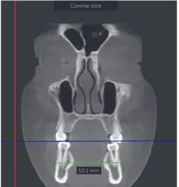
Figure 1 - Coronal slice and transversal width of buccal bone (TWBB).
the manufacturers’s (Aditek) prescription (Damon sys-tem). Each archwire remained in place for two months. Cone-beam computed tomography scans were obtained from all patients at two time intervals prior to orthodontic treatment onset and 7 months after it. All CBCT scans were carried out by a single ex-perienced radiologist using the same scanner (i-Cat Imaging Sciences International, Hatfield, Pennsylva-nia, USA) set up as follows: 22 x 16 cm fov, 40 sec, 120 kVp, 36 mA. This scanner has high-resolution sensors and affords 0.4-mm voxel images.21
CBCT scans were analyzed by one single operator who assessed mandibular bone changes by means of Dolphin 3D software (Version 11.5®, Dolphin Imaging
& Management Solutions, Chatsworth, Calif., USA) with a level of sensitivity set at 25%.
Coronal slices were selected for the bone mea-surements (Fig 1) and 1-mm thick cross-sections were made through the irst molar (M1), second premolar (P2) and irst premolar (P1), in the right and let mandibular arches. As for coronal slices, the mid portion of teeth (molars and premolars) was chosen. The point selected for buccal bone measurement was the most external prominence of the buccal bone (EBB) in the root most apical portion (apex). At this same height, a point was projected from the parallel projection of the cusp point. The distance between the two points was determined as BBT, buccal bone thick-ness (Figs 2 and 3). Thus, changes in BBT were calcu-lated by subtracting T1 from T2 values. For transversal width of the buccal bone, the EBB point was used on the right and let sides. The distance between right and let EBB was the transversal width of buccal bone (TWBB) (Fig 1). Similarly, TWBB changes were cal-culated by subtracting T1 from T2 values. In order to conirm whether transversal width and bone thickness measurements were taken on the same coronal slices, the mid region of each posterior teeth was used as ref-erence to ensure consistency of slices.
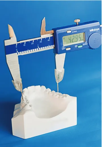
Intermolar distances, intersecond premolar dis-tances, and interfirst premolar distances were mea-sured in dental casts (Fig 4) by means of a previously calibrated digital caliper (Mitutoyo Caliper, Japan). In order to measure the transversal distances, buccal cusp tips were selected for first and second mandibu-lar premomandibu-lars, while mesiobuccal cusp tips were se-lected for first molars.
Statistical analysis
To assess intra and interexaminer reliability, ten CBCT scans were randomly selected and remea-sured four weeks apart by two operators. Intraex-aminer error was assessed by means of paired t-test and Dahlberg’s formula. Interexaminer reliability was assessed by intraclass correlation coefficient (ICC). Data were tested for normal distribution by means of Kolmogorov-Smirnov test. As data were normally distributed, parametric tests were applied. Results were described by parameters of mean and standard deviation of T1 and T2 measurements for both groups. Independent t-tests were used to com-pare the initial demographic data of both groups. Paired and unpaired t-tests were used to compare intra and intergroup changes. Finally, Pearson cor-relation coefficient was calculated to further explore the association between dental expansion and ex-pansion of TWBB. In all statistical tests, the signifi-cance level was set at 5%.22 All statistical analyses
were performed with SPSS software for Windows version 17.0 (SPSS Inc, Chicago Ill.).
Coronal slice
180
Figure 2 - Buccal bone thickness (BBT) measurements. Figure 4 - Intermolar width measured on a dental cast by means of a digital caliper.
Figure 3 - Example of measurements for buccal bone thickness (BBT) of mandibular first molars.
5.0 mm
160
170
190
200
Coronal slice
5.0 mm
RESULTS
Systematic (paired t-test) and casual error (Dahlberg’s formula) showed no intraexaminer dif-ference. Intraclass correlation coefficients for bone thickness and transversal width of buccal bone mea-surements were 0.89 and 0.98, respectively, thereby showing acceptable reliability. Random error ranged from 0.30 to 0.56 mm and from 0.53 to 1.08 mm for dental casts and CBCT measurements, respectively.
Patients’ demographic distribution is presented in Table 1. Both samples were comparable at treat-ment onset regarding the following aspects: initial age, treatment time, intermolar distances, inter-second premolar distances, interfirst premolar dis-tances, TWBB and BBT measurements. Means and standard deviation values for BBT and TWBB mea-surements at pretreatment (T1), 7 months after treat-ment onset (T2) and the changes observed (T2-T1) are shown in Tables 2 to 4.
Mandibular buccal bone thickness (BBT) de-creased from T1 to T2 for both bracket types. BBT in the CLB group significantly decreased for P1L (-1.51 mm; p = 0.016), P1R (-0.9 mm; p = 0.039), P2R (-1.09 mm; p = 0.007) and M1R (-0.79 mm; p = 0.008). BBT in the SLB group significantly de-creased for P1R (-0.88 mm, p = 0.019), P2L (-0.64 mm; p = 0.002), P2R (-1.09 mm, p < 0.001) and M1R (-0.54 mm; p = 0.025). However, changes in TWBB measurements showed a slight decrease and were not considered statistically significant in either one of the groups: for the CLB group, the following measurements decreased: P1 (-0.21 mm; p = 0.613), P2 (-0.66 mm; p = 0.222) and M1 (-0.31 mm; p = 0.611); as for the SLB group, the following mea-surements decreased: P1 (-0.56 mm; p = 0.076) and P2 (-0.01 mm; p = 0.980), with an increase in M1 (0.10 mm; p = 0.750).
Comparison between BBT and TWBB mea-surements from T1 to T2 revealed no significant dif-ferences between groups (Table 4). Additionally, no significant differences were found when compar-ing dental casts at treatment onset (T1) and 7 months later (T2) (Table 5). An average increase of dental transversal distances occurred from T1 to T2, which was considered significant. Bracket type had no sig-nificant influence on changes in mandibular dental arch. Differences between SLB and CLB for interfirst
SLB (G1) (n = 13) CLB (G2) (n = 12) P
Initial mean age (years) 18.58 ± 5.43 21.61 ± 6.69 0.221
Treatment time (days) 210.15 ± 41.44 218.17 ± 46.60 0.654
CBCT scans
P1L BBT (mm) 2.34 ± 2.59 2.31 ± 1.32 0.972
P1R BBT (mm) 2.65 ± 2.23 2.44 ± 1.21 0.788
P2L BBT (mm) 4.64 ± 2.38 4.02 ± 3.56 0.613
P2R BBT (mm) 4.82 ± 2.71 4.92 ± 2.11 0.927
M1L BBT (mm) 6.24 ± 2.29 6.49 ± 2.10 0.783
M1R BBT (mm) 6.70 ± 2.78 6.87 ± 1.48 0.856
P1 TWBB (mm) 40.37 ± 2.43 38.94 ± 2.58 0.175
P2 TWBB (mm) 49.35 ± 4.44 49.17 ± 4.07 0.928
M1 TWBB (mm) 59.04 ± 4.86 59.16 ± 4.45 0.956
Dental cast measurements (mm)
4-4 width (mm) 33.95 ± 1.87 33.37 ± 2.36 0.749
5-5 width (mm) 38.42 ± 2.18 38.57 ± 2.69 0.888
6-6 width (mm) 44.85 ± 1.68 44.37 ± 2.76 0.612
Table 1 - Patients’ demographic distribution.
M1 = first molar, P2 = second premolar and P1 = first premolar.
Table 2 - Mean and standard deviation at the beginning of treatment (T1) and 7
months after treatment onset (T2), regarding changes in buccal bone thickness
and transversal width of buccal bone (CBCT measurements) for the CLB group.
* P < 0.05. M1 = first molar, P2 = second premolar and P1 = first premolar.
Measurements T1 T2 Dif. P value
P1L BBT (mm) 2.31 ± 1.32 0.80 ± 1.86 -1.51 0.016*
P1R BBT (mm) 2.44 ± 1.21 1.54 ± 1.46 -0.90 0.039*
P1 TWBB (mm) 38.94 ± 2.58 38.73 ± 2.88 -0.21 0.613
P2L BBT (mm) 4.02 ± 3.56 3.14 ± 2.31 -0.88 0.165
P2R BBT (mm) 4.92 ± 2.11 3.83 ± 2.01 -1.09 0.007*
P2 TWBB (mm) 49.17 ± 4.07 48.52 ± 3.72 -0.66 0.222
M1L BBT (mm) 6.49 ± 2.10 6.18 ± 1.55 -0.31 0.292
M1R BBT (mm) 6.87 ± 1.48 6.08 ± 1.76 -0.79 0.008*
M1 TWBB (mm) 59.16 ± 4.45 58.90 ± 4.34 -0.26 0.611
Table 3 - Mean and standard deviation at the beginning of treatment (T1) and
7 months after treatment onset (T2), regarding changes in buccal bone
thick-ness and transversal width of buccal bone (CBCT measurements) SLB group.
* P < 0.05. ** P < 0.01. M1 = first molar, P2 = second premolar and P1 = first premolar.
Measurements T1 T2 Dif. P value
P1L BBT (mm) 2.34 ± 2.59 1.69 ± 1.64 -0.66 0.177
P1R BBT (mm) 2.65 ± 2.23 1.77 ± 2.01 -0.88 0.019*
P1 TWBB (mm) 40.37 ± 2.43 39.82 ± 2.67 -0.56 0.076
P2L BBT (mm) 4.64 ± 2.38 4.00 ± 2.42 -0.64 0.002*
P2R BBT (mm) 4.82 ± 2.71 3.73 ± 2.40 -1.09 <0.001**
P2 TWBB (mm) 49.35 ± 4.44 49.34 ± 4.13 -0.01 0.980
M1R BBT (mm) 6.24 ± 2.29 5.93 ± 2.43 -0.32 0.158
M1R BBT (mm) 6.70 ± 2.78 6.16 ± 2.63 -0.54 0.025*
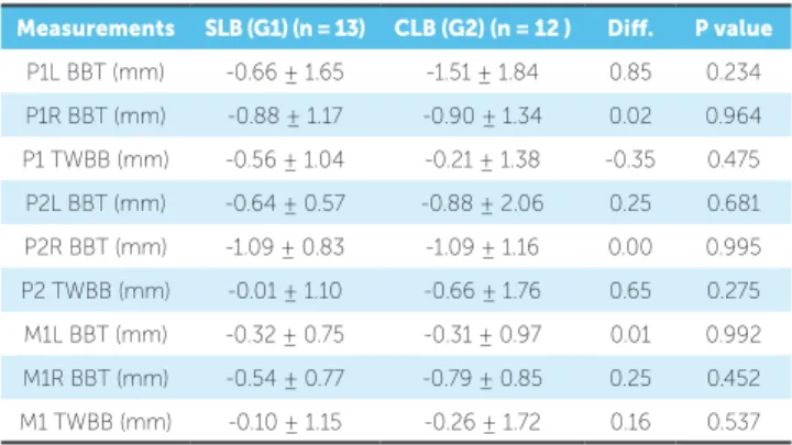
Table 4 - Means and standard deviation at the beginning of treatment (T1) and
7 months after treatment onset (T2) measured by CBCT and comparing CLB
and SLB groups.
M1 = first molar, P2 = second premolar and P1 = first premolar.
Measurements SLB (G1) (n = 13) CLB (G2) (n = 12 ) Dif. P value
P1L BBT (mm) -0.66 ± 1.65 -1.51 ± 1.84 0.85 0.234
P1R BBT (mm) -0.88 ± 1.17 -0.90 ± 1.34 0.02 0.964
P1 TWBB (mm) -0.56 ± 1.04 -0.21 ± 1.38 -0.35 0.475
P2L BBT (mm) -0.64 ± 0.57 -0.88 ± 2.06 0.25 0.681
P2R BBT (mm) -1.09 ± 0.83 -1.09 ± 1.16 0.00 0.995
P2 TWBB (mm) -0.01 ± 1.10 -0.66 ± 1.76 0.65 0.275
M1L BBT (mm) -0.32 ± 0.75 -0.31 ± 0.97 0.01 0.992
M1R BBT (mm) -0.54 ± 0.77 -0.79 ± 0.85 0.25 0.452
M1 TWBB (mm) -0.10 ± 1.15 -0.26 ± 1.72 0.16 0.537
Table 5 - Means and standard deviation at the beginning of treatment (T1) and
7 months after treatment onset (T2) measured in dental casts and comparing
CLB and SLB groups.
Measurements SLB (G1) (n = 13) CLB (G2) (n = 12 ) Dif. p value
4-4 width 1.27 ± 1.95 1.87 ± 2.30 -0.60 0.489
5-5 width 2.10 ± 1.00 1.75 ± 1.33 0.35 0.465
6-6 width 0.92 ± 0.88 0.46 ± 0.77 0.46 0.180
Table 6 - Pearson correlation coefficient between transversal width of buccal bone (TWBB) and dental expansion within the two bracket system groups.
M1 = first molar, P2 = second premolar and P1 = first premolar.
Measurements r P
P1 TWBB 0.15 0.467
P2 TWBB 0.28 0.176
M1 TWBB 0.09 0.676
premolar width, intersecond premolar width and in-termolar width were -0.6 mm (p = 0.489), 0.35 mm (p = 0.465) and 0.46 mm (p = 0.180), respectively.
Furthermore, no statistically significant associa-tion was found between transversal width of buccal bone (TWBB) and dental expansion (Table 6).
DISCUSSION
In this sample, patients were treated by different dentists, but in order to obtain more reliable results, measurements were made by only one previously calibrated examiner. The error of the method used to assess intra and interexaminer reliability proved to
be small. No significant differences were found be-tween measurements made by two operators at two different time points. Interexaminer analysis showed that errors ranged from 0.30 to 1.08 mm. This may have occurred due to high resolution images offering excellent view without overlapping structures.
A disadvantage of the CBCT method is its greater radiation dose in comparison to conventional radio-graphs (periapical and panoramic). However, CBCT is an invaluable tool in orthodontic research. Good to excellent reliability of CBCT scans used for detection of bone defects was demonstrated by Misch et al.23
Furthermore, when compared to bidimensional ra-diographs, CBCT showed great reliability and offered advantages when detecting and quantifying bone fis-sures and fenestrations, as well as periodontal defects in the buccal bone.24
Mandibular arch bone expansion studies with CBCT scans comparing SLB and CLB are rare in the literature. And few studies have assessed the maxillary arch response to SLB and CLB systems.19 Nonetheless,
some studies compared arch expansion on dental casts and on digitized models, which may ofer great
accura-cy.7,10,11 Claims have been made that SLB can result in
broader arch forms in comparison to CLB.4 Thus, this
study aimed at testing the null hypothesis that there are no signiicant diferences in the amount of expan-sion of the mandibular arch (dental and alveolar bone changes) during the irst 7 months of alignment and leveling when either SLB or CLB systems are used, as demonstrated by analysis on CBCT and dental casts.
According to Birnie,25 Damon divulged his theory
that by using SLB with low friction and light forces more stable biological results could be produced. Da-mon,4 based on empirical and anedotical evidence,
at-tributed advantages to self-ligating brackets, among which is the passive expansion of the arches. The Da-mon SLB system claims that post-treatment comput-ed tomography images show transverse arch devel-opment and normal alveolar bone on buccal surface. Low friction and low force are purported to be good to physiologically rebuild the alveolar bone.26
majority of measurements regarding BBT from T1 to T2 for both groups. There was signiicant diference for the following measurements, from T1 to T2, regarding BBT changes: CLB group — P1L (-1.51 mm, p = 0.016), P1R (-0.90 mm, p = 0.039), P2R (-1.09 mm, p = 0.007), M1R (-0.79 mm, p = 0.008); SLB group — P1R (-0.88 mm, p = 0.019), P2L (-0.64 mm, p = 0.002), P2R (-1.09 mm, p < 0.001), M1R (-0.54 mm, p = 0.025). However, no signiicant diferences were found between groups. Furthermore, no signiicant diferences from T1 to T2 were observed between and within groups for TWBB.
The results of the present study conirm indings in the literature showing similar behaviors for both brackets, particularly with regard to dental expansion assessed by means of dental casts. Mandibular arch alignment resulted in transverse expansion irrespec-tive of the appliance system used. Interirst premolar distances, measured on dental casts with a digital caliper in both groups, increased (SLB, 1.27 mm; CLB, 1.87 mm). This result is similar to those found by Fleming et al,7 with an increase of 0.85 mm and
1.17 mm for SLB and CLB, respectively. However, the change was not signiicantly diferent between the two bracket systems. Further corroborating these ind-ings, Vajaria el al11 also found expansion in interirst
premolar distances.As for intersecond premolar dis-tances, there was an increase of 2.10 mm for SLB and 1.75 mm for CLB; however, this increase was similar for both groups. Once again, the results yielded by the present study are similar to those obtained by Fleming et al7 (SLB= 1.43 mm, and CLB= 1.72 mm).
Never-theless, contrary to our indings, Vajaria et al11 found
a larger increase for the self-ligating group (4.35 mm in comparison to 2.6 mm for the conventional group). Regarding intermolar distances, there was an increase ranging from 1.4 to 2.4 mm for SLB, and from 0.43 to 1.85 mm for CLB.7,9,10,11,17,27,28 On the other hand, a
decrease in intermolar distance was observed in only one study in which cases were treated by means of pre-molar extractions.28 We found nonsigniicant increases
of mandibular irst intermolar width for both SLB and CLB groups, and there was no signiicant diference between the two bracket groups. The present study showed molar expansion of 0.92 mm and 0.46 mm for SLB and CLB, respectively. This result is in ac-cordance with the study by Vajaria et al.11
Nonethe-less, Pandis et al10,17 and Fleming et al7 found that SLB
expanded more than CLB in the molars region, and this diference was considered statistically signiicant.
When the Pearson correlation coefficient was as-sessed, we found that the alveolar buccal bone did not follow dental expansion. Therefore, the statements wherein self-ligating brackets produce physiological and passive movements of the arches were not con-firmed in this study, at least 7 months after orthodon-tic treatment onset. Regarding buccal bone changes, it seems that self-ligating appliances do not offer any advantages over the conventional bracket system. Thus, the null hypothesis of the present study was accepted; in other words, no significant differences were found between self-ligating and conventional brackets systems regarding mandibular buccal bone plate expansion or dentoalveolar expansion.
CONCLUSIONS
» There is no difference between patients treated with self-ligating brackets or conventional brackets, regarding mandibular dentoalveolar expansion.
» There is no diference between patients treated with self-ligating brackets or conventional brackets, re-garding buccal bone plate changes (mandibular buccal bone thickness and transversal width of buccal bone).
1. Stolzenberg J. The eiciency of the Russell attachment. Am J Orthod Oral Surg. 1946;32:572-82.
2. Berger J. Self-ligation in the year 2000. J Clin Orthod. 2000;34:74-81. 3. Harradine N. The history and development of self-ligating brackets. Semin
Orthod. 2008;14(1):5-18.
4. Damon DH. The Damon low-friction bracket: a biologically compatible straight-wire system. J Clin Orthod. 1998;32(11):670-80.
5. Fleming PS, DiBiase AT, Lee RT. Self-ligating appliances: evolution or revolution? J Clin Orthod. 2008;42:641-51.
6. Rinchuse DJ, Miles PG. Self-ligating brackets: present and future. Am J Orthod Dentofacial Orthop. 2007;132(2):216-22.
7. Fleming PS, DiBiase AT, Sarri G, Lee RT. Comparison of mandibular arch changes during alignment and leveling with 2 preadjusted edgewise appliances. Am J Orthod Dentofacial Orthop. 2009;136(3):340-7.
8. Fleming PS, Johal A. Self-ligating brackets in orthodontics. A systematic review. Angle Orthod. 2010;80(3):575-84.
9. Chen SS, Greenlee GM, Kim JE, Smith CL, Huang GJ. Systematic review of self-ligating brackets. Am J Orthod Dentofacial Orthop. 2010;137(6):726.e1-726.e18; discussion -7.
10. Pandis N, Polychronopoulou A, Eliades T. Self-ligating vs conventional brackets in the treatment of mandibular crowding: a prospective clinical trial of treatment duration and dental efects. Am J Orthod Dentofacial Orthop. 2007;132(2):208-15.
11. Vajaria R, Begole E, Kusnoto B, Galang MT, Obrez A. Evaluation of incisor position and dental transverse dimensional changes using the Damon system. Angle Orthod. 2011;81(4):647-52.
12. Tai K, Park JH, Mishima K, Shin JW. 3-dimensional cone-beam computed tomography analysis of transverse changes with Schwarz appliances on both jaws. Angle Orthod. 2011;81(4):670-7.
13. Yagci A, Veli I, Uysal T, Ucar FI, Ozer T, Enhos S. Dehiscence and fenestration in skeletal Class I, II, and III malocclusions assessed with cone-beam computed tomography. Angle Orthod. 2012;82(1):67-74.
14. Mah JK, Huang JC, Choo H. Practical applications of cone-beam computed tomography in orthodontics. J Am Dent Assoc. 2010;141 Suppl 3:7S-13S. 15. Sun Z, Smith T, Kortam S, Kim DG, Tee BC, Fields H. Efect of bone thickness on
alveolar bone-height measurements from cone-beam computed tomography images. Am J Orthod Dentofacial Orthop. 2011;139(2):e117-27.
REFERENCES
16. Timock AM, Cook V, McDonald T, Leo MC, Crowe J, Benninger BL, et al. Accuracy and reliability of buccal bone height and thickness measurements from cone-beam computed tomography imaging. Am J Orthod Dentofacial Orthop. 2011;140(5):734-44.
17. Pandis N, Polychronopoulou A, Makou M, Eliades T. Mandibular dental arch changes associated with treatment of crowding using self-ligating and conventional brackets. Eur J Orthod. 2010;32(3):248-53.
18. Tecco S, Tete S, Perillo L, Chimenti C, Festa F. Maxillary arch width changes during orthodontic treatment with ixed self-ligating and traditional straight-wire appliances. World J Orthod. 2009;10(4):290-4.
19. Cattaneo P, Treccani M, Carlsson K, Thorgeirsson T, Myrda A, Cevidanes L, et al. Transversal maxillary dento-alveolar changes in patients treated with active and passive self-ligating brackets: a randomized clinical trial using CBCT-scans and digital models. Orthod Craniofac Res. 2011;14(4):222-33.
20. Pandis N, Polychronopoulou A, Eliades T. Sample size estimation: an overview with applications to orthodontic clinical trial designs. Am J Orthod Dentofacial Orthop. 2011;140(4):e141-6.
21. Leite V, Conti AC, Navarro R, Almeida MR, Oltramari-Navarro P, Almeida R. Comparison of root resorption between self-ligating and conventional preadjusted brackets using cone beam computed tomography. Angle Orthod. 2012;82(6):1078-82.
22. Houston WJ. The analysis of errors in orthodontic measurements. Am J Orthod. 1983;83(5):382-90.
23. Misch KA, Yi ES, Sarment DP. Accuracy of cone beam computed tomography for periodontal defect measurements. J Periodontol. 2006;77(7):1261-6.
24. Mol A, Balasundaram A. In vitro cone beam computed tomography imaging of periodontal bone. Dentomaxillofac Radiol. 2008;37(6):319-24.
25. Birnie D. The Damon passive self-ligating appliance system. Semin Orthod. 2008;16:19-35.
26. Yu YL, Qian YF. The clinical implication of self-ligating brackets. Shanghai Kou Qiang Yi Xue. 2007;16:431-5.
27. Fleming PS, DiBiase AT, Lee RT. Randomized clinical trial of orthodontic treatment eiciency with self-ligating and conventional ixed orthodontic appliances. Am J Orthod Dentofacial Orthop. 2010;137(6):738-42.