Review Article
DOI: http://dx.doi.org/10.1590/2446-4740.03415*e-mail: [email protected]
Received: 09 November 2015 / Accepted: 18 May 2016
Artiicial motor control for electrically stimulated upper limbs of
plegic or paretic people
Elgison da Luz dos Santos, Manuela Cristina Gelain, Eddy Krueger, Guilherme Nunes Nogueira-Neto, Percy Nohama*
Abstract Introduction: Functional Electrical Stimulation (FES) is a technique used in the restoration and generation of movements performed by subjects with neuromuscular disorders such as spinal cord injury (SCI). The purpose of this article is to outline the state of the art and perspectives of the use of FES in artiicial motor control of the upper limbs in paretic or plegic people. Methods: The databases used in papers selection were Google Scholar and Capes’ Portals as well as proceedings of the Annual Conference of the International Functional Electrical Stimulation Society (IFESS). Results: Approximately 85% of the reviewed studies showed FES proile with pulse duration ranging from 1 to 300 μs and modulating (burst) frequency between 10 and 40 Hz. Regarding the type of electrodes, 88% of the studies employed transcutaneous electrodes. Conclusion: We concluded that FES with closed-loop feedback and feedforward are the most used and most viable systems for upper limbs motor control, because they perform self-corrections slowing neuromuscular adaptation, allowing different planes and more range of movement and sensory-motor integration. One of the dificulties found in neuroprosthesis systems are electrical wires attached to the user, becoming uninteresting in relation to aesthetics and break. The future perspectives lead to a trend to miniaturization of the stimulation equipment and the availability of wireless networks, which allow the attachment of modules to other components without physical contact, and will become more attractive for daily use.
Keywords: Functional electrical stimulation, Spinal cord injury, Upper limb, Rehabilitation.
Introduction
The gesture made so that a person opens a door involves efferent systems (leading information to the muscles) and afferent systems (carrying information to the nervous system), which process information at different hierarchical levels of the central nervous system (CNS) and peripheral nervous system. The integration of these systems allows the individual to have motor skills with quality and functionality. People with complete or incomplete spinal cord injury (SCI) suffered interruption or interference in these systems, what makes it dificult or impossible for them to control locomotion (Rodriguez and Clemente, 2008) below the level of lesion (Krueger-Beck et al., 2011). The complete high SCI affects the spinal cord at least at the height of the irst cervical vertebrae and causes quadriplegia, in which the individual loses functional movement of both lower limbs (LL) and upper limbs (UL), consequently, it presents secondary systemic alterations (Rodrigues and Herrera, 2004). When the SCI occurs in lower cervical level, in the region of the C5-C6 vertebrae, the movements of the shoulder and elbow joints can be maintained, but generally provokes lack of control of the intrinsic
and extrinsic muscles of the hand. This hampers or prevents holding of objects, feeding and writing, as well as other activities that require ine motor skills. Thus, regardless of the level of SCI, there is impairment in the individual’s and his/her family’s quality of life. Faced with this problem, mechanisms are studied to artiicially restore the functionality of members such as functional electrical stimulation (FES). FES applied to people with SCI aims to replace the interaction pathways that were discontinued (Nogueira et al., 2010).
Santos EL, Gelain MC, Krueger E, Nogueira-Neto GN, Nohama P
must also be considered for the correct application of FES in individuals with SCI, including the choice of electrodes (Nogueira et al., 2010; Petrofsky, 2004); the kind of pulses (monophasic or biphasic) and the existence or not of automatic feedback of the system (operating in closed or open-loop, respectively).
Within this scenario, this work aimed to describe and evaluate the current status of FES in the motor control of plegic or paretic upper limbs during the performance of functional movements, in order to glimpse their future prospects.
Methods
Papers were selected from the research bases of Google Scholar, periodicals of Capes Portal, and the Proceedings of the Annual Conference of the International Functional Electrical Stimulation Society. Search descriptors were: “upper limb or hand” and “functional electrical stimulation” and “spinal cord injury” and “neuronal plasticity or rehabilitation or control strategy”. Papers were included written in Portuguese or English, published between 1995-2016. After searching the databases, the abstracts were read and applied the exclusion criteria. The exclusion criteria involved duplicate papers and those addressing stimulation devices for other purposes.
Results
The total number of papers retrieved by the research was 88, written in English or in Portuguese: 39 from Google Scholar, 35 from Capes Portal, and 14 from Proceedings of the Annual Conference of the International Functional Electrical Stimulation Society. The exclusion criteria impacted more than the half of all papers (48): 26 duplicates, 15 papers addressing electrical stimulation for purposes such as analgesia, growth and tissue repair and muscle strengthening, as well as 7 papers describing animal studies. Therefore, the analysis was performed with 40 papers.
Table 1 lists the selected papers with the description of some of the stimulatory parameters, type and site of application of the electrodes and the functional activities performed found in some of the papers. It shows the summary of the main parameters found in 21 articles published between the years 1995 to 2016.
Discussion
There is a growing number of people who use FES to restore functions of the upper limbs. However, as the natural movements require coordination of different muscles and multiple joints in muscle groups
combinations, FES systems are still insuficient for certain activities. FES falls short of total independence to the individual with high complete SCI.
The results presented in Table 1 show that 85% of the studies used transcutaneous electrodes. The predominant use of this kind of electrodes, especially hydrogel (Imatz et al., 2013), is justiied by the ease of application, because it requires no electrode implant surgery. This reduces the risk of secondary complications, such as infections and/or rejections (Petrofsky, 2004). This electrodes category presents disadvantages by having reduced muscle selectivity and potential discomfort resulting from sensorial activation (Imatz et al., 2013) and, depending on the movement to be artiicially controlled, the low muscle selectivity may result in limitation in the number of available stimulator channels. It was noticed that artiicially evoked movements did not fail to happen when using this category of electrodes, and the desired movements were performed accurately. Moreover, the size of the electrodes used in FES for upper limbs must be taken into account because, in case of power application via different size electrodes (bipolar), the smaller electrode will have higher energy density and can cause strong local skin irritation (Imatz et al., 2013).
Regarding the principle of modulation of stimulatory pulses, the revised items, listed in Table 1, show pulse durations ranging from 0 to 300 μs, and burst frequencies (modulating signal) ranging from 10 to 40 Hz. Consequently, the values found in the consulted literature were different from the parameters described in Thrasher et al. (2006), that found durations between 100 μs and 500 μs and modulating frequencies from 20 to 100 Hz.
Data presented in Table 1 indicate that in 85% of the studies, feedback systems were used. In closed-loop systems, feedback happens automatically by the system itself, preventing sudden neuromuscular adaptation. In open or closed-loop systems it is important to notice the existence of controller modules. However, in open-loop systems, the input does not depend on the output, unlike closed-loop systems, whose output information is monitored by means of joint angle or neuromuscular force signals and, by means of these output information, an automatic change in some of the system’s parameters occurs. Furthermore, the open-loop neuroprostheses might have limited sensitivity on force and gripping control due to the lack of tactile feedback, as well as it is also observed in robotic prostheses developed in open-loop (Saunders and Vijayakumar, 2011).
Schearer et al. (2012) analyzed the use of FES in feedback systems in closed-loop with feedforward
Ar
tiicial mot
or c
on
tr
ol f
or upper limbs
Table 1. Stimulatory parameters found in the reviewed articles (1995-2016) for artiicial neuromuscular control and activities.
Year / Author Waveform Pulse width Intensity Burst Phase Loop Electrodes Placement of electrodes Performed activities
Q R S M Bi Cl Ol T I H W F E A Sh
Matsushita et al.
(1995) Ns Ns 0 to 250 V Ns X X X X X
Grasping cylindrical objects.
Mulcahey et al.
(1997) Ns
0 to 200 µs (changed after 8
weeks)
20 mA 12 a 16 Hz Ns X X X X X
Open, close and shake hands with control to assist during activities of daily living (ADLs). Prochazka et al.
(1997) X 200 µs 35 mA 30 Hz X X X X X
Open, close and shake hand.
Snoek et al.
(2000) Ns
10 µs a
500 µs 60 mA 18 Hz/36 Hz Ns X X X X
Assistance to hold keys, feeding and other.
Thorsen et al.
(2001) X 300 µs Ns Ns X X X X
Handle extension and thumb force to grasp objects.
Castro and Cliquet
(2001) X Ns Ns 20 Hz X X X X X X Lateral grip.
Peckham et al.
(2001) Ns Ns Ns Ns X X X X X
Open/close hand; grasp objects; assistance in ADLs.
Pfurtscheller et al.
(2003) X 300 µs Ns 16 Hz X X X X X Side handshake and grasp objects.
Alon and McBride
(2003) Ns 10 µs a500 µs 60 mA 18 Hz/36 Hz Ns X X X X
Hold keys and food, among other ADLs.
Schearer et al.
(2012) X 0 to 200 µs Ns 13 Hz X X X X X
Movements in three dimensions aiming to take a hand to mouth during feeding. Berends et al.
(2013) Ns 270 µs Ns 35 Hz X X X X X Hold objects and surface cleaning.
Westerveld et al.
(2013) Ns Ns Ns Ns Ns X X X X X
Open/close hand and thumb and grasp objects.
Štrbac et al.
(2013) Ns 250 µs 0-20 mA 40 Hz Ns X X X X Flexion and extension of the elbow.
Legend: Q: quadratic; R: rectangular; S: sinusoidal; M: monophasic; Bi: biphasic; Cl: closed loop; Ol: open loop; T: transcutaneous; I: implanted; Ha: Hand; W: wrist; F: forearm; E: elbow; A: arm; Sh: shoulder; Ns: nonspeciied.
201
Res
. Biomed
. E
ng
. 2016 J
San
tos EL, G
elain MC, Kr
ueger E
, N
ogueir
a-N
et
o GN, N
ohama P
Table 1. Continued...
Year / Author Waveform Pulse width Intensity Burst Phase Loop Electrodes Placement of electrodes Performed activities
Q R S M Bi Cl Ol T I H W F E A Sh
Koutsou et al.
(2013) Ns 250 µs motor threshold + 2 mA 30 Hz X X X X
Monitored movements: pronation / supination of the forearm/lexion/ extension of the wrist abduction/adduction/ extension/lexion of the ingers.
Imatz et al. (2013) Ns 250 µs Ns 40 Hz X X X X Handle extension.
Cologni et al.
(2013) Ns Ns Ns 20 Hz X X X X X X Grip and release objects.
Exell et al. (2013) X 0 to 300 µs 5 V 40 Hz X X X X
Activities with ine motor skills of the hand and ingers.
Kapadia et al.
(2013) X 250 µs 8-50 mA 40 Hz X X X X
Monitored movements: lexion / extension of the wrist abduction/ adduction/extension/ lexion/oppositors of the ingers – precision grip and power grasp.
Memberg et al.
(2014) Ns 225 µs 0-20 mA 12-16 Hz Ns X X X X X X X X
Eating with a fork and inger food, hand shaking, nose scratching, nose wiping with a tissue, face washing, and teeth brushing. Kitamura et al.
(2015) X 200 µs 0 -20 mA 50 Hz Ns Ns X X
Flexion and extension of the elbow. De Marchis et al.
(2016) Ns 500 µs 9 - 10 - 11 mA Ns X X X X
Movements of ingers and wrist.
Legend: Q: quadratic; R: rectangular; S: sinusoidal; M: monophasic; Bi: biphasic; Cl: closed loop; Ol: open loop; T: transcutaneous; I: implanted; Ha: Hand; W: wrist; F: forearm; E: elbow; A: arm; Sh: shoulder; Ns: nonspeciied.
202
Res
. Biomed
. E
ng
. 2016 J
Artiicial motor control for upper limbs
control. The feedforward control model is important to perform precise movements, because while the limb performs a functional movement, the system is already determining the next movement, anticipating the necessary musculoskeletal actions for the subsequent movement. The objective of Schearer et al.’s (2012) study was to allow a person with high SCI to be able to feed. The authors report that the studied model was viable and showed quality in the analyzed movement. This can be understood by the fact that the feedforward control requires less energy and can be modulated with low frequency. The integration of FES with robotic prostheses emphasizing rehabilitation through functional tasks (Westerveld et al., 2013) is viable for persons with motor sequelae resulting from SCI or Stroke.
The main motor activities found in the consulted literature involving FES or neuroprostheses were: (1) handgrip, (2) hold and release relatively cylindrical objects (cans, bottles, among others) and (3) lateral gripping for thin and smaller objects (keys, paper, pen drives). Thus, even if FES allows artiicial changes for the upper limb, it is possible to realize that the use of neuroprostheses is still limited, because there are people who are not beneited because they do not reach eligible criteria. On the other hand, individuals with SCI that preserve small motor responses might be beneited from the use of FES. This can be explained by the fact that when the individual has the ability to perform concurrent voluntary contraction to the FES, the inal motor response can be more effective and coordinate than when only FES application occurs (Joa et al., 2012).
The complete SCI with “A” rating by American Spinal Injury Association Impairment Scale (AIS A) not only affects the motor function, but also affects the sensory function. Thus, it would be necessary to control motor parameters simultaneously to sensory parameters in artificial rehabilitation systems. However, the provision of somatosensory feedback in myoelectric prosthesis is a goal to be achieved. It has been tried the electrical stimulation on the skin to evoke the tactile sense to restore the missing sensory information and was concluded that it is a possible method for restoring sensory information that is lacking in people with SCI (Dosen et al., 2013).
On the other hand, when people with SCI keep the sensitivity preserved (AIS B), the beneit of FES may be diminished, because these persons have low-tolerance to the unpleasant feeling usually provoked by the passage of the electric current. The discomfort felt by the person can limit the effectiveness of FES (Imatz et al., 2013). Some electrical parameters of the FES, as well as size and mode of activation of the electrodes, aim to
reduce the discomfort. Imatz et al. (2013) compared the sensation perceived by 15 individuals while applied two electrodes activation methods during stimulation in upper limbs. The studied methods were asynchronous (different electrodes were activated, sequentially) and synchronous (different electrodes were activated, simultaneously). The authors report that the synchronous stimulation resulted in greater discomfort than asynchronous one. Therefore, it is understood that it is necessary to know mechanisms that provide greater comfort to the patient during the treatment or even to develop a neuroprosthesis for daily use.
The irst neuroprostheses for upper limbs were developed in the 1960s using surface electrodes. The movements initially run consisted of opening and closing the hand (gripping). Since then, different types of electrodes were used (Nogueira et al., 2010). The search for new approaches with FES in upper limbs was justiied by the fact that quadriplegic people, with SCI in C5-C6 level, preserve the lower motor neuron segment in C7-C8 level, being able to restore some degree of gripping, allowing the performance of activities of daily living (ADLs), such as feeding and handling personal hygiene objects (Ragnarsson, 2008).
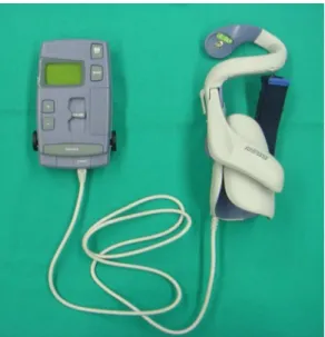
Some neuroprosthesis systems are recognized by the trade name. One of these systems is the Handmaster (Figure 1) recently recognized as HandNess H200 (Ragnarsson, 2008). These neuroprostheses consist of a manually adjustable handle with ive embedded electrodes to enable the forefinger and thumb. The user starts a pre-programmed stimulation of
Figure 1. Handness System. Right: the manually adjustable handle
with ive built-in electrodes to activate the index inger and thumb.
Santos EL, Gelain MC, Krueger E, Nogueira-Neto GN, Nohama P
opening and closing the hand by pressing a button on the system control unit. Snoek et al. (2000) and Peckham and Knutson (2005) describe handmaster assays in tetraplegic subjects. Shibata et al. (2013) also studied the HandNess H200 in tetraplegic subjects. The studies that used the neuroprostheses proposed by Snoek et al. (2000), Peckham and Knutson (2005) and Shibata et al. (2013) show that individuals were able to accomplish movement to some pre-programmed ADLs, increased grip strength, improved inger (Peckham and Knutson, 2005) movement and gained agility in the execution of the proposed activities.
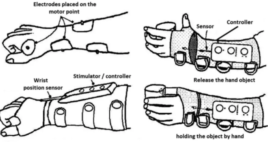
The Bionic Gloves system is a glove without ingers and an extension as forearm, as shown in Figure 2. The adhesive electrodes are positioned on the hand and forearm. The stimulation is controlled by movements which are detected by a sensor located in the handle. The extension of the handle, beyond a certain angle, causes the stimulation (gripping) of the hand and handle lexion triggers the opening of the hand. Individuals who have used this system while performing some ADL, for a period of a year or more, increased grip strength and reduced the time to perform such activities (Prochazka et al., 1997).
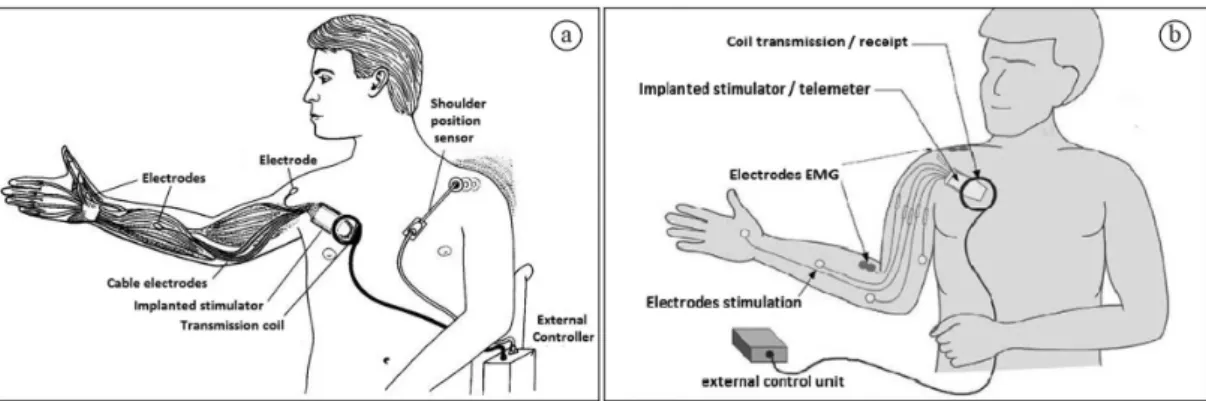
The Freehand and Cleveland systems consist of stimulators implanted in the user’s chest. In these systems, the electrodes are positioned on the motor points of the arm and hand muscles. Externally, there is a programming central of the stimulation parameters with radiofrequency broadcasting (Peckham and Knutson, 2005). The irst Freehand systems
(Figure 3a) contained a shoulder motion sensor controlling the degree of opening and closing the hand (Giszter, 2008; Peckham et al., 2001). Later, in the most advanced Freehand systems (Figure 3b), the function has been provided by additional stimulation channels and it was used for activating the intrinsic hand muscles, brachial triceps and forearm pronators. So, the shoulder (Peckham and Knutson, 2005) motion sensor was eliminated. The development of neuroprosthesis provided improvement in grip movement to subjects with complete SCI at the level C5-C6 (Giszter, 2008). There is another neuroprosthesis system known commercially as FESMate that uses up to 30 percutaneous electrodes placed on the upper limbs and artiicial movements are performed based on natural activation models, with speciic limits and various command (Peckham and Knutson, 2005) sources. It also showed better functional movements after its use.
Moss et al. (2011) studied the feasibility of using innervated muscles below the level of SCI as command sources for neuroprosthesis. They showed that the recording of electromyographic signal in muscles below the injury is possible even in complete SCI (AIS A) with no sign of movement. The authors justiied this fact by having observed the presence of intact axons even after complete SCI. So, a myoelectrically controlled functional electrical stimulator (MeCFES) was developed and applied to one hemiplegic and six quadriplegic individuals. Residual myoelectric signals of the radial extensor
Figure 2. Neuroprosthesis Bionic Gloves. This system is a ingerless glove and an extension as forearm. The four images illustrated indicate
the position of electrodes, sensor and controller in different perspectives. Images at the right focus the wrist sensor operation for gripping and releasing a cylindrical object. Adapted from Prochazka et al. (1997).
Artiicial motor control for upper limbs
muscle were used for controlling the stimulation of ist extension or lexion of the thumb. A screening test based on visual feedback, produced by force or movement in comparison with the track record of benchmarks, was used to quantify the control accuracy. Tracking performance of subjects with and without the MeCFES was compared. The results showed that ist extension improved in three of ive patients with SCI at C5 level. The lexion of the thumb was expanded in an individual with incomplete SCI (C3). The hemiplegic patient showed limited thumb control with MeCFES, but indicated the possibility of a transient effect. It was veriied that low natural residual force resulted in less accurate (Thorsen et al., 2001) movements.
Cologni et al. (2013) developed a complex system integrating interactive learning and neuromuscular activity, as shown in Figure 4. They used FES control based on electromyography (EMG) in order to allow the recovery of motor function of the upper limbs, especially of the ist, of patients who did not obtain great extension angle of this joint. The system was based on FES-EMG interactive learning with stimulation intensity dependent on the EMG values and also conducting a voluntary activity map as the detected EMG signal at a certain joint angular reference. Figure 4 shows that the EMG signal from the forearm musculature is captured by surface electrodes, and is ampliied. Later, having the EMG signal detected to trigger the stimuli, the signal is applied to a low-pass ilter, in order to minimize the effects of noise and artifacts. Afterwards, the signal is forwarded to a controller that will perform the necessary adjustments in the intensity of electrical stimulation according to the magnitude and frequency of the EMG signal received. After the controller make the necessary adjustments, the electrical stimulator delivers current via surface electrodes to the forearm
muscles. The results showed that after the interactive learning, the patient needed less voluntary effort to produce the same range of motion. Even after ceasing the electric current, the volunteer was still able to generate angular movement alone (Cologni et al., 2013), demonstrating that there was motor learning due to repetitive movement using FES.
De Marchis et al. (2016) have been developed a FES-EMG control system with an array of thirty electrodes divided into subgroups, which would be positioned at the proximal and distal part of the forearm. During the experimental protocol, a scanning technique was performed to determine the thresholds of individual stimulation pulses. The stimulation parameters were chosen based on the recording of the M-waves of muscles extensor digitorum communis (EDC), radial extensor carpi (ECR) and
Figure 3. Freehand system. These systems consist of stimulators implanted in the user’s chest. The electrodes are positioned on the motor
points of the arm and hand muscles. Two models were developed: (a) with motion sensor in contralateral shoulder (adapted from Peckham et al. (2001)) and (b) without shoulder sensor (adapted from Peckham and Knutson (2005)).
Figure 4. Block Diagram of a FES system controlled by electromyography.
The igure shows that the EMG signal from the forearm musculature is captured by surface electrodes, and is ampliied. Later, having the
EMG signal detected to trigger the stimuli, the signal is applied to a
low-pass ilter, in order to eliminate noise and artifacts. Afterwards,
the signal is forwarded to a controller that will perform the necessary adjustment in the intensity of electrical stimulation according to the magnitude and frequency of the EMG signal received. After the controller makes the necessary adjustments, the electrical stimulator delivers current through surface electrodes to the forearm muscles. Adapted from Cologni et al. (2013).
Santos EL, Gelain MC, Krueger E, Nogueira-Neto GN, Nohama P
the extensor carpi ulnaris muscles (ECU). These muscles effectively induce the movements on ingers and also the wrist extension and handling deviations. Therefore, it is possible to determine which muscle should be activated to maintain muscle recruitment with M-waves’ recordings. Moreover, the activation of electrodes subgroups happens automatically and very quickly, thus avoiding damage/delay in the movement cycle.
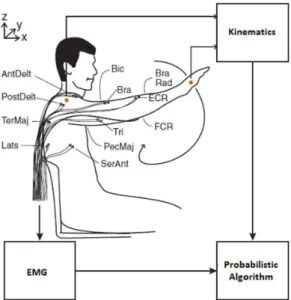
The probabilistic algorithm includes a probabilistic logic to generate a pseudorandom number in which the same input sequence does not necessarily lead to the same inal state. Johnson and Fuglevand (2009) evaluated the capacity of three different probabilistic models, being estimate of unsupervised Bayesian density, polynomial curve itting algorithm and dynamic neural network. The experimental model, shown in Figure 5, consisted in the acquisition of EMG signals of twelve arm muscles. Kinematic data obtained with markers (placed in the hand and shoulder) were recorded during the random movements in the sagittal plane. These data served as inputs for one of the three probabilistic algorithms that characterized the relation between electromyography and kinematics. With the determined probabilistic algorithm, a new set of kinematic data was serving as input for the other algorithms, aiming
to predict the patterns associated with the EMG of all twelve muscles. The analyses indicated that the dynamic neural network approach provided better estimates than the other two methods. It also tested the capacity of the neural network model to predict muscle activity associated with three-dimensional movements. The high correspondence between the muscle activity recorded during the three-dimensional movements conirms that this approach helps in the identiication of complex patterns of muscle activity required to control movements using FES. More recently, Koutsou et al. (2013) also obtained promising results using analysis of muscular selectivity of the forearm.
Castro and Cliquet (2001) applied the FES technique to perform certain functional movements of upper limbs together with an instrumented glove with force sensors which allowed to quantify the movement pattern artiicially exercised. Besides, it served as feedback data for restoring proprioception by the application of electrical stimulation, which allowed the evocation of coded tactile sensations related to artiicial movement. The sensorimotor integration was performed by means of the simultaneous application of both systems. It became possible the restoration of functional patterns of artiicially gripping and the recognition of the movement pattern exercised through evoked sensations.
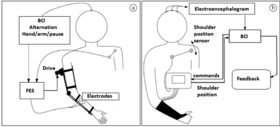
Hybrid neuroprostheses constituted by a combination of FES and its control components with brain-computer interface (BCI) are being studied (Martin Rohm et al., 2013; Pfurtscheller et al., 2003). Research on the use of BCI for neuroprosthesis control has seen tremendous progress in recent years. However, such systems are not ready for the user to use them independently at his/her home. To establish BCI as a neuroprosthesis control for day to day users, some gaps need to be illed up, mainly to delimit the usability difference, reliability, movements in some degree of rotation (Rupp et al., 2015). It was developed the FES controller for prehension restoration in a quadriplegic person through a BCI system using electroencephalographic (EEG) signals. During the experiment, the user was able to grip cylindrical (Pfurtscheller et al., 2003) objects. In a more recent research, it was used a hybrid neuroprosthesis based on FES and BCI (Martin Rohm et al., 2013) control, as shown in Figure 6a and b. The project aimed to control hand and elbow movements. For its functioning, the neuroprosthesis used analog signals from a motion sensor placed on the user contralateral shoulder. This way, according to predetermined coniguration, the user could control the degree of lexion and extension of the elbow and hand opening, performing the elevation, pronation or retraction of the contralateral
Figure 5. Experimental installation diagram of probabilistic models. This model consisted in the acquisition of EMG (12 muscles) and
kinematic data (shoulder – wrist). These data served as inputs to
characterize probabilistic algorithms. With the determined probabilistic algorithm, a new set of kinematic data was serving as input for the other algorithms, aiming to predict the patterns associated with the EMG (Lats: latissimus dorsi; PecMaj: pectoralis major; TerMaj: teres major; Serant: serratus anterior; AntDelt: anterior deltoid; PostDelt: posterior deltoid; Tri: triceps; Bic: biceps; Bra: brachial, BraRad: Brachioradialis; ECR: extensor carpi radial; FCR: lexor carpi radial). Adapted from Johnson and Fuglevand (2009).
Artiicial motor control for upper limbs
shoulder. It was necessary to adjust the pulse width and the current intensity to perform such movements precisely. The analog signal for controlling the hand (opening or closing) and arm (lexion or extension) movements or their cessation was determined by a BCI signal, that is, by means of signals obtained from EEG. In Figure 6a the electrodes control the arm and hand and in Figure 6b only the hand is controlled by the electric current. The results showed that with this neuroprosthesis quadriplegic people could perform pre-established ADLs that they would not perform otherwise (Martin Rohm et al., 2013).
Hallewell et al. (2013) developed a system named Go-Sail, which was composed of eight different interactive components for controlling the upper limb. Go-Sail adjusts the movement through a feedback algorithm’s controller for the electrical stimulation in three muscle groups: anterior deltoid, triceps brachii and extensors of the ingers and ist. In their system, Hallewell et al. (2013) developed a stop button which interrupts the stimulation when pressed. An electrogoniometer and the Microsoft Kinect are used to measure the range of motion of the shoulder, elbow and ist articulations. Data from these sensors are stored and processed through speciic control algorithms. There is an armrest for use during individual activities or for the needs of each user and inally an output monitor, which makes possible to the therapist to analyze graphically the information about electrical stimuli delivered to the muscles. The potential user of this technology performs functional training of closing a drawer, triggering a switch, pressing a button and holding an
object. Although still experimental, their preliminary results showed promising (Hallewell et al., 2013).
Cornwell et al. (2012) have developed two tests to evaluate paraplegics, the irst one was for ADLs, looking for assessing what paraplegics could do as grooming, feeding, dressing, bathing, toileting, general mobility and social relations. The second test was based on the ADLs, about which especiic arm movements were necessary e.g. in order to wash one’s face. The movements which required more training were touching one’s face, drinking from a mug with a straw, eating with ingers from a plate, retrieving an object from a countertop and pressing an elevator button. They used a neuroprotesis with electrodes implanted in the upper limb muscles to create and assist on movements by FES.
Kapadia et al. (2013) used the Compex Motion Stimulator (FES) to restore eight tetraplegics in the subacute phase. Two groups were formed: occupational therapy exercises (control) and FES therapy (intervention). They applied Toronto Rehabilitation Institute–Hand Function Test (TRI-HFT), Graded Redeined Assessment of Strength, Sensibility and Prehension (GRASSP), Functional Independence Measure (FIM) and Spinal Cord Independence Measure (SCIM), to assess the grasp strength and precision grip, before and after training (39 sessions) and six months following up the irst outcome. FES therapy has been tested as a resource to assist and improve functional movements when compared to occupational therapy.
The case study of Memberg et al. (2014) was a neuroprosthesis stimulator implanted in the shoulder peripheral nerves that they were connected to upper limbs muscles, with 24 channels, 4 myoelectric records
Figure 6. Neuroprosthesis hybrid coniguration scheme (use of FES controlled by a BCI) for users with restricted elbow and hand function
(a) and only on hand (b). These neuroprosthesis used analog signals from a motion sensor placed on the user contralateral shoulder. Adapted from Martin Rohm et al. (2013).
Santos EL, Gelain MC, Krueger E, Nogueira-Neto GN, Nohama P
(EMG), as seen in Figure 7. The neuroprosthesis following a mathematical description of limb movement, allowed many different control strategies i.e. during ADLs as eating with a fork and inger food, scratching nose, wipping nose with tissue, washing face, brushing teeth and shaking hands.
Even more recently, in another study with artiicial control by means of FES, Kitamura et al. (2015) showed a FES system using feedback as a bilateral control. The bilateral control can be understood as a form of feedback system based on information from the contralateral limb, for example. The authors used in their studies, self-adhesive electrodes, positioned on the biceps and triceps brachial muscles. The experimental research protocol of Kitamura et al. (2015) included ive healthy male subjects. The aim of the study was to perform elbow lexion and extension with the use of FES, measuring the degree of movement using encoders and proportional-integral-derivative controller, which was implemented to act as a position controller. The results of experimental tests have shown that the proposed method, the bilateral control, allowed movement of the joint and showed good performance in practical viewpoint. Moreover, the authors presented some limitations, as in some volunteers the elbow
movement could not be successfully controlled. This could be due to interference that the electric current occasioned between the stimulated muscles, for example, the electric current used for stimulating the biceps, interfered with the triceps muscle. Another limitation may be related to the ability to affect the sensory functions. However, the authors report that in further studies with the proposed methodology, they will seek to reduce error levels and to develop a system with varying degrees of freedom.
Conclusion and future perspectives
The application of FES provides momentary artiicial movements and, in long term, promotes muscle strengthening and stimulates neuroplasticity, being the best alternative to generate or complement functional upper limb movements in people who have lost or decreased function. The retrieved papers claimed their FES systems to be effective for producing artiicial plegic or paretic upper limb movements. Artiicial movements performed with FES observed in the reviewed papers involved lexion, ist extension, pronation and supination of the forearm; hand grip with or without holding objects, such as writing, cleaningFigure 7. Model of neuroprosthesis with the stimulator implanted near to the shoulder peripheral nerves where they were connected with
upper limbs muscles, with 24 channels of stimulation and 4 myoelectric records of electromyography. Adapted from Memberg et al. (2014).
Artiicial motor control for upper limbs
or activation of keys. The FES parameters found to perform these movements vary from 0 to 300 μs for pulse duration and modulating frequencies between 10 and 40 Hz. The surface electrodes are the most used even when not providing full muscle selectivity in this category. Closed-loop systems have better motor control conditions for upper limbs, because they allow self-correction when needed to avoid torque loss or a decrease in range of motion by delaying the neuromuscular adaptation.
FES does not enable yet the complete independence to an individual without movement of the upper limbs. In addition, the interface of systems in the evaluated studies has many connections and electrical wiring, making dificult the movements themselves and the transportation and it causes confusion to the user and his/her assistants. Because the available technological innovations are becoming more advanced and with the universal tendency to miniaturize electronic devices, the perspective for the near future is that electrical stimulation microsystems or nanosystems are also developed, where the devices connected to the user become almost imperceptible and not lose or reduce their effectiveness. Allied to miniaturization, there are also the wireless networks that allow attaching the module to the electrodes without physical contact, reducing the size of the equipments connected to the user. However, the electromagnetic coupling must be tested on the new devices, in view of the required reliability and its redundancy to faults. Therefore, given the inconsistencies observed in the described systems, arising from differences in the types of protocols and adopted control strategies, inter-subjective differences between users, and the dificulties of placing the implants, implantable microsystems are not yet widespread. Therefore, nowadays, external systems are still the most used and found.
References
Alon G, McBride K. Persons with C5 or C6 tetraplegia achieve selected functional gains using a neuroprosthesis. Archives of Physical Medicine and Rehabilitation. 2003;
84(1):119-24. http://dx.doi.org/10.1053/apmr.2003.50073.
PMid:12589632.
Berends HI, Boer JM, Renzenbrink GJ, Nijlant JMM, Jannink MJA, Prange GB. Clinical pilot study into the effect of multi-channel EMG-triggered electrical stimulation on arm function and dexterity after stroke. In: 18th Annual Conference of the International Functional Electrical Stimulation Society (IFESS); 2013; Belgrade, Serbia. Enschede: University of Twente; 2013.
Castro MCF, Cliquet A Jr. Neuromuscular electrical stimulation and electron-tactile stimulation in rehabilitation of artificial prehension and proprioception in tetraplegic patients. Acta Ortopedica Brasileira. 2001; 9(3):19-28.
Cologni AL, Seel T, Madaschi MG, Previdi F, Werner C, Schauer T. Automatic adjustment of electromyography-based FES control. In: 18th Annual Conference of the International Functional Electrical Stimulation Society (IFESS); 2013; Belgrade, Serbia. Enschede: University of Twente; 2013. Cornwell AS, Liao JY, Bryden AM, Kirsch RF. Standard task set for evaluating rehabilitation interventions for individuals with arm paralysis. Journal of Rehabilitation
Research and Development. 2012; 49(3):395-403. http://
dx.doi.org/10.1682/JRRD.2011.03.0040. PMid:22773199.
De Marchis C, Monteiro T, Simon-Martinez C, Conforto S, Gharabaghi A. Multi-contact functional electrical stimulation for hand opening: electrophysiologically driven identification of the optimal stimulation site. Journal of Neuroengineering and Rehabilitation. 2016; 13(22):1. http://dx.doi.org/10.1186/
s12984-016-0129-6. PMid:26955873.
Dosen S, Krajoski G, Đozić DJ, Farina D, Jorgovanović N. Closed-Loop control of dynamic Systems using electrotactile feedback. In: 18th Annual Conference of the International Functional Electrical Stimulation Society (IFESS); 2013; Belgrade, Serbia. Enschede: University of Twente; 2013. Exell T, Freeman C, Meadmore K, Hughes A-M, Hallewell E, Burridge J. Stimulation of hand postures using an electrode array and iterative learning control. In: 18th Annual Conference of the International Functional Electrical Stimulation Society (IFESS); 2013; Belgrade, Serbia. Enschede: University of Twente; 2013.
Giszter SF. Spinal cord injury: Present and future therapeutic devices and prostheses. Neurotherapeutics; the Journal of the American Society for Experimental NeuroTherapeutics. 2008; 5(1):147-62. http://dx.doi.org/10.1016/j.nurt.2007.10.062. PMid:18164494.
Hallewell E, Exell T, Meadmore K, Freeman C, Kutlu M, Hughes A-M, Burridge J. Goal-orientated functional rehabilitation using electrical stimulation and iterative learning control for motor recovery in the upper extremity post-stroke. In: 18th Annual Conference of the International Functional Electrical Stimulation Society (IFESS); 2013; Belgrade, Serbia. Enschede: University of Twente; 2013. Imatz E, Hoffmann U, Veneman J, Malešević N, Keller T. Stimulation discomfort comparison of asynchronous and synchronous methods with multi-field surface electrodes. In: 18th Annual Conference of the International Functional Electrical Stimulation Society (IFESS); 2013; Belgrade, Serbia. Enschede: University of Twente; 2013.
Joa K-L, Han Y-H, Mun C-W, Son B-K, Lee C-H, Shin Y-B, Ko H-Y, Shin Y-I. Evaluation of the brain activation induced by functional electrical stimulation and voluntary contraction using functional magnetic resonance imaging. Journal of Neuroengineering and Rehabilitation. 2012; 9(1):48.
http://dx.doi.org/10.1186/1743-0003-9-48. PMid:22828165.
Johnson LA, Fuglevand AJ. Evaluation of probabilistic methods to predict muscle activity: Implications for neuroprosthetics. Journal of Neural Engineering. 2009; 6(5):055008. http://
dx.doi.org/10.1088/1741-2560/6/5/055008. PMid:19721180.
Kapadia N, Zivanovic V, Popovic MR. Restoring voluntary grasping function in individuals with incomplete chronic
Santos EL, Gelain MC, Krueger E, Nogueira-Neto GN, Nohama P
spinal cord injury: pilot study. Topics in Spinal Cord Injury Rehabilitation. 2013; 19(4):279-87. http://dx.doi.org/10.1310/
sci1904-279. PMid:24244093.
Kitamura T, Sakaino S, Tsuji T. Bilateral control using functional electrical stimulation. In: Industrial Electronics Society IECON 2015-41st Annual Conference of the IEEE; 2015; Yokohama. EUA: IEE; 2015. p. 002336-41. Koutsou AD, Rocon E, Brunetti F, Moreno JC, Pons JL. A novel method for the analysis of forearm muscle activation by selective sFES. In: 18th Annual Conference of the International Functional Electrical Stimulation Society (IFESS); 2013; Belgrade, Serbia. Enschede: University of Twente; 2013.
Krueger E, Scheeren EM, Nogueira-Neto GN, Neves EB, Button VLSN, Nohama P. Button VLdSN, Nohama P. Relationship between peak and mean amplitudes of the stimulating output voltage for functional control of the knee by spinal cord patients and healthy volunteers. Revista Brasileira de Engenharia Biomédica. 2013; 29(2):144-52. http://dx.doi.org/10.4322/rbeb.2013.013.
Krueger-BeckE, ScheerenEM, Nogueira-NetoGN, Button VLSN, NohamaP. Efeitos da estimulação elétrica funcional no controle neuromuscular artificial.Revista Neurociências. 2011; 19(3):530-41.
Martin Rohm MS, Rupp R, Kreilinger A, Müller-Putz G. Hybrid Brain-Computer Interfaces for control of neuroprosthetic systems for restoration of upper limb functions in high spinal cord injured individuals. In: 18th Annual Conference of the International Functional Electrical Stimulation Society (IFESS); 2013; Belgrade, Serbia. Enschede: University of Twente; 2013.
Matsushita N, Handa Y, Ichie M, Hoshimiya N. Electromyogram analysis and electrical stimulation control of paralysed wrist and hand. Journal of Electromyography and Kinesiology. 1995; 5(2):117-28.
http://dx.doi.org/10.1016/1050-6411(95)00001-G. PMid:20719643.
Memberg WD, Polasek KH, Hart RL, Bryden AM, Kilgore KL, Nemunaitis GA, Hoyen HA, Keith MW, Kirsch RF. Implanted neuroprosthesis for restoring arm and hand function in people with high level tetraplegia. Archives of Physical Medicine and Rehabilitation. 2014; 95(6):1201e1-11e1. http://
dx.doi.org/10.1016/j.apmr.2014.01.028. PMid:24561055.
Moss CW, Kilgore KL, Peckham PH. A novel command signal for motor neuroprosthetic control. Neurorehabilitation
and Neural Repair. 2011; 25(9):847-54. http://dx.doi.
org/10.1177/1545968311410067. PMid:21693772.
Mulcahey M, Betz RR, Smith BT, Weiss AA, Davis SE. Implanted functional electrical stimulation hand system in adolescents with spinal injuries: an evaluation. Archives of Physical Medicine and Rehabilitation. 1997; 78(6):597-607. http://dx.doi.org/10.1016/S0003-9993(97)90425-1. PMid:9196467.
NogueiraGNNo, ManffraEF, NohamaP, ButtonVLSN. Sistemas implantáveis de estimulação elétrica funcional para controle artificial de movimentos funcionais.Revista Brasileira de Engenharia Biomédica. 2010; 26(2):121-42.
http://dx.doi.org/10.4322/rbeb.2012.085.
Peckham PH, Keith MW, Kilgore KL, Grill JH, Wuolle KS, Thrope GB, Gorman P, Hobby J, Mulcahey M, Carroll S, Hentz VR, Wiegner A. Efficacy of an implanted neuroprosthesis for restoring hand grasp in tetraplegia: a multicenter study. Archives of Physical Medicine and Rehabilitation. 2001;
82(10):1380-8. http://dx.doi.org/10.1053/apmr.2001.25910.
PMid:11588741.
Peckham PH, Knutson JS. Functional electrical stimulation for neuromuscular applications. Annual Review of Biomedical Engineering. 2005; 7(1):327-60. http://dx.doi.org/10.1146/
annurev.bioeng.6.040803.140103. PMid:16004574.
Petrofsky JS. Electrical stimulation: neurophysiological basis and application. Basic and Applied Myology. 2004; 14(4):205-13.
Pfurtscheller G, Müller GR, Pfurtscheller J, Gerner HJ, Rupp R. ‘Thought’–control of functional electrical stimulation to restore hand grasp in a patient with tetraplegia. Neuroscience Letters. 2003; 351(1):33-6.
http://dx.doi.org/10.1016/S0304-3940(03)00947-9. PMid:14550907.
Prochazka A, Gauthier M, Wieler M, Kenwell Z. The bionic glove: an electrical stimulator garment that provides controlled grasp and hand opening in quadriplegia. Archives of Physical Medicine and Rehabilitation. 1997;
78(6):608-14. http://dx.doi.org/10.1016/S0003-9993(97)90426-3.
PMid:9196468.
Ragnarsson K. Functional electrical stimulation after spinal cord injury: current use, therapeutic effects and future directions. Spinal Cord. 2008; 46(4):255-74. http://dx.doi. org/10.1038/sj.sc.3102091. PMid:17846639.
RodriguesD, Herrera G. Recursos fisioterapêuticos na prevenção da perda da densidade mineral óssea com lesão medular.Acta Ortopedica Brasileira. 2004; 12(3):183-8. http://dx.doi.org/10.1590/S1413-78522004000300008. RodriguezMR, ClementeFAR. Avaliação das disfunções do controle motor.Universitas Ciências da Saúde.2008; 2(2):259-67.
RuppR, RohmM, SchneidersM, KreilingerA, Muller-PutzGR. Functional rehabilitation of the paralyzed upper extremity after spinal cord injury by noninvasive hybrid neuroprostheses. Proceedings of the IEEE. 2015;
103(6):954-68. http://dx.doi.org/10.1109/JPROC.2015.2395253.
Saunders I, Vijayakumar S. The role of feed-forward and feedback processes for closed-loop prosthesis control. Journal of Neuroengineering and Rehabilitation. 2011; 8(60):60-72. http://dx.doi.org/10.1186/1743-0003-8-60. PMid:22032545.
Schearer EM, Liao Y-W, Perreault EJ, Tresch MC, Memberg WD, Kirsch RF, Lynch KM. System identification for 3D force control of a human arm neuroprosthesis using functional electrical stimulation. In: 2012 IEEE International Conference on Robotics and Automation (ICRA); 2012; Saint Paul, MN. EUA: IEEE; 2012. p. 3698-705. Shibata N, Matsunaga T, Sasaki K, Kudo D, Okudera Y, Sato M, Chida S, Hatakeyama K, Watanabe M, Shimada Y. Improved function of the upper extremity in persons with cervical spine disorders by therapeutic electrical stimulation. In: 18th Annual Conference of the International Functional
Artiicial motor control for upper limbs
Electrical Stimulation Society (IFESS); 2013; Belgrade, Serbia. Enschede: University of Twente; 2013.
Snoek GJ, IJzerman MJ, in ’t Groen F A CG, Stoffers TS, Zilvold G. Use of the NESS handmaster to restore handfunction in tetraplegia: clinical experiences in ten
patients. Spinal Cord. 2000; 38(4):244-9. http://dx.doi.
org/10.1038/sj.sc.3100980. PMid:10822395.
Štrbac MD, Malešević NM, Čobeljić R, Schwirtlich L. Feedback control of the forearm movement of tetraplegic patient based on Microsoft Kinect and multi-pad electrodes. In: 18th Annual Conference of the International Functional Electrical Stimulation Society (IFESS); 2013; Belgrade, Serbia. Enschede: University of Twente; 2013.
Thorsen R, Spadone R, Ferrarin M. A pilot study of myoelectrically controlled FES of upper extremity. IEEE Transactions on Neural Systems and Rehabilitation Engineering. 2001; 9(2):161-8. http://dx.doi.org/10.1109/7333.928576. PMid:11474969.
Thrasher T, Flett H, Popovic M. Gait training regimen for incomplete spinal cord injury using functional electrical stimulation. Spinal Cord. 2006; 44(6):357-61. http://dx.doi.
org/10.1038/sj.sc.3101864. PMid:16249784.
Westerveld AJ, Kuck A, Schouten AC, Veltink PH, Van der Kooij H. Passive reach and grasp with functional electrical stimulation and robotic arm support. Spain: Bridging Mind and Body; 2013.
Authors
Elgison da Luz dos Santos1, Manuela Cristina Gelain1, Eddy Krueger1,2, Guilherme Nunes Nogueira-Neto3,
Percy Nohama1,3*
1 Biomedical Engineering at the Graduate Program in Electrical and Computer Engineering – CPGEI, Universidade
Tecnológica Federal do Paraná – UTFPR, Av. Sete de Setembro, 3165, CEP 80230-901, Curitiba, PR, Brazil.
2 Anatomy Department, Universidade Estadual de Londrina – UEL, Londrina, PR, Brazil.
3 Pontifícia Universidade Católica do Paraná – PUCPR, Curitiba, PR, Brazil.