DOI: http://dx.doi.org/10.1590/2446-4740.04515
*e-mail: [email protected]
Reiceved: 10 November 2015 / Accepted: 02 July 2016
Low-intensity laser therapy improves tetanic contractions in mouse
anterior tibialis muscle injected with
Bothrops jararaca
snake venom
Vania Maria de Araújo Giaretta, Luiz Prudêncio Santos, Ana Maria Barbosa, Stephen Hyslop, Alexandre Pinto Corrado, Milene Sanches Galhardo, Renata Amadei Nicolau*, José Carlos Cogo
Abstract Introduction: Envenomation by Bothrops snakes can produce local pain, edema, hemorrhage and myonecrosis. However, standard antivenom therapy is generally ineffective in neutralizing these effects so that alternative methods of treatment have been investigated. In experimental animals, low-level laser therapy (LLLT) attenuates the local effects of Bothrops venoms, but the beneits of LLLT on muscle function after envenomation are unclear. In this study, we examined the inluence of LLLT on the contractile activity of mouse skeletal muscle injected with venom from Bothrops jararaca, the principal cause of snakebite in southeastern Brazil. Methods: Twenty‑seven male mice were used. Mice were injected with venom (40 μg in 50 μl) in the right anterior tibialis muscle, after which the muscle tendon was exposed, connected to an isometric transducer and subjected to a resting tension of 1 g. A bipolar electrode was attached to the tibial nerve for electrical stimulation. The mice were randomly allocated to ive groups: A – Control (n = 3), B – Venom 3 h (n = 6), C – Venom 9 h (n = 6), D – Venom + Laser 3 h (n = 6), E – Venom + Laser 9 h (n = 6). Results: The two groups that received LLLT post-venom showed improved muscle contraction and contracture in relation to muscle treated with venom alone. Conclusion: These results indicate that LLLT can improve muscle function after damage induced by B. jararaca venom.
Keywords: Bothrops jararaca, Low-level laser therapy, Muscle activity, Muscle fatigue, Snake venom, Tetanus.
Introduction
Bothrops snakes account for 90% of venomous
snakebites in Brazil, with Bothrops jararaca being responsible for most of these cases, especially in southeastern Brazil (Brasil, 2011; França and
Málaque, 2009; Leobas et al., 2016). Envenomation by Bothrops species can result in local effects (pain,
edema, inlammation, hemorrhage and necrosis) and
systemic effects (coagulopathy, systemic hemorrhage, circulatory shock and renal damage) (França and
Málaque, 2009; Warrell, 2004). In severe cases of envenoming, myonecrosis mediated predominantly by basic myotoxic phospholipases A2 (PLA2) in these venoms (Gutiérrez and Ownby, 2003; Montecucco et al., 2008) can result in important loss of muscle mass, with permanent damage to the bitten limb and loss of function. Part of this myotoxicity is attributable to ischemia caused by impaired circulation as a result of microvascular damage (Gutiérrez and Ownby, 2003;
Queiróz et al., 1984).
The most effective treatment for envenomation by Bothrops species is antivenom therapy. However, despite its effectiveness in neutralizing systemic effects such as coagulopathy (França and Málaque, 2009;
Warrell, 2004), antivenom is generally ineffective in neutralizing local reactions (Barbosa et al., 2009; Guimarães-Souza et al., 2011; Nadur-Andrade et
al., 2012; Pereira et al., 2009; Picolo et al., 2002). Indeed, a major problem in the treatment of Bothrops
venom-induced myonecrosis is the rapid onset of this damage (within 15 min of venom injection) that makes treatment virtually impossible (Gutiérrez et al., 2007). Consequently, various alternatives have been investigated to reduce the local effects of myotoxicity caused by bothropic venoms, including the use of medicinal plants (Cavalcante et al., 2007; Collaço et al., 2012a; 2012b; Ferraz et al., 2012; 2014; Pereira et al., 2012; Tribuiani et al., 2014) and substances such as heparin and polyanions (Lomonte et al., 1994; Melo et al.,
1993). In addition, low-intensity laser therapy (LLLT) has been investigated as a potential treatment for experimental envenomation (Barbosa et al., 2008;
2009; 2010; Doin‑Silva et al., 2009; Dourado et al.,
2003; Nadur-Andrade et al., 2012).
The effects of LLLT on biological tissues are very broad and induce trophic-regenerative,
anti‑inlammatory and analgesic actions, as well as
enhanced local microcirculation, as demonstrated by studies in vitro and in vivo (Barbosa-Souza et al., 2011; Oliveira et al., 2013). This beneicial effect on the microcirculation is related to an indirect action of the laser on precapillary sphincters through the release of chemical mediators that maintain a functional local peripheral microcirculation (Maier et al., 1990; Miró
et al, 1984). In spite of various studies have examined
the potentially beneicial effects of LLLT in animal models of pain, edema, local inlammation, hemorrhage
and myonecrosis (Medeiros et al., 2010; Oliveira et al.,
2013), to date there has been no assessment of the
inluence of LLLT on the contractility of venom‑injected
skeletal muscle. In this study, we examined the effect of LLLT on the function of mouse tibial muscle injected with B. jararaca venom.
Methodology
Twenty-seven male Swiss mice (18-22 g), obtained from Animais de Laboratório (Anilab, Paulínia, SP), were housed six/cage in the Laboratory of Physiology and Pharmacodynamics of the Institute for Research
and Development at the Universidade do Vale do Paraíba (UNIVAP). The mice were maintained on a
12 h light/dark cycle at 22-26 °C, with free access to food and water. Prior to use, the mice were randomly
allocated to ive groups: A – Control (n = 3), B – Venom 3 h (n = 6), C – Venom 9 h (n = 6), D – Venom + Laser 3 h (n = 6) and E – Venom + Laser 9 h (n = 6). The animal
experiments approved by the institutional Committee
for Ethics in Animal Use at UNIVAP (protocol no. CEUA/2012/A01). The experiments were done within
the ethical guidelines of the Brazilian Society for Laboratory Animal Science (SBCAL).
Venom was extracted from adult specimens of
B. jararaca maintained at the serpentarium at UNIVAP.
The venom was extracted manually and stored at
–80 °C until lyophilized. Storage was subsequently
at 4 °C until use.
For the experiments, the right anterior tibialis muscle was used, as described by Giaretta et al. (2015). Mice were injected with B. jararaca venom
(40 µg in 50 µl of 0.9% saline); this dose of venom
was chosen based on previous publications by our group (Barbosa et al., 2010; Dourado et al., 2011). The tibialis muscle of control mice was injected with
an equal volume of 0.9% saline. Mice treated with
LLLT received irradiation (InGaAlP laser equipment, Twin Flex Evolution, MMOptics) 30 min after venom injection, operated in direct contact mode (parameters described in Table 1).
At 3 h and 9 h post‑injection, the mice were anesthetized (xylazine – 20 mg/kg, i.p.,
ketamine – 100 mg/kg, i.p., atropine – 0.25 mg/kg, i.p., and diazepam – 5 mg/kg, i.p.), ixed on a surgical table
and the tendon of the right anterior tibialis muscle and the tibial nerve were exposed. In the region of insertion close to the metatarsal plantar region, the muscle tendon was connected to an isometric transducer and the nerve was connected to a bipolar electrode.
The muscle was subjected to a constant tension of 1 g and stimulated indirectly by individual pulses from a Grass Technologies model S48 square pulse stimulator. Pulses (4-8 mV, 0.2 Hz, 2 ms) were applied
for 3 min to determine the minimum and maximum
voltages needed to induce muscle contraction with
recruitment of all ibers (Doin‑Silva et al., 2009; Lopes-Martins et al., 2006). Tetanic stimuli were applied by increasing the frequency to 50 Hz for 10 s and the tetanic response was recorded. Six tetanic
stimuli were applied at 3 min intervals. During these
intervals, muscle intactness and normal contractility were assessed using the stimulus parameters described above (4-8 mV, 0.2 Hz, 2 ms). The tetanic contractions were recorded via an isometric force transducer
(model 7003, Ugo Basile) coupled to a Gemini
7070 lat‑bed recorder (Ugo Basile, Varese, Italy). The tetanic responses were analyzed by measuring
the amplitude of the irst peak of contracture to the
electrical stimulus.
At the end of the stimulation protocols, the anesthetized mice were killed with an overdose of
10% KCl administered intracardially (Barbosa et al.,
2009; 2010; Barbosa-Souza et al., 2011; Santos et al., 2010). The muscles (control and venom-treated) were
removed and ixed in 4% formaldehyde for 24 h,
followed by routine processing for dehydration and
embedding in parafin or paraplast. Sections 5 µm thick
were cut with a microtome, mounted on microscope slides and stained with hematoxylin-eosin (HE).
The sections were examined with a Leica DM2500 microscope itted with a Leica DFC425 camera. Aspects
analyzed included the occurrence of myonecrosis, the intactness of the cell membrane, the position of
the nuclei and the presence of cellular iniltration.
Representative images were photographed and
processed with Leica LASV3 software. In addition,
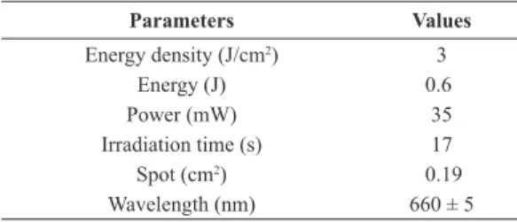
the muscle iber diameters (longitudinal section) Table 1. Laser parameters.
Parameters Values
Energy density (J/cm2) 3
Energy (J) 0.6
Power (mW) 35
Irradiation time (s) 17
Spot (cm2) 0.19
and area (cross‑section) were quantiied, using ImageJ 1.49t software. The changes in muscle iber morphometry were assessed by measuring the iber diameter in 6 non‑overlapping ields per section in
10 non-consecutive sections for each muscle (total
of 60 ibers/mouse). The ibers area was measured in 10 ibers/section/animal.
The results were expressed as the mean ± standard error of the mean (SEM). Statistical comparisons between the control (saline), venom alone and venom
+ laser groups were done using ANOVA followed by the Tukey‑Kramer post‑test for multiple comparisons. Values of p<0.05 were considered signiicant. All data
analyses were done using Prism v.5.0 (GraphPad,
La Jolla, CA, USA).
Results
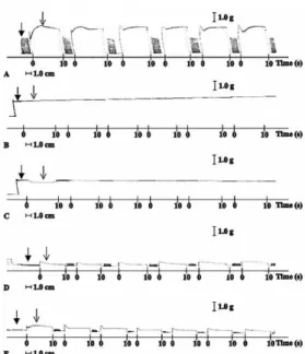
Figure 1 shows representative recordings of the tetanic responses of saline- (control) and venom-injected muscles. Control muscle showed consistent, reproducible responses to a series of six tetanic trains (Figure 1A), indicating that the muscle was not damaged by repeated stimulations. In contrast,
3 h and 9 h after venom injection the tibial muscle
showed no responses to low voltage or tetanic stimuli
(Figure 1B, C), which suggested that the venom had caused important damage to the muscle contractile
machinery. Treatment with LLLT 30 min after venom
injection followed by assessment of muscle function
3 h and 9 h post‑venom revealed small but signiicant recovery of the tetanic response (by 10% and 15%,
respectively) compared to the responses to venom alone (Figures 1D, E, 2 and 3).
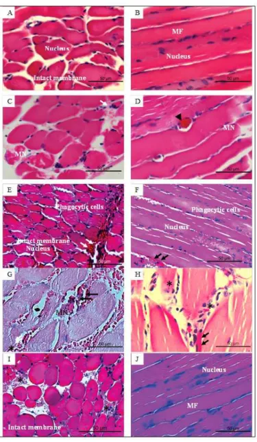
Histological analysis of saline-injected (control) muscle revealed myocytes with a normal appearance
(intact cell membrane and muscle ibers, well‑deined
muscle fascicles, peripherally located nuclei and a normal interstitial space) (Figure 4A, B). In contrast
to this normal appearance, 3 h after venom injection
Figure 1. Myographic records of electrically-induced tetanic contractions in mouse tibialis muscle. (A) Control (saline; n = 3); (B)
Venom 3 h (n = 6); (C) Venom 9 h (n = 6); (D) Venom + Laser 3 h (n = 6); and (E) Venom + Laser 9 h (n = 6). The baseline in control
preparations remained stable throughout the experiment, indicating that the muscle did not go into contracture and was not injured during repeated stimulations. Venom-injected muscles treated with laser
showed contractions and tetanus. In each record: irst (dark) arrow – baseline and second (light) arrow – response to tetanic stimulus.
The records are representative of the number of experiments indicated.
Figure 3. Contractile force generated by tetanic responses in mouse
tibial muscle 9 h after injection of B. jararaca venom (40 µg) (▄) and venom followed by treatment with LLLT (●). In contrast to venom alone, venom-injected muscle exposed to LLLT showed enhanced tetanic responses. The contractile force (muscle tension) was calculated from the initial peak of the tetanic response. The points are the mean ± SEM. **p<0.01 compared to venom alone.
Figure 2. Contractile force generated by tetanic responses in mouse
tibial muscle 3 h after injection of B. jararaca venom (40 µg) (▄) and
there was muscle iber disorganization and degradation
indicative of myonecrosis; there was also hemorrhage,
cellular iniltration and an increase in iber diameter
suggestive of intracellular edema formation (Figures 4C, D, 5A, B). Similar changes occurred at
9 h post‑venom, but were less marked (Figure 4G, H).
In venom-injected muscle treated with LLLT and
analyzed 3 h post‑venom, there was less myonecrosis but inlammatory (phagocytic) cells were observed
(Figure 4E, F). At 9 h post‑venom, LLLT‑treated muscle showed an intact cell membrane (no myonecrosis),
cellular iniltration and a small amount of hemorrhage Figure 4. Histological sections of mouse tibial muscle injected with B. jararaca venom (40 µg). (A, B): Control muscles 3 h after injection
(Figure 4I, J). Overall, at both time intervals, there was attenuation of venom-induced myonecrosis.
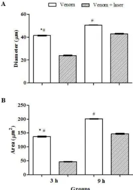
Histomorphometric analysis showed a signiicant decrease in the diameter and area of muscle ibers in mice treated with LLLT (3 h) compared to those treated
with venom alone; this improvement persisted after
9 h, but was less marked than at 3 h, probably because
of partial recovery in the venom-treated muscles, i.e., the diameter and area in venom-treated muscle
were greater at 9 h compared to 3 h (Figure 5A, B).
Discussion
LLLT is a non-invasive, low-cost procedure that has been widely used in clinical practice to relieve pain and stimulate tissue regeneration (Buso, 2006;
Dall‑Agnol et al., 2009; Fikackova et al., 2006; Kato et al., 2006; Medeiros et al., 2010; Oliveira et al., 2013; Piva et al., 2011). Laser light interacts with biological
tissues to produce signiicant beneicial therapeutic
effects in various physiological systems (Leal et al., 2010; Piva et al., 2011). Lopes-Martins et al. (2006)
found that GaAlAs laser at λ 630‑680 nm protected
against muscle damage, with a concomitant decrease in creatine kinase release, stimulated the release of cGMP to cause local vasodilatation and helped to
maintain muscle contractile strength in rats. More recently, Giaretta et al. (2015) observed that LLLT improved muscle function in mice, as assessed by the enhanced tetanic response and resistance to fatigue.
Based on studies of the beneicial effects of LLLT in
a variety of models, various experimental investigations have shown that LLLT can reduce the myonecrosis,
edema and inlammation induced by snake venoms
(Barbosa et al., 2009; 2010; Barbosa-Souza et al., 2011; Doin‑Silva et al., 2009; Oliveira et al., 2013).
Dourado et al. (2003) examined the effects of laser irradiation with GaAs 4 J/cm2 on the myonecrosis
caused by Bothrops moojeni snake venom in mouse gastrocnemius muscle. Subsequently, Barbosa et al. (2008; 2010) observed that GaAs laser signiicantly decreased the myonecrosis caused by Bothrops jararacussu snake venom in mice. Although these studies reported the recovery of muscle cell structures, they did not assess possible improvements in muscle
function. These beneicial effects of laser treatment on
venom-induced damage are apparently not mediated by a direct action of laser light on venom activities since
Barbosa et al. (2009) showed that prior irradiation of B. jararacussu venom did not adversely affect the myotoxicity of this venom.
As shown here, the intramuscular injection of B. jararaca venom adversely affected muscle contractility (electrically-induced contractions and contractures). Treatment with LLLT partially
restored this muscle dysfunction 3 h and 9 h after
venom injection, probably by reducing the muscle
fatigue. This conclusion is supported by the indings
of Leal et al. (2009), who reported that phototherapy (infrared radiation) attenuated the skeletal muscle fatigue in biceps muscles of professional male volleyball players. The possible cellular mechanisms involved in this enhanced resistance to fatigue have been investigated in recent studies (Giaretta et al., 2015; Kelencz et al., 2010; Maciel et al., 2013;
Muñoz et al., 2013; Reis et al., 2015). Passarella and
Karu (2014) reported that mitochondria respond to low intensity light and that this response increases ATP production and facilitates cell multiplication. Albuquerque-Pontes et al. (2015) also observed increased cytochrome c oxidase activity in response to a 660 nm laser with an output of 1 J. These authors
noted a signiicant increase in the percentage of muscle ibers with >50% staining for cytochrome c oxidase
within 5 min after LLLT; the enhanced cytochrome c oxidase activity probably facilitated the transfer of protons and electrons, thereby accelerating the rate of oxidative metabolism. This led to an increase in ATP that in turn enhanced cellular activity and muscle contractility.
Figure 5. Histomorphometric analysis of venom and venom + laser
Histological analysis of muscle injected with
B. jararaca venom alone revealed myonecrosis, similar to but less marked than that caused by the venoms of
B. jararacussu (Barbosa et al., 2009; Doin‑Silva et al.,
2009) and B. moojeni (Dourado et al., 2003). Treatment with LLLT attenuated this myonecrosis, as also observed by Doin‑Silva et al. (2009) for
B. jararacussu venom in rats. Dourado et al. (2003)
reported that GaAs and HeNe laser therapy signiicantly reduced the inlammation and myonecrosis caused
by B. moojeni venom, with GaAs laser being more effective than HeNe laser. Similarly, Doin‑Silva et al.
(2009) found that HeNe laser attenuated the damage to rat gastrocnemius muscle and associated nerve caused by B. jararacussu venom; these authors also noted an improvement in the contractility of irradiated muscle compared to muscle treated with venom alone. Barbosa et al. (2010) reported that GaAs laser reduced
the edema, inlammation and myonecrosis caused by
B. jararacussu venom in rat paw.
As shown here, LLLT reduced the diameter and area
of venom‑treated muscle ibers in relation to venom alone at 3 h, with the attenuation being less marked at 9 h. This attenuation by LLLT was accompanied by
improved tetanic responses compared to venom alone.
This inding suggests that LLLT had a beneicial effect
on muscle contractility in the presence of venom. Overall, the results of this study indicate that treatment with LLLT attenuated the myonecrosis caused by B. jararaca venom and partially but signiicantly
reversed the venom-induced dysfunction in muscle contractility. The ability of LLLT to partially restore the function of envenomed muscle suggests that this
intervention could be a potentially beneicial ancillary
procedure for treating the myonecrosis and muscle dysfunction caused by Bothrops venoms.
References
Albuquerque-Pontes GM, Vieira RP, Tomazoni SS, Caires CO, Nemeth V, Vanin AA, Santos LA, Pinto HD, Marcos RL, Bjordal JM, Carvalho PTC, Leal-Junior ECP. Effect of pre-irradiation with different doses, wavelengths, and application intervals of low-level laser therapy on cytochrome c oxidase activity in intact skeletal muscle of rats. Lasers in Medical Science. 2015; 30(1):59‑66. http://dx.doi.org/10.1007/ s10103‑014‑1616‑2. PMid:24957189.
Barbosa AM, Villaverde AB, Guimarães-Sousa L, Soares AM, Zamuner SF, Cogo JC, Zamuner SR. Low-level laser therapy decreases local effects induced by myotoxins isolated from Bothrops jararacussu snake venom. The Journal of Venomous Animals and Toxins Including Tropical Diseases. 2010; 16(3):470‑9. http://dx.doi.org/10.1590/ S1678‑91992010000300014.
Barbosa AM, Villaverde AB, Guimarães-Souza L, Munin E, Fernandes CM, Cogo JC, Zamuner SR. Effect of the low
level laser therapy in the myonecrosis induced by Bothrops jararacussu snake venom. Photomedicine and Laser Surgery. 2009; 27(4):591‑7. http://dx.doi.org/10.1089/pho.2008.2296. PMid:19530909.
Barbosa AM, Villaverde AB, Guimarães-Souza L, Ribeiro W, Cogo JC, Zamuner SR. Effect of low-level laser therapy in the inflammatory response induced by Bothrops jararacussu snake venom. Toxicon. 2008; 51(7):1236‑44. http://dx.doi. org/10.1016/j.toxicon.2008.02.007. PMid:18439641. Barbosa‑Souza V, Contin DK, Bonventi W Fo, Araújo AL, Irazusta SP, Cruz-Höfling MA. Osteopontin, a chemotactic protein with cytokine-like properties, is up-regulated in muscle injury caused by Bothrops lanceolatus (fer-de-lance) snake venom. Toxicon. 2011; 58(5):398‑409. http://dx.doi. org/10.1016/j.toxicon.2011.07.011. PMid:21839764. Brasil. Manual de diagnóstico e tratamento de acidentes por animais peçonhentos. 2. ed. Brasília: Fundação Nacional de Saúde; 2011 [cited 2011 Jun 16]. 120 p. Available from: http://dtr2004.saude.gov.br/sinanweb/
Buso NC. Avaliação da ação do laser arseneto de gálio-904nm no processo de reparação do tendão de Aquiles em ratos [dissertation]. São José dos Campos: Universidade do Vale do Paraíba; 2006.
Cavalcante WL, Campos TO, Dal Pai‑Silva M, Pereira PS, Oliveira CZ, Soares AM, Gallacci M. Neutralization of snake venom phospholipase A2 toxins by aqueous extract of Casearia sylvestris (Flacourtiaceae) in mouse neuromuscular preparation. Journal of Ethnopharmacology. 2007; 112(3):490‑7. http://dx.doi.org/10.1016/j.jep.2007.04.002. PMid:17540522. Collaço RCO, Cogo JC, Rodrigues-Simioni L, Rocha T, Oshima-Franco Y, Randazzo-Moura P. Protection by Mikania laevigata (guaco) extract against the toxicity of Philodryas olfersii snake venom. Toxicon. 2012b; 60(4):614-22. http:// dx.doi.org/10.1016/j.toxicon.2012.05.014. PMid:22677807. Collaço RCO, Leite GB, Cogo JC, Hyslop S, Rocha T, Randazzo-Moura P, Rodrigues-Simioni L. Neuromuscular activity of Bothrops fonsecai snake venom and its neutralization by commercial Bothropic antivenom. Toxicon. 2012a; 60(2):174-5. http://dx.doi.org/10.1016/j.toxicon.2012.04.157. Dall‑Agnol MA, Nicolau RA, Lima JC, Munin E. Comparative analysis of coherent light action (laser) versus non-coherent light (LED) for tissues repair in diabetic rats. Lasers in Medical Science. 2009; 24(6):909‑16. http://dx.doi.org/10.1007/ s10103‑009‑0648‑5. PMid:19238507.
Doin‑Silva R, Baranauskas V, Rodrigues‑Simioni L, Cruz-Höfling MA. The ability of low level laser therapy to prevent muscle tissue damage induced by snake venom. Photochemistry and Photobiology. 2009; 85(1):63‑9. http:// dx.doi.org/10.1111/j.1751‑1097.2008.00397.x. PMid:18643907. Dourado DM, Fávero S, Baranauskas V, Cruz‑Höfling MA. Effects of the Ga-As laser irradiation on myonecrosis caused by Bothrops moojeni snake venom. Lasers in Surgery and Medicine. 2003; 33(5):352‑7. http://dx.doi.org/10.1002/ lsm.10237. PMid:14677163.
26. http://dx.doi.org/10.1111/j.1751‑1097.2010.00878.x. PMid:21166811.
Ferraz MC, Parrilha LAC, Moraes MSD, Amaral J Fo, Cogo JC, Santos MG, Franco LM, Groppo FC, Puebla P, Feliciano AS, Oshima-Franco Y. The effect of lupane triterpenoids (Dipteryx alata Vogel) in the in vitro neuromuscular blockade and myotoxicity of two snake venoms. Current Organic Chemistry. 2012; 16(1385‑2728):2717‑23. http://dx.doi. org/10.2174/138527212804004481.
Ferraz MC, Yoshida EH, Tavares RVS, Cogo JC, Belo CAD, Franco LM, Santos MG, Hyslop S, Puebla P, Feliciano AS, Oshima-Franco Y. An isoflavone from Dipteryx alata vogel is active against paralysis of Bothrops jararacussu snake venom and Bothrops venom-induced myonecrosis. Molecules (Basel, Switzerland). 2014; 19(5):5790‑805. http://dx.doi. org/10.3390/molecules19055790. PMid:24806579. Fikackova H, Dostalova L, Vosicka R, Peterova V, Navratil L, Lesak J. Arthralgia of the temporomandibular joint and low-level laser therapy. Photomedicine and Laser Surgery. 2006; 24(4):522-7. http://dx.doi.org/10.1089/pho.2006.24.522. PMid:16942435.
França FOS, Málaque CMS. Acidente botrópico. In: Cardoso JLC, França FOS, Wen FH, Málaque CMS, Haddad V Jr, editors. Animais peçonhentos no Brasil: biologia, clínica e terapêutica dos acidentes. São Paulo: Sarvier/FAPESP; 2009. p. 81‑95.
Giaretta VMA, Santos LP, Barbosa AM, Hyslop S, Corrado AP, Nicolau RA, Cogo JC. Effect of low-level laser therapy (GaAlAs ‑ λ660 nm) on muscle function. Research in Biomedical Engineering. 2015; 31(3):241‑8. http://dx.doi. org/10.1590/2446‑4740.0736.
Guimarães‑Souza L, Dale CS, Nadur‑Andrade N, Barbosa AM, Cogo JC, Zamuner SR. Low-level laser therapy reduces edema, leukocyte influx and hyperalgesia induced by Bothrops jararacussu snake venom. Clinical and Experimental Medical Letters. 2011; 52(3‑4):97‑102.
Gutiérrez JM, Lomonte B, León G, Rucavado A, Chaves F, Ângulo Y. Trends in snakebite envenomation therapy: scientific, technological and public health considerations. Current Pharmaceutical Design. 2007; 13(28):2935‑50. http:// dx.doi.org/10.2174/138161207782023784. PMid:17979738. Gutiérrez JM, Ownby CL. Skeletal muscle degeneration induced by venom phospholipases A2: insights into the mechanisms of local and systemic myotoxicity. Toxicon. 2003; 42(8):915‑31. http://dx.doi.org/10.1016/j.toxicon.2003.11.005. PMid:15019491.
Kato MT, Kogawa EM, Santos CN, Conti PCR. TENS and low‑ level laser therapy in the management of temporomandibular disorders. Journal of Applied Oral Science. 2006; 14(2):130‑ 5. http://dx.doi.org/10.1590/S1678‑77572006000200012. PMid:19089044.
Kelencz CA, Munoz ISS, Amorim CF, Nicolau RA. Effect of low-power gallium-aluminium-arsenium non-coherent light (640 nm) on muscle activity: a clinical study. Photomedicine and Laser Surgery. 2010; 28(5):647-52. http://dx.doi. org/10.1089/pho.2008.2467.
Leal ECP Jr, Lopes-Martins RÁB, Almeida P, Ramos L, Iversen VV, Bjordal JM. Effect of low-level laser therapy (GaAs 904 nm) in skeletal muscle fatigue and biochemical
markers of muscle damage in rats. European Journal of Applied Physiology. 2010; 108(6):1083‑8. http://dx.doi.org/10.1007/ s00421‑009‑1321‑1. PMid:20024577.
Leal ECP Jr, Lopes-Martins RÁB, Baroni BM, Marchi T, Taufer D, Manfro DS, Rech M, Danna V, Grosselli D, Generosi RA, Marcos RL, Ramos L, Bjordal JM. Effect of 830 nm low‑level laser therapy applied before high‑intensity exercises on skeletal muscle recovery in athletes. Lasers in Medical Science. 2009; 24(6):857‑63. http://dx.doi.org/10.1007/ s10103‑008‑0633‑4. PMid:19057981.
Leobas GF, Feitosa SB, Seibert CS. Accidents by venomous animals in tocantins state: clinical and epidemiological aspects. DESAFIOS: Revista Interdisciplinar da Universidade Federal do Tocantins. 2016; 2(2):269‑82. http://dx.doi.org/10.20873/ uft.2359.
Lomonte B, Moreno E, Tarkowski LA, Hanson LA, Maccarana M. Neutralizing interaction between heparins and myotoxin II, a lysine 49 phospholipase A2 from Bothrops asper snake
venom. Identification of a heparin-binding and cytolytic toxin region by the use of synthetic peptides and molecular modeling. The Journal of Biological Chemistry. 1994; 269(47):29867‑73. PMid:7961981.
Lopes-Martins RÁB, Marcos RL, Leonardo PS, Prianti AC Jr, Muscará MN, Aimbire F, Frigo L, Iversen VV, Bjordal JM. Effect of low-level laser (Ga-Al-As 655 nm) on skeletal muscle fatigue induced by electrical stimulation in rats. Journal of Applied Physiology. 2006; 101(1):283‑8. http:// dx.doi.org/10.1152/japplphysiol.01318.2005. PMid:16627677. Maciel TS, Muñoz ISS, Nicolau RA, Nogueira DV, Hauck LA, Osório R, Paula AR Jr. Phototherapy effect on the muscular activity of regular physical activity practitioners. Lasers in Medical Science. 2013; 29:1145‑52.
Maier M, Haina D, Landthaler M. Effect of low energy laser on the growth and regeneration of capillaries. Lasers in Medical Science. 1990; 5(4):381‑6. http://dx.doi.org/10.1007/ BF02032595.
Medeiros JL, Nicolau RA, Nicola EMD, dos Santos JN, Pinheiro ALB. Healing of surgical wounds made with λ970‑nm diode laser associated or not with laser phototherapy (λ655 nm) or polarized light (λ400‑2000 nm). Photomedicine and Laser Surgery. 2010; 28(4):489‑96. http://dx.doi.org/10.1089/ pho.2009.2592. PMid:19857050.
Melo PA, Homsi‑Brandeburgo MI, Giglio JR, Suarez‑Kurtz G. Antagonism of the myotoxic effects of Bothrops jararacussu venom and bothropstoxin by polyanions. Toxicon. 1993; 31(3):285‑91. http://dx.doi.org/10.1016/0041‑0101(93)90146‑A. PMid:8470133.
Miró L, Coupe M, Charras C, Jambon C, Chevalier JM. Estudio capiloroscópico de la acción de um láser de AsGa sobre la microcirculación. Investigacion y Clinica Laser. 1984; 1(2):9‑14.
próximo sobre a atividade muscular esquelética estudo clínico. Revista Brasileira de Engenharia Biomédica. 2013; 29:262‑8. Nadur-Andrade N, Barbosa AM, Carlos FP, Lima CJ, Cogo JC, Zamuner SR. Effects of photobiostimulation on edema and hemorrhage induced by Bothrops moojeni venom. Lasers in Medical Science. 2012; 27(1):65-70. http://dx.doi. org/10.1007/s10103‑011‑0914‑1. PMid:21484453. Oliveira RA, Matos AF, Barros NRB, Fernandes GA, Lima ACG, Nicolau RA. Low‑intensity laser therapy and LED (light emitting diode) therapy in mechanical resistance and heating of Rattus norvegicus chest incision with implant of steel wire for sternal suture. Revista Brasileira de Engenharia Biomédica (Impresso). 2013; 29(2):166‑74. http://dx.doi. org/10.4322/rbeb.2013.016.
Passarella S, Karu T. Absorption of monochromatic and narrow band radiation in the visible and near IR by both mitochondrial and non-mitochondrial photoacceptors results in photobiomodulation. Journal of Photochemistry and Photobiology. B, Biology. 2014; 140:344‑58. http://dx.doi. org/10.1016/j.jphotobiol.2014.07.021. PMid:25226343. Pereira DM, Cogo JC, Zamuner SR. Efeito antiedematogênico da planta Vernonia scorpioides frente ao veneno de Bothrops fonsecai e carragenina. ConScientiae Saúde (Impresso). 2012; 11(1):46-52. http://dx.doi.org/10.5585/ConsSaude.v11n1.2862. Pereira IC, Barbosa AM, Salvador MJ, Soares AM, Ribeiro W, Cogo JC, Zamuner SR. Anti-inflammatory activity of Blutaparon portulacoides ethanolic extract against the inflammatory reaction induced by Bothrops jararacussu venom and isolated myotoxins BthTX-I and II. Journal of Venomous Animals and their Toxins including Tropical Diseases. 2009; 15(3):527‑45. http://dx.doi.org/10.1590/ S1678‑91992009000300013.
Picolo G, Chacur M, Gutiérrez JM, Teixeira CFP, Cury Y. Evaluation of antivenoms in the neutralization of hyperalgesia and edema induced by Bothrops jararaca and Bothrops asper snake venoms. Brazilian Journal of Medical and Biological Research. 2002; 35(10):1221‑8. http://dx.doi.org/10.1590/ S0100‑879X2002001000016. PMid:12424496.
Piva JAAC, Abreu EMC, Silva VS, Nicolau RA. Ação da terapia com laser de baixa potência nas fases iniciais do reparo tecidual: princípios básicos. Anais Brasileiros de Dermatologia. 2011; 86(5):947‑54. http://dx.doi.org/10.1590/ S0365‑05962011000500013. PMid:22147035.
Queiróz LS, Santo-Neto H, Rodrigues-Simioni L, Prado-Franceschi J. Muscle necrosis and regeneration after envenomation by Bothrops jararacussu snake venom. Toxicon. 1984; 22(3):339‑46. http://dx.doi.org/10.1016/0041-0101(84)90077‑1. PMid:6474488.
Reis MCR, Andrade EAF, Borges ACL, Souza DQ, Lima FPS, Nicolau RA, Andrade AO, Lima MO. Immediate effects of low level laser (808 nm) on fatigue and strength of spastic muscle. Lasers in Medical Science. 2015; 30(3):1089‑96. http://dx.doi.org/10.1007/s10103‑014‑1702‑5.
Santos CM, Pereira ES, Rabelo NS, Zamuner SR, Barbosa AM, Cogo JC. Efeito do laser de baixa intensidade e LED na atividade hemorrágica induzida pelo veneno da serpente Bothrops jararaca. In: Anais do 14° Encontro Latino Americano de Iniciação Científica e 10° Encontro Latino Americano de Pós-Graduação; 2010; São José dos Campos, Brasil. São José dos Campos: UNIVAP; 2010. p. 1‑4.
Tribuiani N, Da Silva A, Ferraz M, Silva M, Bentes AP, Graziano T, Dos Santos M, Cogo JC, Varanda E, Groppo F, Cogo K, Oshima‑Franco Y. Vellozia lavicans Mart. ex Schult. hydroalcoholic extract inhibits the neuromuscular blockade induced by Bothrops jararacussu venom. BMC Complementary and Alternative Medicine (Online). 2014; 14(1):48. http://dx.doi.org/10.1186/1472-6882-14-48. PMid:24507387.
Warrell DA. Snakebites in Central and South America: epidemiology, clinical features and clinical management. In: Campbell JA, Lamar WW, editors. Venomous reptiles of the Western Hemisphere. Ithaca: Comstock Publishers/Cornell University Press; 2004. vol. 2, p. 709‑61.
Authors
Vania Maria de Araújo Giaretta1, Luiz Prudêncio Santos2, Ana Maria Barbosa3, Stephen Hyslop4, Alexandre Pinto Corrado5, Milene Sanches Galhardo6, Renata Amadei Nicolau7*, José Carlos Cogo1
1 Laboratório de Fisiologia e Farmacodinâmica, Instituto de Pesquisa e Desenvolvimento – IP&D, Universidade do Vale do Paraíba – UNIVAP, São José dos Campos, SP, Brazil.
2 Escola de Enfermagem Wenceslau Braz – EEWB, Itajubá, MG, Brazil.
3 Faculdade Anhanguera Taubaté, Taubaté, SP, Brazil.
4 Departamento de Farmacologia, Faculdade de Ciências Médicas, Universidade Estadual de Campinas – UNICAMP, Campinas, SP, Brazil.
5 Departamento de Farmacologia, Faculdade de Medicina de Ribeirão Preto, Universidade de São Paulo – USP, Ribeirão Preto, SP, Brazil.
6 Laboratório de Histologia, Instituto Básico de Biociências, Universidade de Taubaté – UNITAU, Taubaté, SP, Brazil.