O
h
r
c
i
r
g
a
in
e
a
s
l
R
e
Can Huzmeli1, Ferhan Candan1, Gokhan Bagci2, Demet Alaygut3, Binnur Bagci4, Esin Yildiz5, Ayse Seker Kockara1, Mansur Kayatas1 1Department of Nephrology, Faculty of Medicine, 2Department of Medical Genetics, Faculty of Medicine, 3Department of Pediatric Nephrology, Faculty of Medicine, 4Department of Nutrition and Dietetics, Faculty of Health Sciences, 5Department of Pathology, Faculty of Medicine, Cumhuriyet University, Sivas, Turkey Evaluation of 61 Secondary Amyloidosis Patients
Evaluation of 61 Secondary Amyloidosis Patients:
A Single-Center Experience from Turkey
61 Sekonder Amiloidoz Hastasının Değerlendirilmesi:
Türkiyeden Tek Merkez Deneyimi
DOI: 10.4328/JCAM.4481 Received: 12.03.2016 Accepted: 29.03.2016 Printed: 01.09.2016 J Clin Anal Med 2016;7(5): 695-700 Corresponding Author: Binnur Bağcı, Department of Nutrition and Dietetics, Faculty of Health Sciences, Cumhuriyet University, Sivas, Turkey.
T.: +90 3462192523 F.: +90 3462191261 E-Mail: binnur.koksal@hotmail.com Özet
Amaç: Sekonder amiloidozlu hastaların demografik, klinik ve laboratuar özel-liklerini, hastalık sebeplerini, MEFV gen mutasyonlarını ve mortalite oranla-rını değerlendirmek. Gereç ve Yöntem: 2007-2013 arasında Cumhuriyet Üni-versitesi, Tıp Fakültesi, Nefroloji kliniğine başvuran böbrek yada rektal doku biyopsisiyle sekonder amiloidoz tanısı alan toplam 61 hasta çalışmaya dahil edildi. Demografik özellikler, sekonder amiloidoz sebepleri, MEFV gen mu-tasyonları, ve son dönem böbrek yetmezliği (SDBY), renal transplantasyon ve mortalite oranları retrospektif olarak değerlendirildi. Bulgular: Etiyolojik açı-dan hastaların %62.2’si (38) FMF, %9.8’i (6) bronşiyektazi ve amfizem, %4.9’u (3) tüberküloz, %3.2’si (2) FMF ve ankilozan spondilit birlikteliği, %1.6’sı (1) FMF ve romatoid artrid birlikteliği, %1.6’sı (1) FMF ve sistemik lupus erite-matozus birlikteliği, %1.6’sı (1) osteomiyelit, %1.6’sı (1) septik artrit, %1.6’sı (1) crohn hastalığı, %1.6’sı (1) kolon kanseri, %1.6’sı (1) bronşiyektazi ve tü-berküloz birlikteliği, %1.6’sı (1) romatoid artrit ve %6.5’i (4) idiyopatik olarak değerlendirildi. Sekonder amiloidozu olan 47 hastanın 32’sinde (%68) nefro-tik düzeyde proteinuri saptandı. Sekonder amiloidozlu 45 hastanın MEFV gen mutasyonları incelendi. Hastaların çoğunda M694V mutasyonu vardı. Şaşır-tıcı bir şekilde, 3 vakada heterozigot E148Q mutasyonu saptadık. 12 vaka öldü ve bu hastaların 9’u SDBY’idi. SDBY olan 5 vaka böbrek nakli oldu. Tar-tışma: Bu çalışmada sekonder AA amiloidoz için en yaygın sebep olarak FMF hastalığını bulduk. Daha büyük yada çok merkezli kohortlarda daha ileri ça-lışmalar yapılmalıdır.
Anahtar Kelimeler
AA Amiloidoz; Ailevi Akdeniz Ateşi; MEFV
Abstract
Aim: To evaluate demographic,clinical and laboratory characteristics, causes, MEFV gene mutations, and mortality rates of patients with secondary amy-loidosis. Material and Method: 61 patients who had been diagnosed with secondary amyloidosis by renal and rectal biopsy between 2007 and 2013 in the nephrology clinic of Cumhuriyet University, Faculty of Medicine, were included in the study. Demographic characteristics, causes of secondary amyloidosis, MEFV gene mutations, end-stage renal failure (ESRF), renal transplantation, and mortality rates were examined retrospectively. Results: In etiological terms, Familial Mediterranean Fever (FMF) occurrence was 62.2% (38), bronchiectasis and emphysema 9.8% (6), tuberculosis 4.9% (3), coexistence of FMF and ankylosing spondylitis 3.2% (2), coexistence of FMF and rheumatoid arthritis 1.6% (1), coexistence of FMF and systemic lupus erythematosus (SLE) 1.6% (1), osteomyelitis 1.6% (1), septic arthritis 1.6% (1), Crohn’s disease 1.6% (1), colon cancer 1.6% (1), coexistence of bronchiec-tasis and tuberculosis 1.6% (1), rheumatoid arthritis 1.6% (1), and idiopathic cases 6.5% (4). Proteinuria was determined at nephrotic level among 68% (32) of 47 patients who had secondary amyloidosis. MEFV gene mutation of 45 patients with secondary amyloidosis was assessed. Most patients had M694V gene mutation. Surprisingly, we detected heterozygous E148Q muta-tion in 3 cases. 12 cases died; of these, 9 had ESRF. Five cases with ESRF underwent renal transplantation. Discussion: We found FMF as the most common cause for secondary AA amyloidosis in this study. Further studies should be done with larger or multicenter cohorts.
Keywords
Introduction
Amyloidosis is a large group of diseases caused by extracel-lular and/or intracelextracel-lular accumulation of amyloid ibril proteins due to diferent etiologies [1]. Accumulations are regarded as localized if seen in one organ or as systemic if seen in mul-tiple organs. Accumulations are mostly seen in the brain, heart, kidney, liver, and digestive systems. Amyloid accumulations in organs other than the brain are generally observed as systemic. In time, the increase of amyloid accumulation causes pressure on the cells. Then atrophic changes in cells and tissue destruc-tion occur. Also, organ dysfuncdestruc-tions occur due to the direct toxic efects of accumulated ibril proteins. Diferent clinical indings may be observed depending on the amyloid accumulation areas (localized and systemic forms) [1, 2].
Systemic amyloidosis types are mainly classiied as primary (immunoglobulin light chain or AL) amyloidosis, secondary (AA) amyloidosis, and familial (hereditary) amyloidosis. To date, ap-proximately 31 diferent proteins have been identiied as caus-ing amyloidosis. These proteins are structurally diferent from each other [3, 4].
The cause of secondary amyloidosis is an acute phase reactant known as serum amyloid A (SAA) which is the precursor protein of AA amyloidosis. The most common causes of secondary AA amyloidosis are rheumatic diseases, chronic infections, chronic arthritis, auto-inlammatory diseases, and malignancies and rheumatoid arthritis (RA) in particular [5]. The common feature of secondary amyloidosis is chronic inlammation, which is seen in all cases. SAA is synthesized in the liver through the efect of cytokines such as interleukin 1 (IL-1), (IL-6), and tumor necrosis factor (TNF)-alpha and circulates in the bloodstream as a part of high-density lipoprotein 3 fractions (HDL3) [6]. Although the main source of SAA protein is the liver, it is also synthesized in endothelial cells, macrophages, atherosclerotic smooth muscle cells, the brain, and synovium. A high SAA level alone does not cause amyloid accumulation, however it leads to macrophage activation and increased secretion of cytokines such as IL-1, IL-6, and TNF-alpha. This, in turn, is followed by amyloid accu-mulation. Amyloid accumulation is primarily seen in the spleen, liver and kidneys, and AA amyloidosis can lead to nephrotic syn-drome and ESRD [6, 7].
Clinical indings of amyloidosis vary based on the organ in-volved. Proteinuria develops in renal involvement as a result of accumulation of amyloid ibrils. Proteinuria is more commonly encountered at nephrotic level. Occasionally, renal dysfunction may be observed without proteinuria. Hematuria is observed in some cases. Renal involvement eventually leads to ESRF. Systolic or diastolic dysfunction, heart failure, cardiomyopathy, various arrhythmia, and heart blocks are observed in cardiac involvement. Constrictive pericarditis and restrictive cardiomy-opathy may cause accumulation in coronary arteries as well as ischemic cardiac disease. Accumulation of amyloid in the gastrointestinal system and autonomic nerves may cause com-plaints. In particular, macroglossia is observed [8-10].
The MEFV gene, located on chromosome 16p13.3 and encod-ing a protein called pyrin, is responsible for Familial Mediter-ranean Fever (FMF) disease. Mutations in the MEFV gene have been found in the majority of FMF patients [11-14]. The vast majority of MEFV mutations identiied in FMF patients include
mutations (M680I, M694V, M694I, and V726A) clustered in exon 10 and a mutation in exon 2 (E148Q) [15]. The main concern in FMF is the development of renal amyloidosis. The severe dis-ease phenotypes of amyloidosis were found to be associated with M694V mutation and the compound E148Q-V726A mu-tation [16]. Furthermore, renal amyloidosis has been reported in asymptomatic individuals who do not experience attacks of serositis (phenotype II) [17]. Colchicine is the drug of choice for the treatment of FMF and can prevent amyloidosis [11, 16]. In this study, demographic, clinical and laboratory characteris-tics of patients, causes of secondary amyloidosis, MEFV gene mutations, end-stage renal failure (ESRF), renal transplanta-tion, and mortality rates were examined retrospectively.
Material and Method
Sixty-one patients diagnosed as biopsy-proven AA amyloidosis at the nephrology clinic of Cumhuriyet University, Faculty of Medicine between 2007 and 2013 were assessed retrospec-tively. In the process of studying patient proteinuria or renal dysfunctions in our clinic, AA amyloidosis was determined by renal and rectal biopsy. By screening patient iles, we analyzed their laboratory examinations including blood urea nitrogen (BUN), serum creatinine, serum albumin, total protein, alanine aminotransferase enzyme (ALT), aspartate aminotransferase enzyme (AST), low density lipoprotein (LDL), cholesterol, high density lipoprotein (HDL), triglyceride, C-reactive protein (CRP), sedimentation, hemoglobin (Hb), hematocrit (HCT), complete urinalysis, microalbuminuria and proteinuria levels, as well as hepatomegaly and splenomegaly results through abdominal ul-trasonography, and we recorded information related to their di-agnoses and clinical courses. Among these patients, those diag-nosed with FMF were classiied as phenotype I or phenotype II. MEFV gene mutations of 45 patients with secondary amyloido-sis were examined. The common mutations of MEFV gene (exon 2, 3, 5 and 10) were detected using Sanger sequencing or Strip assay methods. Patients were categorized according to their proteinuria values as < 1 gr/day, 1-3.5 gr/ day and > 3.5 gr/ day (nephrotic proteinuria).
Biopsy samples were primarily examined in a light microscope, then stained with Congo red and evaluated in a polarized micro-scope. An additional chemical process was conducted in cases who were observed to have amyloid in tissue, and cases were diagnosed with AA amyloidosis when the tissue with amyloid was exposed to potassium permanganate and amyloid dis-solved from the tissue.
Statistical Analysis
All statistical analyses were performed by SPSS version 22.0 (SPSS IBM, Armonk, NY, USA). All demographic data and clini-cal indings were expressed as mean ± standard deviation (and range) with a 95% conidence interval.
Results
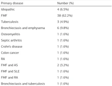
1 shows demographic and laboratory indings of patients. Forty-two of the patients included in the study were diagnosed with FMF. Of those, 2 cases were diagnosed with ankylosing spondylitis (AS), one case was diagnosed with RA and one case was diagnosed with (SLE) (Table 2).
MEFV mutation analysis was performed in 45 cases with sec-ondary amyloidosis. Table 3 illustrates MEFV gene mutations of patients. Homozygous or heterozygous M694V gene muta-tions were determined in most of the cases. There was also coexistence of several gene mutations. R202Q mutation, rarely analyzed, was identiied in 4 cases and only one case had RA in addition to FMF. We identiied 3 patients with E148Q mutation. Fourteen patients had one heterozygous mutation. In mutation analysis of 9 phenotype II patients, it was found as homozygous M694V and homozygous R202Q (1), heterozygous M694V (3),
heterozygous E148Q (1), heterozygous R202Q (1), heterozy-gous M694V and heterozyheterozy-gous R761H (1), heterozyheterozy-gous M680I (G/C), and heterozygous V726A (1).
Of 61 amyloid patients, hemodialysis was started for a total of 35 patients (57.4%) with the diagnosis of end-stage renal failure (ESRF). 27 of them were diagnosed with FMF, 2 were diagnosed idiopathic, one was diagnosed with emphysema, 2 were diagnosed with bronchiectasis, one was diagnosed with septic arthritis, and one was diagnosed with RA. Twenty two of the FMF patients were determined to bephenotype I and 5 were determined to be phenotype II. Five of the 9 patients with phenotype II were diagnosed with ESRF.
Twelve patients were diagnosed with amyloidosis ater their exitus. Seven of these exitus patients died during diagnostic evaluations, 4 patients died approximately within 6 months to one year ater diagnosis, and one patient died approximately 5 years ater diagnosis. Four of the exitus patients had FMF, one had FMF plus RA, 3 had idiopathic and 4 had bronchiectasis plus emphysema. Seven of the exitus patients had proteinuria at a nephrotic level, one had 1-3.5 gr/day and 1 had < 1 gr/day proteinuria. Proteinuria could not be examined for the other 3 exitus patients. Nine of the exitus patients had anemia. 9 of the 12 exitus patients were undergoing hemodialysis.
Thirty-nine patients included in the study had anemia; of these, 26 were undergoing hemodialysis. Of the 39 anemic patients, 23 had FMF (18 had phenotype I, 5 had phenotype II), one had RA plus FMF, 6 had bronchiectasis plus emphysema, 2 had tu-berculosis, one had osteomyelitis, one had septic arthritis, one had colon cancer and one patient was diagnosed with RA. Of 61 amyloidosis patients, 11 patients had hypothyroid. Seven of them were diagnosed with proteinuria at nephrotic level and 2 were diagnosed with proteinuria at non-nephrotic level. The proteinuria level of one patient could not be examined. Six of the cases with hypothyroid were undergoing hemodialysis. CRP test was conducted for 56 patients. The results of 43 of Table 1. Demographic and laboratory indings of patients.
Variable Number Mean±SD Range
Age 61 49.81 ± 17.93 (17- 84)
Male 41 47.6 ± 16.6 (17-78)
Female 20 54.35 ± 20.07 (23-84) BUN (mg/dl) 61 39.77± 34.26 (8-210) Creatinine (mg/dl) 61 3.82 ± 3.92 (0.3-24) Total protein (gr/dl) 61 5.57 ± 1.24 (2.6-7.8) Albumin (gr/dl) 61 2.31 ± 1.00 (0.2-4.2) ALT (IU/L) 61 19.36 ± 27.99 (3-225) AST (IU/L) 61 22.75 ± 26.91 (4-219) Hb (gr/dl) 61 11.41 ± 2.46 (5.4-16.7) HCT (%) 61 34.48 ± 6.85 (16-48.5)
Sedimentation (mm/hour) 54 76.31 ± 35.72 (7-145) CRP (mg/L) 56 44.42 ± 47.23 (3-220) LDL (mg/dl) 58 120.74 ± 20.85 (36-425) HDL (mg/dl) 53 33.3 ± 13.1 (13-83) Cholesterol (mg/dl) 56 185.19 ± 85.4 (91-95) Microalbuminuria (gr/day) 47 3.24 ± 3.25 (0.1-13) Proteinuira (gr/day) 47 6.49 ± 5.02 (0.5-22.8) BUN: Blood urea nitrogen, ALT: Alanine aminotransferase,
AST: Aspartate aminotransferase, Hb: Hemoglobin, HCT: Hematocrit, CRP: C-reactive protein, LDL: Low density lipoprotein,
HDL: High density lipoprotein.
Table 2. Primary diseases causing amyloidosis Primary disease Number (%)
Idiopathic 4 (6.5%)
FMF 38 (62.2%)
Tuberculosis 3 (4.9%) Bronchiectasis and emphysema 6 (9.8%) Osteomyelitis 1 (1.6%) Septic arthritis 1 (1.6%) Crohn’s disease 1 (1.6%)
Colon cancer 1 (1.6%)
RA 1 (1.6%)
FMF and AS 2 (3.2%)
FMF and SLE 1 (1.6%)
FMF and RA 1 (1.6%)
Bronchiectasis and tuberculosis 1 (1.6%)
FMF: Familial Mediterranean Fever, AS: Ankylosing Spondylitis, SLE: Systemic Lupus Erythematosus, RA: Rheumatoid Arthritis
Table 3. MEFV Gene Mutations of Patients
Mutation Patients (n=61) (%)
Mutation analysis not conducted 16 (26.2%)
No mutation 8 (13.1%)
Homozygous M694V 7 (11.4%) Heterozygous M694V 5 (8.1%) Homozygous M694V and Homozygous R202Q 1 (1.6%) Heterozygous M694V and Heterozygous R202Q 1 (1.6%) Heterozygous M694V and Heterozygous M680I (G/C) 3 ( 4.9%) Heterozygous M694V and Heterozygous E148Q 3 (4.9%) Heterozygous M694V and Heterozygous R761H 1 (1.6%) Heterozygous M694V and Heterozygous V726A 1 (1.6%) Heterozygous M694V, Heterozygous R202Q
and Heterozygous V726A 1 (1.6%)
these patients were considered as high. Sedimentation was examined for 54 patients. The results of 49 of these patients were found to be high because of the level exceeded 25 mm/h. All patients except for 2 had low serum albumin levels (serum albumin < 4 gr/dl).
Lipid levels of patients with secondary amyloidosis were found to be generally normal; however, the HDL levels were found to be low in 73.6% (39) of patients. Twenty-eight of 38 cases with FMF had low HDL levels, 3 patients had high HDL levels, and 7 had normal HDL levels. Eight of the exitus cases had low HDL levels, 2 had normal HDL levels and one had high HDL level. Two of 10 cases whose LDL levels were above 160 mg/dl had proteinuria above 1-3.5 gr and 8 had proteinuria above 3.5 gr. All of the 8 patients whose triglyceride level was above 200 mg/ dl had proteinuria at nephrotic level. Ten of 16 patients whose cholesterol level was above 200 mg/dl had proteinuria at ne-phrotic level and 5 had 1-3.5 gr proteinuria. The cholesterol level of one patient was not measured.
In the current study, of 61 amyloidosis patients, 56 patients had complete urine analyses, 39 of these patients had proteinuria, 16 had hematuria in addition to proteinuria, and one patient had only hematuria. Proteinuria was determined at nephrotic level among 68% (32) of 47 patients who had secondary amy-loidosis. All patients except for 2 had hypoalbuminemia (serum albumin < 4 gr/dl). While hepatomegaly was found in 5 (8.3%) cases, splenomegaly was found in 9 (15%) cases.
In the current study, of 61 amyloidosis patients, 5 patients had renal transplants and also all of the cases who had renal trans-plants were diagnosed with FMF phenotype I. MEFV mutation results of cases who underwent transplantations were identi-ied as: homozygous M694V (1), heterozygous M694V and het-erozygous E148Q (2), hethet-erozygous V726A and hethet-erozygous F479L (1). Mutation analysis of one patient was not performed. Three patients had proteinuria at nephrotic level, one patient had proteinuria between 1-3 gr/day, and proteinuria of one pa-tient was not examined. None of these papa-tients had repeated proteinuria in renal allograt.
Discussion
Secondary amyloidosis is deined as the accumulation of amy-loid on tissues and organs as a result of increasing SAA pro-tein, which is an acute phase reactant developing in conjunction with inlammation. SAA production increases in inlammatory conditions [6]. In the current study, we evaluated demographic characteristics, etiology and mortality rates, and MEFV gene mutations of 61 secondary amyloidosis patients who were ad-mitted to our clinic.
Although in Europe the most common cause of amyloidosis is RA, in Turkey the most common cause of amyloidosis is FMF [18]. Amyloid may develop in a period between 2 months and 14 years ater FMF episodes. The period leading to ESRF ater proteinuria may be 2-13 years [19].Secondary AA amyloidosis remains the most serious manifestation of FMF. The incidence rate of secondary AA amyloidosis among FMF patients is 37% in sephardic Jews, 24% in Armenians, 8% in Ashkenazi Jews, 12% in Turks and 10% in Arabs [20, 21]. Amyloidosis develop-ment rates among FMF patients were found as 25.7% in ho-mozygous M694V mutation, 15.4% in M694V compound
het-erozygous and 12.3% in other mutations. Amyloidosis did not develop in E148Q mutations [22].
Although heterozygous E148Q mutation was found in non-am-yloid glomerular diseases [23, 24], there is still no data about whether heterozygous E148Q mutation is an amyloidosis-causing mutation. Topaloglu et al. analyzed 26 patients ho-mozygous for E148Q, 10 compound heterozygous for E148Q, and 8 complex cases. They found that none of their patients had amyloidosis except that 2 cases with E148Q/E148Q muta-tion had a family history of amyloidosis and one had rapidly progressive glomerulonephritis [25]. Balci et al. investigated AA amyloidosis patients (25 phenotype II) in terms of 4 MEFV mutations (M694V, M680I (G/C), V726A and E148Q), and they observed M694V mutation as the most frequent mutation in homozygous, heterozygous and compound heterozygous states. In their study, E148Q was found as compound heterozygous with M694V mutation in 2 cases [26]. In a study conducted with 507 children with FMF in the southeast of Turkey, renal amyloidosis developed in 1.4% (n=7) of patients. Five of the pa-tients with renal amyloidosis had homozygous M694V mutation and two of them were compound heterozygous with M694V/ V726A and M694V/M680I (G/C) mutations. Two of these pa-tients with amyloidosis were phenotype II FMF papa-tients [27]. In a study evaluating frequency of MEFV mutations in FMF pa-tients, it was found that 16 patients had a history of amyloido-sis (3.55%). Among the cases with amyloidoamyloido-sis, 12 cases had M694V homozygous, one case had compound heterozygous for M694V and M680I (G/C) mutations, and one case had a com-plex allele for E148Q/E148Q/M694V mutations. No mutation could be detected in 2 cases with amyloidosis [28]. In Jewish renal amyloidosis patients, M694V/M694V genotype was found as the most frequent mutation. However, E148Q mutation was found as compound heterozygous state together with exon 10 mutation of MEFV gene [16, 17].
higher among heterozygous persons than in the healthy control group [30, 31]. Similarly, amyloid accumulation was determined in non-compound heterozygous cases in our study. In addition, 9 cases with FMF phenotype II were determined. During diag-nosis, ESRF was observed in 5 of these patients.
The frequency of amyloidosis in autopsies in Western countries has varied between 0.50% and 0.86%. According to the report-ed series in England and USA, more than half of the cases have juvenile and adult RA. Ankylosing spondylitis is the second most frequent cause with rates up to 12%. It is followed by psoriatic arthritis with the rate of 4-5% [32-34]. AA amyloidosis is a rare complication of RA. However, there have been signiicant difer-ences among publications. AA amyloid prevalence in RA ranges between 4% and 26% in literature [35]. AA amyloid was higher (14-61%) in RA patients in the post-mortem period [36]. Simi-larly, one patient had RA and one patient had coexistence of FMF and RA in our study. The most common cause of secondary amyloidosis in Turkey is reported as FMF [18]. Furthermore, the RA-related secondary amyloidosis frequency has been found to be low. In 2 studies conducted in ankylosing spondylitis patients who lived in Europe, amyloidosis rates were 7% and 9% re-spectively, as determined by rectal and fat biopsy screenings [37, 38]. In the current study, we found 2 (3.2%) cases who had coexistence of ankylosing spondylitis and FMF.
In the current study, there was 3 occurrences of tuberculosis, one of osteomyelitis, one of septic arthritis, and one coexis-tence of bronchiectasis and tuberculosis among patients with infectious diseases. As in other studies, the most common cause of infectious diseases was found to be tuberculosis in our study. Tuberculosis is the most commonly seen of the chronic infections that cause secondary AA amyloidosis; it is followed by osteomyelitis, chronic bronchitis, chronic mucocutaneous ul-cer, leprosy, Q fever, and subacute bacterial endocarditis [1]. In a study that included 287 secondary amyloidosis patients from 11 centers in Turkey, the etiological causes were deter-mined as FMF in 64% of patients, tuberculosis in 10%, bron-chiectasis and chronic obstructive pulmonary disease in 6%, RA in 4%, spondyloarthritis in 3%, chronic osteomyelitis in 2%, other causes in 4%, and idiopathic in 7%. While hepatomegaly was 17%, splenomegaly was 11% [18]. In another study that evaluated 59 renal amyloidosis cases, 18 patients (30.5%) were found to have FMF, 12 (20.3%) had pulmonary tuberculosis, 8 (13.5%) had chronic osteomyelitis, 9 (15.2%) had bronchiecta-sis, 4 (11.8%) had RA, one (1.6%) had Castleman disease, and 7 (11.8%) were idiopathic [39]. In a study including 40 amy-loidosis patients between 2003 and 2009 in Egypt, secondary amyloidosis was determined in 32 of patients and primary amyloidosis was determined in 8 patients. 30% of secondary amyloidosis cases (12) were FMF, 20% (8) were pulmonary tu-berculosis, 10% (4) were chronic osteomyelitis, 10% (4) were bronchiectasis, 7% (3) were RA, and 2% (1) were rheumatic heart disease [40]. In another study, the most common cause of secondary amyloidosis was RA, which was followed by recurrent pulmonary infections at the rate of 11%, Crohn’s disease at 5%, ankylosing spondylitis at 5%, tuberculosis at 3%, osteomyelitis at 2%, FMF at 2%, Hodgkin’s lymphoma at 2%, and idiopathic cases at 5% [41].
In a multicenter study conducted in Australia and New Zealand,
58,422 patients who underwent renal replacement treatments between 1963 and 2010 were examined; 490 (0.8%) of these patients had problems related to secondary amyloidosis. Sur-vival rates of patients with dialysis-induced secondary amyloi-dosis were found to be worse and renal allograt survival and renal allograt recurrence were worse compared to those re-lated to other diseases. A signiicant number of deaths were observed due to amyloidosis complications in amyloidosis-re-lated ESRF. When amyloidosis-reamyloidosis-re-lated ESRF and ESRF reamyloidosis-re-lated to other causes were compared, cardiac death rates were 37% and 41%, respectively. However, death from heart failure was more common in the amyloidosis-related group (8.6% vs. 2.2%). No diference was found between the 2 groups in the 10-year follow-up in terms of dialysis interruption, infection, cancer, and other causes of death [42]. One, 2 and 6-year survival rates in the retrospective analysis of 48 ESRF patients with systemic amyloidosis (72%, 62%, and 44% respectively) were signii-cantly lower compared to non-diabetic controls (95%, 91%, and 81%) [43]. In our study, 35 patients were diagnosed with ESRF and transferred to hemodialysis. In a retrospective study performed with 23 amyloidosis patients who underwent renal transplant operations, 10-year patient and grat survival rates were found as 66% and 68% respectively, and no signiicant diference was found between them and 47 non-amyloidosis control patients (57% and 87%). Recurrent amyloid rates in re-nal allograt were 4.3% [44]. In a study comparing 45 amyloido-sis transplant receivers and 45 control patients, 3-year patient survival rates were 51%, compared to 79% in the control group. Grat survival was 45% as opposed to 38% in the control group. Recurrent amyloidosis rate in renal allograt was 9% [45]. In this study, 5 patients underwent renal transplant. No amyloid developed in renal allograt cases.
It is important to form a clinical picture of FMF in Turkey, espe-cially in the province of Sivas where FMF is most frequently ob-served [12-14]. Screening of individuals who have FMF in their family histories and evaluating the treatments of phenotype III cases that have mutations but do not show clinical symp-toms are prominent issues to be examined. Although studies have been conducted on phenotype II cases, when we consider that secondary amyloidosis can also develop in non-compound heterozygous mutations, it should be clariied through future studies whether phenotype III cases should be detected in early stages and treated through the evaluation of acute phase re-actants.
As stated in other studies conducted in Turkey, we found FMF to be the most common cause of secondary amyloidosis. Sur-prisingly, we detected heterozygous E148Q mutation in 3 cases with amyloidosis. This inding can bring a new perspective to the literature about E148Q mutation and the development of amyloidosis. We suggest that further studies should be done with larger or multicenter cohorts.
Compliance with Ethical Standards
Competing interests
The authors declare that they have no competing interests.
References
1. Lachmann HJ, Hawkins PN. Systemic amyloidosis. Curr Opin Pharmacol 2006;(6):214-20.
2. Merlini G, Bellotti V. Molecular mechanisms of amyloidosis. N Engl J Med 2003;(349):583-96.
3. Kyle RA. Amyloidosis: a convoluted story. Br J Haematol 2001;(114):529-38. 4. Sipe JD, Benson MD, Buxbaum JN, Ikeda S, Merlini G, Saraiva MJ, et al. Nomen-clature 2014: Amyloid ibril proteins and clinical classiication of the amyloidosis. Amyloid 2014;(21):221-4.
5. Obici L, Merlini G. AA amyloidosis: basic knowledge, unmet needs and future treatments. Swiss Med Wkly 2012;(142):13580.
6. Gillmore JD, Lovat LB, Persey MR, Pepys MB, Hawkins PN. Amyloid load and clinical outcome in AA amiloidosis in relation to circulating concentration of se-rum amyloid A protein. Lancet 2001;(358):24-9.
7. Kanat O, Evrensel T, Filiz G, Usta M, Baskan E, Dilek K, et al. Systemic AA amyloidosis and nephrotic syndrome associated with small cell carcinoma of the bladder. Nephrol Dial Transplant 2003;(18):2453-4.
8. Obici L, Perfetti V, Palladini G, Moratti R, Merlini G. Clinical aspects of systemic amyloid diseases. Biochim Biophys Acta 2005;(1753):11-22.
9. Gertz MA, Lacy MQ, Dispenzieri A, Hayman SR. Amyloidosis. Best Pract Res Clin Haematol 2005;(18):709-27.
10. Pepys MB. Amyloidosis. Annu Rew Med 2006;(57):223-41.
11. Centola M, Wood G, Frucht DM, Galon J, Aringer M, Farrell C, et al. The gene for familial Mediterranean fever, MEFV, is expressed in early leukocyte development and is regulated in response to inlammatory mediators. Blood 2000;(95):3223-31.
12. Ozdemir O, Sezgin I, Kurtulgan HK, Candan F, Koksal B, Sumer H, et al. Preva-lence of known mutations in the MEFV gene in a population screening with high rate of carriers. Mol Biol Rep 2011;(38):3195-200.
13. Koksal B, Nur N, Sari M, Candan F, Acemoglu M, Kocak N, et al. Clinical and molecular analysis of common MEFV gene mutations in familial Mediterranean fever in Sivas population. Biologia 2009;(64):388-93.
14. Ozdemir O, Kayatas M, Cetinkaya S, Yildirim ME, Silan F, Kurtulgan HK, et al. Bcii-RFLP proiles for serum amiloid A1 and mutated MEFV gene prevalence in chronic renal failure patients requiring long-term hemodialysis. Renal Failure 2015;(37):292-6.
15. Shinawi M, Brik R, Berant M, Kasinetz L, Gershoni-Baruch R. Familial Mediter-ranean fever: high gene frequency and heterogeneous disease among an Israeli-Arab population. J Rheumatol 2000;27:1492-5
16. Livneh A, Langevitz P, Shinar Y, Zaks N, Kastner DL, Pras M, et al. MEFV mu-tation analysis in patients sufering from amyloidosis of familial Mediterranean fever. Amyloid 1999; 6:1-6.
17. GershoniBaruch R, Brik R, Zacks N, Shinawi M, Lidar M, Livneh A. The contri-bution of genotypes at the MEFV and SAA1 loci to amyloidosis and disease sever-ity in patients with familial Mediterranean fever. Arthritis Rheum 2003;(48):1149-55.
18. Tuglular S, Yalcinkaya F, Paydas S, Oner A, Utas C, Bozfakioglu S, et al. A retro-spective analysis for aetology and clinical indings of 287 secondary amiloidosis cases in Turkey. Nephrol Dial Transplant 2002;(17):2003-5.
19. Zemer D, Livneh A, Pras M, Sohar E. The kidney in familial Mediterranean fever. Contrib Nephrol 1993;(102):187-97.
20. Rocken C, Sletten K. Amyloid in surgical pathology. Virchows Arch 2003;(443):3-16.
21. Ozen S. Renal amyloidosis in familial mediterranean fever. Kidney Int 2004;(65):1118-27. 22. Mimouni A, Magal N, Stofman N, Shohat T, Minasian A, Krasnov M, et al. Familial Mediterranean fever: Efects of Genotype and Ethnicity on inlammatory Attacks and Amyloidosis. Pediatrics 2000;(105):e70.
23. Eroglu E, Kocyigit I, Ates O, Unal A, Sipahioglu MH, Akgun H, et al. Mesan-gial proliferative glomerulonephritis in familial Mediterranean fever patient with E148Q mutation: the irst case report. Int Urol Nephrol 2013;(45):591-4. 24. Hüzmeli C, Koçkara AŞ, Candan F, Kayataş M Biopsy Proven Non-Amyloid Glomerular Diseases in Patients With Familial Mediterranean Fever. Journal of Nephrology Research 2015;(1):34-9.
25. Topaloglu R, Ozaltin F, Yilmaz E, Ozen S, Balci B, Besbas N, Bakkaloglu A. E148Q is a disease-causing MEFV mutation: a phenotypic evaluation in patients with familial Mediterranean fever. Ann Rheum Dis 2005;(64):750-2.
26. Balci B, Tinaztepe K, Yilmaz E, Guçer S, Ozen S, Topaloğlu R, et al. MEFV gene mutations in familial Mediterranean fever phenotype II patients with renal amyloidosis in childhood: a retrospective clinicopathological and molecular study. Nephrol Dial Transplant 2002;(17):1921-3.
27. Uluca Ü, Ece A, Şen V, Coşkun S, Güneş A, Yel S, et al. High frequency of E148Q sequence variation in children with familial Mediterranean fever in southeast Tur-key. Arch Argent Pediatr 2015;(113):133-9.
28. Yilmaz, E, Ozen S, Balci B, Duzova A, Topaloglu R, Besbas N, et al. Mutation frequency of familial Mediterranean fever and evidence for a high carrier rate in the Turkish population. Eur J Hum Genet 2001;(9):553-5.
29. Livneh A, Langevitz P, Zemer D, Padeh S, Migdal A, Sohar E, et al. The changing face of familial Mediterranean fever. Semin Arthritis Rheum 1996;(26):612-27. 30. Berkun Y, Padeh S, Reichman B, Zaks N, Rabinovich E, Lidar M, et al. A single
testing of serum amyloid a levels as a tool for diagnosis and treatment dilemmas in familial Mediterranean fever. Semin Arthritis Rheum 2007;(37):182-8. 31. Grateau G. Clinical and genetic aspects of the hereditary periodic fever syn-dromes. Rheumatology 2004;(43):410-5.
32. Simms RW, Prout MN, Cohen AS.The epidemiology of AL and AA amyloidosis. Baillieres Clin Rheumatol 1994;(8):627-34.
33. Real de Asúa D, Costa R, Galván JM, Filigheddu MT, Trujillo D, Cadiñanos J. Systemic AA amyloidosis: epidemiology, diagnosis, and management. Clin Epide-miol 2014;(6):369-77.
34. Gertz MA, Kyle RA. Secondary systemic amyloidosis: Response and survival in 64 patients. Medicine 1991;(70):246-56.
35. Wakhlu A, Krisnani N, Hissaria P, Aggarwal A, Misra R. Prevalence of secondary amyloidosis in Asian North Indian patients with rheumatoid arthritis. J Rheumatol 2003;(30):948-51.
36. Dhillon V, Woo P, Isenberg D. Amyloidosis in the rheumatic diseases. Ann Rheum Dis 1989;(48):696-701.
37. Jayson MI, Salmon PR, Harrison W Amyloidosis in ankylosing spondylitis. Rheu-matol Phys Med 1971;(11):78-82.
38. Gratacos J, Orellana C, Sanmarti R, Sole M, Collado A, Gomez-Casanovas E, et al. Secondary amyloidosis in ankylosing spondylitis. A systematic survey of 137 patients using abdominal fat aspiration. J Rheumatol 1997;(24):912-5. 39. Paydas S. Report on 59 patients with renal amyloidosis. Int Urol Nephrol 1999;(31):619-31.
40. Abdullah E, Waked E. Incidence and clinical outcome of renal amyloidosis: a retrospective study. Saudi J Kidney Dis Transpl 2013;(24):950-8.
41. Hazenberg BPC, Van Rijswijk MH. Clinical and therapeutic aspects of AA amy-loidosis. Baillieres Clin Rheumatol 1994;(8):661-90.
42. Tang W, McDonald SP, Hawley CM, Badve SV, Boudville N, Brown FG, et al. End-stage renal failure due to amyloidosis: outcomes in 490 ANZDATA registry cases Nephrol Dial Transplant 2013;(28):455-61.
43. Martinez-Vea A, Garcia C, Carreas M, Revert L, Oliver JA. End stage renal disease in systemic amyloidosis: clinical course and outcome on dialysis. Am J Nephrol 1990;(10):283-9.
44. Sherif AM, Refaie AF, Sobh MA, Mohamed NA, Sheashaa HA, Ghoneim MA. Long term outcome of live donor kidney transplantation for renal amyloidosis. Am J Kindey Dis 2003;(42):370-5.
45. Haq A, Hussain S, Meskat B, Mohan P, Conlon P, Hickey DP. Complications of re-nal transplantation in patients with amyloidosis. Transplant Proc 2007;(39):120-4.
How to cite this article: