751
DOI: 10.1590/0004-282X20150113
ARTICLE
Cerebral autosomal dominant
arteriopathy with subcortical infarcts and
leukoencephalopathy (CADASIL) in Argentina
Arteropatia cerebral autosomal dominante com infartos subcorticais e
leucoencefalopatia (CADASIL) na Argentina
Maximiliano A Hawkes1, Miguel Wilken1, Verónica Bruno1, Virginia Pujol-Lereis1, Guillermo Povedano1, María
Saccoliti2, Analia Taratuto2, Sebastián F Ameriso1
Cerebral Autosomal Dominant Arteriopathy with Subcortical Infarcts and Leucoencephalopathy (CADASIL) is the most common cause of hereditary stroke and vascu-lar dementia1. It is caused by mutations in the NOTCH3 gene
located in chromosome 192. Subcortical ischemic strokes
(60-85%), cognitive impairment (40-50%), migraine with aura (20-40%), depression (20%), and apathy (40%) are the most prevalent symptoms1. An increased risk of intracerebral
hem-orrhage (ICH) has also been reported in these patients3,4,5,6.
Clinical suspicion is based on typical symptoms, family
history, and speciic indings in magnetic resonance imaging
(MRI)1. While family history is almost always present, de novo
mutations have also been described7,8.
Genetic test assessing for NOTCH3 mutations or skin bi-opsy investigating the presence of granular osmiophilic
mate-rial (GOM) are required for deinitive diagnosis1. Management
strategies consist of vascular risk factor (VRF) control, anti-aggregation, and, when appropriate, genetic counseling.
1Neurological Research Institute Raúl Carrea, FLENI, Departament of Neurology, Buenos Aires, Argentina;
2Neurological Research Institute Raúl Carrea, FLENI, Departament of Pathology, Buenos Aires, Argentina.
Correspondence: Maximiliano A Hawkes; Montañeses 2325; Postal Code: 1428 Ciudad de Buenos Aires, Argentina; E-mail: [email protected]
Conlict of interest: There is no conlict of interest to declare.
Received 10 March 2015; Received in inal form 24 April 2015; Accepted 14 May 2015.
AbStrACt
CADASIL is the most common cause of hereditary stroke and vascular dementia. Published information about this disease in South America is scant. We describe clinical and demographic characteristics of 13 patients (10 families) with CADASIL from Argentina. Methods: Medical records, diagnostic tests and family history of patients with CADASIL were reviewed. results: Thirteenpatients with CADASIL (10 families) were included. All patients had European ancestry. Initial presentation was stroke in most patients (n = 11). Stroke patients later developed cognitive complaints (n = 9), migraine with aura (n = 1), apathy (n = 4) and depression (n = 6). External capsule and temporal lobe involvement on MRI were characteristic imaging indings. Two patients died after intracerebral hemorrhage. Conclusion: This is the irst report of non-related patients with CADASIL in South America addressing ancestry. Since European ancestry is not highly prevalent in all South American countries, there may be variable incidence of CADASIL within this region.
Keywords: CADASIL, stroke, South America, leukoencephalopathy, vascular dementia.
reSuMO
752 Arq Neuropsiquiatr 2015;73(9):751-754
To the best of our knowledge this is the irst case series
report of CADASIL in South America.
We describe clinical and demographic characteristics of 13 patients from 10 families with diagnosis of CADASIL from a single center in Argentina.
METHOD
Retrospective review of medical records and diagnostic tests of patients with CADASIL. Genetic test was not widely available in Argentina at the time of this report. hus, most patients were diagnosed by skin biopsy samples. A single specialized laboratory studied all samples. Family history was documented in all cases. Patients or patient’s relatives were contacted to complete missing information and to update vital status. All subjects provided consent. he eth -ics committee of the institution approved the study.
RESULTS
We report 13 patients (7 men) with diagnosis of
CADASIL from 10 families, aged 48 ± 9 (40-73) years old at time of diagnosis (Table). Follow up was between 3 months and 10 years. Diagnosis was established by skin biopsy in 11 patients, and by genetic testing performed abroad in two
cases. Ultrastructural study of the skin biopsies showed
GOM deposits, mainly in the thickened basement mem -brane of arteries or capillaries (Figure 1).
All patients had European ancestry: Spanish (n = 8), Greek (n = 1), Russian/Polish (n = 2), Portuguese (n = 1), Ukrainian (n = 1). hree patients had no familiar history of symptoms suggestive of CADASIL. Initial clinical pre-sentations were: ischemic stroke (n = 11), migraine with aura (n = 1), and cognitive complaint (n = 1). All stroke patients later developed other symptoms: cognitive com-plaint (n = 9), migraine with aura (n = 1), apathy (n = 4), and depression (n = 6).
Cognitive impairment was diagnosed by neurologi
-cal examination in six patients and by neuropsychologi-cal testing in four. Five patients were men and ive women.
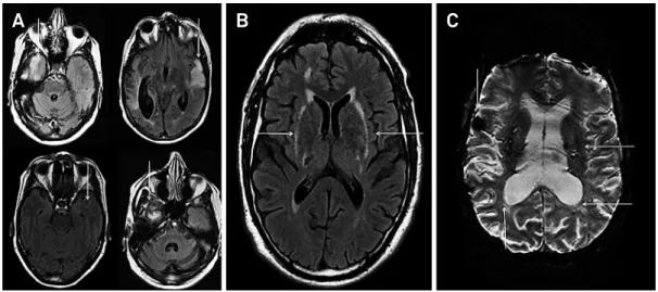
All patients had brain MRI. Typical lesions were found in external capsule (n = 11) and temporal lobes (n = 10). Only two had microbleeds (Figure 2). Two patients reveled asymptomatic acute infarcts in routine follow-up MRIs. Associated vascular risk factor were hypertension (n = 7), dyslipemia (n = 9), smoking (n = 4), type 2 diabetes melli-tus (n = 2) and patent foramen ovale (PFO) (n = 2). All pa -tients received risk factor control and antiplatelet treat-ment. Two of them were empirically anticoagulated. Two patients died during follow up as result of intracranial hemorrhage (ICH), one was on aspirin and the other was on warfarin. Currently eleven patients are on aspirin or
clopidogrel. Modiied Rankin scale was < 3 in 9 surviving subjects and ≥ 3 in 2.
Six asymptomatic family members were evaluated (physical examination and brain MRI). One had chronic
headache. A 38 year-old woman presented white matter le
-sions not-typical of CADASIL. hree relatives in two families underwent skin biopsy after MRI. It was normal in all cases.
DISCUSSION
CADASIL clinical and imaging patterns in this
Argentinean population of European ancestry appear simi-lar to those described in Northern hemisphere populations.
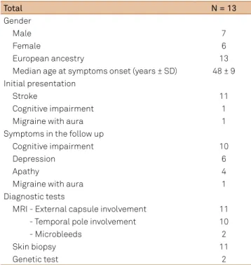
Table. Population characteristics.
total N = 13
Gender
Male 7
Female 6
European ancestry 13
Median age at symptoms onset (years ± SD) 48 ± 9 Initial presentation
Stroke 11
Cognitive impairment 1
Migraine with aura 1
Symptoms in the follow up
Cognitive impairment 10
Depression 6
Apathy 4
Migraine with aura 1
Diagnostic tests
MRI - External capsule involvement 11
- Temporal pole involvement 10
- Microbleeds 2
Skin biopsy 11
Genetic test 2
753
Maximiliano A Hawkes et al. CADASIL in Argentina
hree patients had no familiar history of CADASIL. False-negative family history is commonly documented in individuals presenting with features of CADASIL and is
as-sociated with initial misdiagnosis9.
Stroke was the leading presenting symptom1. As no
treatment to modify disease progression is available, our patients are under risk factor control and single antiplate-let treatment.
Cognitive impairment afects up to 50% of patients1. We
found it in ten patients and it was the initial symptom in two. he latter indings are consistent with those reported
in a single large family in Colombia10. Depression and
apa-thy are described in 20-50% and 40% of CADASIL patients
respectively1. Incidences in our population were similar to
those described in the literature.
he proportion of migraine with aura in CADASIL pa-tients is ive times higher than in general population1. Five
of our patients had migraine, two with and three without aura. Our series supports indings reporting that migraine without aura frequency is slightly higher in CADASIL pa-tients compared with the general population11.
Increased risk of ICH in CADASIL patients is based on case reports and it has not been proved with con-trolled studies. Approximately 22 cases of ICH in CADASIL
patients have been reported3,4. In our series, one hyperten
-sive male treated with warfarin and one non-hyperten-sive woman treated with aspirin died after ICH.
Genetic test screening for the 23 exons of the EGFR is the gold standard for the diagnosis of CADASIL (sen-sitivity and speciicity of almost 100%)1. Skin biopsy has
been restricted to patients with negative genetic test with highly suggestive features of CADASIL or identi-ication of a new sequence of unknown signiicance1,
with a 100% of speciicity and 96% or more sensitivity12,13.
Operator skills, number of arteries examined, and adhe-sion to guidelines for GOM identiication could inluence
the sensitivity of the technique14. Skin biopsy examined
by experienced observers was a suitable option in our population as complete genetic test of all 23 exons is not available in Argentina.
GOM deposits are located both in small and medium sized arteries and capillaries, in contact with vascular
smooth muscle cells (VSMC) or within the thickened base
-ment membrane. Vein walls may also be involved.
Cerebral MRI adds valuable information to clini-cal suspicion. Changes in external capsule and temporal pole were found in eleven and ten patients respective -ly. While the irst one is an earlier less speciic inding, temporal pole involvement has been described as a
ra-diologic marker of CADASIL15. Microbleeds are detected
in 31-69% of CADASIL patients4. We found them in two
subjects (15%). he number of young patients (83% were under 55 year-old), an adequate hypertension control could explain the low incidence in our population. Two patients showed asymptomatic acute cerebral infarcts in DWI/MRI during follow-up. his inding has been report-ed in 10% of CADASIL patients comparreport-ed with 8% of
pa-tients with small-vessel disease16.
Prevalence of PFO in CADASIL is up to 47%17. We
found it in two patients. his inding may be related to NOTCH receptor family participation in the
cardiovascu-lar system development18.
Brain MRI was performed in six asymptomatic relatives of 3 diferent families. Only a 38 year-old female showed white matter lesions. hree of them underwent skin biop-sy. It was normal in all cases. hese indings are consistent with a recent report in which only 33% of patients that had undergone a multistep genetic counseling inally took the genetic test. Reasons for drop out were not reported19.
In conclusion, we report the irst case series of patients
with CADASIL in South America. Published data in our
re-gion is scant. his can be due to low suspicion threshold and
limited access to diagnostic tools. As European ancestry is not highly prevalent in all South American countries there
Figure 2. MRI indings. Temporal pole (A) and external capsule involvement (B). Microbleeds (C).
754 Arq Neuropsiquiatr 2015;73(9):751-754
may be a variable incidence of CADASIL within this region.
he Amerindian and African contributions to the gene ad -mixture in the population of Buenos Aires are 15% and 4% respectively20. Hence European ancestry in this population is
an important factor to take into account.
If MRI is not available the number of misdiagnosis may be high. In countries where genetic test screening for all the
References
1. Chabriat H, Joutel A, Dichgans M, Tournier-Lasserve E, Bousser MG: Cadasil. Lancet Neurol 2009;8:643-653.
2. Joutel A, Corpechot C, Ducros A, Vahedi K, Chabriat H, Mouton P et al. Notch3 mutations in cadasil, a hereditary adult-onset condition causing stroke and dementia. Nature. 1996;383(6602):707-10. doi:10.1038/383707a0
3. Lian L, Li D, Xue Z, Liang Q, Xu F, Kang H et al. Spontaneous intracerebral hemorrhage in CADASIL. J Headache Pain. 2013;14(1):98. doi:10.1186/1129-2377-14-98
4. Rinnoci V, Nannucci S, Valenti R, Donnini I, Bianchi S, Pescini F et al. Cerebral hemorrhages in CADASIL: report of four cases and a brief review. J Neurol Sci. 2013;330(1-2):45-51. doi:10.1016/j.jns.2013.04.002
5. Dichgans M, Holtmannspötter M, Herzog J, Peters N, Bergmann M, Yousry TA. Cerebral microbleeds in CADASIL: a gradient-echo magnetic resonance imaging and autopsy study. Stroke. 2002;33(1):67-71. doi:10.1161/hs0102.100885
6. Lesnik Oberstein SA, Boom R, Buchem MA, Houwelingen HC, Bakker E, Vollebregt E et al. Cerebral microbleeds in cadasil. Neurology. 2001;57(6):1066-70. doi:10.1212/WNL.57.6.1066
7. Joutel A, Dodick DD, Parisi JE, Cecillon M, Tournier-Lasserve E, Bousser MG. De novo mutation in the Notch3 gene causing CADASIL. Ann Neurol. 2000;47(3):388-91. doi:10.1002/1531-8249(200003)47:3<388::AID-ANA19>3.0.CO;2-Q
8. Coto E, MenÉndez M, Navarro R, GarcÍa-Castro M, Alvarez V. A new de novo Notch3 mutation causing CADASIL. Eur J Neurol. 2006;13(6):628-31. doi:10.1111/j.1468-1331.2006.01337.x
9. Razvi SS, Davidson R, Bone I, Muir KW. Is inadequate family history a barrier to diagnosis in CADASIL? Acta Neurol
Scand.2005;112(5):323-26. doi:10.1111/j.1600-0404.2005.00495.x
10. Lopera F, Arboleda J, Moreno S, Almeida N, Cuartas M, Arcos-Burgos M. [Clinical characteristics of hereditary cerebrovascular disease in a large family from Colombia]. Rev Neurol. 2000;31(10):901-7.
11. Liem MK, Oberstein SA, van der Grond J, Ferrari MD, Haan J. CADASIL and migraine: a narrative review. Cephalalgia. 2010;30(11):1284-9. doi:10.1177/0333102410370870
12. Kalaria RN, Viitanen M, Kalimo H, Dichgans M, Tabira T. The pathogenesis of CADASIL: an update. J Neurol Sci. 2004;226(1-2):35-9. doi:10.1016/j.jns.2004.09.008 13. Tikka S, Mykkänen K, Ruchoux MM, Bergholm R, Junna M,
Pöyhönen M et al. Congruence between NOTCH3 mutations and GOM in 131 CADASIL patients. Brain. 2009;132(4):933-9. doi:10.1093/brain/awn364
14. Morroni M, Marzioni D, Ragno M, Di Bella P, Cartechini E, Pianese L et al. Role of electron microscopy in the diagnosis of cadasil syndrome: A study of 32 patients. PLoS One. 2013;8(6):e65482. doi:10.1371/journal.pone.0065482
15. O’Sullivan M, Jarosz JM, Martin RJ, Deasy N, Powell JF, Markus HS. MRI hyperintensities of the temporal lobe and external capsule in patients with cadasil. Neurology. 2001;56(5):628-34. doi:10.1212/WNL.56.5.628
16. O’Sullivan M, Rich PM, Barrick TR, Clark CA, Markus HS. Frequency of subclinical lacunar infarcts in ischemic leukoaraiosis and cerebral autosomal dominant arteriopathy with subcortical infarcts and leukoencephalopathy. AJNR Am J Neuroradiol. 2003;24(7):1348-54. 17. Zicari E, Tassi R, Stromillo ML, Pellegrini M, Bianchi S, Cevenini
G et al. Right-to-left shunt in CADASIL patients: prevalence and correlation with clinical and MRI indings. Stroke. 2008;39(7):2155-7. doi:10.1161/STROKEAHA.107.506311
18. Niessen K, Karsan A. Notch signaling in the developing
cardiovascular system. Am J Physiol Cell Physiol. 2007;293(1):C1-11. doi:10.1152/ajpcell.00415.2006
19. Reyes S, Kurtz A, Hervé D, Tournier-Lasserve E, Chabriat H. Presymptomatic genetic testing in CADASIL. J Neurol. 2012;259(10):2131-6. doi:10.1007/s00415-012-6468-8 20. Avena SA, Goicochea AS, Rey J, Dugoujon JM, Dejean CB, Carnese
FR. [Gene mixture in a population sample from Buenos Aires City]. Medicina (B. Aires). 2006;66(2):113-8. Spanish.
23 exons of EFGR is not available, skin biopsy assessed by trained pathologists is a suitable option.
Studies to clarify the risk of ICH with antiaggregation and anticoagulation treatment in CADASIL patients are needed. Since CADASIL is heterogeneous regarding clinical manifes-tations and severity grade depending on genotype, studies