University of São Paulo
“Luiz de Queiroz” College of Agriculture
Transcriptome changes associated with muscle and intramuscular
connective tissue growth in cull cows under different recovery gain
rates
Daiane Aparecida Fausto
Thesis presented to obtain the degree of Doctor in Science. Area: Animal Science and Pastures
Animal Scientist
Transcriptome changes associated with muscle and intramuscular connective tissue growth in cull cows under different recovery gain rates
versão revisada de acordo com a resolução CoPGr 6018 de 2011
Advisor:
Prof. Dr. EDUARDO FRANCISQUINE DELGADO
Thesis presented to obtain the degree of Doctor in Science. Area: Animal Science and Pastures
my parents Remi Fausto and Marilena Stedille
Fausto, for all support and love!
ACKNOWLEDGMENTS
To the “Luiz de Queiroz” College of Agriculture (ESALQ), University of São
Paulo, and the Animal Science and Pastures Graduate Program for the training and support provided by faculty and staff.
To my advisor, Dr. Eduardo Francisquine Delgado, for his professional and personal example and exemplary guidance.
To Coordenação de Aperfeiçoamento de Pessoal de Nível Superior (Capes) and Conselho Nacional de Pesquisa (CNPq), for their financial and technical support. To EMBRAPA Gado de Corte (Campo Grande, MS, BR), represented by Dr. Gelson Luís Dias Feijó, for letting me be part of the meat science team and for his technical support.
To Dr. Sónia C. S. Andrade for her bioinformatics support, attention, and knowledge.
To Dr. Luiz Lehmann Coutinho of the Animal Science Department at ESALQ, for his support and guidance.
To Dr. Steven Lonergan, my supervisor at Iowa State University (Ames, Iowa,
United States), for the opportunity to do this training. Also to Dr. Lonergan’s team, for
allowing me to use their infrastructure for the development experiment.
To Dr. André Luiz Julien Ferraz of Mato Grosso do Sul State University, for his support, friendship, guidance, suggestions, and assistance since the conception of my project.
To Dr. Gustavo Gasparin, for all his help and patience in the preparation of the sample for sequencing.
To my lovely husband, Diógenes Marcelo Cassiano Coriguazi, for all his love and support.
To my family, especially to my parents Remi Fausto and Marilena Stedille Fausto, my brothers Marcelo Paulo Fausto and José Antônio Fausto, and my sister Vanessa Regina Fausto.
To all my friends and colleagues in the Animal Science Department, Piracicaba-SP, Ames-IA, Campo Grande for all their support and love. Especially to my friends from the meat and animal biotechnology laboratory at ESALQ for their friendship and knowledge shared during this time.
To all those who directly or indirectly contributed to this work and supported
me somehow, my sincere…
CONTENTS
RESUMO...11
ABSTRACT...13
1 INTRODUCTION ... ...15
References...16
2 LITERATURE REVIEW...19
2.1 Influence of growth rate on muscle remodeling………...19
2.2 Components of the extracellular matrix………...20
2.2.1 Proteoglycans………...20
2.2.1.2 Decorin………...21
2.2.2 Adhesive glycoproteins………...22
2.3 Studies related to the extracellular matrix and its impact on meat………..22
2.4 Hypotheses.……….23
2.5 Objectives……….23
2.6 Specific objectives………..23
References………24
3 TRANSCRIPTOME CHANGES IN MUSCLE OF NELLORE CULL COWS SUBJECTED TO RECOVERY WEIGHT GAIN UNDER GRAZING CONDITIONS.……….....29
Abstract………...29
3.1 Introduction………..29
3.2 Material and methods……….30
3.2.1 Treatments………30
3.2.2 RNA Extraction, library and sequencing………..31
3.2.3 Mapping and Counting Reads.………..32
3.2.4 Enrichment analysis………33
3.2.5 Uncharacterized proteins, microRNA-targets, and Ingenuity Pathway Analysis (IPA)……… ………33
3.4 Results and Discussion………..34
3.5 Conclusion………39
Acknowledgments..………...…39
4 RECOVERY FROM UNDERNUTRITION CHANGES MUSCLE GENE EXPRESSION IN MATURE NELLORE CULL COWS DIFFERENTIALLY DEPENDING ON THE RATE OF WEIGHT GAIN
Abstract………45
4.1 Introduction………..46
4.2 Material and methods……….47
4.2.1 Treatments………....47
4.2.2 RNA Extraction, library, and sequencing.…..………..47
4.2.3 Mapping and Counting Reads..……….48
4.2.4 Enrichment analysis………49
4.2.5 Novel transcripts, Uncharacterized proteins, microRNA-targets, and Ingenuity Pathway Analysis (IPA)……….49
4.3 Results and Discussion………..50
4.3.1 Differential expression……….50
4.3.2 Upregulated genes common to both recovery gain rate groups ………...52
4.3.3 Upstream regulator gene analysis by IPA ………..57
4.4 Base x MG………59
4.5 Base x HG………61
Conclusion………..63
Acknowledgments.………63
References...64
5 GENERAL CONCLUSIONS………...69
RESUMO
Mudanças no transcriptoma associadas com o crescimento muscular e do tecido conjuntivo intramuscular em vacas de descarte submetidas a diferentes
taxas de recuperação do ganho
A taxa de renovação do estroma e proteínas miofibrilares define o crescimento muscular, e pode afetar a qualidade da carne, afetando turnover de colágeno e taxa proteolítica. Há uma falta de informação sobre as mudanças no processo de remodelação de proteína muscular em resposta à taxa de recuperação
do ganho de peso, observada durante a “realimentação” depois de subnutrição, o
que pode ser alterado em animais mais velhos. Alterações no tecido muscular durante o período de recuperação pode ser indicada pelo perfil de expressão diferencial de genes, pela técnica de sequenciamento de RNA. Os objetivos deste trabalho foram avaliar as alterações do transcriptoma na musculatura de vacas de descarte Nelore submetidas a: 1) a recuperação do peso em condições de pastejo; e 2) a recuperação da desnutrição em diferentes taxas de ganho de pesos. No primeiro experimento, os animais foram divididos em: grupo de manutenção (manutenção de peso e de alta escore corporal); Recuperação do ganho (recuperação de baixo escore corporal com o ganho de peso corporal moderado em 0,6 kg / dia sob pastejo). No segundo experimento, os animais foram divididos em três grupos em confinamento: Controle (abatidos com baixo escore de condição corporal), ganho moderado (0,6 kg de ganho de peso vivo por dia) durante a estação seca e alta recuperação de ganho (1,2 kg de ganho de peso vivo por dia) durante a estação seca. Nos dois estudos, amostras do Longissimus dorsi foram coletadas
após o abate e imediatamente congeladas até à análise de sequenciamento ser realizada. No primeiro experimento, os genes encontrados no tratamento de recuperação do ganho foram relacionados com a resposta inflamatória como: 4A semaphorin (SEMA4A), solute carrier family 11 member 1 (SLC11A1), Ficolin 2
(FCN2) e placental growth factor (PGF). No segundo experimento, o osteonectina
(SPARC), colágeno tipo IV subunidades 1 (COL4A1) foram alguns dos genes mais
expressos para ambas as taxas de ganho de recuperação de remodelação do tecido conjuntivo. Para ganho moderado, foram identificadas proteínas miofibrilares estruturais como: Myosin IE (MYO1E), Myosin, Heavy Chain 11 (MYH11), (MYOG)
and actinin, alpha 4 (ACTN4). No tratamento de alta recuperação de ganho, genes como CLL de célula B / 9 linfoma (BCL9), peroxisome proliferator-activated receptor alpha (PPARA), Diacylglycerol O-Acyltransferase 2 (DGAT2) and
Phosphatidylinositol 4-Kinase, Catalytic, Beta (PI4KB) indicam maior deposição de
tecido adiposo. Em resumo, observou-se que a deposição muscular durante a recuperação do ganho de peso envolve a regulação da expressão de vários genes relacionados com a matriz extracelular (ECM), corroborando com modelos de inflamação e similar a fibrose observada em animais maduros. Além disso, no grupo HG, genes relacionados com a síntese de colágeno e a deposição de gordura, também foram encontrados, indicando contribuição do tecido conjuntivo durante o crescimento muscular. Estes resultados são importantes para a compreensão do desenvolvimento do tecido como um todo, e auxiliam no progresso do conhecimento científico sobre a remodelação muscular durante a recuperação do ganho de peso e sua influência sobre estruturas de proteínas e vias intracelulares.
ABSTRACT
Transcriptome changes associated with muscle and intramuscular connective tissue growth in cull cows under different recovery gain rates
The renewal rate of stromal and myofibrillar proteins defines muscle growth, and can affect the quality of meat, by affecting collagen turnover and proteolytic rate. There is a lack of information on changes in the muscle protein remodeling process in response to the recovery weight gain rate observed during “realimentation” after
undernutrition, which may be altered in older animals. Changes in muscle tissue during the recovery period may be indicated by the differential expression profile of genes after RNA sequencing. The objectives of this study were to evaluate transcriptome changes in the muscle of Nellore cull cows subjected to: 1) recovery weight gain under grazing conditions; and 2) recovery from undernutrition at different weight gain rates. In the first experiment, the animals were divided into two groups and subjected to one of two nutritional managements under grazing conditions: maintenance (maintenance of weight and high body condition score under grazing conditions) and recovery gain (recovery from low body condition score with moderate body weight gain of 0.6 kg/day under grazing conditions). In the second experiment, the animals were divided into three groups and subjected to one of three nutritional managements under feedlot conditions: control (slaughtered at low body condition score), moderate recovery gain (MG; 0.6 kg of daily live weight gain) during the dry season, and high recovery gain (HG; 1.2 kg of daily live weight gain) during the dry season. In both experiments, samples of longissimus dorsi muscle were collected after slaughter and immediately frozen until sequencing analysis could be performed. In the first experiment, genes related to inflammatory response, such as semaphorin 4A (SEMA4A), solute carrier family 11 member 1 (SLC11A1), ficolin-2 (FCN2), and
placental growth factor (PGF), were expressed at higher levels during recovery gain.
In the second experiment, osteonectin (SPARC) and collagen type IV subunits 1
(COL4A1) were expressed at higher levels in both recovery gain and connective
tissue remodeling. For MG, structural myofibrillar proteins such as myosin IE (MYO1E), myosin, heavy chain 11 (MYH11), myogenin (MYOG), and actinin, alpha 4
(ACTN4) were identified. In the HG treatment, the B-cell CLL/lymphoma 9 (BCL9),
peroxisome proliferator-activated receptor alpha (PPARA), diacylglycerol
O-Acyltransferase 2 (DGAT2), and phosphatidylinositol 4-Kinase, catalytic, and beta
(PI4KB) genes indicated more deposition of adipose tissue. In summary, we
observed that muscular deposition during recovery weight gain involved the regulation of expression of several genes related to the extracellular matrix (ECM), corroborating the inflammatory and -like models observed in mature animals. Moreover, in the HG group, genes related to collagen synthesis and fat deposition were also found, indicating the important contribution of connective tissue during muscle growth. These results are important for understanding tissue development as a whole, and will assist in the progress of scientific knowledge on muscle remodeling during recovery weight gain and its influence on protein structures and intracellular routes.
1 INTRODUCTION
It has already been established that growth alters rates of DNA, RNA, and protein accretion, ultimately affecting skeletal muscle (BEERMANN, 2004) and myofibrillar protein turnover (KOOHMARAIE et al., 2002), which is involved with certain cell routes related to meat tenderness. This quality attribute has been a problem for the meat industry (MORGAN et al., 1991; SAVELL; SHACKELFORD, 1992), since consumers identify it as a primary sensory trait in purchasing decisions (DELGADO et al., 2006; MENNECKE et al., 2007). In addition, tenderness involves complex interdependent biochemical mechanisms (RILEY et al., 2007) that involve protein degradation (LONERGAN, 2001; HUFF-LONERGAN et al., 1996; HO et al., 1997; GEESINK; KOOHMARAIE, 1999), lipid content (WOOD et al., 2003), connective tissue maturity and content (PURSLOW, 2005; MCCORMICK et al., 2009), and heat shock proteins (BERNARD et al., 2007), which have a strong genetic component. These mechanisms comprising the different deposition patterns of tissues interact in response to nutritional management strategies (BOLEMAN et al., 1996) and affect tenderness, which makes it difficult to predict this particular quality trait (OUALI, 1990; KOOHMARAIE, 1996).
Several studies have evaluated changes in the transcriptome of genes associated with muscle remodeling in response to nutritional challenges (LEE; HOSSNER, 2002; LEE; ENGLE; HOSSNER, 2002; BYRNE et al., 2005). These changes indicate the possibility of modifying proteins that are part of the muscle structure involved in meat tenderness, such as components of the ECM and cytoskeleton. However, no information exists on muscle remodeling in mature cows, even though they contribute significantly to meat production, especially in Brazil, where a significant portion of slaughters fall into this category. In 2015, cull cows represented close to 34% of the Brazilian bovine herd slaughtered (IBGE, 2015). Despite advances in our understanding of the impact of growth rate on myofibrillar metabolism and tissue deposition in muscle, the basics of these processes are still not fully understood.
relevant to meat scientists, providing potential information on the biological characteristics of muscle (HOCQUETTE et al., 2007; BALDWIN et al., 2012).
Moreover, a major contribution to the process involving accelerated proteolysis via the proteasome-ubiquitin pathway has been recognized as the principal cause of muscle atrophy under a number of catabolic conditions (JAGOE; GOLDBERG, 2001). Genes involved in connective tissue remodeling, such as SPARC, have also been identified; some in relation to tissue injury or high turnover rates with an important role within the ECM (BRADSHAW; SAGE, 2001), and others as serine protease inhibitors (SENTANDREU; COULIS; OUALI, 2002).
References
BALDWIN, R.L.V.; LI, R.W.; LI, C.; THOMSON, J.M.; BEQUETTE, B.J.
Characterization of the longissimus lumborum transcriptome response to adding propionate to the diet of growing Angus beef steers. Physiological genomics,
Bethesda, v. 44, p. 543–550, 2012.
BEERMANN, D.H. Physiology. In: JENSEN, W.K; DEVINE, C.; DIKEMAN, M.
Encyclopedia of meat sciences. Cambridge: Woodhead Publ., 2004. v. 2: Growth
of meat animals, p. 511-518.
BERNARD, C.; CASSAR-MALEK, I.; CUNFF, M.L.; DUBROEUCO, H.; RENAND, G.; HOCQUETTE, J.F. New indicators of beef sensory quality revealed by expression of specific genes. Journal of Agricultural and Food Chemistry, Washington, v. 55,
p. 5229–5237, 2007.
BOLEMAN, S.J.; MILLER, R.K.; BUYCK, M.J.; CROSS, H.R.; SAVELL, J.W.
Influence of realimentation of mature cows on maturity, color, collagen solubility, and sensory characteristics. Journal of Animal Science, Champaign, v. 74, p. 2187–
2194, 1996.
BRADSHAW, A.D.; SAGE, E.H. Sparc, a matricellular protein that functions in cellular differentiation and tissue response to injury. Journal of Clinical Investigation, Michigan, v. 107, p. 1049–1054, 2001.
BYRNE, K.A.; WANG, Y.H.; LEHNERT, S.A.; HARPER, G.S.; MCWILLIAM, S.M.; BRUCE, H.L.; REVERTER, A. Gene expression profiling of muscle tissue in Brahman steers during nutritional restriction. Journal of Animal Science,
Champaign, v. 83, p. 1-12, 2005.
DAUNCEY, M.J.; KATSUMATA, M.; WHITE, P. Nutrition, hormone receptor
expression and gene interactions. In: PAS, M.F.W. te; EVERTS, M.E.; HAAGSMAN.
Muscle development of livestock animals: physiology, genetics and meat quality.
DELGADO, E.F.; AGUIARII, A.P.; ORTEGAIII, E.M.M.S.; SPOTO, M.H.F.; CASTILLO, C.J.C. Brazilian consumers' perception of tenderness of beef steaks classified by shear force and taste. Science Agricola, Piracicaba, v. 63, n. 3, p.
232-239, 2006.
IBGE. (2015). Indicadores. Pesquisa da pecuária. Retrieved in December, 2015 from http://www.ibge.gov.br/home/estatistica/
GEESINK, G.H.; KOOHMARAIE, M. Effect of calpastatin on degradation of
myofibrillar proteins by μ-calpain under postmortem conditions. Journal of Animal Science, Champaign, v. 77, p. 2685-2692, 1999.
HO, C.Y.; MARVIN, H.; STROMER, M.H.; GENE ROUSE, G.; RICHARD, M.;
ROBSON, R.M. Effects of electrical stimulation and postmortem storage on changes
in titin, nebulin, desmin, troponin-t, and muscle ultrastructure in Bos indicus
crossbred cattle. Journal of Animal Science, Champaign, v. 75, p. 366–376, 1997.
HOCQUETTE, J.F.; LEHNERT, S.A.; BARENDSE, W.; CASSAR-MALEK, I.; PICARD, B. Recent advances in cattle functional genomics and their application to beef quality. Animal, Cambridge, v. 1, p. 159-173, 2007.
HUFF-LONERGAN, E.; MITSUHASHI, T.; BEEKMAN, D.D.; PARISH, F.C.; OLSON,
D.G.; ROBSON, R.M. Proteolysis of specific muscle structural proteins by μ-calpain at low pH and temperature is similar to degradation in post-mortem Bovine muscle.
Journal of Animal Science, Champaign, v. 74, p. 993-1008, 1996.
KOOHMARAIE, M. Biochemical factors regulating the toughening tenderization processes of meat. Meat Science, Barking, v. 43, p. 193-201, 1996.
KOOHMARAIE, M.; KENT, M.P.; SHACKELFORD, S.D.; VEISETH, E.; WHEELER, T.L. Meat tenderness and muscle growth: is there any relationship? Meat Science,
Barking, v. 62, p. 345–352, 2002.
JAGOE, R.T.; GOLDBERG, A.L. What do we really know about the
ubiquitin-proteasome pathway in muscle atrophy? Current Opinion in Clinical Nutrition and Metabolic Care, Beijing, v. 4, p.183-190, 2001.
LEE, S.H.; HOSSNER, K.L. Coordinate regulation of ovine adipose tissue gene expression by propionate. Journal of Animal Science, Champaign, v. 80, p. 2840–
2849, 2002.
LEE, S.H.; ENGLE, T.E.; HOSSNER, K.L. Effects of dietary copper on the expression of lipogenic genes and metabolic hormones in steers. Journal of Animal Science,
Champaign, v. 80, p. 1999–2005, 2002.
LONERGAN, S.M.; HUFF-LONERGAN, E.; WIEGAND, B.R.; KRIESE-ANDERSON, L.A. Postmortem proteolysis and tenderization of top loin steaks from Brangus cattle.
MCCORMICK, R.J. Collagen. In: MIN, D.; MCCORMICK, R.J. Collagen applied
muscle biology and meat science. Boca Raton: CRC Press, 2009. chap. 7,
p. 129−148.
MENNECKE, B.E.; TOWNSEND, A.M.; HAYES, D.J.; LONERGAN, S.M. A study of the factors that influence consumer attitudes toward beef products using the conjoint market analysis tool. Journal of Animal Science, Champaign, v. 85, p. 2639-2659,
2007.
MORGAN, J.B.; SAVELL, J.W.; HALE, D.S.; MILLER, R.K.; GRIFFIN, D.B.; CROSS, H.R.; SHACKELFORD, S.D. National beef tenderness survey, Journal of Animal Science, Champaign, v. 69, p. 3274-3283, 1991.
OUALI, A. Meat tenderization: possible cause and mechanisms; a review. Journal of Muscle Foods, Trumbull, v. 1, p. 129-165, 1990.
PURSLOW, P.P. Intramuscular connective tissue and its role in meat quality. Meat Science, Barking, v. 70, p. 435–447, 2005.
RILEY, D.G.; JOHNSON, D.D.; CHASE Jr., C.C.; WEST, R.L.; COLEMAN, S.W. OLSON, T.A.; HAMMOND, A.C. Factors influencing tenderness in steaks from Brahman cattle. Meat Science, Barking, v. 70, p. 347-356, 2007.
SAVELL, J.; SHACKELFORD, S.D. Significance of tenderness to the meat industry. In: RECIPROCAL MEAT CONFERENCE, 45., 1992, Ft. Collins. Proceedings… Ft.
Collins: Colorado State University, 1992. p. 43-46.
SENTANDREU, M.A.; COULIS, G.; OUALI, A. Role of muscle endopeptidases and their inhibitors in meat tenderness. Trends in Food Science & Technology, New
York, v. 13, p. 400–421, 2002.
2 LITERATURE REVIEW
2.1 Influence of growth rate on muscle remodeling
During animal development and growth prior to maturity, body tissues grow in specific waves, starting with nervous tissue and followed by bone, muscle, and adipose tissue, which accumulates at a faster rate with higher weight gain in mature animals (OWENS et al., 1993). This differential tissue accretion is also modified by nutritional restriction, which alters muscle fiber type profiles by increasing the contribution of oxidative myofibers. These are spared during restrictive conditions (BYRNE et al., 2005). In addition, collagen renewal processes are directly related to the rate of muscle growth (FISHELL et al., 1985).
When muscle is subjected to regeneration after atrophy, the first phase is characterized by the release of growth factors and cytokines to signal the proliferation of cells and the infiltration of inflammatory cells (TIDBALL; VILLALTA, 2010). In the first stage, recovery is characterized by an inflammatory process, where macrophages are recruited (MCLENNAN, 1996) to promote muscle regeneration as mediators of tissue remodeling. The support of myogenesis and myofiber growth is described as a second phase of muscle recovery from atrophy (CHAZAUD et al., 2009).
The inflammatory reaction is followed by the activation of satellite cells, maturation of newly formed myofibers (CICILIOT; SCHIAFFINO, 2010), and remodeling. Simultaneously, angiogenesis and the proliferation of fibroblasts occur, the latter enabling the muscle to synthesize ECM components that are degraded during the formation of new myofibers (SERRANO; MUÑOZ-CÁNOVES, 2010). However, if stem cells (satellite cells) lose their repair capacity during skeletal muscle remodeling, excessive ECM accumulation takes place, leading to fibrosis, which is characterized by increased amounts of collagen types I and III, fibronectin, elastin, proteoglycans, and laminin (UEZUMI et al., 2011). It can also provoke the infiltration of adipocytes (NATARAJAN; LEMOS; ROSSI, 2010), and may be accentuated by age (ALEXAKIS; PARTRIDGE; BOU-GHARIOS, 2007). In addition, some researchers have found that transforming growth factor-beta1 (TGF-β1) may be
2.2 Components of the extracellular matrix
The main constituent of connective tissue is the ECM, which is primarily composed of connective fibers made up of collagen (95%), elastin, and an amorphous substance composed of proteoglycans and glycoproteins (JUNQUEIRA; CARNEIRO, 2004). The collagen molecules are composed of a combination of triple alpha-helix subtypes that define the collagen types. The primary types involved in skeletal muscle development are I, III, IV, V, VI, XI, XII, XIV, XV, and XVIII (NISHIMURA et al., 1997).
During intramuscular connective tissue remodeling, the enzymes responsible for degrading almost all ECM components are the metalloproteinases, or MMPs (NAGASE; VISSE; MURPHY, 2006). This enzyme system is formed primarily of zinc-dependent endopeptidases that degrade collagen, elastin, fibronectin, laminin, and proteoglycans and can regulate the turnover of ECM macromolecules that are involved in the regulation of preadipocyte growth, thus contributing to adipose tissue development in skeletal muscle (BAILEY; LIGHT, 1989). Among these is MMP-2, which plays an important role in regulating the integrity and composition of the ECM (CARMELI et al., 2004). The MMP system is regulated by specific inhibitors, known as TIMPs (tissue inhibitors of matrix metalloproteinases). Most notably, TIMP-1 and TIMP-2 are capable of inhibiting the activity of any MMP, with a preference for the inhibition of MMP-2 and MMP-9, respectively (KJAER, 2004). The occurrence of co-expression between the various major components of the MMP system in bovine muscle, particularly MMP-2, TIMP-1, and TIMP-2 (BALCERZAK et al., 2001), may indicate the presence of an important regulatory mechanism in the alteration of the ECM. The types of collagen expressed during this process have also been studied.
2.2.1 Proteoglycans
(ROUGHLEY; LEE 1994; PINS et al., 1997). There are reports of the degradation of proteoglycans in the ECM, which would be a weakening factor in the intramuscular connective tissue of bovine muscle (NISHIMURA et al., 1996).
Proteoglycan expression changes as development in embryonic chicken muscle occurs, changing a matrix rich in chondroitin sulfate to a complex array containing a lower level of chondroitin sulfate, heparan, and dermatan (YOUNG et al., 1989; FERNANDEZ et al., 1991). This process has also been reported in other species (VELLEMAN et al., 1999; YOUNG et al., 1990). Different types of proteoglycans expressed in skeletal muscle have special functions during the process of differentiation, but the expression pattern appears to be conserved among them.
2.2.1.2 Decorin
Decorin, the main proteoglycan in skeletal muscles, is a macromolecule belonging to the family of leucine-rich proteoglycans, which is divided into subfamilies according to amino acid sequences and genes (IOZZO, 1997). It participates in cell growth by modulating some growth factors (LI; MCFARLAND; VELLEMAN, 2008), and regulates the formation and stability of collagen fibrils and fibers (NISHIMURA, 2015).
The decorin subfamily contains an N-terminal domain substituted with chondroitin sulfate/dermatan chains. It is the main proteoglycan in skeletal muscles, and interacts with collagen (primarily types I and II) during fibrogenesis (VOGEL et al., 1984; HEDBOM; HEINEGARD, 1989; BROWN; VOGEL, 1989). This interaction appears to be mediated by the central protein and dermatan sulfate chains (FONT et al., 1993). In previous studies, beef muscles with divergent tenderness differed in their content of dermatan sulfate (PEDERSEN et al., 1999), a glycosaminoglycan present in decorin. In the muscles of adult bovine animals, decorin is found in the endomysium and perimysium (EGGEN et al., 1994). The architecture of collagen fibrils can be modified by decorin in bovines (NISHIMURA et al., 2003) and mice (DANIELSON et al., 1997). Other in vitro studies on extracellular components found
2.2.2 Adhesive glycoproteins
Adhesive glycoproteins are so named because they participate in adhesion between cells, fibers, and ECM macromolecules. These molecules contain a predominant protein that is associated with carbohydrates (JUNQUEIRA; CARNEIRO, 2004). Fibronectin is one of the main adhesive glycoproteins in the intramuscular connective tissue matrix. It binds to integrins, which act as transmembrane receptors of information between cells and the matrix. Such communication or signaling between cells and the matrix results in modulation of the growth and function of tissues, including migration, cell survival or death, and responsiveness to growth factors. Different tissues have different matrices, which change as the animal ages (VELLEMAN, 2003).
2.3 Studies related to the extracellular matrix and its impact on meat
Skeletal muscle is mainly composed of muscle fibers and surrounding intramuscular connective tissues of three types: endomysium, epimysium, and perimysium (NISHIMURA, 2015). In theory, rapid growth increases the synthesis of new soluble collagen that dilutes the mature existing collagen (ABERLE et al., 1981; FISHELL et al., 1985). However, the relationship between nutrition management and changes in connective tissue is difficult to determine (HALL; HUNT, 1982). MMP-2 activity has also been associated with collagen degradation and tenderness of meat in sheep with a high growth rate (SYLVESTRE et al., 2002). However, this effect may be related to turnover from the collagen fraction, which would influence meat tenderness independent of the action of MMPs during the postmortem. On the other
connective tissue accumulates when its rate of synthesis is greater than its rate of degradation.
In mice, age has been associated with fibrosis (BRACK et al., 2007) and with accumulation of ECM (ALEXAKIS, PARTRIDGE; BOU-GHARIOS, 1981). Moreover, the synthesis of glycosaminoglycan is high in young tissues but decreases with growth and maturation (SCHOFIELD; WEIGHTMAN, 1978); changes in proteoglycans may also change with age. However, during severe fibrosis, the level of decorin is reduced (ZANOTTI et al., 2005). Decorin has been related to anti-fibrosis agents in skeletal muscle (SATO et al., 2003). It can inhibit myostatin (MSTN) and TGF-β1, which probably act together to amplify the fibrotic process (ZHU et al., 2007).
2.4 Hypotheses
1. A moderate recovery weight gain under grazing conditions during body condition recovery is sufficient to elicit changes in proteins and protease gene expression that are associated with the renewal of major ECM components in cull cows.
2. The greater gain rate achieved under feedlot conditions is necessary to increase connective tissue growth and turnover of ECM major proteins by altering the transcription of associated proteins and protease genes.
2.5 Objective
To evaluate transcriptome profile changes related to protein turnover in the muscle of cull cows under different recovery gain rates.
2.6 Specific objectives
1. To evaluate transcriptome changes in muscle of Nellore cull cows subjected to recovery weight gain under grazing conditions.
References
ABERLE, E.D.; REEVES, E.S.; JUDGE, M.D.; HUNSLEY, R.E.; PERRY, T.W. Palatability and muscle characteristics of cattle with controlled weight gain: time on a high energy diet. Journal of Animal Science, Champaign, v. 52, p. 757-763, 1981.
ALEXAKIS C, PARTRIDGE T, BOU-GHARIOS G. Implication of the satellite cell in
dystrophic muscle fibrosis: a self-perpetuating mechanism of collagen
overproduction. American Journal of Physiology, Bethesda, v. 293, C661–C669,
2007.
BALCERZAK, D., QUERENGESSER, L.; DIXON, W.T.; BARACOS, V.E. Coordinate expression of matrix-degrading proteinases and their activators and inhibitors in bovine skeletal muscle. Journal of Animal Science, Champaign, v. 79, p. 94–107,
2001.
BAILEY, A.J.; LIGHT, N.D. The connective tissue of meat and meat products.
London: Elsevier, 1989. 356 p.
BRACK, A.S.; CONBOY, M.J.; ROY, S.; LEE, M.; KUO, C.J.; KELLER, C.; RANDO, T.A. Increased Wnt signaling during aging alters muscle stem cell fate and increases fibrosis. Science, New York, v. 317, p. 807-810, 2007.
BROWN, D.C.; VOGEL, K.G. Characteristics of the in vitro interaction of a small proteoglycan (PGII) of bovine tendon with type I collagen. Matrix, Philadelphia, v. 9,
p.468-478, 1989.
BYRNE, K.A. WANG, Y.H.; LEHNERT, S.A.; HARPER, G.S.; MCWILLIAM, S.M.; BRUCE, H.L.; REVERTER, A. Gene expression profiling of muscle tissue in Brahman steers during nutritional restriction. Journal of Animal Science,
Champaign, v. 83, p. 1-12, 2005.
CARMELI, E.; MOAS, M.; REZNICK, A.Z.; COLEMAN, R. Matrix metalloproteinases and skeletal muscle: a brief review. Muscle Nerve, Malden, v. 29, p. 191-197, 2004.
CHAZAUD, B.; BRIGITTE, M.; YACOUB-YOUSSEF, H.; ARNOLD, L.; GHERARDI, R.; SONNET, C.; LAFUSTE, P.; CHRETIEN, F. Dual and beneficial roles of
macrophages during skeletal muscle regeneration. Exercise and sport sciences
reviews, New York, v. 37, p. 18-22, 2009.
CICILIOT, C.; SCHIAFFINO, S. Regeneration of mammalian skeletal muscle: basic mechanisms and clinical implications. Current Pharmaceutical Design, Schiphol,
v. 16, 906-914, 2010.
DANIELSON, K.G.; BARIBAULT, H.; HOLMES, D.F.; GRAHAM, H.; KADLER, K.E.; IOZZO, R.V. Targeted disruption of decorin leads to abnormal collagen fibril
morphology and skin fragility. Journal of Cell Biology, New York, v. 136, p.
EGGEN, K.H.; MALMSTRØM. A.; KOLSET, S.O. Decorin and a large dermatan sulfate proteoglycan in bovine striated muscle. Biochimica et Biophysica Acta,
Amsterdam, v. 1204, p. 287–297, 1994.
ESKO, J. Genetic analysis of proteoglycan structure, function and metabolism.
Current Opinion in Cell Biology, Bethesda, v. 3, p. 805-816, 1991.
FERNANDEZ, M.S.; DENNIS, J.E.; DRUSHEL, R.F.; CARRINO, D.A.; KIMATA, K.; YAMAGATA, M.; CAPLAN, A.I. The dynamics of compartmentalization of embryonic muscle by extracellular matrix molecules. Developmental Biology, Pasadena,
v. 147, p. 46-61, 1991.
FISHELL, V.K.; ABERLE, E.D.; JUDGE, M.D.; PERRY, T.W. Palatability and muscle properties of beef as influenced by pre slaughter growth rate. Journal of Animal Science, Champaign, v. 61, p. 151-157, 1985.
FONT, B.; AUBERT-FOUCHER, E.; GOLDSCHMIDT, D.; EICHENBERGER, D. Binding of collagen XIV with the dermatan sulfate side chain of decorin. Journal of Biological Chemistry, Rockville, v. 268, p. 25015-25018, 1993.
GALLAGHER, J. The extended family of proteoglycan; social residents of the pericellular zone. Current Opinion in Cell Biology, Bethesda, v. 1, p. 1201-1218,
1989.
HALL, J.B.; HUNT, M.C. Collagen solubility of A-maturity bovine Longissimus muscle as affected by nutritional regimen. Journal of Animal Science, Champaign, v. 55,
p. 321-328, 1982.
HAUS, J.M.; CARRITHERS, J.A.; TRAPPE, S.W.; TRAPPE, T.A. Collagen, cross-linking, and advanced glycation end products in aging human skeletal muscle.
Journal of Applied Physiology, Bethesda, v. 103, p. 2068-2076, 2007.
HEDBOM, E.; HEINEGARD, D. Interaction of a 59-kDa connective tissue matrix protein with collagen I and collagen II. Journal of Biological Chemistry, Rockville,
v. 264, p. 6898-6905, 1989.
IOZZO, R.V. The family of small leucine-rich proteoglycans: key regulators of matrix assembly and cellular growth. Critical Reviews in Biochemistry and Molecular Biology, Boca Raton, v. 32, p. 141-174, 1997.
JUNQUEIRA, C.L; CARNEIRO, J. Histologia básica. 10. ed. São Paulo: Guanabara
Koogan, 2004. 488 p.
KJAER, M. Role of extracellular matrix in adaptation of tendon and skeletal muscle to mechanical loading. Physiological Reviews, Bethesda, v. 84, p. 649-698, 2004.
KJELLEN, L.; LINDAHL, U. Proteoglycans: structures and interactions. Annual Review of Biochemistry, Palo Alto, v. 60, p. 443-475, 1991.
LI, Y.; FOSTER, W.; DEASY, B. M.; CHAN, Y.; PRISK, V.; TANG, Y.; CUMMINS, J.; HUARD, JOHNNY. Transforming Growth Factor-β1 induces the differentiation of myogenic cells into fibrotic cells in injured skeletal muscle. a key event in muscle fibrogenesis. The American Journal of Pathology, New York, v. 164, p. 1007–
1019, 2004.
LI, X.; MCFARLAND, D.C.; VELLEMAN, S.G. Extracellular matrix proteoglycan decorin-mediated myogenic satellite cell responsiveness to transforming growth factor-β1 during cell proliferation and differentiation: Decorin and transforming growth
factor-β1 in satellite cells. Domestic Animal Endocrinology, New York, v. 35,
p. 263–273, 2008.
MCLENNAN, I. S. Degenerating and regenerating skeletal muscles contain several subpopulations of macrophages with distinct spatial and temporal distributions.
Journal of Anatomy and Physiology, New York, v. 188, p. 17–28, 1996.
MCCORMICK, R.J. The flexibility of the collagen compartment of muscle. Meat Science, Barking, v. 36, p. 79-91, 1994.
NAGASE, H.; WOESSNER JR., J.F. Matrix metalloproteinases: minireview. The Journal of Biological Chemistry, Rockville, v. 274, n. 3, p. 21491-21494, 1999.
NAGASE, H.; VISSE, R.; MURPHY, G. Structure and function of matrix
metalloproteinases and TIMPs. Cardiovascular Research, London, v. 69, p. 562-73,
2006.
NATARAJAN, A.; LEMOS, D.R.; ROSSI, F.M. Fibro/adipogenic progenitors: a double-edged sword in skeletal muscle regeneration. Cell Cycle, Georgetown, v. 9,
p. 2045-2046, 2010.
NISHIMURA, T. Role of extracellular matrix in development of skeletal muscle and postmortem aging of meat. Meat Science, Barking, v. 109, p. 48–55, 2015.
NISHIMURA, T.; HATTORI, A., TAKAHASHI, K. Relationship between degradation of proteoglycans and weakening of the intramuscular connective tissue during post-mortem ageing of beef. Meat Science, Barking, v. 42, p. 251-260, 1996.
NISHIMURA, T.; OJIMA, K.; HATTORI, A.; TAKAHASHI, K. Developmental
expression of extracellular matrix components in intramuscular connective tissue of
bovine semitendinosus muscle. Histochemistry and Cell Biology, Berlin, v. 107,
p. 215–221, 1997.
NISHIMURA, T.; TANEICHI, A.; WAKAMATSU, J.; HATTORI, A. Effect of skeletal muscle decorin on collagen fibrillogenesis in vitro. Animal Science Journal, Malden,
OWENS, F.N.; BUBESKI, P.; HANSON, C.F. Factors that alter the growth and
development of ruminants. Journal of Animal Science, Champaign, v. 71, n. 11,
p. 3138-3150, 1993.
PEDERSEN, M.E.; KOLSET, S.O.; SRENSEN, T.; EGGEN, K.H. Sulfated
Glycosaminoglycans and collagen in two bovine muscles (M. Semitendinosus and M. Psoas Major) differing in texture. Journal of Agricultural and Food Chemistry,
Davis,v. 47, p. 1445-1452, 1999.
PINS, G.D.; CHRISTIANSEN, D.L.; PATEL, R.; SILVER, F.H. Selfassembly of collagen fibers. Influence of fibrillar alignment and decorin on mechanical properties.
Biophysical Journal, Bethesda, v. 73, p. 2164-2172, 1997.
ROUGHLEY, P.J.; LEE, E.R. Cartilage proteoglycans: structure and potential functions. Microscopy Research and Technique, New York, v. 28, p. 385-397,
1994.
RUOSLAHTI, E. Proteoglycans in cell regulation. Journal of Biological Chemistry,
Rockville, v. 264, p. 13369-13372, 1989.
SATO, K.; LI, Y.; FOSTER, W.; FUKUSHIMA, K.; BADLANI, N.; ADACHI, N.; USAS, A.; FU, F.H.; HUARD, J. Improvement of muscle healing through enhancement of
muscle regeneration and prevention of fibrosis. Muscle Nerve, New York, v. 28,
p. 365–372, 2003.
SCHOFIELD, J.D.; WEIGHTMAN, B. Structural and metabolic disease: new
knowledge of connective tissue ageing. Journal of Clinical Pathology, London,
v. 3/12, p. 174-179, 1978.
SCOTT, J.E. Proteoglycan-fibrillar collagen interactions. Biochemical Journal
Bethesda, v. 252, p. 313-323, 1988.
SERRANO, A.L.; MUÑOZ-CÁNOVES, P. Regulation and dysregulation of fibrosis in skeletal muscle. Experimental Cell Research, New York, v. 316, p. 3050-3058,
2010.
SYLVESTRE, M.N.; BALCERZAK, D.; FEIDT, C.; BARACOS, V.E.; BRUN BELLUT, J. Elevated rate of collagen solubilization and postmortem degradation in muscles of lambs with high growth rates: possible relationship with activity of matrix
metalloproteinases. Journal of Animal Science, Champaign, v. 80, p. 1817-1878,
2002.
TAYLOR, R.G.; KOOHMARAIE, M. Effects of post mortem storage on the
ultrastructure of the endomysium and myofibrils in normal and callipyge longissimus.
Journal of Animal Science, Champaign, v. 76, p. 2811-2817, 1998.
TIDBALL, J. G.; VILLALTA, S. A. Regulatory interactions between muscle and the immune system during muscle regeneration. American Journal of Physiology,
TORNBERG, E. Biophysical aspects of meat tenderness. Meat Science, Barking,
v. 43, p. 175-191, 1996.
UEZUMI, A.; ITO, T.; MORIKAWA, D.; SHIMIZU, N.; YONEDA, T.; SEGAWA, M.; YAMAGUCHI, M.; OGAWA, R.; MATEV, M.M.; MIYAGOE-SUZUKI; TAKEDA, S.; TSUJIKAWA, K.; TSUCHIDA, K.; YAMAMOTO, H.; FUKADA, S. Fibrosis and adipogenesis originate from a common mesenchymal progenitor in skeletal muscle.
Journal of Cell Science, London, v. 124, 3654–3664, 2011
VELLEMAN, S. The role of the extracellular matrix in skeletal muscle development.
Poultry Science, Ithaca, v. 78, p. 778-784, 1999.
______. Extracellular matrix and growth. In: SCANES, C.G. Biology of growth of domestic animals. Ames:Blackwell Publ., 2003. p. 408.
VOGEL, K.G.; PAULSSON, M.; HEINEGARD, D. Specific inhibition of type I and II collagen fibrillogenesis by the small proteoglycan of tendon. Biochemical Journal,
Bethesda, v. 223, p. 587-597, 1984.
WEBER, I.T.; HARRISON, R.W.; IOZZO, R.V. Model structure of decorin and
implications for collagen fibrillogenesis. Journal of Biological Chemistry, Rockville,
v. 271, p. 31767-31770, 1996.
YOUNG, H.E.; CARRINO, D.A.; CAPLAN, A.I. Change in synthesis of sulfated glycol conjugates during muscle development, maturation, and aging in embryonic to
senescent CBF-1 mouse. Mechanisms of Ageing and Development, Lausanne,
v. 53, p. 179-193, 1990.
YOUNG, H.E.; YOUNG, V.E.; CAPLAN, A.I. Comparison of fixatives for maximal retention of radiolabeled glycol conjugate for autoradiography, including use of sodium sulfate to release unincorporated (35S)-sulfate. Journal of Histochemistry & Cytochemistry, Thousand Oaks, v. 37, p. 223-228, 1989.
ZANOTTI, S.; NEGRI, T.; CAPPELLETTI, C.; BERNASCONI, P.; CANIONI, E.; DI BLASI, C.; PEGORARO, E.; ANGELINI, C.; CISCATO, P.; PRELLE, A.; MANTEGAZZA, R.; MORANDI, L.; MORA, M. Decorin and biglycan expression is differentially altered in several muscular dystrophies. Brain, London, v. 128, p.
2546-2555, 2005.
3 TRANSCRIPTOME CHANGES IN MUSCLE OF NELLORE CULL COWS
SUBJECTEDTORECOVERYWEIGHTGAINUNDERGRAZINGCONDITIONS
Abstract
The rate of tissue structural remodeling is related to muscle growth, and may affect some routes involved in meat quality attributes. However, it is uncertain which main routes are changed in mature animal muscle, as in Bos taurus indicus cull cows
under grazing conditions. In grazing production systems, there is a period of undernutrition followed by realimentation, which affects muscle protein turnover and the remodeling of the connective and muscle tissues. Our hypothesis was that a moderate growth rate during the body condition recovery period would elicit changes in the expression of genes involved in ECM renewal. The aim of this study was to evaluate transcriptome changes in the muscle tissue of Bos taurus indicus cull cows
subjected to recovery weight gain under grazing conditions. Thirty-eight Nellore cull cows were divided randomly into two different management groups: 1) Maintenance (MA) of cows with high body condition score (BCS = 8.0); and 2) Recovery gain (RG) from weight loss by moderate growth under high forage availability. After slaughter, RNA analysis was performed on the longissimus dorsi muscle. A total of 13,145
genes were identified in the transcriptome after quality filtering, alignment, and normalization. Only 17 genes were differentially expressed (Padj < 0.2), and these
were grouped by biological process, indicating tissue remodeling. In the MG group, a greater abundance of myostatin (MSTN), insulin-like growth factor binding protein 5
(IGFBP5), and methyltransferase-like 7A (METTL7A) transcripts was observed.
Semaphorin 4A (SEMA4A), solute carrier family 11 member 1 (SLC11A1), and
ficolin-2 (FCN2) were expressed at higher levels in the RG, which may indicate an
inflammatory response during tissue regrowth. Signaling factors related to the negative control of satellite cell proliferation in adults and muscle protein synthesis were less abundant in the RG group. The only gene related to anabolic processes that was more abundant in the MA group was related to fat deposition. On the other hand, transcriptome analysis did not reveal any differences in expression of proteases involved in muscle protein turnover and related to collagen in Bos taurus indicus cull cows experiencing recovery gain from low body condition scores. The few
genes that were differentially expressed in the experiment showed muscle repair-related changes during RG based on the greater expression of genes involved in inflammatory responses and the lower expression of negative regulators of muscle cell proliferation and hypertrophy.
Keywords: Connective tissue; Growth; Protein metabolism; Proteases; Extracellular matrix; Inflammatory process
3.1 Introduction
the impact of these genes on the phenotypic characteristics of animals. The changes reported in the literature point to the possibility of modifying structures that are part of the muscle physiological pathways. These are ultimately involved in meat tenderization, regulating genes from the intermediary metabolism as well as genes involved in connective tissue turnover, which may be compromised by caloric restriction (BYRNE et al., 2005).
Compensatory growth alters the response of cells based on endocrine status and nutrient availability. These integrated signals are reflected in one of the key hormones related to growth rate, insulin-like growth factor I (IGF-I), which plays a role in protein synthesis (ELLENBERGER et al., 1989) and satellite cell proliferation and differentiation (BARTON-DAVIS; SHOTURMA; SWEENEY, 1999). The IGF-I action mode involves its binding proteins (IGFBP), which transport it in the blood and are also altered during feed restriction (LEE et al., 2005). The IGFBPs protect IGFs against proteolysis and potentiate or inhibit its biological actions (CLEMMONS, 1998) or function through IGF-independent mechanisms (XI et al., 2006).
In theory, diets that promote rapid growth can lead to increased rates of protein turnover, including turnover of collagen molecules (FISHELL et al., 1985; ABERLE et al., 1981; ARCHILE-CONTRERAS, MANDELL; PURSLOW, 2010). This increase in collagen turnover has been observed during recovery gain after a period of body weight loss (ALLINGHAM; HARPER; HUNTER, 1998). Our hypothesis was that a moderate growth rate during body condition recovery is sufficient to elicit changes in gene expression that are related to muscle ECM renewal. The aim of this study was to evaluate transcriptome changes in muscle of Nellore cull cows subjected to recovery weight gain under grazing conditions.
3.2 Material and Methods
3.2.1 Treatments
the environmental (CEAP; protocol #66) and animal (CEUA) ethical committees of the Luiz de Queiroz College of Agriculture, University of São Paulo.
The animals were randomly divided into two groups and subjected to one of two nutritional managements: 1) Maintenance group (MA): high forage (Brachiaria decumbens) availability to promote weight maintenance of cows with high body
condition scores (BCS = 8.0); and 2) Recovery gain group (RG): low forage availability causing weight loss, followed by high forage (Brachiaria decumbens)
availability and supplementation to induce compensatory gain. The supplementation in the dry season (0.6 kg/animal/day) consisted of 20.6% corn grain, 20% soybean meal, 25% soybean hull, 12% urea, 0.4% monensin sodium, and mineral salt. Calculations of nutritional supplementation value were based on the recommendations of the National Research Council (1996). Animals were serially slaughtered after 1, 41, 103 and 137 days (considering the presented increment in body condition score within the slaughter intervals) at the Meat Laboratory of the Beef Cattle Research Center/EMBRAPA in Campo Grande, MS, Brazil. Immediately after slaughter, samples of longissimus dorsi muscle were collected and frozen in
liquid nitrogen (-80°C). They were kept frozen at -80°C until sequencing analysis could be performed. The differences between groups used the gene expression data of all the animals from each treatment, not considering the slaughter time. Our approach was to identify genes were consistently changed in the growth curve during the recovery gain.
3.2.2 RNA Extraction, library, and sequencing
Extraction of RNA from longissimus dorsi muscle samples (100 mg) from each
being purified and fragmented, the first cDNA tape was synthesized using random primers (hexamers) and reverse transcriptase enzyme; once synthesized, it was passed to the messenger RNA removal step for synthesis of the second cDNA tapes, which were purified using magnetic beads (Agencourt® Ampure XP, Beckman Coulter).
The double cDNA tapes were repaired to stand “blunt end,” followed by
adenylation of the 3' extremities and the correct connection of the adapters, which are necessary for correct hybridization in the flow cell and allow molecule sequencing. Next, the DNA fragments were enriched via polymerase chain reaction (PCR); only the fragments with adapters were selected and amplified using specific primers that connected themselves to the end of the adapters. This library was then purified and validated in a Bioanalyzer, which measured the quantity and size of the fragments present in the sample. All samples were diluted to 10 nM, since in this stage, it is possible to multiplex the samples through specific index usage (7 bp oligonucleotides). After the preparation phase, the samples were clustered in flow cell cBot (Illumina, San Diego, USA). For this step, we used an SR TruSeq Kit, a Cluster Kit v2-CBOT-SH, and a set of capillaries that transferred the samples and reagents into the flow cell (HiSeq cBot manifold). After clustering, the flow cell with 38 libraries was sequenced in HiScanSQ (Illumina) at the Genomics Center, University of Sao Paulo/ESALQ, Piracicaba, São Paulo, Brazil. A read was defined as a 100 bp cDNA fragment sequenced from a paired end.
3.2.3 Mapping and Counting Reads
The sequencing data quality was evaluated with FastQC [http://www.bioinformatics.bbsrc.ac.uk/projects/fastqc/]. Seqclean software [https://bitbucket.org/izhbannikov/seqyclean/downloads] was used with 24 Phred quality parameters for maximum average error. Vector and adaptor sequences from the UniVec database [https://www.ncbi.nlm.nih.gov/tools/vecscreen/univec/] were used as a guide to remove possible contaminants from the quality filter. The reads were mapped using TopHat 2.0.10 (TRAPNELL; PACHTER; SALZBERG, 2009) and Bowtie2 v2.1.0 (LANGMEAD et al., 2009) against the UMD3.1 Bos taurus taurus
masked genome available at Ensembl
allowed. To quantify the read counts, the HTSeq v0.5.4p2 program (ANDERS; HUBER, 2010) was used with the model nonempty intersection; reads that aligned on more than one gene were considered ambiguous and were not counted. Normalization of the expression analysis data was performed by the R differential gene expression analysis package (DESeq2) (LOVE; HUBER; ANDERS, 2014), considering growth rates (maintenance and recovery gain) and age of animal in the model, followed by the Benjamini-Hochberg (1995) correction for multiple tests on the obtained P values, which was used to control the false discovery rate (FDR).
3.2.4 Enrichment analysis
The Functional Annotation Clustering function of the Database for Annotation, Visualization and Integrated Discovery (DAVID) v. 6.7 (HUANG; SHERMAN; LEMPICKI, 2009) used the GOTERM, Sp_Pir_KEYWORDS, and KEGG_PATHWAY analyses to create clusters that showed decreasing values of enrichment scores for the genes. The Benjamini and Hochberg correction applied to the DAVID enrichments was the P-value adjusted to ≤ 0.09. Only genes that were differentially
expressed (P-value ≤ 0.001 and P-value adjusted ≤ 0.2) were submitted to
enrichment analysis.
3.2.5 Uncharacterized proteins, microRNA-targets, and Ingenuity Pathway
Analysis (IPA)
The uncharacterized, differentially expressed proteins were annotated by comparison with orthologous genes at BioMart Ensembl [http:www.ensemble.org/biomart]. Analysis of the microRNA targets was performed by the TargetScanHuman database (http://www.targetscan.org/) and miRBase
(http://www.mirbase.org/). In addition, enrichment analysis was performed using
3.3 Results and Discussion
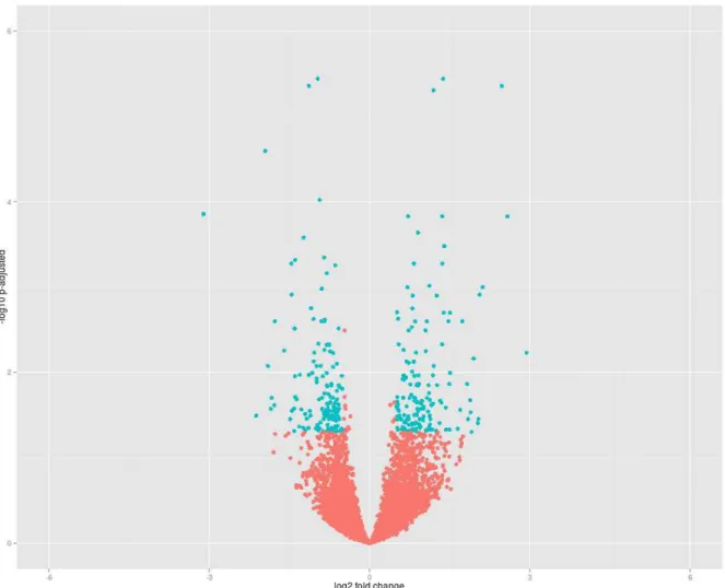
Identification of 13,145 genes in the muscle transcriptome was possible following quality filtering, alignment, and normalization procedures. Seventeen genes that were differentially expressed (Padj < 0.2) were grouped by biological function
through functional annotation clustering analysis (Table 1). The distribution of fold-change data of the genes expressed in both treatments was homogeneous (Figure 1).
Figure 1. MA plot of longissimus dorsi muscle of mature Bos taurus indicus cull cows
subjected to recovery weight gain under grazing conditions, showing gene distribution according to log2foldchange
In the group of differentially expressed genes annotated by Biomart Ensembl (Table 1), the METTL7A gene that was expressed in two transcripts in the MA group
droplet formation (BOUCHOUX et al., 2011). This is to be expected since these animals presented high body score and weight. A greater abundance of MSTN
transcripts (Table 1) was also observed in the MA group. This protein is a member of the transforming growth factor-β superfamily, which has been considered a novel and
unique negative regulator of muscle mass (RODGERS; GARIKIPATI, 2008; MCCROSKERY et al., 2003). MSTN inhibits the expression of myogenic regulatory
factors (RIOS et al., 2002) and satellite cell proliferation (FRY et al., 2014). The latter authors showed that proliferation is important in regulating muscle adaptations to hypertrophic growth in adults. Therefore, it seems plausible that cows under recovery weight gain, which are experiencing muscle hypertrophy, would present lower amounts of this negative growth regulator.
Transcripts of another protein related to growth factors, IGFBP5, were also
more abundant in the muscle of cows from the MA group. In these animals, we expected to find signs of cessation of muscle growth. IGFBP5 has been shown to be
an important regulator of IGF-I local action (JACKMAN; KANDARIAN, 2004) by
sequestering this growth factor, leading to decreases in protein synthesis (BAXTER, 2000). However, IGFBP-5 may have effects that are unrelated to its IGF-regulatory
role (TRIPATHI et al., 2009; MIYAKOSHI et al. 2001; TANNO et al., 2005). IPA analysis showed that IGF-I was the upstream gene regulating the greater expression
of IGFBP5 and MSTN (P = 0.023) in the MG group. This reinforces the idea that IGF-I, IGFBP5, and MSTN regulated muscle gain in this group of cows. Betacellulin (BTC)
has been reported in fibroblastic cells (WATANABE et al., 1994) with mitogenic action.
SEMA4A, Slc11A1, PGF, and FCN2 were more strongly expressed in the RG
group (Table 1). SEMA4A is part of a large family of extracellular proteins (ROTH et
al., 2009), and is involved in the regulation of cell migration and muscle angiogenesis (MEDA et al., 2012). It has also been linked to immune response (KUMANOGOH et al., 2002; KUMANOGOH et al., 2005), an important pathway related to renewal.
Slc11A1 is related to immune response and defense as well (DING et al., 2014), and PGF is related to the inflammatory process (CLAUS et al., 1996), pointing to
macrophage secretory and physiological function in the RG group. FCN2 is related to
apoptosis of myofibroblasts, endothelial cells, and macrophages (JENSEN et al., 2007), which can also indicate immune responses during tissue regrowth.
In summary, inflammatory responses seem to increase during regeneration in recovery growth as a response to remodeling. Tissue remodeling of the ECM has been shown to involve an initial inflammatory process (FIELDING et al., 1993) followed by interactions between fibroblasts and satellite cells that normally lead to fibrogenic processes (MURPHY et al., 2011).
Table 1 – Differentially genes expressed# in muscle from mature Bos taurus indicus
cull cows between maintenance of high body score and recovery gain conditions under grazing
Gene name Gene ID Base Mean log2(FC)* P-Value Padj# 1 IGFBP5 ENSBTAG00000007062 144.26 -1.5 <0.0001 0.1 2 SEMA4A ENSBTAG00000012228 10.00 1.3 <0.0001 0.1 3 FCN2 ENSBTAG00000048155 8.84 2.1 <0.0001 0.1 4 SLC11A1 ENSBTAG00000015520 14.13 1.5 <0.0001 0.1 5 NAP1L3 ENSBTAG00000019407 7.39 -1.4 <0.0001 0.2 6 UNC80 ENSBTAG00000015415 8.71 -2.2 0.0001 0.2 7 NEPN ENSBTAG00000039574 38.94 -1.7 0.0002 0.2 8 CIB2 ENSBTAG00000010981 52.88 -1.3 0.0001 0.2 9 *U. PROTEIN ENSBTAG00000032899 44.35 -1.3 0.0002 0.2 10 METTL7A ENSBTAG00000025005 337.68 -1.0 0.0001 0.2 11 PGF ENSBTAG00000013688 112.45 1.0 0.0001 0.2 12 RAB15 ENSBTAG00000003474 37.00 1.2 0.0002 0.2 13 KIF3C ENSBTAG00000019138 33.57 -1.1 0.0002 0.2 14 BTC ENSBTAG00000004237 877.91 -0.6 0.0002 0.2 15 INTS7 ENSBTAG00000018548 172.50 0.5 0.0002 0.2 16 TMLHE ENSBTAG00000011648 25.50 -0.8 0.0003 0.2 17 MSTN ENSBTAG00000011808 86.34 -1.8 0.0003 0.2
#annotated by BioMart Ensembl; *log2 (FC): log2(foldchange); Negative values:
upregulation in maintenance; Positive values: upregulation in recovery gain; #Padj:
adjusted P value for multiple testing with the Benjamini-Hochberg procedure (FDR);
&U. protein – uncharacterized protein
bonds. PGF and SMA4A were also included in the disulfide bonds. From the
enrichment data, it appears that NEPN was related to extracellular space (P < 0.1)
and MSTN to disulfide bonds (P < 0.1) in the MG group. In addition, BTC and IGFBP5 were found in both pathways.
Table 2 – Functional Annotation Clustering& of genes from mature Bos taurus indicus
cull cows during maintenance of high body score or recovery gain under grazing conditions
Annotation Cluster* Representative Annotation Terms Count P-Value Padj#
GOTERM_CC_ALL extracellular space 4 0.003 0.09
SP_PIR_KEYWORDS disulfide bond 6 0.004 0.09
SP_PIR_KEYWORDS Signal 6 0.009 0.10
SP_PIR_KEYWORDS Glycoprotein 6 0.013 0.11
GOTERM_CC_ALL extracellular region 6 0.002 0.12
SP_PIR_KEYWORDS Secreted 5 0.008 0.12
SP_PIR_KEYWORDS growth factor 3 0.004 0.16
GOTERM_CC_ALL extracellular region part 4 0.011 0.20 GOTERM_MF_ALL growth factor activity 3 0.004 0.25
GOTERM_MF_ALL receptor binding 4 0.009 0.28
GOTERM_MF_ALL protein binding 9 0.032 0.53
GOTERM_BP_ALL positive regulation of biological process 4 0.054 1.00 GOTERM_BP_ALL biological regulation 8 0.055 1.00 GOTERM_BP_ALL regulation of cellular process 7 0.093 1.00 GOTERM_BP_ALL regulation of biological process 7 0.120 1.00 GOTERM_BP_ALL positive regulation of cellular process 3 0.180 1.00
&analyzed by DAVID tool (annotation clusters had a group of enrichment scores of
1.82); *Annotation Cluster; SP_PIR: single protein of protein information resource; MF: molecular function; BP: biological process; CC: cellular component; #P
adj:
adjusted P value for multiple testing with the Benjamini-Hochberg procedure (FDR)
A group of cytokines, oncostatin M (OSM), chemokine ligand 2 (CCL2), and
interleukin 6 (IL6), were identified as upstream regulators of SEMA4A, PGF, and SLC11A1 (Appendixe A) in the RG group (P < 0.0076, Table 3). The cytokines
degradation (KJAER, 2004) and increased collagen transcription in response to remodeling of the connective tissue. This produces a large number of advanced glycation end products, reducing the impact on the synthesis of other types of collagen (DEGROOT et al., 2001).
Table 3 – Upstream regulators& of genes from mature Bos taurus indicus cull cows
during recovery gain under grazing conditions Upstream
regulator Molecule Type P value Target molecules
RBPJ transcription regulator 0.0028 PGF
SHH Peptidase 0.0045 PGF
EPAS1 transcription regulator 0.0054 PGF MTF1 transcription regulator 0.0061 PGF DLX3 transcription regulator 0.0104 PGF FOXD1 transcription regulator 0.0067 PGF HIF1A transcription regulator 0.0192 PGF
mir-182 microRNA 0.0242 PGF
miR-182-5p mature microRNA 0.0218 PGF AGT (SerpinA8) growth factor 0.0279 PGF
OSM Cytokine 0.0393 PGF
HRAS Enzyme 0.0439 PGF
SMARCA4 transcription regulator 0.0150 SLC11A1
Tnf (family) Group 0.0048 SLC11A1
PIAS2 transcription regulator 0.0061 SLC11A1 ATF3 transcription regulator 0.0391 SLC11A1
CCL2 Cytokine 0.0432 SLC11A1
IL6 Cytokine 0.0076 SEMA4A
&Analysis by Ingenuity Pathway Analysis (IPA)
We also found miR-182-5p (Table 3), a miRNA upstream regulator of the PGF
Table 4. Major extracellular matrix-related genes identified as unaltered in mature
Bos taurus indicus cull cows between maintenance of high body score
and recovery gain conditions Gene name
Gene ID Base Mean log2(FC)* P Value Padj #
1 ADAMTS2 ENSBTAG00000014665 86.474 0.1 0.718 0.9 2 COL1A2 ENSBTAG00000013472 1507.5 -0.1 0.993 0.9 3 COL3A1 ENSBTAG00000021466 2934.1 -0.1 0.966 0.9 4 COL4A1 ENSBTAG00000012849 5880.2 0.1 0.476 0.9 5 COL4A2 ENSBTAG00000025210 3351.4 0.1 0.503 0.9 6 DCN ENSBTAG00000003505 2546.5 0.4 0.214 0.9 7 ELASTIN ENSBTAG00000019517 433.35 0.1 0.762 0.9 8 HSPG2 ENSBTAG00000017122 2694.9 -0.1 0.963 0.9 9 SPINT2 ENSBTAG00000000182 336.72 0.4 0.008 0.5 10 SERPINH1& ENSBTAG00000001027 1304.2 0.1 0.735 0.9 11 MMP2 ENSBTAG00000019267 604.3 0.5 0.048 0.8 12 MMP14 ENSBTAG00000014824 42.949 0.2 0.517 0.9 13 TIMP2 ENSBTAG00000010899 488.1 0.1 0.560 09 *log2 (FC): log2(foldchange); Negative values: upregulation in maintenance; Positive values: upregulation in recovery gain; #Padj: adjusted P value for multiple testing with
the Benjamini-Hochberg procedure (FDR); &HSP47
3.4 Conclusion
Recovery gain provoked changes in genes associated with growth factors that are involved in the control of muscle cell proliferation and protein synthesis during recovery gain.
However, the transcriptome for proteases possibly involved in muscle protein turnover and related to collagen and other major ECM components was not affected in mature females experiencing recovery gain.
Acknowledgments
References
ABERLE, E.D.; REEVES, E.S.; JUDGE, M.D.; HUNSLEY, R.E.; PERRY, T.W. Palatability and muscle characteristics of cattle with controlled weight gain: time on a high energy diet. Journal of Animal Science, Champaign, v. 52, p. 757-763, 1981.
ALLINGHAM, P.G.; HARPER, G.S.; HUNTER, R.A. Effect of growth path on the tenderness of the semitendinosus muscle of Brahman-cross steers. Meat Science,
Barking, v. 48, p. 65–73, 1998.
ANDERS, S.; HUBER, W. Differential expression analysis for sequences count data.
Genome Biology, London, v. 11, p. R106.1-R.106.12, 2010.
ARCHILE-CONTRERAS, A.; MANDELL, I.B.; PURSLOW, P.P. Disparity of dietary effects on collagen characteristics and toughness between two beef muscles. Meat Science, Barking, v. 86, p. 491–497, 2010.
BAXTER , R.C. Insulin-like growth factor (IGF)-binding proteins: interactions with IGFs and intrinsic bioactivities. American Journal of Physiology: Endocrinology and Metabolism, Bethesda, v. 278, p. E967-E976, 2000.
BARTON-DAVIS, E.R.; SHOTURMA, D.I.; SWEENEY, H.L. Contribution of satellite cells to IGF-I induced hypertrophy of skeletal muscle. Acta Physiologica,
Stockholm, v. 167, p. 301-305, 1999.
BENJAMINI, Y.; HOCHBERG, Y. Controlling the false discovery rate: a practical and powerful approach to multiple testing. Journal of the Royal statistical society. Series B., London, v. 57, p. 289–300, 1995.
BOUCHOUX, J.; BEILSTEIN, F.; PAUQUAI, T.; GUERRERA, C.; CHATEAU, D.; LY, N.; ALQUB, M.; KLEIN, C.; CHAMBAZ, J.; ROUSSET, M.; LACORTE, J.; MOREL, E.; DEMIGNOT, S. The proteome of cytosolic lipid droplets isolated from
differentiated Caco-2/TC7 enterocytes reveals cell-specific characteristics. Biology of the Cell, Paris, v. 103, p. 499–517, 2011.
BRASAEMLE, D.L.; WOLINS, N.E. Packaging of fat: an evolving model of lipid
droplet assembly and expansion. Journal of Biological Chemistry, Bethesda,
v. 287, p. 2273–2279, 2012.
BYRNE, K.A.; WANG, Y.H.; LEHNERT, S.A.; HARPER, G.S.; MCWILLIAM, S.M.; BRUCE, H.L.; REVERTER , A. Gene expression profiling of muscle tissue in Brahman steers during nutritional restriction. Journal of Animal Science,
Champaign, v. 83, p. 1-12, 2005.
CHANG, H.Y.; YANG, X. Proteases for cell suicide: functions and regulation of
caspases. Microbiology and Molecular Biology Reviews, Washington, v. 64,
CLAUS, M.; WEICH, H.; BREIER, G.; KNIES, U.; ROCKL, W.; WALTENBERGER, J.; RISAU, W. The vascular endothelial growth factor receptor flt-1 mediates biological.
The Journal of Biological Chemistry, Bethesda, v. 271, n. 30, p. 17629–17634,
1996.
CLEMMONS, D.R. Role of insulin like growth factor binding proteins in controlling IGF action. Molecular and Cellular Endocrinology, London, v. 140, p. 19-24, 1998.
CROMBRUGGHE, B.; VUORIO, T.; KARSENTY, G. Control of type I collagen genes in scleroderma and normal fibroblasts. Rheumatic Diseases Clinics of North America, Maryland Heights, v. 16, p. 109-123, 1990.
DEGROOT, J.; VERZIJL, N.; BUDDE, M.; BIJLSMA, J.W.J.; LAFEBER, F.P.; TEKOPPELE, J.M. Accumulation of advanced glycation end products decreases
collagen turnover by bovine chondrocytes. Experimental Cell Research, Orlando,
v. 266, p. 303–310, 2001.
DING, XIAOLING; ZHANG, XIAODONG; YANG, YONG; DING, YUEYUN; XUE, WEIWEI; MENG, YUN; ZHU, WEIHUA; YIN, ZONGJUN. Polymorphism, expression of natural resistance-associated macrophage protein 1 encoding gene (NRAMP1) and its association with immune traits in pigs. Asian-Australasian Journal of Animal Sciences, Seoul, v. 27, p. 1189-1195, 2014.
ELLENBERGER, M.A.; JOHNSON, D.E.; CARSTENS, G.E.; HOSSNER, K.L.; HOLLAND, M.D.; NETT, T.M.; NOCKELS, C.F. Endocrine and metabolic changes during altered growth rates in beef cattle. Journal of Animal Science, Champaign,
v. 67, p. 1446-1454, 1989.
FIELDING R.A.; MANFREDI, T.J.; DING, W.; FIATARONE, M.A.; EVANS, W.J.; CANNON, J.G. Acute phase response in exercise. III. Neutrophil and IL-1 beta
accumulation in keletal muscle. The American Journal of Physiology, Washington,
v. 265, R166–R172, 1993.
FISHELL, V.K.; ABERLE, E.D.; JUDGE, M.D.; PERRY, T.W. Palatability and muscle properties of beef as influenced by pre slaughter growth rate. Journal of Animal Science, Champaign, v. 61, p. 151-157, 1985.
FRY, C.S., LEE, J.D.; JACKSON, J.R.; KIRBY, T.J.; STASKO, S.A.; LIU, H.;
DUPONT-VERSTEEGDEN, E.E.; MCCARTHY, J.J.; PETERSON, C.A. Regulation of the muscle fiber microenvironment by activated satellite cells during hypertrophy. The FASEB Journal, Bethesda, v. 28, p. 1654-1665, 2014.
GARRED, P.; HONORÉ, C.; MA, Y.J.; MUNTHE-FOG, L.; HUMMELSHOJ, T. MBL2, FCN1, FCN2 and FCN3: the genes behind the initiation of the lectin pathway of complement. Molecular Immunology, Elmsford, v. 46, n. 14, p. 2737-2744, 2009.
HUANG, D.W.; SHERMAN, B.T.; LEMPICKI, R.A. Systematic and integrative
analysis of large gene lists using DAVID bioinformatics resources. Nature Protocols,