Volume 2012, Article ID 619530,11pages doi:10.1155/2012/619530
Review Article
Nanophotonics for Molecular Diagnostics
and Therapy Applications
Jo˜ao Conde,
1, 2Jo˜ao Rosa,
1, 3Jo˜ao C. Lima,
3and Pedro V. Baptista
11CIGMH, Departamento de Ciˆencias da Vida, Faculdade de Ciˆencias e Tecnologia, Universidade Nova de Lisboa, Campus de Caparica,
2829-516 Caparica, Portugal
2Instituto de Nanociencia de Arag´on, Universidad de Zaragoza, Campus R´ıo Ebro, Edif´ıcio I+D, Mariano Esquillor s/n,
50018 Zaragoza, Spain
3REQUIMTE, Departamento de Qu´ımica, Faculdade de Ciˆencias e Tecnologia, Universidade Nova de Lisboa,
Campus de Caparica, 2829-516 Caparica, Portugal
Correspondence should be addressed to Pedro V. Baptista,[email protected]
Received 15 June 2011; Accepted 10 July 2011
Academic Editor: Danuta Wrobel
Copyright © 2012 Jo˜ao Conde et al. This is an open access article distributed under the Creative Commons Attribution License, which permits unrestricted use, distribution, and reproduction in any medium, provided the original work is properly cited.
Light has always fascinated mankind and since the beginning of recorded history it has been both a subject of research and a tool for investigation of other phenomena. Today, with the advent of nanotechnology, the use of light has reached its own dimension where light-matter interactions take place at wavelength and subwavelength scales and where the physical/chemical nature of nanostructures controls the interactions. This is the field of nanophotonics which allows for the exploration and manipulation of light in and around nanostructures, single molecules, and molecular complexes. What is more is the use of nanophotonics in biomolecular interactions—nanobiophotonics—has prompt for a plethora of molecular diagnostics and therapeutics making use of the remarkable nanoscale properties. In this paper, we shall focus on the uses of nanobiophotonics for molecular diagnostics involving specific sequence characterization of nucleic acids and for gene delivery systems of relevance for therapy strategies. The use of nanobiophotonics for the combined diagnostics/therapeutics (theranostics) will also be addressed, with particular focus on those systems enabling the development of safer, more efficient, and specific platforms. Finally, the translation of nanophotonics for theranostics into the clinical setting will be discussed.
1. Introduction
Nanophotonics deals with the interaction of light with matter at a nanometer scale, providing challenges for fun-damental research and opportunities for new technologies, encompassing the study of new optical interactions, materi-als, fabrication techniques, and architectures, including the exploration of natural and synthetic, or artificially engi-neered, structures such as photonic crystals, holey fibers, quantum dots, subwavelength structures, and plasmonics
[1,2]. The use of photonic nanotechnologies in medicine
is a rapidly emerging and potentially powerful approach for disease protection, detection, and treatment. The high speed of light manipulation and the remote nature of optical methods suggest that light may successfully connect diag-nostics, treatment, and even the guidance of the treatment
in one theranostic procedure combination of therapeutics with diagnostics (including patient prescreening and therapy monitoring).
Limitations in medical practice are closely associated with the fact that diagnostics, therapy, and therapy guidance are three discrete and isolated stages. In order to overcome some of the sensitivity and specificity of current medicines, theranostics unites the three above stages in one single process, supporting early-stage diagnosis and treatment [3, 4]. Nowadays, there is an ever increasing need to enhance the capability of theranostics procedures where nanophotonics-based sensors may provide for the simultaneous detection of several gene-associated conditions and nanodevices utilizing light-guided and light-activated therapy with the ability to
Biobar-codes
Colorimetric assays
Plasmonics
Nanophototherapy
Photons
Nanoshells for thermal ablation
Cell death
FRET FRET
QDs
Near/far
Field effects
SERS Reporters
Surface enhanced raman scattering
Bioimaging
− → E
−→
E Labels for immunoassays
Fluorescence-based systems
Nanophotonics for theranostics
200 400 600 800 1000 1200
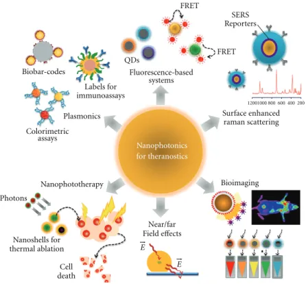
Figure1:Nanophotonics for theranostics. Nanoparticles-based strategies can be used for biosensing usingplasmonic nanosensors, such as
metal nanoparticles functionalized with nucleic acid strand for colorimetric assays and biobar codes for protein detection or intense labels for
immunoassays. Some nanoparticle systems can also be used for sensing by exploring a typicalFRET systemor can be surrounded withRaman
reportersin order to providein vivodetection and tumour targeting. In fact, NPs symbolize an important class of materials with unique
features suitable forbiomedical imagingapplications such as increased sensitivity in detection and high quantum yields for fluorescence.
Alternatively, NPs can surveynear/far fieldenhancing qualities that hold promise for a bounty of novel applications in optics and photonics.
Engineered NPs can also act asphototherapeutic agentsthat can be attached to specific targets for selective damage to cancer cells.
2. Nanophotonics For Diagnostics
2.1. Surface Plasmons on Nanoparticles and Surfaces. Surface plasmons are collective charge oscillations that occur at the interface between conductors and dielectrics. They can take various forms, ranging from freely propagating electron density waves along metal surfaces to localized electron
oscillations on metal nanoparticles (NPs) [5, 6]. When
light passes through a metal nanoparticle, it induces dipole moments that oscillate at the respective frequency of the incident wave, consequently dispersing secondary radiation in all directions. This collective oscillation of the free conduc-tion electrons is called localized surface plasmon resonance (LSPR). Light on NP induces the conduction electrons to oscillate collectively with a resonant frequency that depends on the nanoparticles’ size, shape, composition, interparticle distance, and environment (dielectric properties) [7–10]. As a result of these SPR modes, the nanoparticles absorb and scatter light so intensely that single NPs are easily observed by eye using dark-field (optical scattering) microscopy. Plasmonic NPs provide a nearly unlimited photon resource for observing molecular binding for longer periods of time, once they do not blink or bleach like fluorophores [11].
Nanoparticle-based colorimetric assays for diagnostics have been a subject of intensive research, where LSPR can
be used to detect DNA or proteins by the changes in the local index of refraction upon adsorption of the target molecule to the metal surface. Due to the intense SPR in the visible yielding extremely bright colors, gold nanoparticle colloids have been widely used of molecular diagnostics. In fact, gold nanoparticles (AuNPs) functionalized with ssDNA capable of specifically hybridizing to a complementary target for the detection of specific nucleic acid sequences in biological samples have been extensively used [12–27]. Other approaches use the AuNPs’ plasmonic as a core/seed that can be tailored with a wide variety of surface functionalities to provide highly selective nanoprobes for diagnostics [28] or the SPR scattering imaging or SPR absorption spectroscopy generated from antibody-conjugated AuNPs in molecular biosensor techniques for the diagnosis of oral epithelial living
cancer cellsin vivoandin vitro [29] and the use of
multi-functional AuNPs which incorporate both cytosolic delivery and targeting moieties on the same particle functioning as intracellular sensors to monitor actin rearrangement in live fibroblasts [30].
Plasmonic NPs have also been used as extremely intense labels for immunoassays [31–34] and biochemical sensors
[19,35–37]. Also, the use of colloidal silver plasmon resonant
particles (PRPs) coated with standard ligands as
immunocytology assays [34]. Most notably, a nanoparticle-based Biobar code has been developed for the detection of proteins that relies on magnetic microparticle probes with antibodies that specifically bind a target of interest and nanoparticle probes that are encoded with DNA that is unique to the protein target of interest and antibodies that can sandwich the target captured by the microparticle probes [33]. Haes and coworkers have reported on an optical biosensor based on localized surface plasmon resonance spectroscopy developed to monitor the interaction between
the antigen, amyloid-β-derived diffusible ligands (ADDLs),
and specific anti-ADDL antibodies, used in the detection of a biomarker for Alzheimer’s Disease [35].
2.2. Raman-Spectroscopy-Based Systems. When light inter-acts with a substance, it can be absorbed, transmitted, or scattered. Scattered radiation can result from an elastic col-lision (Rayleigh scattering) or inelastic (Raman scattering). Raman spectroscopy is based on a change of frequency when light is inelastically scattered by molecules or atoms resulting in a molecular fingerprint information on molec-ular structure or intermolecmolec-ular interaction of a specific process or molecule. The potential of Raman spectroscopy as biomedical diagnostics tool is rather low due to its low
cross-section (∼10−30cm2) that results in low sensitivity [38].
However, in 1977, two groups independently described the use of noble metal surfaces to enhance the Raman scattering
signal of target molecules [39, 40]—Surface enhancement
raman spectroscopy (SERS). Jeanmaire and Van Duyne proposed a twofold electromagnetic field enhancement that was later associated with the interaction between the incident and scattered photons with the nanostructure’s LSPR [41]. Simultaneously, Albrecht and Creighton suggested the source of the enhancement to be caused by a specific interaction between an adsorbate and the nanoparticle surface, briefly, a charge transfer from the adsorbate into the empty energetic levels on the metal surface or from the occupied levels of the nanoparticle’s surface to the adsorbate [42–44].
Generally, SERS requires that the biological analyte reaches a suitable surface where the substrates are treated as two-dimensional macroscopic surfaces onto which adsorbed
molecules suffer a local-field enhancement. Despite direct
adsorption not being a good solution because of its
depen-dence on the affinity between substrate and analyte,
a method to identify and distinguish different strains of virus
based on signal differences generated by the surface
amino-acids using silver nanorods has been successfully developed [45]. Using a similar approach of direct adsorption, Pˆınzary
et al. used naked silver nanoparticles to differentiatein situ
healthy colon from carcinoma colon tissue [46]. Nanotags have been widely employed to address the lack of specificity
[47,48]. These nanotags usually possess a metallic colloidal
core functionalized with a Raman reporting molecule and the specific molecule used to capture the analyte and have
been used to directly detect DNA sequences [49, 50] and
amplified DNA products of epizootic pathogens using com-plementary DNA strands so that only the comcom-plementary target hybridizes with the probes [51]. Using a similar
system, but exploring the distance-dependent enhancement of the electromagnetic field with a hairpin probe molecule, Wabuyele has also been used to distinguish single nucleotide polymorphisms in cancer-related genes [52]. Combining nanotags with other nanoparticles or binding surfaces that target the same analyte in a sandwich conformation proved useful to detect antibodies in serum [53]. A similar approach using a flat substrate instead of NPs had already been
proposed to detect DNA, RNA, and proteins [54, 55].
However, in this approach, the substrate is used only to immobilize the analyte; a gold-nanoparticle-based nanotag is used to identify the analyte and the surface enhancement is obtained by silver coating of the nanotag. miRNA profiling
has also been pursued via a slightly different approach based
on the hybridization of the target molecules with a thiolated oligonucleotide and subsequent functionalization on a silver substrate [56]. SERS have also been explored to identify changes in the analyzed system such as interaction between DNA and xenobiotic molecules like cisplatin [57] or
DNA-binding proteins [58, 59]. The combination of magnetic
iron/gold core-shell nanoparticles with gold nanorods has
also been used to specifically enumerate E. coli in water
samples in a rapid and sensitive test [60]. In this case, the magnetic nanoparticles are used to concentrate the bacteria, improving the Raman signal by concentration and the posteriorly added gold nanorods serve as Raman signal enhancers.
SERS can also be used in conjunction with colloidal
gold to detect and target tumorsin vivo, where the AuNPs
are surrounded with Raman reporters that provide light
emission 200 times brighter than quantum dots [61, 62].
It was also found that the Raman reporters became more stable and yielded larger optical enhancements when NPs were encapsulated with a thiol-modified polyethylene glycol coat, which also allows for increased biocompatibility and
circulation times in vivo. When conjugated to
tumor-targeting ligands, these conjugated SERS-NPs were able to target tumor markers at surface of malignant cells, such as epidermal growth factor receptor (EGFR) that is sometimes overexpressed in cells of certain cancer types [29] and used to locate the tumor in xenograft tumor models [50].
2.3. Fluorescence-Based Systems. Quantum dots (QDs) are semiconductor nanoparticles with narrow, tunable, symmet-rical emission spectra, and high quantum yields [63–65], and together with compatibility with DNA and proteins, make QDs exceptional substitutes as fluorescence labels. The use of QDs for nucleic acid characterization has long been proposed, for example, CdSe/ZnS QDs for SNP identification on human TP53 gene, multiallele detection
of hepatitis B and C viruses [66], and in situdetection of
adenine itself to quench emission of the quantum-dot-like nanoparticles [69].
Several studies report on the modulation of fluorophores at the vicinity of nanoparticles (e.g., gold, silver, and
quan-tum dots) [70,71], an interaction that has found application
in a variety of systems to detect biologically relevant targets with particular focus upon AuNPs due to their ease in func-tionalization with biomolecules [72–75]. Several methods based on the quenching of fluorescence have been proposed for DNA detection consisting of fluorophore-labeled ssDNA electrostatically adsorbed onto gold nanoparticles [76], car-bon nanotubes [77], and carcar-bon nanoclots [78], where the presence of a complementary target triggers desadsorption of the newly formed dsDNA from the nanostructures due to the electrostatic variation between ssDNA and dsDNA, and fluorescence emission is restored. Also, fluorescence quenching of fluorophores close to metal nanoparticles functionalized with thiol-modified oligonucleotides has been
explored in different conformations. Tang and co-workers
proposed a method to probe hydroxyl radicals using an AuNP-oligonucleotide-FAM system where the hydroxyl rad-ical promotes strand breakage and consequent release of FAM, restoring the previously quenched fluorescence [79]. The same quenching mechanism was used to detect specific DNA strands using two probes (one with an AuNP label and another labeled with TAMRA) that hybridize to two DNA sequences near each other [80], bringing the fluorophore and AuNP close enough to quench fluorescence emission.
Proteins have also been probed through nanoparticle-fluorescence-mediated systems, for example, human blood proteins have been let to interact with fluorescent AuNPs and detected through quenching [81]. In another example, a sandwich immunoassay using AuNPs quenching has been proposed for the detection of the protein cardiac troponin T
by its interaction with two different antibodies, one attached
to AuNPs and the other labeled with fluorescent dyes [82]. By means of an opposite modulation, infrared fluorescent nanoparticles showed enhanced fluorescence when interact-ing with protein [83].
A popular application of fluorescence modulation by nanoparticles has been specific ion sensing. Su and co-workers developed a copper sensor by covering fluores-cent DNA-Cu/Ag nanoclusters with mercaptopropionic acid which quenches the intrinsic fluorescence of the
nanopar-ticle; in the presence of Cu2+, the capping agent is
oxi-dized to form a disulfide compound resulting in release of the nanoparticle and restoration of emission suitable for quantification between 5 and 200 nM [84]. A very specific colorimetric and fluorimetric method to detect
Hg2+ions was developed with porphyrin-modified Au@SiO2
nanoparticles, where the intensively fluorescent red complex
turns green and weakly fluorescent in presence of Hg2+[85].
Another examples include sensing of Pb2+ and adenosine
by combining an adenosine aptamer and a DNAzyme with an abasic site where 2-amino-5,6,7-trimethyl-1,8-naphthyridine is trapped to quench its fluorescence [86].
When in solution, Pb2+ enables the DNAzyme to cleave its
substrate thus removing the fluorescent compound from the abasic site restoring its fluorescence. Similarly, the presence
of adenosine induces structural change of the aptamer, resulting in the release of the fluorescent molecule from the DNA duplex and a subsequent fluorescence enhancement.
2.4. Nanophotonics Bioimaging. Nanoparticles show unique features suitable for biomedical imaging applications, such as an increased sensitivity in detection through amplification of signal changes (e.g., magnetic resonance imaging); high fluorescence quantum yields and large magnetic moments; properties that induce phagocytosis and selective uptake by macrophages (e.g., liposomes); physicochemical manip-ulations of energy (i.e., quantum dots); among others [87]. Because light absorption from biologic tissue com-ponents is minimized at near infrared (NIR) wavelengths, most nanoparticles (e.g., noble metal and magnetic NPs, nanoshells, nanoclusters, nanocages, nanorods and quantum
dots) forin vivoimaging and therapy have been designed to
strongly absorb in the NIR and used forin vivodiagnostics
[83, 88, 89]. Ex vivo and in vivo imaging applications of
nanoparticles have included their use as contrast agents for magnetic resonance imaging (MRI) [90], optical coherence tomography (OCT) [91–93], photoacoustic imaging (PAI) [94], and two-photon luminescence (TPL) spectroscopy [95].
2.4.1. Magnetic Resonance Imaging. Magnetic resonance imaging (MRI) is based heavily on nuclear magnetic reso-nance (NMR), first described by R. Damadian. Magnetic
res-onance measurements cause no obvious deleterious effects
on biological tissue, and the incident radiation consists of common radio frequencies at right angles to a static magnetic field [96]. Iron oxide nanoparticles show super-paramagnetism, allowing for the facile alignment of the magnetic moments to an applied magnetic field, thus of great interest as contrast agents for MRI [97]. Presently, magnetic iron oxide nanoparticles are routinely used as contrast agents to enhance an MRI image, providing sharper contrast between soft and hard tissue in the body (e.g. liver and spleen or lymph nodes) [98]. Jun et al. pre-sented a synthetically controlled magnetic nanocrystal model system that led to the improvement of high-performance nanocrystal—antibody probe systems for the diagnosis of breast cancer cells via magnetic resonance imaging [99].
Also, MnFe2O4nanocrystalsfunctionalizedwith an antibody
conjugate (herceptin) capable of specific targeting of
can-cerous cells was successfully used for in vivo MRI in mice
[88]. Driehuysb et al. developed an imaging method to detect submillimeter-sized metastases with molecular specificity by targeting cancer cells with iron oxide nanoparticles functionalized with cancer-binding ligands, demonstrating
in vivo detection of pulmonary micrometastases in mice injected with breast adenocarcinoma cells [100]. Hybrid NPs with a superparamagnetic iron oxide/silica core and a gold nanoshell, with significant absorbance and scattering in the
NIR region, have been used in vivoas contrast agents for
2.4.2. Optical Coherence Tomography. Optical Coherence Tomography (OCT) is an imaging modality that provides cross-sectional subsurface imaging of biological tissue with micrometer scale resolution which is based on a broadband light source and a fiber-optic interferometer. It captures three-dimensional images from within optical scattering media, typically employing near-infrared light. The use of relatively long wavelength light allows it to penetrate into the scattering medium [102–104]. The extra scattering provided by Au-nanoshells enhances optical contrast and brightness for improved diagnostic imaging of tumors in mice due to the preferential accumulation of the nanoshells
in the tumor [105]. Tseng et al.developed nanorings with
a localized surface plasmon resonance covering a spectral range of 1300 nm that produced both photothermal and
image contrast enhancement effects in OCT when delivered
into pig adipose samples [106]. Additionally, the image
con-trast enhancement effect could be isolated by continuously
scanning the sample with a lower scan frequency, allowing to
effectively control the therapeutic modality. In the same way,
gold capped nanoroses have been used in photothermal OCT
to detect macrophages inex vivorabbit arteries [107].
2.4.3. Photoacoustic Imaging. In photoacoustic imaging (PAI) and photoacoustic tomography (PAT), a pulse of NIR laser light, typically 757 nm, is used in resonance with the surface plasmon instead of a continuous NIR source. With this technique causing rapid thermal expansion of the surrounding media, the generated sound wave can be detected on the surface of the subject. NIR reduces the amount of absorption that occurs, but absorption of light
by various other organs is unavoidable [108, 109]. Yang
et al. demonstrated the feasibility of using poly(ethylene
glycol)-coated Au nanocages as a newin vivoNIR
contrast-enhancing agent for photoacoustic tomography and image their distribution in the vasculature of rat brain. These Au-nanocages enhanced the contrast between blood and the surrounding tissues by up to 81%, achieving a more detailed image of vascular structures at greater depths. Additionally, they were shown to present slight advantages over
Au-nanoshells, being better suited for in vivo applications,
specially due to their more compact size (<50 nm compared
to>100 nm for Au-nanoshells) and larger optical absorption
cross-sections [110]. Due to the ability of gold-nanorods to have the maximum of the plasmon resonance tuned
further into the NIR, Motamedi et al.reported a contrast
agent for a laser optoacoustic imaging system for in vivo
detection of gold nanorods and to enhance the diagnostic
power of optoacoustic imaging [111]. Song et al.proposed
a noninvasive in vivo spectroscopic photoacoustic sentinel
lymph node mapping in a rat model using gold nanorods as lymph node tracers [112].
2.4.4. Two-Photon Luminescence. In two-photon lumines-cence (TPL) spectroscopy, an electron is excited from the conductance band to the valence band of the metal nanoparticles using two photons. As the electron relaxes to the conductance band, light is released and amplified
due to a resonant coupling with localized surface plasmons, enhancing a variety of linear and nonlinear optical
prop-erties [113, 114]. TPL was first described by Boyd et al.
that found that roughened metal surfaces exhibited much
higher induced luminescence efficiency than smooth surfaces
[115]. In fact, TPL is a potentially powerful technique for noninvasive imaging at the micron scale hundreds of microns deep into scattering tissue. This way, it ought to be possible to discriminate cancerous and healthy tissue based on two-photon imaging from endogenous fluorophores. For enhanced imaging, two-photon contrast agents have been developed showing the ability to increase signal-to-noise ratio and targeted to molecular signatures of interest that are not fluorescent. Because imaging of intrinsic fluorophores
is often difficult due to their relatively weak signals, the use
of such a bright contrast agent holds the promise to enable
in vivo applications of two photon imaging in a clinical
setting [113,116,117]. Wang et al. collected images of single
gold nanorods flowing in the mouse ear blood vessels with luminescence three times stronger than background [114]. It is worth mentioning that the TPL signal from a single nanorod is 58 times that of the two-photon fluorescence signal from a single rhodamine molecule.
2.4.5. QDs for In Vivo Imaging. In the last decade, water soluble bioconjugated QDs have been increasingly applied
for imaging [63,64,118] However, QD probes for imaging
show poor stability once inside cytosolic environment and reduced biocompatibility in living organisms [119], which
constitutes a serious drawback for widespread in vivo
application.
Despite the serious concerns related to thein vivouse of
QDs, these nanocrystals show remarkable imaging properties that may be judged of value for improved diagnostics. In fact, QDs have proven of great value when imaging vascular networks of mammals such as lymphatic and cardiovascular systems [120–124]. Also, Kim et al. demonstrated that quan-tum dots allowed a major cancer surgery to be performed in large animals (mice and pigs) under complete image guidance, by locating the position of sentinel lymph nodes [125]. With similar potential to that observed when imaging the lymph system, the imaging of cardiovascular systems
has also been achieved using QDs [126, 127]. Larson et
al. demonstrated that QDs retained their fluorescence after injection and could be detected in the capillaries of skin and adipose tissue of a mouse [128]. The fluorescent emission and multiplexing capabilities of QDs are being exploited to improve the sensitivity and selectivity in the early detection
of tumors [61,129,130]. ˚Akerman et al. described for the
demonstrate the potential of QDs for ultrasensitive and
multiplexed imaging of molecular targetsin vivo.
3. Nanophotonics for Therapy
Nanophototerapy uses pulsed lasers and absorbing nanopar-ticles attached to specific targets for selective damage to cancer cells. Plasmonic photothermal therapy (PPTT) and photodynamic therapy (PDT) are two of the main techniques that take advantage of the selective absorbance of the surface plasmon resonance and the fact that the nanoparticles relax by liberating heat into their surrounding environment.
3.1. Plasmonic Photothermal Therapy. Plasmonic photother-mal therapy is a less invasive experimental technique that holds great promise for the treatment of cell malignancies and, in particular, of cancer. It combines two key compo-nents: (i) light source, specifically lasers with a spectral range of 650–900 nm for deep tissue penetration and (ii) optical absorption of AuNPs which release the optical irradiation as heat in the picoseconds time scale, thereby inducing
photothermal ablation [133–135]. Kirui et al.reported the
use of gold and iron oxide hybrid nanoparticles in targeting, imaging, and selective thermal killing of colorectal cancer cells [136]. Huang and colleagues have demonstrated that gold nanorods have a longitudinal absorption band in the
NIR on account of SPR oscillations and are effective as
photothermal agents [137]. Gold nanorods aspect ratios allow tuning the SPR band from the visible to the NIR (transmits readily through human skin and tissue), mak-ing them suitable for photothermal converters of near
infrared light for in vivo applications [138,139]. Effective
photothermal destruction of cancer cells and tissue have been demonstrated for other gold nanostructures, such as branched gold nanoparticles [140], gold nanoshells [141– 143], gold nanocages [134], and gold nanospheres [144].
3.2. Photodynamic Therapy. Photodynamic therapy employs chemical photosensitizers that generate reactive oxygen
species (ROS), such as a singlet oxygen (1O2), capable of
tumor destruction [145,146]. This technique is noninvasive
and can be applied locally or systemically without noticeable
cumulative toxicity effects without high costs. To attain
maximal killing efficiency of tumor cells, the photosensitizer
must be in close proximity to the tumor cells, thus requiring specific targeting when administered systemically. One of the major limitations is the poor tissue penetration of high-energy light and the systemic dispersal of the photosensitizer
[147,148].
Aiming at circumventing some of the limitations of
pho-todynamic therapy, Zhang et al.reported a new type of
pho-tosensitizers based on photon upconverting nanoparticles (a process where low energy light, usually near-infrared (NIR) or infrared (IR), is converted to higher energies, ultraviolet (UV), or visible, via multiple absorptions or energy transfer
steps) [149,150]. One year later, Yong et al. reported the
use of NPs modified with zinc phthalocyanin photosensitizer that produce green/red emission on near-infrared (NIR)
excitation and is capable of singlet oxygen sensitization; upon targeted binding to cancer cells, significant cell destruction
was induced [151]. Recently, Qian et al.published similar
results with the use of zinc phthalocyanine nanocrystals coated with a uniform layer of mesoporous silica [152].
4. Conclusions
Light is an amazing intermediate with a gargantuan capacity for carrying multiple information and functions. Instinc-tively, we view light as rays, which propagate in a single direc-tion, either being absorbed or reflected to some extent by any object on which it impinges. However, the propagation of
light through a material is itself a quantum effect, involving
the excitation and relaxation of electrons in the material. It is well known that light has the facility to act through biological, chemical, mechanical, and thermal pathways at molecular/cellular levels in diagnostic and therapeutic applications.
Currently, we are in the dawn of a new age in therapy driven by nanotechnology vehicles. Although there are tech-nical challenges associated with the therapeutic application of nanodevices, the integration of therapy with diagnostic profiling has accelerated the pace of discovery of new nanotechnology methods. In addition to continuing to push forward on the above challenges, nanotechnology together with photonics can be used both for identifying useful target candidates and for validating their importance in disease states. Nanophotonics may present new opportunities for personalized medicine in which diagnosis and treatment are based on each individual’s molecular profile. Further
research into the fundamental mechanisms that efficiently
control light using nanodevices could unveil new dimensions of nanoparticle-mediated theranostic systems.
Here, we have attempted to give the reader a limited overview of some aspects of the current state of research into the fascinating aspects and control over nanophotonics in molecular diagnostics and therapy applications.
Acknowledgment
The authors acknowledge FCT/MCTES—CIGMH(Portugal) for financial support.
References
[1] M. Ohtsu, K. Kobayashi, T. Kawazoe, T. Yatsui, and M.
Naruse, Principles of Nanophotonics, Series in Optics and
Optoelectronics, Taylor & Francis, CRC Press, 2008. [2] Y. Shen and P. N. Prasad, “Nanophotonics: a new
multidisci-plinary frontier,”Applied Physics B, vol. 74, no. 7-8, pp. 641–
645, 2002.
[3] T. Lammers, S. Aime, W. E. Hennink, G. Storm, and F.
Kiessling, “Theranostic nanomedicines,”Accounts of
Chem-ical Research. In press.
[4] F. Pene, E. Courtine, A. Cariou, and J. P. Mira, “Toward
ther-agnostics,”Critical Care Medicine, vol. 37, no. 1, pp. S50–S58,
[5] W. L. Barnes, A. Dereux, and T. W. Ebbesen, “Surface
plasmon subwavelength optics,”Nature, vol. 424, no. 6950,
pp. 824–830, 2003.
[6] U. Kreibig and M. Vollmer,Optical Properties of Metal
Clus-ters, vol. 25 ofSpringer Series in Materials Science, Springer,
Berlin, Germany, 1995.
[7] M. C. Daniel and D. Astruc, “Gold nanoparticles: assembly, supramolecular chemistry, quantum-size-related properties, and applications toward biology, catalysis, and
nanotechnol-ogy,”Chemical Reviews, vol. 104, no. 1, pp. 293–346, 2004.
[8] S. Eustis and M. A. El-Sayed, “Why gold nanoparticles are more precious than pretty gold: Noble metal surface plasmon resonance and its enhancement of the radiative and nonradiative properties of nanocrystals of different shapes,”
Chemical Society Reviews, vol. 35, no. 3, pp. 209–217, 2006. [9] S. K. Ghosh and T. Pal, “Interparticle coupling effect on
the surface plasmon resonance of gold nanoparticles: from
theory to applications,”Chemical Reviews, vol. 107, no. 11,
pp. 4797–4862, 2007.
[10] W. Zhao, M. A. Brook, and Y. Li, “Design of gold
nanoparticle-based colorimetric biosensing assays,”
Chem-BioChem, vol. 9, no. 15, pp. 2363–2371, 2008.
[11] J. N. Anker, W. P. Hall, O. Lyandres, N. C. Shah, J. Zhao, and R. P. Van Duyne, “Biosensing with plasmonic nanosensors,”
Nature Materials, vol. 7, no. 6, pp. 442–453, 2008.
[12] P. Baptista, G. Doria, D. Henriques, E. Pereira, and R. Franco, “Colorimetric detection of eukaryotic gene expression with
DNA-derivatized gold nanoparticles,”Journal of
Biotechnol-ogy, vol. 119, no. 2, pp. 111–117, 2005.
[13] P. Baptista, E. Pereira, P. Eaton et al., “Gold nanoparticles for
the development of clinical diagnosis methods,”Analytical
and Bioanalytical Chemistry, vol. 391, no. 3, pp. 943–950, 2008.
[14] Y. C. Cao, R. Jin, C. S. Thaxton, and C. A. Mirkin, “A two-color-change, nanoparticle-based method for DNA
detection,”Talanta, vol. 67, no. 3, pp. 449–455, 2005.
[15] M. M. C. Cheng, G. Cuda, Y. L. Bunimovich et al., “Nanotechnologies for biomolecular detection and medical
diagnostics,”Current Opinion in Chemical Biology, vol. 10,
no. 1, pp. 11–19, 2006.
[16] J. Conde, J. M. de la Fuente, and P. V. Baptista, “RNA quantification using gold nanoprobes - application to cancer
diagnostics,”Journal of Nanobiotechnology, vol. 8, article no.
5, 2010.
[17] P. Costa, A. Amaro, A. Botelho, J. In´acio, and P. V. Baptista, “Gold nanoprobe assay for the identification of mycobacteria
of the Mycobacterium tuberculosis complex,”Clinical
Micro-biology and Infection, vol. 16, no. 9, pp. 1464–1469, 2010. [18] G. Doria, R. Franco, and P. Baptista, “Nanodiagnostics:
fast colorimetric method for single nucleotide
polymor-phism/mutation detection,” IET Nanobiotechnology, vol. 1,
no. 4, pp. 53–57, 2007.
[19] R. Elghanian, J. J. Storhoff, R. C. Mucic, R. L. Letsinger, and C. A. Mirkin, “Selective colorimetric detection of polynucleotides based on the distance-dependent optical
properties of gold nanoparticles,”Science, vol. 277, no. 5329,
pp. 1078–1081, 1997.
[20] H. Li and L. Rothberg, “Colorimetric detection of DNA sequences based on electrostatic interactions with
unmodi-fied gold nanoparticles,”Proceedings of the National Academy
of Sciences of the United States of America, vol. 101, no. 39, pp. 14036–14039, 2004.
[21] C. A. Mirkin, R. L. Letsinger, R. C. Mucic, and J. J. Storhoff, “A DNA-based method for rationally assembling
nanoparticles into macroscopic materials,”Nature, vol. 382,
no. 6592, pp. 607–609, 1996.
[22] W. J. Qin and L. Y. L. Yung, “Nanoparticle-based detection and quantification of DNA with single nucleotide
poly-morphism (SNP) discrimination selectivity,” Nucleic Acids
Research, vol. 35, no. 17, article no. e111, 2007.
[23] K. Sato, K. Hosokawa, and M. Maeda, “Rapid aggregation of gold nanoparticles induced by non-cross-linking DNA
hybridization,”Journal of the American Chemical Society, vol.
125, no. 27, pp. 8102–8103, 2003.
[24] K. Sato, K. Hosokawa, and M. Maeda, “Non-cross-linking gold nanoparticle aggregation as a detection method for
single-base substitutions,”Nucleic Acids Research, vol. 33, no.
1, article e4, 2005.
[25] J. J. Storhoff, A. D. Lucas, V. Garimella, Y. P. Bao, and
U. R. M¨uller, “Homogeneous detection of unamplified genomic DNA sequences based on colorimetric scatter of
gold nanoparticle probes,”Nature Biotechnology, vol. 22, no.
7, pp. 883–887, 2004.
[26] T. A. Taton, C. A. Mirkin, and R. L. Letsinger, “Scanometric
DNA array detection with nanoparticle probes,”Science, vol.
289, no. 5485, pp. 1757–1760, 2000.
[27] C. S. Thaxton, D. G. Georganopoulou, and C. A. Mirkin, “Gold nanoparticle probes for the detection of nucleic acid
targets,”Clinica Chimica Acta, vol. 363, no. 1-2, pp. 120–126,
2006.
[28] C. C. You, O. R. Miranda, B. Gider et al., “Detection and identification of proteins using nanoparticle-fluorescent
polymer ’chemical nose’ sensors,” Nature Nanotechnology,
vol. 2, no. 5, pp. 318–323, 2007.
[29] I. H. El-Sayed, X. Huang, and M. A. El-Sayed, “Surface plasmon resonance scattering and absorption of anti-EGFR antibody conjugated gold nanoparticles in cancer
diagnos-tics: applications in oral cancer,”Nano Letters, vol. 5, no. 5,
pp. 829–834, 2005.
[30] S. Kumar, N. Harrison, R. Richards-Kortum, and K. Sokolov, “Plasmonic nanosensors for imaging intracellular
biomark-ers in live cells,”Nano Letters, vol. 7, no. 5, pp. 1338–1343,
2007.
[31] W.-C. Law, K.-T. Yong, A. Baev, and P. N. Prasad, “Sensitivity improved surface plasmon resonance biosensor for cancer
biomarker detection based on plasmonic enhancement,”ACS
Nano, vol. 5, no. 6, pp. 4858–4864, 2011.
[32] J. S. Mitchell and T. E. Lowe, “Ultrasensitive detection of testosterone using conjugate linker technology in a nano-particle-enhanced surface plasmon resonance biosensor,”
Biosensors and Bioelectronics, vol. 24, no. 7, pp. 2177–2183, 2009.
[33] J. M. Nam, C. S. Thaxton, and C. A. Mirkin, “Nanoparticle-based bio-bar codes for the ultrasensitive detection of
proteins,”Science, vol. 301, no. 5641, pp. 1884–1886, 2003.
[34] S. Schultz, D. R. Smith, J. J. Mock, and D. A. Schultz, “Single-target molecule detection with nonbleaching multicolor
optical immunolabels,”Proceedings of the National Academy
of Sciences of the United States of America, vol. 97, no. 3, pp. 996–1001, 2000.
[35] A. J. Haes, L. Chang, W. L. Klein, and R. P. Van Duyne, “Detection of a biomarker for Alzheimer’s disease from synthetic and clinical samples using a nanoscale optical
biosensor,”Journal of the American Chemical Society, vol. 127,
[36] A. D. McFarland and R. P. Van Duyne, “Single silver nanoparticles as real-time optical sensors with zeptomole
sensitivity,”Nano Letters, vol. 3, no. 8, pp. 1057–1062, 2003.
[37] G. Raschke, S. Kowarik, T. Franzl et al., “Biomolecular recog-nition based on single gold nanoparticle light scattering,”
Nano Letters, vol. 3, no. 7, pp. 935–938, 2003.
[38] M. K. Hossain and Y. Ozaki, “Surface-enhanced Raman
scattering: facts and inline trends,”Current Science, vol. 97,
no. 2, pp. 192–201, 2009.
[39] D. L. Jeanmaire and R. P. Van Duyne, “Surface Raman spectroelectrochemistry Part I. Heterocyclic, aromatic, and aliphatic amines adsorbed on the anodized silver electrode,”
Journal of Electroanalytical Chemistry, vol. 84, no. 1, pp. 1–20, 1977.
[40] M. G. Albrecht and J. A. Creighton, “Anomalously intense
Raman spectra of pyridine at a silver electrode,”Journal of
the American Chemical Society, vol. 99, no. 15, pp. 5215–5217, 1977.
[41] B. Pettinger, “Light scattering by adsorbates at Ag particles: quantum-mechanical approach for energy transfer induced interfacial optical processes involving surface plasmons,
multipoles, and electron-hole pairs,”The Journal of Chemical
Physics, vol. 85, no. 12, pp. 7442–7451, 1986.
[42] A. M. Michaels, M. Nirmal, and L. E. Brus, “Surface enhanced Raman spectroscopy of individual rhodamine 6G
molecules on large Ag nanocrystals,”Journal of the American
Chemical Society, vol. 121, no. 43, pp. 9932–9939, 1999. [43] P. Etchegoin, H. Liem, R. C. Maher et al., “A novel
amplifi-cation mechanism for surface enhanced Raman scattering,”
Chemical Physics Letters, vol. 366, no. 1-2, pp. 115–121, 2002. [44] A. Otto, “On the electronic contribution to single molecule
surface enhanced Raman spectroscopy,” Indian Journal of
Physics, vol. 77B, pp. 63–73, 2003.
[45] S. Shanmukh, L. Jones, J. Driskell, Y. Zhao, R. Dluhy, and R. A. Tripp, “Rapid and sensitive detection of respiratory virus molecular signatures using a silver nanorod array SERS
substrate,”Nano Letters, vol. 6, no. 11, pp. 2630–2636, 2006.
[46] S. C. Pˆınzaru, L. M. Andronie, I. Domsa, O. Cozar, and S. Astilean, “Bridging biomolecules with nanoparticles: surface-enhanced Raman scattering from colon carcinoma
and normal tissue,”Journal of Raman Spectroscopy, vol. 39,
no. 3, pp. 331–334, 2008.
[47] S. P. Mulvaney, M. D. Musick, C. D. Keating, and M. J. Natan, “Glass-coated, analyte-tagged nanoparticles: a new tagging system based on detection with surface-enhanced Raman
scattering,”Langmuir, vol. 19, no. 11, pp. 4784–4790, 2003.
[48] W. E. Doering, M. E. Piotti, M. J. Natan, and R. G. Freeman, “SERS as a foundation for nanoscale, optically detected
biological labels,” Advanced Materials, vol. 19, no. 20, pp.
3100–3108, 2007.
[49] L. Sun, C. Yu, and J. Irudayaraj, “Surface-enhanced Raman scattering based nonfluorescent probe for multiplex DNA
detection,”Analytical Chemistry, vol. 79, no. 11, pp. 3981–
3988, 2007.
[50] X. Qian, X. Zhou, and S. Nie, “Surface-enhanced raman nanoparticle beacons based on bioconjugated gold
nanocrys-tals and long range plasmonic coupling,” Journal of the
American Chemical Society, vol. 130, no. 45, pp. 14934– 14935, 2008.
[51] K. K. Strelau, A. Brinker, C. Schnee, K. Weber, R. M¨oller, and J. Popp, “Detection of PCR products amplified from DNA of epizootic pathogens using magnetic nanoparticles
and SERS,”Journal of Raman Spectroscopy, vol. 42, no. 3, pp.
243–250, 2011.
[52] M. B. Wabuyele, F. Yan, and T. Vo-Dinh, “Plasmonics nanoprobes: detection of single-nucleotide polymorphisms
in the breast cancer BRCA1 gene,”Analytical and
Bioanalyti-cal Chemistry, vol. 398, no. 2, pp. 729–736, 2010.
[53] J. Neng, M. H. Harpster, H. Zhang, J. O. Mecham, W. C. Wilson, and P. A. Johnson, “A versatile SERS-based immunoassay for immunoglobulin detection using antigen-coated gold nanoparticles and malachite green-conjugated
protein A/G,”Biosensors and Bioelectronics, vol. 26, no. 3, pp.
1009–1015, 2010.
[54] Y. C. Cao, R. Jin, and C. A. Mirkin, “Nanoparticles with Raman spectroscopic fingerprints for DNA and RNA
detec-tion,”Science, vol. 297, no. 5586, pp. 1536–1540, 2002.
[55] Y. C. Cao, R. Jin, J. M. Nam, C. S. Thaxton, and C. A. Mirkin,
“Raman dye-labeled nanoparticle probes for proteins,”
Jour-nal of the American Chemical Society, vol. 125, no. 48, pp. 14676–14677, 2003.
[56] J. D. Driskell and R. A. Tripp, “Label-free SERS detection of microRNA based on affinity for an unmodified silver
nanorod array substrate,”Chemical Communications, vol. 46,
no. 19, pp. 3298–3300, 2010.
[57] A. Barhoumi, D. Zhang, F. Tam, and N. J. Halas,
“Surface-enhanced raman spectroscopy of DNA,” Journal of the
American Chemical Society, vol. 130, no. 16, pp. 5523–5529, 2008.
[58] D. Graham, R. Stevenson, D. G. Thompson, L. Barrett, C. Dalton, and K. Faulds, “Combining functionalised nanopar-ticles and SERS for the detection of DNA relating to disease,”
Faraday Discussions, vol. 149, pp. 291–299, 2011.
[59] C. V. Pagba, S. M. Lane, H. Cho, and S. Wachsmann-Hogiu, “Direct detection of aptamer-thrombin binding via
surface-enhanced Raman spectroscopy,”Journal of Biomedical Optics,
vol. 15, no. 4, Article ID 047006, 2010.
[60] B. Guven, N. Basaran-Akgul, E. Temur, U. Tamer, and I. H. Boyaci, “SERS-based sandwich immunoassay using antibody coated magnetic nanoparticles for Escherichia coli
enumeration,”Analyst, vol. 136, no. 4, pp. 740–748, 2011.
[61] W. Cai, D. W. Shin, K. Chen et al., “Peptide-labeled near-infrared quantum dots for imaging tumor vasculature in
living subjects,”Nano Letters, vol. 6, no. 4, pp. 669–676, 2006.
[62] J. Kneipp, H. Kneipp, M. McLaughlin, D. Brown, and K. Kneipp, “In vivo molecular probing of cellular compartments
with gold nanoparticles and nanoaggregates,”Nano Letters,
vol. 6, no. 10, pp. 2225–2231, 2006.
[63] W. C. W. Chan and S. Nie, “Quantum dot bioconjugates for
ultrasensitive nonisotopic detection,”Science, vol. 281, no.
5385, pp. 2016–2018, 1998.
[64] M. Bruchez Jr., M. Moronne, P. Gin, S. Weiss, and A. P. Alivisatos, “Semiconductor nanocrystals as fluorescent
biological labels,”Science, vol. 281, no. 5385, pp. 2013–2016,
1998.
[65] H. Weller, “Colloidal semiconductor Q-particles: chemistry in the transition region between solid state and molecules,”
Angewandte Chemie - International Edition, vol. 32, no. 1, pp. 41–53, 1993.
[66] D. Gerion, F. Chen, B. Kannan et al., “Room-temperature single-nucleotide polymorphism and multiallele DNA
detec-tion using fluorescent nanocrystals and microarrays,”
Analyt-ical Chemistry, vol. 75, no. 18, pp. 4766–4772, 2003. [67] S. Pathak, S. K. Choi, N. Arnheim, and M. E. Thompson,
“Hydroxylated quantum dots as luminescent probes for in
situ hybridization,”Journal of the American Chemical Society,
[68] I. L. Medintz, A. R. Clapp, H. Mattoussi, E. R. Goldman, B. Fisher, and J. M. Mauro, “Self-assembled nanoscale
biosensors based on quantum dot FRET donors,” Nature
Materials, vol. 2, no. 9, pp. 630–638, 2003.
[69] L. M. Devi and D. P. S. Negi, “Sensitive and selective detection
of adenine using fluorescent ZnS nanoparticles,”
Nanotech-nology, vol. 22, no. 24, Article ID 245502, 2011.
[70] J. Gersten and A. Nitzan, “Spectroscopic properties of
molecules interacting with small dielectric particles,” The
Journal of Chemical Physics, vol. 75, no. 3, pp. 1139–1152, 1981.
[71] K. A. Kang, J. Wang, J. B. Jasinski, and S. Achilefu, “Fluorescence manipulation by gold nanoparticles: from
complete quenching to extensive enhancement,” Journal of
Nanobiotechnology, vol. 9, article 16, 2011.
[72] A. Quarta, R. D. Corato, L. Manna, A. Ragusa, and T. Pellegrino, “Fluorescent-magnetic hybrid nanostructures:
preparation, properties, and applications in biology,” IEEE
Transactions on Nanobioscience, vol. 6, no. 4, pp. 298–308, 2007.
[73] J. R. Lakowicz, J. Malicka, E. Matveeva, I. Gryczynski, and Z. Gryczynski, “Plasmonic technology: novel approach to
ultrasensitive immunoassays,”Clinical Chemistry, vol. 51, no.
10, pp. 1914–1922, 2005.
[74] J. R. Lakowicz, “Plasmonics in biology and
plasmon-controlled fluorescence,”Plasmonics, vol. 1, no. 1, pp. 5–33,
2006.
[75] I. L. Medintz and H. Mattoussi, “Quantum dot-based resonance energy transfer and its growing application in
biology,”Physical Chemistry Chemical Physics, vol. 11, no. 1,
pp. 17–45, 2009.
[76] P. C. Ray, G. K. Darbha, A. Ray, J. Walker, and W. Hardy, “Gold nanoparticle based FRET for DNA detection,”
Plasmonics, vol. 2, no. 4, pp. 173–183, 2007.
[77] Y. Liu, Y. Wang, J. Jin, H. Wang, R. Yang, and W. Tan, “Fluorescent assay of DNA hybridization with label-free molecular switch: reducing background-signal and
improv-ing specificity by usimprov-ing carbon nanotubes,”Chemical
Com-munications, no. 6, pp. 665–667, 2009.
[78] W. Bai, H. Zheng, Y. Long, X. Mao, M. Gao, and L. Zhang, “A carbon dots-based fluorescence turn-on method for DNA
determination,”Analytical Sciences, vol. 27, no. 3, pp. 243–
246, 2011.
[79] B. Tang, N. Zhang, Z. Chen et al., “Probing hydroxyl radicals and their imaging in living cells by use of FAM-DNA-Au
nanoparticles,”Chemistry, vol. 14, no. 2, pp. 522–528, 2008.
[80] Z. S. Wu, J. H. Jiang, L. Fu, G. L. Shen, and R. Q. Yu, “Optical detection of DNA hybridization based on fluorescence quenching of tagged oligonucleotide probes by
gold nanoparticles,”Analytical Biochemistry, vol. 353, no. 1,
pp. 22–29, 2006.
[81] S. H. De Paoli Lacerda, J. J. Park, C. Meuse et al., “Interaction of gold nanoparticles with common human blood proteins,”
ACS Nano, vol. 4, no. 1, pp. 365–379, 2010.
[82] S. Mayilo, M. A. Kloster, M. Wunderlich et al., “Long-range fluorescence quenching by gold nanoparticles in a sandwich
immunoassay for cardiac troponin T,”Nano Letters, vol. 9,
no. 12, pp. 4558–4563, 2009.
[83] X. He, J. Gao, S. S. Gambhir, and Z. Cheng, “Near-infrared fluorescent nanoprobes for cancer molecular imaging: status
and challenges,”Trends in Molecular Medicine, vol. 16, no. 12,
pp. 574–583, 2010.
[84] Y. T. Su, G. Y. Lan, W. Y. Chen, and H. T. Chang, “Detection of copper ions through recovery of the fluorescence of
DNA-templated copper/silver nanoclusters in the presence of
mercaptopropionic acid,”Analytical Chemistry, vol. 82, no.
20, pp. 8566–8572, 2010.
[85] Y. Cho, S. S. Lee, and J. H. Jung, “Recyclable fluorimetric and colorimetric mercury-specific sensor using
porphyrin-functionalized Au@SiO2 core/shell nanoparticles,”Analyst,
vol. 135, no. 7, pp. 1551–1555, 2010.
[86] Y. Xiang, A. Tong, and Y. Lu, “Abasic site-containing DNAzyme and aptamer for label-free fluorescent detection
of Pb2+and adenosine with high sensitivity, selectivity, and
tunable dynamic range,”Journal of the American Chemical
Society, vol. 131, no. 42, pp. 15352–15357, 2009.
[87] R. Gill, M. Zayats, and I. Willner, “Semiconductor quantum
dots for bioanalysis,” Angewandte Chemie - International
Edition, vol. 47, no. 40, pp. 7602–7625, 2008.
[88] J. H. Lee, Y. M. Huh, Y. W. Jun et al., “Artificially engineered magnetic nanoparticles for ultra-sensitive molecular
imag-ing,”Nature Medicine, vol. 13, no. 1, pp. 95–99, 2007.
[89] E. I. Altinoˇglu and J. H. Adair, “Near infrared imaging
with nanoparticles,”Wiley Interdisciplinary Reviews:
Nano-medicine and Nanobiotechnology, vol. 2, no. 5, pp. 461–477, 2010.
[90] P. Vartholomeos, M. Fruchard, A. Ferreira, and C. Mavroidis, “MRI-guided nanorobotic systems for therapeutic and
diag-nostic applications,”Annual Review of Biomedical
Engineer-ing, vol. 13, pp. 157–184, 2011.
[91] E. V. Zagaynova, M. V. Shirmanova, M. Y. Kirillin et al., “Contrasting properties of gold nanoparticles for optical coherence tomography: phantom, in vivo studies and Monte
Carlo simulation,”Physics in Medicine and Biology, vol. 53,
no. 18, pp. 4995–5009, 2008.
[92] J. C. Kah, M. Olivo, T. H. Chow et al., “Control of optical contrast using gold nanoshells for optical coherence tomography imaging of mouse xenograft tumor model in
vivo,”Journal of Biomedical Optics, vol. 14, no. 5, Article ID
054015, 2009.
[93] A. L. Oldenburg, M. N. Hansen, T. S. Ralston, A. Wei, and S. A. Boppart, “Imaging gold nanorods in excised human breast carcinoma by spectroscopic optical coherence tomography,”
Journal of Materials Chemistry, vol. 19, no. 35, pp. 6407–6411, 2009.
[94] X. Yang, E. W. Stein, S. Ashkenazi, and L. V. Wang,
“Nanopar-ticles for photoacoustic imaging,” Wiley Interdisciplinary
Reviews. Nanomedicine and nanobiotechnology, vol. 1, no. 4, pp. 360–368, 2009.
[95] M. B. Mohamed, V. Volkov, S. Link, and M. A. El-Sayed, “The ’lightning’ gold nanorods: fluorescence enhancement of
over a million compared to the gold metal,”Chemical Physics
Letters, vol. 317, no. 6, pp. 517–523, 2000.
[96] R. Damadian, “Tumor detection by nuclear magnetic
reso-nance,”Science, vol. 171, no. 3976, pp. 1151–1153, 1971.
[97] D. S. Mathew and R. S. Juang, “An overview of the structure and magnetism of spinel ferrite nanoparticles and their
synthesis in microemulsions,”Chemical Engineering Journal,
vol. 129, no. 1–3, pp. 51–65, 2007.
[98] S. Laurent, D. Forge, M. Port et al., “Magnetic iron oxide nanoparticles: synthesis, stabilization, vectorization, physic-ochemical characterizations and biological applications,”
Chemical Reviews, vol. 108, no. 6, pp. 2064–2110, 2008. [99] Y. W. Jun, Y. M. Huh, J. S. Choi et al., “Nanoscale size effect
of magnetic nanocrystals and their utilisation for cancer
diagnosis via magnetic resonance imaging,” Journal of the
[100] R. T. Branca, Z. I. Cleveland, B. Fubara et al., “Molecular MRI for sensitive and specific detection of lung metastases,”
Proceedings of the National Academy of Sciences of the United States of America, vol. 107, no. 8, pp. 3693–3697, 2010. [101] D. Kim, M. K. Yu, T. S. Lee, J. J. Park, Y. Y. Jeong, and S.
Jon, “Amphiphilic polymer-coated hybrid nanoparticles as
CT/MRI dual contrast agents,”Nanotechnology, vol. 22, no.
15, Article ID 155101, 2011.
[102] D. Huang, E. A. Swanson, C. P. Lin et al., “Optical coherence
tomography,” Science, vol. 254, no. 5035, pp. 1178–1181,
1991.
[103] J. M. Schmitt, “Optical Coherence Tomography (OCT):
a review,” IEEE Journal on Selected Topics in Quantum
Electronics, vol. 5, no. 4, pp. 1205–1215, 1999.
[104] H. G. Bezerra, M. A. Costa, G. Guagliumi, A. M. Rollins, and D. I. Simon, “Intracoronary optical coherence tomography: a comprehensive review. Clinical and research applications,”
JACC: Cardiovascular Interventions, vol. 2, no. 11, pp. 1035– 1046, 2009.
[105] A. M. Gobin, M. H. Lee, N. J. Halas, W. D. James, R. A. Drezek, and J. L. West, “Near-infrared resonant nanoshells for combined optical imaging and photothermal cancer
therapy,”Nano Letters, vol. 7, no. 7, pp. 1929–1934, 2007.
[106] H. Y. Tseng, C. K. Lee, S. Y. Wu et al., “Au nanorings for enhancing absorption and backscattering monitored with
optical coherence tomography,”Nanotechnology, vol. 21, no.
29, Article ID 295102, 2010.
[107] A. S. Paranjape, R. Kuranov, S. Baranov et al., “Depth resolved photothermal OCT detection of macrophages in
tissue using nanorose,”Biomedical Optics Express, vol. 1, no.
1, pp. 2–16, 2010.
[108] Y. Su, F. Zhang, K. Xu, J. Yao, and R. K. Wang, “A photoa-coustic tomography system for imaging of biological tissues,”
Journal of Physics D, vol. 38, no. 15, pp. 2640–2644, 2005. [109] K. S. Valluru, B. K. Chinni, and N. A. Rao, “Photoacoustic
imaging: opening new frontiers in medical imaging,”Journal
of Clinical Imaging Science, vol. 1, article 24, 2011.
[110] X. Yang, S. E. Skrabalak, Z. Y. Li, Y. Xia, and L. V. Wang, “Photoacoustic tomography of a rat cerebral cortex in vivo
with Au nanocages as an optical contrast agent,”Nano Letters,
vol. 7, no. 12, pp. 3798–3802, 2007.
[111] M. Eghtedari, A. Oraevsky, J. A. Copland, N. A. Kotov, A. Conjusteau, and M. Motamedi, “High sensitivity of in vivo detection of gold nanorods using a laser optoacoustic
imaging system,”Nano Letters, vol. 7, no. 7, pp. 1914–1918,
2007.
[112] K. H. Song, C. Kim, K. Maslov, and L. V. Wang, “Noninvasive in vivo spectroscopic nanorod-contrast photoacoustic
map-ping of sentinel lymph nodes,”European Journal of Radiology,
vol. 70, no. 2, pp. 227–231, 2009.
[113] M. B. Mohamed, V. Volkov, S. Link, and M. A. El-Sayed, “The ‘lightning’ gold nanorods: fluorescence enhancement of
over a million compared to the gold metal,”Chemical Physics
Letters, vol. 317, no. 6, pp. 517–523, 2000.
[114] H. Wang, T. B. Huff, D. A. Zweifel et al., “In vitro and in vivo two-photon luminescence imaging of single gold nanorods,”
Proceedings of the National Academy of Sciences of the United States of America, vol. 102, no. 44, pp. 15752–15756, 2005. [115] G. T. Boyd, Z. H. Yu, and Y. R. Shen, “Photoinduced
luminescence from the noble metals and its enhancement on
roughened surfaces,”Physical Review B, vol. 33, no. 12, pp.
7923–7936, 1986.
[116] N. J. Durr, T. Larson, D. K. Smith, B. A. Korgel, K. Sokolov, and A. Ben-Yakar, “Two-photon luminescence imaging of
cancer cells using molecularly targeted gold nanorods,”Nano
Letters, vol. 7, no. 4, pp. 941–945, 2007.
[117] K. Imura, T. Nagahara, and H. Okamoto, “Plasmon mode
imaging of single gold nanorods,” Journal of the American
Chemical Society, vol. 126, no. 40, pp. 12730–12731, 2004. [118] K. T. Yong, I. Roy, H. Ding, E. J. Bergey, and P. N. Prasad,
“Biocompatible near-infrared quantum dots as ultrasensitive
probes for long-term in vivo imaging applications,”Small,
vol. 5, no. 17, pp. 1997–2004, 2009.
[119] A. M. Smith, H. Duan, A. M. Mohs, and S. Nie, “Biocon-jugated quantum dots for in vivo molecular and cellular
imaging,”Advanced Drug Delivery Reviews, vol. 60, no. 11,
pp. 1226–1240, 2008.
[120] E. G. Soltesz, S. Kim, R. G. Laurence et al., “Intraoperative sentinel lymph node mapping of the lung using near-infrared
fluorescent quantum dots,”Annals of Thoracic Surgery, vol.
79, no. 1, pp. 269–277, 2005.
[121] C. P. Parungo, Y. L. Colson, S. W. Kim et al., “Sentinel lymph
node mapping of the pleural space,”Chest, vol. 127, no. 5, pp.
1799–1804, 2005.
[122] J. P. Zimmer, S. W. Kim, S. Ohnishi, E. Tanaka, J. V. Frangioni, and M. G. Bawendi, “Size series of small indium arsenide-zinc selenide core-shell nanocrystals and their application to
in vivo imaging,”Journal of the American Chemical Society,
vol. 128, no. 8, pp. 2526–2527, 2006.
[123] H. Kobayashi, Y. Hama, Y. Koyama et al., “Simultaneous multicolor imaging of five different lymphatic basins using
quantum dots,”Nano Letters, vol. 7, no. 6, pp. 1711–1716,
2007.
[124] Y. Hama, Y. Koyama, Y. Urano, P. L. Choyke, and H. Kobayashi, “Simultaneous two-color spectral fluorescence lymphangiography with near infrared quantum dots to map two lymphatic flows from the breast and the upper
extremity,”Breast Cancer Research and Treatment, vol. 103,
no. 1, pp. 23–28, 2007.
[125] S. Kim, Y. T. Lim, E. G. Soltesz et al., “Near-infrared fluorescent type II quantum dots for sentinel lymph node
mapping,”Nature Biotechnology, vol. 22, no. 1, pp. 93–97,
2004.
[126] Y. T. Lim, S. Kim, A. Nakayama, N. E. Stott, M. G. Bawendi, and J. V. Frangioni, “Selection of quantum dot wavelengths
for biomedical assays and imaging,”Molecular Imaging, vol.
2, no. 1, pp. 50–64, 2003.
[127] J. D. Smith, G. W. Fisher, A. S. Waggoner, and P. G. Campbell, “The use of quantum dots for analysis of chick CAM
vasculature,”Microvascular Research, vol. 73, no. 2, pp. 75–
83, 2007.
[128] D. R. Larson, W. R. Zipfel, R. M. Williams et al., “Water-soluble quantum dots for multiphoton fluorescence imaging
in vivo,”Science, vol. 300, no. 5624, pp. 1434–1436, 2003.
[129] X. Yu, L. Chen, K. Li et al., “Immunofluorescence detection with quantum dot bioconjugates for hepatoma in vivo,”
Journal of Biomedical Optics, vol. 12, no. 1, Article ID 014008, 2007.
[130] H. Tada, H. Higuchi, T. M. Wanatabe, and N. Ohuchi, “In vivo real-time tracking of single quantum dots conjugated with monoclonal anti-HER2 antibody in tumors of mice,”
Cancer Research, vol. 67, no. 3, pp. 1138–1144, 2007. [131] M. E. ˚Akerman, W. C. W. Chan, P. Laakkonen, S. N. Bhatia,
of the National Academy of Sciences of the United States of America, vol. 99, no. 20, pp. 12617–12621, 2002.
[132] X. Gao, Y. Cui, R. M. Levenson, L. W. K. Chung, and S. Nie, “In vivo cancer targeting and imaging with semiconductor
quantum dots,”Nature Biotechnology, vol. 22, no. 8, pp. 969–
976, 2004.
[133] X. Huang, P. K. Jain, I. H. El-Sayed, and M. A. El-Sayed, “Determination of the minimum temperature required for selective photothermal destruction of cancer cells with the
use of immunotargeted gold nanoparticles,”Photochemistry
and Photobiology, vol. 82, no. 2, pp. 412–417, 2006.
[134] J. Chen, D. Wang, J. Xi et al., “Immuno gold nanocages with tailored optical properties for targeted photothermal
destruction of cancer cells,”Nano Letters, vol. 7, no. 5, pp.
1318–1322, 2007.
[135] Y. Haba, C. Kojima, A. Harada, T. Ura, H. Horinaka, and K. Kono, “Preparation of poly(ethylene glycol)-modified poly(amido amine) dendrimers encapsulating gold
nanopar-ticles and their heat-generating ability,”Langmuir, vol. 23, no.
10, pp. 5243–5246, 2007.
[136] D. K. Kirui, D. A. Rey, and C. A. Batt, “Gold hybrid nanoparticles for targeted phototherapy and cancer
imag-ing,”Nanotechnology, vol. 21, no. 10, Article ID 105105, 2010.
[137] X. Huang, I. H. El-Sayed, W. Qian, and M. A. El-Sayed, “Cancer cell imaging and photothermal therapy in the
near-infrared region by using gold nanorods,” Journal of the
American Chemical Society, vol. 128, no. 6, pp. 2115–2120, 2006.
[138] X. Huang, I. H. El-Sayed, and M. A. El-Sayed, “Applications of gold nanorods for cancer imaging and photothermal
therapy,”Methods in Molecular Biology, vol. 624, pp. 343–357,
2010.
[139] W. S. Kuo, C. N. Chang, Y. T. Chang et al., “Gold nanorods in photodynamic therapy, as hyperthermia agents, and in
near-infrared optical imaging,”Angewandte Chemie - International
Edition, vol. 49, no. 15, pp. 2711–2715, 2010.
[140] B. Van De Broek, N. Devoogdt, A. Dhollander et al., “Specific cell targeting with nanobody conjugated branched gold
nanoparticles for photothermal therapy,”ACS Nano, vol. 5,
no. 6, pp. 4319–4328, 2011.
[141] L. R. Hirsch, R. J. Stafford, J. A. Bankson et al., “Nanoshell-mediated near-infrared thermal therapy of tumors under
magnetic resonance guidance,” Proceedings of the National
Academy of Sciences of the United States of America, vol. 100, no. 23, pp. 13549–13554, 2003.
[142] C. Loo, L. Hirsch, M. H. Lee et al., “Gold nanoshell
biocon-jugates for molecular imaging in living cells,”Optics Letters,
vol. 30, no. 9, pp. 1012–1014, 2005.
[143] C. Loo, A. Lowery, N. Halas, J. West, and R. Drezek, “Immunotargeted nanoshells for integrated cancer imaging
and therapy,”Nano Letters, vol. 5, no. 4, pp. 709–711, 2005.
[144] X. Huang, P. K. Jain, I. H. Sayed, and M. A. El-Sayed, “Plasmonic photothermal therapy (PPTT) using gold
nanoparticles,”Lasers in Medical Science, vol. 23, no. 3, pp.
217–228, 2008.
[145] S. B. Brown and S. H. Ibbotson, “Photodynamic therapy and
cancer,”BMJ, vol. 339, Article ID b2459, 2009.
[146] J. Cadet, “The photodynamic therapy of cancer cells,”
Photo-chemistry and photobiology, vol. 87, no. 1, p. 1, 2011. [147] A. Lin and S. M. Hahn, “Photodynamic therapy: a light in
the darkness?”Clinical Cancer Research, vol. 15, no. 13, pp.
4252–4253, 2009.
[148] M. A. MacCormack, “Photodynamic therapy,”Advances in
Dermatology, vol. 22, pp. 219–258, 2006.
[149] P. Zhang, W. Steelant, M. Kumar, and M. Scholfield, “Ver-satile photosensitizers for photodynamic therapy at infrared
excitation,” Journal of the American Chemical Society, vol.
129, no. 15, pp. 4526–4527, 2007.
[150] B. Ungun, R. K. Prud’homme, S. J. Budijono et al., “Nanofab-ricated upconversion nanoparticles for photodynamic
ther-apy,”Optics Express, vol. 17, no. 1, pp. 80–86, 2009.
[151] D. K. Chatterjee and Z. Yong, “Upconverting nanoparticles as nanotransducers for photodynamic therapy in cancer cells,”
Nanomedicine, vol. 3, no. 1, pp. 73–82, 2008.
[152] H. S. Qian, H. C. Guo, P. C. L. Ho, R. Mahendran, and Y. Zhang, “Mesoporous-silica-coated up-conversion
fluores-cent nanoparticles for photodynamic therapy,”Small, vol. 5,
Submit your manuscripts at
http://www.hindawi.com
Hindawi Publishing Corporation
http://www.hindawi.com Volume 2014
Inorganic Chemistry
International Journal ofHindawi Publishing Corporation
http://www.hindawi.com Volume 2014
Photoenergy
Hindawi Publishing Corporation
http://www.hindawi.com Volume 2014
Carbohydrate
Chemistry
International Journal ofHindawi Publishing Corporation
http://www.hindawi.com Volume 2014
Journal of
Chemistry
Hindawi Publishing Corporation
http://www.hindawi.com Volume 2014
Physical Chemistry
Hindawi Publishing Corporation http://www.hindawi.com
Analytical Methods in Chemistry
Journal of
Volume 2014
Bioinorganic Chemistry and Applications
Hindawi Publishing Corporation
http://www.hindawi.com Volume 2014
Spectroscopy
International Journal of Hindawi Publishing Corporationhttp://www.hindawi.com Volume 2014
The Scientific
World Journal
Hindawi Publishing Corporation
http://www.hindawi.com Volume 2014
Medicinal Chemistry
Hindawi Publishing Corporation
http://www.hindawi.com Volume 2014
Chromatography Research International
Hindawi Publishing Corporation
http://www.hindawi.com Volume 2014
Applied Chemistry
Journal of Hindawi Publishing Corporationhttp://www.hindawi.com Volume 2014
Hindawi Publishing Corporation
http://www.hindawi.com Volume 2014 Theoretical Chemistry
Journal of
Hindawi Publishing Corporation
http://www.hindawi.com Volume 2014
Journal of
Spectroscopy
Analytical Chemistry
Hindawi Publishing Corporation
http://www.hindawi.com Volume 2014
Journal of
Hindawi Publishing Corporation
http://www.hindawi.com Volume 2014
Quantum Chemistry
Hindawi Publishing Corporationhttp://www.hindawi.com Volume 2014
International
Electrochemistry
International Journal ofHindawi Publishing Corporation
http://www.hindawi.com Volume 2014
Hindawi Publishing Corporation
http://www.hindawi.com Volume 2014