João Alberto Cunha Barbosa
Development of a poly(L-Lactic acid) based
drug release system.
João Alberto Cunha Barbosa
De
velopment of a pol
y(L
-Lactic acid) based dr
ug r
elease sys
Dissertação de Mestrado
Mestrado em Biofísica e Bionanossistemas
Trabalho realizado sob a orientação do
Professor Doutor Senentxu Lanceros-Mendez
Doutora Clarisse Ribeiro
João Alberto Cunha Barbosa
Development of a poly(L-Lactic acid) based
drug release system.
AKNOWLEDGMENTS
I would like to greatly thank Professor Senentxu Lanceros-Mendez, my supervisor, for always being present, for all the advices, for all the guidance and also for all the support in all stages of my thesis, since my integration in the group until the final step to conclude this journey. Thank you for everything, your leadership is truly inspiring…
I am also grateful to both my co-supervisors, Vitor Sencadas and Clarisse Ribeiro. It was a pleasure to work with Vitor and learn from his great experience and ideas, even though it was for a short time. Also, thank you Clarisse for all the support. Even though you were not working directly with me since the begging of my thesis, you were always available to help any way you could.
I would also like to thank Professor Paul Cox, from University of Portsmouth, for all his help and hospitality during my Erasmus exchange.
A special big thank you to Daniela Correia. Firstly, for you unconditional support in every single moment. Even when you were struggling to do your own work, you always tried to find time to help me as well. You were honestly a fantastic help and a great company. Also, a thank you to Renato Gonçalves, that alongside Daniela helped me to perform part of my experiments when I was abroad and was always ready to help.
A warm thank you to all the ESM group for continuously integrating new members the best way you can and for the great work environment you all create. I never felt left out when I was working there. Thank you all for always being available to help when I needed something.
To my closest friends who sometimes had to deal with my bad mood and my crisis due to this master, thank you. A special word to Diana Leite, who is still the only friend that truly understands what I do and with whom I can discuss my work and my problems – thank you so much for still being there for me!
Finally, a singular thank you and apologies to my parents and sister. Thank you for the support, for trying to understand why I spend so much time working, for letting me pursue my dream and create the possibilities for me to keep studying. You always were, and are, a safe harbor, a place of calm.
Thank you to all people who, in some way, helped me and contributed to my suc-cess during this journey. As Jim Morrison put it “There are things known and things
RESUMO
A nanomedicina e os sistemas de entrega controlada de fármacos iniciaram recentemente a sua utilização terapêutica. Várias práticas utilizadas pela medicina convencional apresentam diversas limita-ções e são, muitas vezes, ineficazes, sendo necessária a sua substituição por novos materiais que asse-gurem que um fármaco é entregue no local certo, no momento adequado. Apesar de vários materiais poliméricos serem já utilizados para produzir sistemas de entrega de fármacos com características mo-duláveis é no entanto necessário ter um controlo ativo da taxa de libertação. Assim, a adição de um componente sensível a um estímulo que pudesse iniciar ou acelerar a libertação seria uma grande van-tagem. Deste modo, neste trabalho foi desenvolvida uma plataforma polimérica contendo um veículo no qual o fármaco esta integrado (zeólito) e um componente sensível a campos magnéticos (Terfenol-D).
Inicialmente, foi realizado um estudo teórico e experimental envolvendo diversos zeólitos com dife-rentes características (estrutura, tamanho do cristal, razão Si/Al e contra-ião) e métodos de incorporação do fármaco usando diferentes solventes (hexano, etanol e acetona), de maneira a entender a influência dos diferentes parâmetros na incorporação de um fármaco modelo – ibuprofeno. Seguidamente, foi oti-mizada a preparação das membranas de ácido poli(L-láctico) para a incorporação dos restantes compo-nentes, tendo sido testadas quatro concentrações de polímero.
Os resultados demonstraram que a as estruturas faujasite e beta polimorph-a foram as que apresen-taram maior mobilidade para a molécula do fármaco e o hexano mostrou ser o solvente que permite maiores taxas de encapsulação. Dos zeólitos testados, foi selecionado o que apresentava libertação total
do seu conteúdo às 24h (faujasite com tamanho de cristal de ~250nm). Os testes às membranas
mos-traram que a membrana com 5 (m/vol.)% de polímero apresentava as características morfológicas e mecânicas mais adequadas, mantendo-as após a incorporação do zeólito e das partículas de Terfenol-D Comparando as cinéticas de libertação dos quatro sistemas de libertação preparados (zeólitos e membranas carregados; membranas com zeólitos carregados; e membranas com zeólitos carregados e Terfenol-D sob campos magnéticos) claras diferenças são observadas. A libertação de IBU das membra-nas contendo zeólitos carregados aparenta ser uma combinação das cinéticas de libertação dos zeólitos e das membranas carregados. Os ensaios de libertação com as membranas contendo zeólito e Terfenol-D sob campos magnéticos confirmaram a influência do estímulo na taxa de libertação. O resultado foi um transporte denominado super caso-II, indicando que o mecanismo de libertação é controlado sobre-tudo pelas variações na matriz polimérica, causada pelo movimento das partículas magnetostritivas.
ABSTRACT
Nanomedicine and controllable drug delivery systems have recently initiated their way into therapeu-tics. Faulty and, many times, ineffective approaches that conventional medicine uses need to be replaced by novel and smart materials that assure that a drug is delivered in the right place, at the right time. Polymeric materials are now widely used to produce drug delivery vehicles with tunable characteristics and, if needed, triggered releases. Although several polymeric materials are already being used to pro-duce drug delivery systems it is, however, necessary to reach an active control of the drug release rate. Hence, the addition of a stimuli-sensitive component to the system that could trigger or increase the drug release rate would be of great interest. Therefore, during this work a polymeric platform containing a drug carrier (zeolite) and a stimuli-sensitive component (Terfenol-D) was developed.
Firstly, a theoretical and experimental screening involving different zeolites with different characteris-tics (structure, crystal size, Si/Al ratio and counter ion) and loading methods with different solvents (hex-ane, ethanol and acetone) was performed in order to understand their influence in the loading of a model drug – Ibuprofen. Next, preparation of poly(L-lactic acid) membranes was optimized by testing three different polymer concentration. The membranes were prepared by freeze-drying method.
Preliminary results from the molecular modelling studies indicated that faujasite and beta polymorph-A structures were the ones allowing a greater displacement of the drug inside the pores. Experimental trials indicated that hexane was the solvent providing greater loadings. From the tested zeolites, the
na-nosized faujasite (crystal size of ~250nm) was selected due to its complete drug release at 24h.
Moreo-ver, membranes prepared with 5(wt/vol.)% presented the best morphological and mechanical character-istics that were maintained after the incorporation of the zeolite and Terfenol-D particles.
Comparing the four different drug release systems prepared (loaded zeolites; loaded membranes; membranes with loaded zeolites; and membranes with loaded zeolites and Terfenol-D under magnetic fields) it is clear that the systems present significant differences in the release kinetics and mechanisms. The membranes containing IBU-loaded zeolites appear to present a combination between the release of IBU-loaded membranes and the IBU-loaded zeolites. The release assays with the membranes containing loaded zeolite and Terfenol-D particles confirmed the influence of the applied magnetic fields in the re-lease ratio. When the trigger is applied the Korsmeyer-Peppas model indicates a super case-II transport, indicating that the release of the drug is being driven mostly by a swelling or erosion mechanism, ex-plained by the movement of the magnetostrictive particles when subject to the magnetic field.
TABLE
OF
CONTENTS
AKNOWLEDGMENTS ... V
RESUMO ... VII
ABSTRACT ... IX
TABLEOFFIGURES ... XIII
LISTOFTABLES ... XV
LISTOFABBREVIATIONS ... XVI
1.1. INTRODUCTION ... 19
1.2. COMPOSITIONANDCHEMICALSTRUCTURE ... 20
1.3. INCORPORATIONOFGUESTCOMPOUNDS ... 21
1.4. ZEOLITESASCONTROLLEDRELEASEVEHICLES ... 22
1.4.1. IMPORTANCE OF THE SI/AL RATIO ... 22
1.4.2. STRUCTURE AND PORE SIZE INFLUENCE ... 23
1.4.3. EXTERNAL PH AND IONIC STRENGTH INFLUENCE ... 24
1.5. INTEGRATIONOFZEOLITESINATRIGGEREDDRUGDELIVERYPLATFORM ... 30
1.6. HYPOTHESIS ... 31
CHAPTER2:MATERIALSANDMETHODS ... 33
2.1. SELECTIONOFASUITABLEZEOLITE ... 35
2.1.1. MOLECULAR MODELLING OF DRUG-HOST INTERACTIONS ... 35
2.1.2. EXPERIMENTAL VALIDATION ´ ... 37
2.2. POLY(L-LACTICACID)MEMBRANESPREPARATION ... 39
2.2.1. POLYMER CONCENTRATION OPTIMIZATION ... 39
2.2.2. INCORPORATION OF IBUPROFEN ... 39
2.2.3. INCORPORATION OF THE ZEOLITE ... 40
2.2.4. INCORPORATION OF TERFENOL-D PARTICLES AND LOADED ZEOLITES ... 40
2.3. RELEASEASSAYSFROMTHEPREPAREDMEMBRANES... 41
2.3.1. EXPERIMENTAL DESIGN ... 41
2.3.2. RELEASE ASSAYS UNDER MAGNETIC FIELD ... 41
2.3.3. DRUG RELEASE MATHEMATICAL MODELS ... 42
2.4. CHARACTERIZATIONTECHNIQUES ... 43
2.4.1. SCANNING ELECTRON MICROSCOPY ... 43
2.4.2. DIFFERENTIAL SCANNING CALORIMETRY ... 43
2.4.3. THERMO-GRAVIMETRIC ANALYSIS ... 44
CHAPTER3:EXPERIMENTALSECTION ... 45
3.1. THEORETICALMODELLINGOFIBUENCAPSULATION ... 47
3.2. EXPERIMENTALSTUDIESOFIBUPROFENENCAPSULATION ... 50
3.2.1. FTIR STUDIES ... 51
3.2.2. THERMO-GRAVIMETRIC ANALYSIS ... 51
3.2.3. DRUG RELEASE PROFILES ... 52
3.3. POLY(L-LACTICACID)MEMBRANESPREPARATION ... 56
3.3.1. POLYMER CONCENTRATION OPTIMIZATION ... 56
3.3.2. RELEASE OF IBUPROFEN FROM THE PREPARED MEMBRANES ... 58
3.4. INCORPORATIONOFTHEZEOLITEINTHEPOLYMERICMEMBRANES ... 59
3.5. PROOFOFCONCEPT–MAGNETICALLYMODULATEDDRUGRELEASE ... 61
3.6. COMPARISONOFTHEPREPAREDDRUGRELEASESYSTEMS ... 613
CONCLUSIONSANDFUTUREPERSPECTIVES ... 65
TABLE
OF
FIGURES
Figure 1.1: Zeolite structures. Silicon atoms are placed at the vertices ... 20
Figure 1.2: Zeolitic pore unidimensional (A), bidimensional (B) and tridimensional (C) frameworks. ... 20
Figure 1.3: Ibuprofen (IBU) delivery from tested zeolites and from MCM-41 (2.5nm pore), included as reference.. ... 23
Figure 1.4: Paraquat release from unmodified (black markers) and modified (white markers) zeolites for 14 days in the presence of different ion concentrations ... 24
Figure 1.5: pH-dependent ketoprofen release from zeolite X. ... 25
Figure 2.1: Schematic representation of the four key contributions to a molecular mechanics force field. ... 36
Figure 2.2: Disassembled (left) and assembled (right) modified glass vial used in drug release assays, with a submerged membrane (red rectangle). ... 41
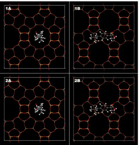
Figure 3.1: Zeolitic structures used in the theoretical modelling studies. ... 47
Figure 3.2: Mean Square Displacement of the Ibuprofen molecule in each zeolitic structure. ... 49
Figure 3.3: Structures of the studied zeolites ... 49
Figure 3.4: FTIR spectra of the zeolite, the drug and drug loaded carrier. ... 51
Figure 3.5: Thermo-gravimetric analysis of nano-FAU zeolites loaded with ibuprofen using different solvents. .. 51
Figure 3.6: Ibuprofen (IBU) release from nanosized Na+-form faujasite with a 5.1 Si/Al ratio. Hexane (C6), ethanol (EtOH) and acetone (Act) were used as loading solvents ... 54
Figure 3.7: Ibuprofen (IBU) release from Na+-form Faujasite with a 5.1 Si/Al ratio. Hexane (C6), ethanol (EtOH) and acetone (Act) were used as loading solvents. ... 54
Figure 3.8: Ibuprofen (IBU) release from Beta polymorph alpha (BEA) and Faujasite (FAU) with different Si/Al ratios and counter-ions. Hexane was used as loading solvent. ... 55
Figure 3.9: Ibuprofen (IBU) release from different zeolitic structures with different Si/Al ratios. Hexane was used as loading solvent. ... 56
Figure 3.10: Scanning electron micrographs of poly(L-lactic acid) (PLLA) membranes. ... 57
Figure 3.11: Differential scanning calorimetry thermogram of the prepared poly(L-lactic acid) membranes. .... 58
Figure 3.12: IBU release from PLLA membranes during 48 hours. Average ± STD; n=3. ... 59
Figure 3.13: Scanning electron micrographs of 5 (wt./vol.)% poly(L-Lactic acid) (PLLA) membranes embedded with the nanosized FAU zeolites. ... 60
Figure 3.14: Differential scanning calorimetry thermogram of simple PLLA membranes and PLLA membranes containing zeolite (PLLA:FAU ratio of 2:1) ... 61
Figure 3.15: IBU release from PLLA membranes containing IBU-loaded FAU and Terfenol-D. One sample was subject to an AC field (0 to 0.2T) during the first 8hours of the experiment. Average ± STD; n=2. ... 61
LIST
OF
TABLES
Table 1.1 – Studies using zeolites as drug delivery systems. ... 26
Table 2.1 – Drug release mathematical models. ... 42
Table 2.2 – Exponent n of the Korsmeyer-Peppas model and drug release mechanism from cylindrical polymeric drug release systems. ... 43
Table 3.1 – Zeolites tested during the optimization stage. ... 50
Table 3.2 – Drug loading of all tested samples of zeolites. ... 53
Table 3.3 – Release kinetics results from the tested ibuprofen-loaded membranes. ... 59
Table 3.4 – Release kinetics results from the tested membranes containing IBU-loaded FAU and Terfenol-D ... 62
LIST
OF
ABBREVIATIONS
5-FU 5 - Fluorouracil
Act Acetone
AMX Amoxicillin
ASA- Acetylsalicylic acid
BEA Beta polymorph-A
BSE Backscattered electrons
CEX Cephalexin
CHCA α-cyano-4-hydroxycinnamic acid
Diox 1,4 – Dioxane
DRS Drug release system
DSC Differential scanning calorimetry
EtOH Ethanol
FAU Faujasite
FER Ferrierite
FTIR Fourier-transformed Infrared spectroscopy
HCT-15 Human colon carcinoma cell line
HEU Heulandite
IBU Ibuprofen
KET Ketoprofen
LTA Linde-type A
LTT Linde-type T
MCF7 Human breast adenocarcinoma cell line
MFI Socony Mobil Five
MIT Mitoxantrone
MOR Mordenite
MRSA Methicillin-resistant Staphylococcus aureus
MSD Mean square displacement
MSSA Methicillin-sensitive Staphylococcus aureus
Nano FAU Nanosized (~250nm) faujasite
NO Nitric oxide
PAAc Poly(acrylic acid)
PAAm Poly(acrylamide)
PBS Phosphate buffer saline
PC-3 Human prostate adenocarcinoma cell line
PEG Polyethylene glycol
PLLA Poly(L-lactic acid)
PTFE Poly(tetrafluoroethylene)
RKO Human colon carcinoma cell line
RSV Resveratrol
SE Secondary electrons
SEM Scanning electron microscopy
TGA Thermo-gravimetric analysis
UV/Vis Ultraviolet/visible spectroscopy
Z Atomic number
C
HAPTER
1:
ZEOLITES
-
NEW
POSSIBILITIES
FOR
DRUG
DELIVERY
SYSTEMS
1.1. INTRODUCTION ... 19
1.2. COMPOSITIONANDCHEMICALSTRUCTURE ... 20
1.3. INCORPORATIONOFGUESTCOMPOUNDS ... 21
1.4. ZEOLITESASCONTROLLEDRELEASEVEHICLES ... 22
1.4.1. IMPORTANCE OF THE SI/AL RATIO ... 22
1.4.2. STRUCTURE AND PORE SIZE INFLUENCE ... 23
1.4.3. EXTERNAL PH AND IONIC STRENGTH INFLUENCE ... 24
1.5. INTEGRATIONOFZEOLITESINATRIGGEREDDRUGDELIVERYPLATFORM ... 30
1.1. INTRODUCTION
Conventional therapy is based on the administration of the therapeutic agent frequently without any protection (other than possible excipients) or targeting moiety. Even though occasionally, a prodrug form is administered to improve some characteristics such as the drug’s half-life in the organism, some drugs are poorly soluble in water or have low molecular weight, leading to an accumulation in unwanted body compartments and sometimes failing to successfully reach the target [1]. All these factors are important contributions to the poor drug bioavailability and consequently, a reduced therapeutic response. Thus, conventional therapy has been revealing itself ineffective, increasing the demand for more adequate so-lutions.
Nanomedicine plays an important role filling the breaches in a faulty therapeutic approach by improv-ing the drug bioavailability. This is achieved by protectimprov-ing it inside a carrier (e.g. hydrogel, nanocapsule or micelle) and by using targeting moieties or exploiting physiological flaws caused by the subjacent dis-ease (such as the enhanced permeability and retention effect in cancer) to accumulate the drug in the desired location [2]. Another important factor is the capacity of a given carrier to reach and deliver the content specifically in the affected site or cells with any, or negligible, leakage during its path, therefore reducing undesirable side-effects. This can be accomplished not only with targeting moieties, but also with stimuli responding carriers [2–4]. Current literature presents numerous studies regarding drug re-lease systems (DRSs) in a wide variety of forms: polymeric nanoparticles and membranes, liposomes, hydrogels, lipid nanoparticles, among many others [1,5,6].
In the latest years, zeolites have also seized some attention in the area of drug delivery. These inor-ganic particles were traditionally used as molecular sieves in several applications to capture numerous compounds. Exploring this characteristic, several groups exploited the highly porous matrix of the zeolites to incorporate different molecules, using them as drug delivery vehicles [7–11]. Zeolites are inexpensive, are present in numerous forms and structures and present tuneable characteristics such as its hydropho-bicity and its pore size. Also, reports recognize zeolites as biologically inert particles and consider them appropriate for biological applications [11–13].
1.2. COMPOSITIONANDCHEMICALSTRUCTURE
Zeolites are inorganic crystalline materials composed of SiO44- and AlO45- groups, which are arranged
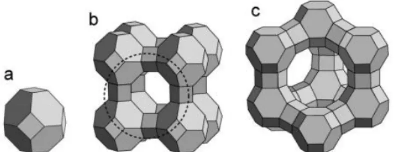
in channels and cages with well-defined structures (Figure1.1). These channels are accessible from the surrounding medium allowing the access by numerous molecules, thus providing zeolites the ability to adsorb considerable amounts of diverse compounds [14].
Figure 1.1: Zeolite structures. Silicon atoms are placed at the vertices.
Keys: (a) Sodalite or (β-) cage; (b) zeolite A: the β-cages are linked to each other via double four-membered rings and form an α-cage denoted by dashed circle; (c) Faujasite structure (FAU): the β-cages are connected via double six-membered rings and arranged as in the diamond lattice, forming a “super cage”. Adapted from [15]
The AlO45-group provides an extra negative charge when replacing the SiO44- group, thus zeolites with
a decreased Si/Al ratio present higher hydrophilicity. Therefore, the degree of the zeolite hydrophobicity can be controlled by modifying the Si/Al ratio, with high Si/Al ratios ranging up to 500 and the lowest
ratio being 1 [16]. The negative charge caused by the AlO45- groups has to be neutralized with organic or
inorganic cations to assure the electroneutrality of the solid. Moreover, the arrangement of the zeolite channel system is also important when incorporating molecules, with zeolites being classified in uni-, bi- or tri-directional, if the pores are organized in one, two or three axes (Figure 1.2). Due to wider pore systems, tri-directional zeolites present higher diffusion coefficients, allowing an easier incorporation but also a fast release of compounds [17].
Figure 1.2: Zeolitic pore unidimensional (A), bidimensional (B) and tridimensional (C) frameworks. Only main pore frameworks are shown. Small pore systems with diameter <4Å are ignored due to negligible access by compounds of interest. From [18]. Keys: Yellow pores - diameter 4-6Å; Orange pores - diameter > 6Å.
Additionally to pore organization, pore size and volume are also important to modulate the entrapment or release of compounds and can be controlled during zeolite synthesis or with post-synthetic reactions [8].
Currently, a zeolite database exists with over 200 entries, where zeolites are denoted as a 3-letter code [19]. This database presents a wide number of zeolites with their structure fully characterized [18], accessible as an online database of structures and with a web tool for a 3D automated approach freely available.
1.3. INCORPORATIONOFGUESTCOMPOUNDS
There are several techniques to incorporate different compounds in zeolites. The chosen technique depends on the zeolite and on the substance to incorporate. Neutral molecules can be incorporated simply by dissolving them in an inert solvent in contact with the zeolite and, with continuous stirring, diffusion-guided migration will occur. The type of solvent will influence the diffusion coefficients due to different polarities and possible interactions with the zeolite [14]. Also, in hydrophilic zeolites water must be removed before the inclusion of any other molecule by heating the zeolite above 100ºC [14].
For the incorporation of cationic guests to take place an ion exchange with the stabilizing cation is needed. The success and the efficiency of the exchange depends on the compound to incorporate and also on the stabilizing cation. Usually the incorporation is carried in water and often repeated with in-creasing concentrations of the molecule to incorporate to assure high exchange ratios [20].
Guests can also be incorporated in the gas phase using chemical vapour deposition chambers. This method is only possible at low reactional pressures or by heating the compound to incorporate at ade-quate temperatures (if possible). When conceivable, this method facilitates the inclusion of the desired molecule due to the absence of solvents and also prevents solvent contamination in the final product [14,21,22].
In some cases, for example when faujasite (FAU) is used, guest molecules are small enough to fit in
the “super cage” (with ~1.3nm in diameter) but oversized to pass through the pore network (with
~0.7nm). When this occurs, it is possible to add a precursor into the zeolite that will eventually be
trans-formed in the final guest. This method is usually termed “ship-in-a-bottle synthesis” [14]. Once synthe-sized, the guest can no longer exit the zeolite, remaining entrapped without any covalent linkage. Several cases are reported in the literature using this method, for example to immobilize fluorescent dyes for
cellular imaging [23] or to be used as catalysts in hydroxylation processes [24,25] and electro [26] or photo catalysis [27].
1.4. ZEOLITESASCONTROLLEDRELEASEVEHICLES
Zeolites were already reported as possible vehicles for controlled release of fertilizers [28], pesticides [20], preservatives [7] and, more recently, several therapeutic agents such as anti-inflammatory [6,8,22,29], antibiotics [10,30] or anticancer drugs [9,31,32]. Moreover, studies have shown the biolog-ical inertia of these compounds [12,13], allowing their use in biologbiolog-ical applications. Thus, their unique characteristics (rigid structure, high adsorption ratios, tuneable hydrophobicity, controlled pore size and biological inertia) allow zeolites to be a promising alternative as carriers in controlled release DRS, provid-ing an increase of the drug loadprovid-ings and also delayprovid-ing the initial release of the drug creatprovid-ing a sustained and prolonged release [33].
There are, however, some parameters to revise when using zeolites as drug carriers. The type of drug-host interaction is of great importance. If, for example, the interaction is based on ion exchanges, the external pH and the Si/Al ratio will greatly influence the release rate. Also, the drug to incorporate must be properly chosen because, due to the zeolites’ well-defined and rigid structure (with restricted pore and cage diameters), the possible drugs to encapsulate are size-limited. Surface modifications are sometimes useful to slow the release by decreasing pore size, delaying the diffusion rate from minutes to days. Some examples illustrate the effect of distinct zeolite characteristics and their importance for the selection of both zeolite and drug to yield a functional and balanced DRS.
1.4.1. IMPORTANCE OF THE SI/AL RATIO
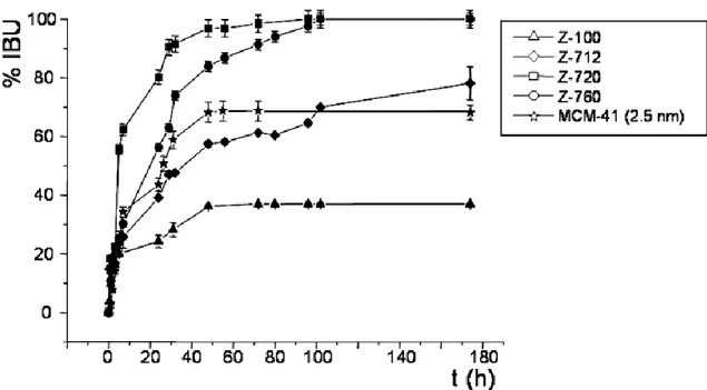
Horcajada and colleagues studied the influence of the Si/Al ratio on the release of ibuprofen (IBU) from FAU [8]. In this study, four zeolites were used: Z100, Z712, Z720 and Z760, with Si/Al ratios of 7, 13, 22 and 62, respectively. The Z100 zeolite showed the lowest IBU internalization (72mg IBU/g) with 4 IBU molecules per unit cell, while the other tested zeolites presented IBU internalization of 150-160mg/g (approx. 9 IBU molecules per unit cell). These results suggest that there is a maximum amount of drug capable of fitting in the zeolite structure. The drug release assay (Figure 1.3) showed a slower IBU release from Z100 and Z712 comparing to Z720 and Z760. FTIR analysis revealed that in Z100 and Z712 the interaction with IBU occurs due to coordination with the extra-framework aluminium, while in
Z720 and Z760 this interaction occurs via hydrogen bonding (weaker bond), explaining the difference in the drug release rates.
Figure 1.3: Ibuprofen (IBU) delivery from tested zeolites and from MCM-41 (2.5nm pore), included as reference. Zeolites Z100, Z712, Z720 and Z760, with Si/Al ratios of 7, 13, 22 and 62, respectively. From [8].
1.4.2. STRUCTURE AND PORE SIZE INFLUENCE
Showing the importance of structure and pore size of the zeolites, Fisher and co-workers studied the uptake of different species of fluorescein (fluorescein sodium salt, fluorescein free acid, and fluorescein diacetate) by Zeolite X (FAU framework with a low Si/Al ratio) and MCM-41 (a mesoporous silicate material with pores with 2 - 6.5nm) [34]. The results indicated that there was little uptake of this compounds within the zeolite pores, possibly due to the excessive size and rigidity of the fluorescein molecule, result-ing only in surface adsorption of small amounts. Contrarily to expected, the increased pore size of MCM-14 did not cause a substantial increase in the uptake of all species of fluorescein. Other studies have already reported that materials with larger pores do not always result in higher uptakes [35] due to the influence of the loading solvent and the hydrophobicity of the loaded molecule and the carrier structure itself.
In a different study, pore size of Zeolite Y was modified by Zhang et al. using 1,1,3,3-tetramethyldisi-lazane [20] to assess its influence on the release of paraquat, a herbicidal drug. After the incorporation of the drug, the zeolites were dehydrated and modified with the silanol groups. Without the modification, the authors observed that the release of paraquat reached a plateau after 20min. However, with the
coupling of the silanol groups, the release occurred at different rates, with a plateau being observed only after 7days of release (Figure 1.3). Hence, pore engineering is an efficient pathway to control the drug release from zeolites, with different possible release rates depending on the modifying agent used.
1.4.3. EXTERNAL PH AND IONIC STRENGTH INFLUENCE
In the same study, Zhang and co-workers also verified the importance of the ionic strength of the release medium [20]. When [NaCl] = 1M, all paraquat was released from the interior of the tested zeolites, however, when the salt concentration decreased to 0.1M and 0.01M, only 65% and 14% of the loaded paraquat was released, respectively (Figure 1.4).
Figure 1.4: Paraquat release from unmodified (black markers) and modified (white markers) zeolites for 14 days in the presence of different ion concentrations (circles: 1.0M Na+; squares: 0.1M Na+; triangles: 0.01M Na+). From [20].
Taking into account that zeolites possess negative charges, which are stabilized by counter-ions,
Payne and Abdel-Fattah studied the effects of pH and ionic strength on the adsorption of Pb2+ ions onto
Zeolite A and Zeolite X (both with Na+ as counter-ion) [36]. The study was conducted by simply adding
the zeolites to an aqueous lead solution (at different pH’s and with different amounts of KNO3) at room
temperature with aliquots being collected at different time periods. A more extensive ion-exchange was observed when the pH reached 4 and above. Also, the presence of competing ions in the medium revealed to significantly decrease the lead adsorption.
Furthermore, Rimoli and colleagues studied the release of ketoprofen (KET), an anti-inflammatory drug, from Zeolite X at different pH values, mimicking the pH changes in the digestive tube [6]. The authors found that at highly acidic pH, there was only a residual release of the drug, while at pH5 a progressive release started which was accelerated when the pH increased to 6.5 (Figure 1.5). This be-haviour was related to the ionization of the carboxylic group of the drug at pH 5, causing an enhanced repulsion between the drug and the zeolite (both holding negative charges) leading to KET dissociation and release. This system was suggested as a possible drug delivery system with the release occurring only after the carrier has passed through the stomach acidic pH, contributing to the reduction of the adverse effects caused by oral ingestion of anti-inflammatory drugs.
Figure 1.5: pH-dependent ketoprofen release from zeolite X. From [6]. Keys: 0-90min: pH1; 90-135min: pH5, 135-165min: pH6.8.
Hence, when considering zeolites for drug delivery several parameters ought to be considered: Drug size and hydrophobicity, loading methodology and solvent, zeolite structure, pore size, hydrophobicity and Si/Al ratio, drug-zeolite interaction and ionic strength and pH of the medium. Table 1.1 summarizes important studies involving zeolites as DRS, with some particular references to the above-mentioned char-acteristics. Despite the existence of several studies, occasionally comparing different frameworks or Si/Al ratios, exhaustive studies showing how the loading of a model drug is influenced by particular character-istics are scarce. Being able to understand to what extent the Si/Al ratio, the counter-ion and the zeolitic structure influence the loading of a drug is an important step to the design of a tuneable drug release platform.
Table 1.1 –Studies using zeolites as drug delivery systems.
1. COMPARATIVE STUDIES Substance Zeolite
framework
Si/Al
ratio Loading Results Observations Refs.
IBU FAU
7 72mg/g IBU adsorption studies revealed an occupation of 4 IBU molecules per unit cell for FAU-7 and 8-9 IBU molecules for FAU-13, -22 and -62. All the samples possess a micropore volume close to 0.30 cm3/g,
except FAU-7 (0.20 cm3/g). FAU-7 presents a
negli-gible mesopore volume, while the other samples present volumes of 0.15-0.17 cm3/g. FAU 7 and
-13 showed an incomplete release of IBU in the 174h study, with a drug retention of 63% and 22%, respec-tively. FAU-22 fully released the drug in 96h and FAU-62 in 102h.
FAU-7 high extra-framework aluminium justifies both its low pore volume (partial pore blockage) and its decreased loading capacity. The similar IBU adsorption capacity of FAU-13, -22 and -62 indicates that there is a maximum amount of drug loadable into the structure. The total pore volume of FAU-7 is nearly 50% of the remaining samples, similarly to its loading capacity. Thus, a ratio of 1.5-1.7mmol IBU/cm3for all samples was
ob-tained. In samples with lower Si/Al ratios the drug is covalently bound to the structure, delaying its diffusion, while in samples with lower Al content, the retention occurs due to weaker interactions (H-bonds and van der Waals).
[8] 13 162mg/g 22 152mg/g 62 148mg/g 5-FU FAU <1.5 161.8mg/g
Release of 5-FU from FAU and BEA was significantly different. Full release occurred at 10min (FAU) and at 120min (BEA). Also, the release profiles are mark-edly different with FAU showing a 80% burst release in the first 3min while BEA shows a multi-step re-lease with 15% rere-leased in the first 10min, up to 50% in the next 40min and the remaining drug within an hour. Unloaded FAU presents noticeable cell growth alterations at 8h (concentration dependent) while unloaded BEA showed no toxicity. Loaded particles showed high toxicity, similarly to the free drug.
Larger pores and pore organization are the main reasons for the difference on the loading and also release profiles, with FAU presenting larger pore diameter. The tighter pores in BEA originate much stronger short-ranged van der Waals’ interactions between the drug and the zeolite, dampening the release. The difference in the toxicity of unloaded particles may be explained by their morphology, with multi-faceted FAU being able to penetrate cell membranes more easily than spherical BEA parti-cles.
[37]
BEA 250 60.2mg/g
Keys: BEA – Beta Polymorph A; FAU – Faujasite; IBU – Ibuprofen; 5-FU – 5-fluorouracil.
C H AP TER 1 Z EO LIT ES : NE W P OS SIBIL IT IE S FO R DR UG D EL IVE RY SYS TE M S 26
(Table 1.1 continued)
Substance Zeolite framework
Si/Al
ratio Loading Results Observations Refs.
5-FU
FAU
5 110 mg/g FAU-5 presented a negligible release (<5%) over a period of 6h, whereas the other 2 samples pre-sented a release of around 60%.
5-FU forms strongly bonded complexes with the extra-framework aluminium sites, extensively ham-pering the release of the drug.
[38] 30 105 mg/g
60 90 mg/g
FAU 2.83 7.2mmol/g (936mg/g)
FAU presented a higher encapsulation efficiency (71.3%) than the other samples (~50%). The release profiles revealed an 80%, 94% and 89% maximum release in 48h for FAU, nano-sized FAU and LTL re-spectively. A burst release occurred in the first 10minutes with 80-90% of 5-FU being released. No toxicity was found to be caused by all the samples in HCT-15 and RKO cell lines. The system showed to potentiate in 1.6-7.6 times the drug effect.
Owning a larger micropore volume, FAU presents a higher 5-FU loading capacity. The rapid release is explained by the drug’s small size facilitating the diffusion from the micropores. The potentiation is explained by the increased bioavailability of 5-FU and the facilitated cell uptake. The extremely high drug loadings indicate that significant amounts of drug may be adsorbed onto the zeolite surface and not inside the pores.
[39] FAU (nano-sized) 2.25 5.5mmol/g (715mg/g) LTL 3.4 5.2mmol/g (676mg/g) CHCA
FAU 2.83 78.9mg/g No decrease in metabolic activity of HCT-15 cells caused by FAU or LTA (24h). Increase in efficiency of the drug up to 585-fold (FAU) and 146-fold (LTA) when compared to the non-encapsulated drug.
The wider structure of FAU allowed a better diffu-sion of the drug out of the zeolite pores, justifying the difference in the results.
[9,31] LTA 1.24 69.7mg/g
ASA FAU
5 106mg/g Lower Si/Al ratio zeolite showed a complete release of the drug in 5hours, whereas the other two sam-ples released only 63% of the drug in the same time. Aluminium-acetylsalicylate formed at the FAU-5 ex-tra-framework aluminium sites due to the nation at pH 7.4. With FAU-30 and -60 the deproto-nation did not occur drug was bound to silanol sites via H-bonds.
As hydrophobicity increases with higher Si/Al ra-tios, van der Waals interactions become a contrib-uting factor to the release profile, hindering the drug release. Aluminium-acetylsalicylate species are more weakly bound to the zeolite than the pro-tonated form of the drug, creating the conditions for a complete release of the drug.
[40] 30 78mg/g
60 69mg/g
Keys: ASA - Acetylsalicylic acid; CHCA – α-cyano-4-hydroxycinnamic acid; FAU – Faujasite; HCT-15 – human colon carcinoma cell line; LTA – Linde Type A; LTT – Linde Type T; n.g. – not given; RKO – human
C H AP TER 1 Z EO LIT ES : NE W P OS SIBIL IT IE S FO R DR UG D EL IVE RY SYS TE M S 27
(Table 1.1 continued)
2. ISOLATED STUDIES
Substance Zeolite type Si/Al
ratio Loading Results Observations Refs.
MIT BEA n.g. 3.02mg/g
MIT structure does not allow a full incorporations onto the zeolitic pore system, however there is a di-rect interaction with the extra-framework aluminium. The 72h release studies revealed that no drug was released from the zeolite. After incubation of the sys-tem with MCF-7 and PC-3 cell lines, lower toxicity was observed when comparing to mitoxantrone alone. In the presence of the system morphological changes were observable in both cell types.
The authors hypothesize that, after ruling out the possible toxicity of the zeolite, the drug was only released once inside the cell. The delayed release justifies the decreased toxicity of the loaded zeolite comparing to the drug alone. The nanoparticles re-tained the optical characteristics of the mitoxan-trone, yielding a useful hybrid bearing both thera-peutic and cell-marking properties.
[41]
CEX HEU n.a. 10.5mg/g
Hexadecyl-trimethylammonium surfactant increased the adsorption of CEX on HEU. Un-buffered and phosphate buffered release mediums presented in-verse release profiles with increasing pH values. At pH 2 (un-buffered) a maximum desorption was ob-tained, suitable for drug releasing platforms to work at gastric pH.
With a surfactant-modified zeolite the ion exchang-ing process between the drug and the carrier is favoured, thus increasing the loading capacity. The adsorption and desorption are affected by the presence of different ions, hence the distinct be-haviour in the presence of buffered (tri-, di- and monovalent phosphate species) and un-buffered (Cl- ions) solutions.
[42]
RSV BEA n.g. 484 mg/g
The release profiles showed that a total release was achieved in about 3h, contrarily to free resveratrol. The encapsulation on the zeolite prevented the transformation of resveratrol from trans to cis (less active form).
The system showed an increased resveratrol solu-bility compares to the free drug. Also, due to the rigidity of the zeolitic pores, the isomeric conver-sion was impaired, preserving the bioactive form of the molecule.
[43]
Keys: BEA – Beta Polymorph A; CEX – Cephalexin; HEU – Heulandite; MIT – Mitoxantrone; MCF-7 – human breast adenocarcinoma cell line; n.a. – not applicable; n.g. – not given; PC-3 – human prostate
adeno-carcinoma cell line; RSV – Resveratrol.
C H AP TER 1 Z EO LIT ES : NE W P OS SIBIL IT IE S FO R DR UG D EL IVE RY SYS TE M S 28
(Table 1.1 continued)
3. COOPERATION WITH DIFFERENT MATERIALS Substance Zeolite type Si/Al
ratio Loading Results Observations Refs.
ZER + Gelatine FAU >5 n.g.
After incorporating ZER in the zeolite, a gelatine coat-ing layer was successfully added to the system, prov-ing to delay the release of the drug. The system showed a sustained release for 24h (100% release) compared to the 3h release from the non-coated ze-olite.
The gelatine layer swelled in contact with the re-leasing medium and was further eroded. During this process, the diffusion of the drug was ham-pered.
[32]
AMX + PEG +
PAAc + PAAm LTA n.g. n.g.
The loaded zeolite was incorporated on a hydrogel. The release studies were conducted by at pH 7.8 and 6.8 and at different temperatures. The composite ex-hibited a temperature and pH dependent release, with higher temperatures and pH causing an en-hanced release.
PAAc and PAAm are polymers both responsive to temperature and pH. At higher temperatures structural changes occur, increasing the release of AMX. At higher pH values, the polymer’s func-tional groups become ionized, increasing the re-pulsive forces and causing the polymer to swell. This effect increases the diffusion of the drug.
[11]
4. ZEOLITE BASED DELIVERY DEVICES
Saline solution MFI n.a. n.a.
Zeolite walled micro-needles effectively penetrated through the upper layers of 8 month old domestic pig skin. The poration of the skin caused a 1000x in-crease in the permeability to saline solution.
The tested system demonstrated promising re-sults as a transdermal drug delivery system. The drug can be embedded on the zeolite’s surface/ pores or dissolved in a solution (e.g. saline).
[44]
NO LTA n.g. n.g.
Antibacterial disks with 50 w% of PTFE and of Zn2+
-exchanged zeolite revealed to release micromolar concentrations of NO over approx. 1h when in aque-ous medium. The composite showed a bactericidal effect when in contact with P.aeruginosa and a bac-teriostatic effect when in contact with MSSA, MRSA and C.difficile.
The system showed an improved bactericidal ac-tivity when compared to NO alone. The bacterio-static effect on the Gram positive strains may be due to Zn2+ions leaching from the zeolites.
[45]
Keys: AMX – Amoxicillin; FAU – Faujasite; LTA – Linde Type A; MFI – Zeolite Socony Mobil – 5; MRSA – Methicillin-resistant Staphylococcus aureus; MSSA – Methicillin-sensitive Staphylococcus aureus; n.a. – not
C H AP TER 1 Z EO LIT ES : NE W P OS SIBIL IT IE S FO R DR UG D EL IVE RY SYS TE M S 29
1.5. INTEGRATIONOFZEOLITESINATRIGGEREDDRUGDELIVERYPLATFORM
As mentioned previously, several zeolite-based systems for drug delivery are reported and, whereas most of the works report the use of the zeolitic material alone, there are cases of zeolites combined with different materials [45] or incorporated in a platform [11]. The incorporation of these materials in other platforms is advantageous due to the possibility of tailoring the material.
Polymers are excellent choices to build quite distinct and unique DRSs due to their wide variety (nat-ural and synthetics), thus offering the advantage of being highly tunable in terms of size, structure and physical-chemical proprieties (hydrophobicity/hydrophilicity, reactive groups, etc.) and degree of degra-dability depending on the application. Reports have been made of polymeric DRSs that can serve as drug carriers with sustained releases up to several months or even years [46].
Other than hydrogels or nanoparticles, polymeric membranes are also good candidates to develop drug release systems. This type of system can be extremely useful to prepare drug releasing platforms to be used, for example, as skin patches [47] or even as temporary interfaces during the healing process after surgery [48]. When a system is going to be integrated in the body, its safety has to be proven and its degradability into non-toxic sub-products is essential. Poly(L-lactic acid) (PLLA) is a well-known polymer already used in drug delivery applications [48–50] and recent reports show that exposure to PLLA mem-branes presents no significant cytotoxicity or genotoxicity [51] making them an excellent option as to be used as DRS.
Polymer-based DRSs can become stimuli-responsive with the insertion of, for example, an ionisable group (pH sensitivity) [52], disulphide bridges (redox sensitivity) [3] or an azobenzene derivative (light sensitivity) [53] into its backbone. This type of systems can sometimes be responsive to more than one stimulus, hence improving drug release performances [54]. The most studied and important stimuli are often divided in two categories [55]: endogenous triggers (pH, redox, glucose, enzymes, etc.) and exoge-nous (temperature, light, magnetic field, ultrasounds, etc.).
Polymeric structures are affected by magnetic fields when magnetic nanoparticles are incorporated onto the matrix. Several studies have reported the incorporation of magnetic particles in polymeric sys-tems, such as hydrogels [56,57] or polymeric nanoparticles [58,59]. These systems can be used as cancer therapy agents [60], artificial muscles [61], magnetic separation [62], drug delivery systems [59], among others [3]. Magnetostriction is, by definition, the variation of the strain of a material as a quadratic function of an applied magnetic field [63,64], i.e. when in presence of a magnetic field, the material will
alter its dimensions, with Terfenol-D and CoFe2O4 as good examples of extensively described
magneto-strictive particles [63]. The application of polymeric systems bearing magnetomagneto-strictive particles are there-fore useful in drug delivery, adding an additional feature that acts as a trigger for the release of the drug.
1.6. HYPOTHESIS
Even though a variety of studies have reported the use of different zeolites as drug release platforms, the integration of drug loaded zeolites onto polymeric membranes was not yet fully accomplished. This combination can improve the DRS’s features as the inclusion of a drug in a carrier that, in its turn, is going to be incorporated in a platform allows a more controlled and prolonged release. This prolonged release occurs because the drug has first to travel first through the zeolitic pores and only then exit the membrane. Also, a wider variety of guests can be incorporated in a zeolite due to the diversity of existing zeolitic structures and their tunable characteristic e.g. the hydrophobicity of a zeolite can be controlled by changing its Si/Al ratio and the counter-ion can be changed in order to allow a better ion exchange with the guest. The addition of magnetostrictive particles to the system would allow the triggered release of the guest from the platform, or at least would allow to greatly increase its release ratio.
The combination of zeolites and magnetostrictive particles in a polymer based platform presents itself as a novelty for the active release of desired drugs, hence a hypothesis was developed: the design of a polymeric drug delivery platform containing a zeolite as the drug carrier and Terfenol-D as the magneto-strictive element.
The specific objectives of this thesis are:
1. Theoretical and experimental studies the encapsulation of a model drug (IBU) onto zeolites; 2. Preparation of IBU-loaded PLLA membranes;
3. Incorporation of the zeolite in PLLA membranes;
4. Incorporation of loaded zeolite and Terfenol-D particles onto PLLA membranes and prove the magnetically-driven release of IBU.
C
HAPTER
2:
MATERIALS
AND
METHODS
2.1. SELECTIONOFASUITABLEZEOLITE ... 35
2.1.1. MOLECULAR MODELLING OF DRUG-HOST INTERACTIONS ... 35
2.1.2. EXPERIMENTAL VALIDATION ´ ... 37 2.2. PLLAMEMBRANESPREPARATION ... 39
2.2.1. POLYMER CONCENTRATION OPTIMIZATION ... 39
2.2.2. INCORPORATION OF IBUPROFEN ... 39
2.2.3. INCORPORATION OF THE ZEOLITE ... 40
2.2.4. INCORPORATION OF TERFENOL-D PARTICLES AND LOADED ZEOLITES ... 40 2.3. RELEASEASSAYSFROMTHEPLLAMEMBRANES ... 41
2.3.1. EXPERIMENTAL DESIGN ... 41
2.3.2. RELEASE ASSAYS UNDER MAGNETIC FIELD ... 41
2.3.3. DRUG RELEASE MATHEMATICAL MODELS ... 42 2.4. CHARACTERIZATIONTECHNIQUES ... 43
2.4.1. SCANNING ELECTRON MICROSCOPY ... 43
2.4.2. DIFFERENTIAL SCANNING CALORIMETRY ... 43
2.4.3. THERMO-GRAVIMETRIC ANALYSIS ... 44
2.1. SELECTIONOFASUITABLEZEOLITE
More than two hundred zeolitic structures are already described, each with particular characteristics suitable for different goals. As already presented in Table 1.1, only a few structures are usually selected when using zeolites as drug carriers. In order to being able to properly adequate the choice of the zeolite to the desired loading and release parameters, a screening of different structures with different counter-ions and Si/Al ratio was conducted.
In a first phase, molecular simulations were conducted with five different frameworks: FAU, BEA, MFI, Mordenite (MOR) and Ferrierite (FER). The aim was to study the influence of the zeolitic structure on the drug diffusion through the pores and channels, possibly allowing to understand how the structure could influence the loading and the release of the drug.
In a second stage, experimental tests were performed with the structures selected by the molecular modelling. This would allow to confirm the results of the simulations, but also to test further variables, such as the influence of the loading solvent, counter-ions and Si/Al ratio.
2.1.1. MOLECULAR MODELLING OF DRUG-HOST INTERACTIONS
When incorporating molecules inside well characterized and rigid zeolitic pore systems it is of great importance to understand not only how much of that compound can be fitted in the zeolite but also how will the drug interact with the host. Using a molecular modelling software, some interactions and behav-iours can be studied. The modelling can be achieved by two computational mechanics: quantum me-chanics and molecular meme-chanics.
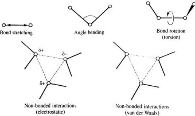
Quantum mechanics explicitly models the electrons of each atom, being possible to derive proprieties that depend upon the electronic distribution, particularly when investigation chemical reactions or for-mations and cleavage of bonds. The starting point of all quantum mechanics calculations is, therefore, the Schrödinger equation. This consideration is the principal differentiation between quantum mechanics and the empirical force fields used in molecular mechanics – the modelling of individual electrons [65]. Molecular mechanics, on the other hand, ignores the electronic contribution and calculates the energy of a system based on the nuclear positions only, using empirical force fields. This is particularly useful when dealing with systems with a significant number of atoms, since when ignoring the electrons, a substantially smaller number of particles are considered, thus largely reducing the computation time. The
majority of the force fields used for molecular systems can be depicted using a simple four-component representation of the intra and intermolecular forces (Figure 2.1).
Figure 2.1: Schematic representation of the four key contributions to a molecular mechanics force field. From [65]
The total energy of the system can then be calculated summing the contributions of bond stretching, angle bending, bond torsion and non-bonded interactions (electrostatic and van der Waals interactions) [65] as depicted in:
𝐸𝑡𝑜𝑡 = ∑𝐸𝑏𝑜𝑛𝑑+ ∑𝐸𝑎𝑛𝑔+ ∑𝐸𝑡𝑜𝑟𝑠𝑖𝑜𝑛+ ∑𝐸𝑛𝑜𝑛−𝑏𝑜𝑛𝑑 (Equation 2.1)
Where ∑𝐸𝑏𝑜𝑛𝑑 represents the bond stretching energies, ∑𝐸𝑎𝑛𝑔 represents the angle bending
ener-gies, ∑𝐸𝑡𝑜𝑟𝑠𝑖𝑜𝑛 represents the energy of torsional movement (bond rotation) and ∑𝐸𝑛𝑜𝑛−𝑏𝑜𝑛𝑑
repre-sents non-bonded interactions (electrostatic and van der Waals).
The calculation the energy of a system is not, however, enough to model the movement of molecules in contact with a solvent or imprisoned in a carrier. To do so, molecular dynamics simulations are needed. In these simulations, successive configurations of the system are generated by the applications of New-ton’s laws of movement, generating a trajectory that determines how the position and velocities of the particles in the system vary with time. The trajectory is obtained by solving the differential equations resulting from Newton’s second law [65]:
𝑑2𝑥𝑖
𝑑𝑡2
=
𝐹𝑥𝑖
𝑚𝑖 (Equation 2.2)
Where 𝑚𝑖 is motion of a particle of mass along a coordinate (𝑥𝑖), with 𝐹𝑥𝑖 being the force on the
Throughout the literature it is possible to find studies in which theoretical and experimental results are performed in parallel [15,16,37,66], allowing to better understand some of the experimentally ob-tained data, or even to eliminate some possibilities prior to the experimental studies.
The molecular dynamics simulations can show how a specific drug molecule will move inside the zeolitic framework over time and, in the specific case of zeolite based drug delivery systems, this can be used to explain different loading efficiencies or different drug release rates obtained when using different zeolitic structures or different drugs [37].
Materials Studio molecular modelling software was used to model distinct drug-host interactions. The study was conducted by placing a drug molecule inside each zeolitic structure, followed by a geometry optimization in order to reduce the energy (and consequently, the interactions). The drug-host complex was then subject to a molecular dynamic simulation, at 310K, using the Forcite module with a universal force field.
2.1.2. EXPERIMENTAL VALIDATION ´
After the molecular modelling simulations, an experimental phase was planned. In this stage, the
previously selected structures were used, bearing different counter-ions (Na+, NH4+ and H+) and also
different Si/Al ratios.
2.1.2.1. LOADING TECHNIQUE
The chosen loading technique consisted in placing the zeolite in an IBU solution for 24h as described [37,38]. Aiming the optimization of the process, this method was conducted using three different solvents. Ethanol (EtOH, 95% v/v, extra pure, Fisher Scientific), Acetone (Act, Laboratory Reagent, ≥99.5%, Sigma-Aldrich) and n-Hexane (Hex, analytical reagent grade, Fisher Scientific) were chosen due to their different polarities and functional groups and also because they were already reported as loading solvents [8,37,39]. Before the utilization of the zeolite, a dehydration step was performed by placing the zeolite at 100-110ºC overnight. This step is important to remove the water trapped in the pores of the zeolite, which otherwise would severely decrease the drug loading [14]. Briefly, 500mg of the zeolite and 250mg of IBU were placed in a glass vial and 10mL of the selected solvent was added, always ensuring that completely solubility of IBU. The mixture was kept under stirring for 24h. Next, a filtration process was conducted, with a Whatman® Ashless, grade 40 filter paper. After the procedure, the zeolite was retrieved and kept
at 50º-60ºC for 4-6h for the complete removal of the remaining solvent. The final product was labelled IBU@(zeolite structure) e.g. IBU@FAU (ibuprofen at faujasite).
2.1.2.2. RELEASE ASSAY
In order to evaluate both the loading and the release kinetics, release assays were conducted using a USP-1 dissolution apparatus (Erkwa instruments; basket; 50rpm). In this apparatus, the zeolite was placed in 1000mL of Phosphate buffer saline (PBS) pH 7.4 at 37ºC. Aliquots were taken at specific time-points by a flow system, using a peristaltic pump, which was connected to a UV/Vis-spectrophotometer (Thermo Spectronic UV-500) and the released IBU was quantified. After the readings, the collected sam-ple returned to the release medium, keeping a constant volume throughout the experiment.
IBU Quantification by UV/Vis-spectroscopy (UV/Vis)
UV/Vis is a widely used technique to quantify different analytes. This method is based on Beer-Lam-bert law which relates the light transmitted through a solution with, among other parameters, the concen-tration of an existing substance [67]:
𝐴 = 𝛼 × 𝑙 × 𝑐
(Equation 2.3)Where 𝐴 is the absorbance, 𝛼 is the molar extinction coefficient, 𝑙 is the optical path length and 𝑐 is the concentration of a present substance.
The first step was the acquisition of a calibration curve within a linear range of concentrations and with values that would fit the concentrations used during the work. Initially a stock solution of 600mg/L of ibuprofen in PBS was prepared which was then properly diluted in order to obtain the desired standard samples used in the calibration curve. The measurements were performed using the same spectropho-tometer described in the above section, reading the absorbance at a wavelength of 264nm. A linear
regression was applied to the obtained calibration curve and presented an R2=0.9946 and the equation
Y = 0.002X+0.0027.
PBS solution preparation
For the release kinetic assays, a 10xPBS 0.1M stock solution was prepared. The following quantities
were used to prepare 1000mL of 10xPBS: 9.94g (70mM) of Disodium hydrogen phosphate (Na2HPO4
anhydrous, ChemAlert); 4.14g (30mM) of Sodium dihydrogen phosphate (KH2PO4 H2O, ChemAlert) and
Prior to use, the 10xPBS solution was diluted 10 times with deionized water and the final PBS had a pH 7.4 and a final concentration of 10 mM phosphate and 130 mM NaCl.
2.1.2.3. DETERMINATION OF IBU LOADING ONTO ZEOLITE
After the quantification of the drug, its loading amount (% wt.) was calculated using equation 2.4:
𝐿𝑜𝑎𝑑𝑖𝑛𝑔 (% 𝑤𝑡. ) =𝐷𝑟𝑢𝑔 𝑚𝑎𝑠𝑠 𝑖𝑛 𝑧𝑒𝑜𝑙𝑖𝑡𝑒
𝐿𝑜𝑎𝑑𝑒𝑑 𝑧𝑒𝑜𝑙𝑖𝑡𝑒 𝑚𝑎𝑠𝑠 × 100 (Equation 2.4)
2.2. POLY(L-LACTICACID)MEMBRANESPREPARATION
Poly(L-lactic) acid (PLLA, average molecular weight of 217,000–225,000g/mol) membranes were prepared through an adapted freeze-drying method [68,69]. In this technique, the polymer is placed in a solvent, the solution is casted on a surface and then freeze dried. During this work, 1,4-dioxane (Diox, anhydrous, 99.8% grade, Sigma-Aldrich) was used as solvent for the preparation of the membranes. Due to a melting point of 11.8ºC, 1,4-dioxane will rapidly freeze at the low temperatures of an ice bath (approx. 0ºC) or a simple freezer (approx. -20ºC). This feature is important to keep the homogeneity of the mem-brane when incorporating the zeolite or the magnetic nanoparticles. The prompt freezing maintains a good dispersion of the particles, which could be lost if the dispersion is kept in the liquid state for a long period. Also, the solvent plays an important part in the pore system development, acting as porogenic. As the temperature decreases and solvent freezes, the polymer will “surround” the solvent crystals, thus giving rise to a pore when the solvent is removed.
2.2.1. POLYMER CONCENTRATION OPTIMIZATION
In order to assess the suitable membranes to be used as drug carriers, several optimizations were performed. The first was the determination of the amount of polymer to be used during the preparation of the membranes. To do so, several polymer concentrations were used: 1, 3, 5 and 10 (wt/vol.)%. Briefly, the desired amount of polymer was placed on a glass vial with 2.3cm of internal diameter, 1mL of solvent was added and kept under stirring until complete dissolution. The solutions were then placed at -20ºC and later freeze-dried. After the freeze-drying process, the membranes were characterized.
2.2.2. INCORPORATION OF IBUPROFEN
During the preparation of the membranes with IBU (purity ≥98%, Sigma-Aldrich), the additional step was the incorporation of the drug in the polymer solution, followed by the same procedure described in
the above section. In this phase, different quantities of IBU were added to study the effect of different loadings on the drug release profile. The obtained membranes contained a final concentration of 3, 5 and 10 wt% of IBU and were later characterized and subject to drug release assays.
2.2.3. INCORPORATION OF THE ZEOLITE
The following step was the development of PLLA membranes containing the zeolite (Freeze-dryed Na+-form of Faujasite (Nano FAU), particles sizes averaging 150nm, NanoScape). This required the dis-persion of the zeolite in the solvent in an ultrasound bath for 4h prior to the addition of the polymer. Again, different amounts of Nano FAU were added to 1,4-dioxane, now aiming to assess the influence of the solid on the membrane pore structure. The prepared membranes contained a final zeolite:PLLA mass proportion of 20, 10, 6, 4 and 2:1. Prior to all preparations, the zeolite was placed at 180ºC overnight in order to remove any adsorbed water.
2.2.4. INCORPORATION OF TERFENOL-D PARTICLES AND LOADED ZEOLITES
The last step involving the development of the PLLA membranes was the incorporation of the Terfenol-D particles (from ETREMA) and drug loaded zeolite. Terfenol-Terfenol-D particles are not dispersible in Terfenol-Diox alone due to their high molecular weight. Consequently, previous to the dispersion, the polymer was added to the solvent in order to increase the viscosity of the medium. Next, the Terfenol-D particles were added and placed on an ultrasound bath for 10min. Next, drug loaded zeolite was added to the dispersion and kept under ultrasounds until complete dispersion of both magnetic and zeolitic particles. While the sample was in the ultrasound bath, the water temperature was decrease with the addition of ice. This caused the solvent to freeze, thus maintaining a good dispersion of the particles when the preparation was removed from the ultrasounds. The final product was then placed at -20ºC and freeze-dried. Due to more desirable characteristics, the membranes chosen for this phase were prepared with 5 (wt/vol.)% of polymer in 1mL of solvent, and the membrane’s components final concentration were 65 wt% of PLLA, 15 wt% of Terfenol-D and 20 wt% of IBU@FAU. The amount of Terfenol-Terfenol-D used was previously optimized in order to obtain the desired magnetostritive effect without compromising the stability of the membrane [70] . These mem-branes were then used to study the drug release profile in the presence and absence of magnetic fields.
2.3. RELEASEASSAYSFROMTHEPREPAREDMEMBRANES
After the loading of the membranes, an evaluation of the release kinetics from membranes loaded with IBU or with IBU@FAU was conducted. The release was again conducted in PBS pH7.4 at 37ºC.
2.3.1. EXPERIMENTAL DESIGN
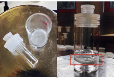
During the release it is essential that the membrane is fully immersed in the PBS solution allowing the diffusion of the drug in all directions and not only through the parts in contact with the release medium. Thus, a glass vial was modified in order to keep the membrane submerse as shown in Figure 2.2. The experiment was conducted by placing the membranes in 5mL of PBS solution under agitation for 48h. At specific time points, the solution was completely collected and stored at 4ºC and replaced by fresh PBS solution. The samples were taken every hour during the first 6h and then at 8h, 24h and 48h time points. The stored samples were next analysed by UV/Vis-spectroscopy for IBU quantification. All experiments were conducted in triplicate. The spectrophotometer used was a Shimadzu UV-Visible 2401 PC and a
new calibration curve was prepared as previously described, yielding an R2=1 and the equation
Y = 1.6005X+0.0008.
Figure 2.2: Disassembled (left) and assembled (right) modified glass vial used in drug release assays, with a submerged membrane (red rectangle).
2.3.2. RELEASE ASSAYS UNDER MAGNETIC FIELD
In order to understand the influence of the Terfenol-D particles in the release profile, the final experi-ence was conducted under magnetic fields. The magnetic fields was applied in the first 8h of the assay. During the first hour no magnetic field was applied for two reasons. The first reason was the necessity to
second reason was the time needed for the drug to exit the zeolite towards the polymeric matrix. The filed was applied by a home-made setup with an applied AC field of 0.3Hz of frequency from 0 to 0.2T.
2.3.3. DRUG RELEASE MATHEMATICAL MODELS
When studying drug release profiles it is important to better understand how the release occurs and of how it is being controlled. Therefore, some mathematical models are used to reach some conclusions. The most widely used are the zero-order and first-order models, the Higuchi model and the Korsmeyer-Peppas model. In Table 2.1 are summarized the equations of each model and how to plot the obtained results in order to fit the models.
Table 2.1 – Drug release mathematical models. From [71,72].
MODEL EQUATION PLOT
Zero-order 𝑄𝑡= 𝑄0+ 𝐾0𝑡
Cumulative drug release vs. time
First-order log 𝐶 = 𝑙𝑜𝑔𝐶0−
𝐾1𝑡
2.303
Log cumulative percentage of drug remaining vs. time
Higuchi 𝑀𝑡
𝑀∞
= 𝐾𝐻 √𝑡
Cumulative percentage of drug re-leased vs. square root of time
Korsmeyer-Peppas 𝑀𝑡
𝑀∞
= 𝐾 𝑡𝑛 Log cumulative percentage of
drug released vs. log time
Keys: Qt is the amount of drug dissolved in time t; Q0 is the initial amount of drug in the solution; K0 is the zero order
release constant; C is the calculated concentration; C0 is the initial concentration of drug; K1 is the first order release constant;
Mt/M∞ is a fraction of drug released at time t; KH is the Higuchi dissolution constant; K is the release rate constant and n is
the release exponent.
A zero-order release kinetic indicates that the release rate is independent of time [72], whereas a first-order kinetic specifies that the release rate is dependent of the concentration of drug still remaining inside the platform [73]. The Higuchi model was the first mathematical model to explain the release of a drug from a matrix system. Due to the extreme simplicity of its equation, Higuchi’s model is one of the most used models and, even though it contains six important assumptions [71] that are rarely fully verified in drug delivery systems (initial drug concentration in the matrix is much higher than drug solubility; drug diffusion takes place only in one dimension (edge effect must be negligible); drug particles are much smaller than system thickness; matrix swelling and dissolution are negligible; drug diffusivity is constant; and perfect sink conditions are always attained in the release environment), it can still provide a rough
idea of the primary release mechanism [72]. Lastly, the Korsmeyer-Peppas model described the drug release from polymeric systems. This model can provide information allowing to distinguish between different release mechanisms. In Table 2.2 are summarized the n values of the Korsmeyer-Peppas model and the associated release mechanisms.
Table 2.2 – Exponent n of the Korsmeyer-Peppas model and drug release mechanism from cylindrical polymeric drug re-lease systems. Adapted from [71,72].
EXPONENT,n DRUG RELEASE MECHANISM
0.45 Fickian diffusion
0.45 < n < 0.89 Anomalous transport 0.89 Case-II transport (zero-order) > 0.89 Super Case-II transport
2.4. CHARACTERIZATIONTECHNIQUES
2.4.1. SCANNING ELECTRON MICROSCOPY
Aiming to study the membrane’s pore system, Scanning electron microscopy (SEM) was the chosen imaging technique. The information provided by the secondary electrons (SE) and the backscattered elec-trons (BSE) allows the visualization of sample with different outputs. The SE, which are generated from the sample’s surface (500-2000nm in depth) provide topographic information, with a resolution between 1-10nm. On the other hand, the most important information provided by BSE is related with the backscat-tered coefficient dependence on the atomic number (Z) [74]. This allows phases with different atomic numbers to be recognized, generating contrast if elements with different Z are present.
During this work, the microscope used was a JEOL-JSM-6300 in SE and BSE modes. To enable the visualization of the membrane’s pore system, the samples were freeze-fractured in liquid nitrogen. Prior to the analysis, a thin gold layer was deposited in the samples to increase the conductivity, thus preventing the formation of static electric fields during the electron irradiation which would interfere with the acquired data
2.4.2. DIFFERENTIAL SCANNING CALORIMETRY
Differential scanning calorimetry (DSC) is a calorimetric technique used to measure thermal proper-ties of a material, determining the temperature and heat flow associated with phase transitions as a function of time and temperature. During the analysis, the calorimeter measures the heat that is radiated or absorbed (exothermic or endothermic process, respectively) by the sample, comparing to a reference

![Figure 1.5: pH-dependent ketoprofen release from zeolite X. From [6].](https://thumb-eu.123doks.com/thumbv2/123dok_br/17598325.819954/25.892.143.757.439.725/figure-ph-dependent-ketoprofen-release-from-zeolite-from.webp)
![Table 2.1 – Drug release mathematical models. From [71,72] .](https://thumb-eu.123doks.com/thumbv2/123dok_br/17598325.819954/42.892.156.766.439.702/table-drug-release-mathematical-models-from.webp)