Wild
Morchella conica
Pers. from different origins: A comparative
study of nutritional and bioactive properties
Vanessa Vieiraa, Ângela Fernandesa, Lillian Barrosa,*, Jasmina Glamočlijab, Ana Ćirićb,
Dejan Stojkovićb, Anabela Martinsa, Marina Sokovićb, Isabel C.F.R. Ferreiraa,*
a
Mountain Research Center(CIMO), ESA, Polytechnic Institute of Bragança, Campus
de Santa Apolónia, Ap. 1172, 5301-855 Bragança, Portugal.
b
University of Belgrade, Department of Plant Physiology, Institute for Biological
Research “Siniša Stanković”, Bulevar Despota Stefana 142, 11000 Belgrade, Serbia
* Authors to whom correspondence should be addressed (Isabel C.F.R. Ferreira; e-mail:
[email protected]; telephone +351-273-303219; fax +351-273-325405 and Lillian Barros;
email: [email protected]; telephone +351-273-303219; fax +351-273-325405).
Abstract
Background: Morchella conica Pers. is a species of fungus that belongs to the
Morchellaceae family and was studied in order to obtain more information about this
species, by comparing Portuguese and Serbian wild samples. Free sugars, fatty acids,
tocopherols, organic and phenolic acids were analysed by chromatographic techniques.
M. conica methanolic extracts were tested regarding antioxidant and antimicrobial
properties. The absence of hepatoxicity was confirmed in porcine liver primary cells.
Results: The nutrition value varied between the two samples and the Serbian one gave
the highest energy contribution and content in sugars, polyunsaturated fatty acids and
phenolic compounds, while the Portuguese sample was richer in saturated and
monounsaturated fatty acids, tocopherols and organic acids. In general, the Portuguese
sample revealed the highest antioxidant potential (unless lipid peroxidation inhibition),
while the Serbian one displayed higher antimicrobial activity. None of the samples
revealed toxicity towards liver cells.
Conclusion: The present study provides new data concerning chemical characterization
and bioactivity of Morchella conica Pers.
Keywords: Morchella conica; Edible mushroom; Nutrients/Non-nutrients; Antioxidant
Introduction
For several centuries wild mushrooms are part of the human diet due to their
organoleptic characteristics (e.g., texture and flavour) and nutrition value.1 In fact, mushrooms have excellent nutritional value, low lipid content and large amounts of
carbohydrates and proteins, also presenting many essential amino acids.2 Different species have also been valued for being an important source of fat-soluble vitamins
(vitamins A, D, E and K).3
The abundance of bioactive compounds has sparked interest in this natural matrix.4 Examples of those compounds present in mushrooms are tocopherols,5 phenolic compounds6 and organic acids,7 some of them also responsible for their nutraceutical potential.8
Several mushroom species have been studied by the scientific community in order to
develop new therapies, and the results have proven their bioactive properties.9 The nutraceuticals present in mushrooms have been related with their antioxidant properties4 and antimicrobialactivity.10
The free radicals formation is associated with the normal metabolism of the organism.
Nevertheless, the protection against free radicals can be increased by the introduction of
antioxidants in the diet and subsequent role in the prevention of several diseases.11 In fact, antioxidants can help the endogenous defense system, playing an important role as
potential protective agents, reducing oxidative damage. In this context, the possibility of
including mushrooms, which contain significant amounts of bioactive phytochemicals,
in our diet can provide desirable benefits to health, beyond basic nutrition.4 Furthermore, despite the wide diversity of antibacterial compounds, bacterial resistance
to the antibiotic of first choice has dramatically increased. The association between
solutions.12 Therefore, the discovery of new antimicrobial compounds effective against pathogens resistant to conventional drugs is crucial, and mushrooms seem to be a
possibility.10
Morchella conica Pers. is a fungus belonging to the Ascomycota phylum.13 The head is distinctly conical in shape; the surface of head comprises a honeycomb of sharp ridges
and deep pits and is rich brown in color. The texture is sponge-like; the head and stem is
generally hollow. It grows generally on chalky soil in grassy woodlands, field margins
ad roadside verges.14 There are reports on its bioactivity, including antioxidant15,16 and antimicrobial14,17 activity. However, these studies do not include the chemical and nutritional characterization of the species.
In the present work, wild M. conica from two different origins (Portugal and Serbia)
was evaluated regarding chemical composition and bioactivity. The chemical
characterization was achieved through the evaluation of nutrients (macronutrients, free
sugars, fatty acids and tocopherols) and non-nutrients (organic acids and phenolic
acids); the bioactivity of its methanolic extract was evaluated in terms of antioxidant
and antimicrobial potential, as was the confirmation of non-toxicity tested in a primary
cell culture from porcine liver cells.
Material and Methods
Sampling of mushroom species
Morchella conica Pers.wild samples were collected in Bragança (Northeast of Portugal)
and in Lipovica Forest, near Belgrade (Serbia), in October 2012. The authentications
were done at the Polytechnic Institute of Bragança and at the Institute for Biological
Unit of the Mycological Laboratory, Department for Plant Physiology, Institute for
Biological Research “Siniša Stanković”, Belgrade, Serbia, respectively.
The samples were immediately lyophilised (FreeZone 4.5, Labconco, Kansas City, MO,
USA and LH Leybold, Lyovac GT2, Frenkendorf, Switzerland, respectively), reduced
to a fine dried powder (20 mesh), mixed to obtain homogenate samples and stored in a
desiccator, protected from light, until further analysis.
Standards and Reagents
Acetonitrile 99.9%, n-hexane 95% and ethyl acetate 99.8% were of HPLC grade from
Fisher Scientific (Lisbon, Portugal). The fatty acids methyl ester (FAME) reference
standard mixture 37 (standard 47885-U) was purchased from Sigma (St. Louis, MO,
USA), as also other individual fatty acid isomers, trolox
(6-hydroxy-2,5,7,8-tetramethylchroman-2-carboxylic acid), sugar (D(-)-fructose, D(-)-mannitol,
D(+)-raffinosepentahydrate, and D(+)-trehalose), tocopherol (α-, β-, γ-, and δ-isoforms),
organic acid (oxalic acid, quinic acid, and fumaric acid), cinnamic acid and phenolic
compound (gallic acid, protocatechuic acid, p-hydroxybenzoic acid, p-coumaric acid)
standards. Racemic tocol, 50 mg/mL, was purchased from Matreya (PA, USA).
2,2-Diphenyl-1-picrylhydrazyl (DPPH) was obtained from Alfa Aesar (Ward Hill, MA,
USA). Dimethylsulfoxide (DMSO) (Merck KGaA, Germany) was used as a solvent in
antimicrobial assays. Fetal bovine serum (FBS), Hank’s balanced salt solution,
nonessential amino acids solution (2 mM), penicillin/streptomycin solution (100 U/mL
and 100 µg/mL, respectively), DMEM medium was from Hyclone (Logan, Utah, USA).
Acetic acid, ellipticine and sulforhodamine B (SRB) were from Sigma Chemical Co.
purchased from common sources. Water was treated in a Milli-Q water purification
system (TGI Pure Water Systems, Greenville, SC, USA).
Nutrients composition of M. conica fruiting bodies
Nutritional value. The samples were analysed for the chemical composition (moisture,
proteins, fat, carbohydrates and ash) using the standard procedures.18 The crude protein content (N×4.38) of the samples was estimated by the macro-Kjeldahl method; the
crude fat was determined by extracting a known weight of powdered sample with
petroleum ether, using a Soxhlet apparatus; the ash content was determined by
incineration at 600±15ºC. Total carbohydrates were calculated by difference. Energy
was calculated according to the following equation: Energy (kcal) = 4 × (g protein + g
carbohydrate) + 9 × (g fat).18
Free sugars. Free sugars were determined by a high performance liquid chromatograph
(HPLC) system consisted of an integrated system with a pump (Knauer, Smartline
system 1000, Berlin, Germany), degasser system (Smartline manager 5000) and an
auto-sampler (AS-2057 Jasco, Easton, MD, USA), coupled to a refraction index
detector (RI detector Knauer Smartline 2300) as previously described by the authors.19 The chromatographic separation was achieved with a Eurospher 100-5 NH2 column (5
µm, 250 mm × 4.6 mm i.d., Knauer) operating at 35 ºC (7971 R Grace oven). The
mobile phase was acetonitrile/deionized water, 70:30 (v/v) at a flow rate of 1 mL min-1.
Sugars identification was made by comparing the relative retention times of sample
peaks with standards. Data were analyzed using Clarity 2.4 Software (DataApex,
Prague, Czech Republic). Quantification was based on the RI signal response of each
curves obtained from the commercial standards of each compound. The results were
expressed in g kg-1 of dry weight.
Fatty acids. Fatty acids were determined after a trans-esterification procedure as
described previously by the authors.19 The fatty acid profile was analysed using a gas chromatographer (DANI 1000, Contone, Switzerland) equipped with a split/splitless
injector and a flame ionization detector (GC-FID, 260 ºC) and a Macherey–Nagel
(Düren, Germany) column (50% cyanopropyl-methyl-50% phenylmethylpolysiloxane,
30 m × 0.32 mm i.d. × 0.25 µm df). The oven temperature program was as follows: the
initial temperature of the column was 50 ºC, held for 2 min, then a 30 ºC/min ramp to
125 ºC, 5 ºC/min ramp to 160 ºC, 20 ºC/ min ramp to 180 ºC, 3 ºC/min ramp to 200 ºC,
20 ºC/min ramp to 220 ºC and held for 15 min. The carrier gas (hydrogen) flow-rate was
4.0 mL/min (0.61 bar), measured at 50 ºC. Split injection (1:40) was carried out at 250
ºC.). Fatty acids identification was made by comparing the relative retention times of
FAME peaks from samples with standards. The results were recorded and processed
using Clarity 4.0.1.7 Software (DataApex, Prague, Czech Republic) and expressed in
relative percentage of each fatty acid.
Tocopherols. Tocopherols were determined following a procedure previously optimized
and described by the authors.5 Analysis was performed by HPLC (equipment described above), and a fluorescence detector (FP-2020; Jasco) programmed for excitation at 290
nm and emission at 330 nm. The chromatographic separation was achieved with a
Polyamide II normal-phase column (5 mm, 250 mm × 4.6 mm i.d., YMC Waters),
operating at 35 °C. The mobile phase used was a mixture of n-hexane and ethyl acetate
chromatographic comparisons with authentic standards. Quantification was based on the
fluorescence signal response of each standard, using the IS (tocol) method and by using
calibration curves obtained from commercial standards of each compound. The results
were expressed in µg kg-1 of dry weight.
Non-nutrients composition of M. conica fruiting bodies
Organic acids. Organic acids were determined by ultra-fast liquid chromatography
(UFLC, Shimadzu 20A series, Shimadzu Corporation, Kyoto, Japan) coupled with a
photodiode array detector (PDA) as previously optimized and described by the authors.7 Separation was achieved on a SphereClone (Phenomenex) reverse phase C18 column (5
µm, 250 mm × 4.6 mm i.d) thermostatted at 35 ºC. The elution was performed with
sulphuric acid 3.6 mM using a flow rate of 0.8 mL min-1. Detection was carried out in a
PDA, using 215 nm as preferred wavelength. The organic acids were quantified by
comparison of the area of their peaks recorded at 215 nm with calibration curves
obtained from commercial standards of each compound: oxalic acid (𝑦 = 9×10⁶ 𝑥 +
377946; 𝑅2=0.994); quinic acid (𝑦 = 612327𝑥 + 16563; 𝑅2=1) and fumaric acid (𝑦 =
148083𝑥 + 96092, 𝑅2=1). The results were expressed in g kg-1 of dry weight.
Phenolic acids and related compounds. Phenolic acids determination was performed
using a Shimadzu 20A series ultra-fast liquid chromatograph (UFLC, Shimadzu,
equipment described above) as previously described by Stojković et al.20 Separation was achieved on an Aqua (3 µm, 150 mm x 4.6 mm i.d., Phenomenex, Torrance, CA,
USA) reverse phase C18 column (5 µm, 250 mm × 4.6 mm i.d.) thermostatted at 35 ºC.
The mobile phase was: (A) 0.1% formic acid in water, (B) 100% HPLC-grade
A in B over 5 min, 25–35% A in B over 10 min, isocratic 50% B for 10 min, and
re-equilibration of the column, using a flow rate of 0.5 ml min-1. The phenolic compounds
were characterized according to their UV and mass spectra and retention times, and
comparison with authentic standards when available. The phenolic compounds were
quantified by comparison of the area of their peaks recorded at 280 and 370 nm with
calibration curves obtained from commercial standards of each compound: gallic acid
(𝑦 = 224587𝑥 - 129097; 𝑅2=1); protocatechuic acid (𝑦 = 116749 𝑥 - 38733; 𝑅2=1), p
-hydroxybenzoic acid (𝑦 = 164204𝑥 + 12917; 𝑅2=1), p-coumaric acid (𝑦 = 511958𝑥 -
1×10⁶; 𝑅2=0.992), cinammic acid (𝑦 = 863668 𝑥 - 884517; 𝑅2=0.999). The results were
expressed in mg kg-1 of dry weight.
Bioactivity of M. conica methanolic extract
Extracts preparation. Each lyophilized sample (1 g) was extracted by stirring with 40
mL of methanol for 1h and subsequently filtered through Whatman No. 4 paper. The
residue was then extracted with 20 mL of methanol for 1h. The combined methanolic
extracts were evaporated at 40 ºC (rotary evaporator Büchi R-210, Flawil, Switzerland)
to dryness and re-dissolved in a) methanol for antioxidant activity assays (20 mg mL-1),
b) 5% solution of DMSO in distilled water for antimicrobial activity assays (100 mg
mL-1), and c) distillated water for the toxicity assay in porcine liver primary cells (8 mg
mL-1).20
Antioxidant activity assays. Successive dilutions (5, 2.5, 1.25, 0.625 and 0.3125 mg mL -1
) were made from the stock solution and submitted to the in vitro assays already
absorbance (EC50) were calculated from the graphs of antioxidant activity percentages
(DPPH, β-carotene/linoleate and TBARS assays) or absorbance at 690 nm (ferricyanide/Prussian blue assay) against sample concentrations. Trolox was used as a
positive control.
Folin-Ciocalteu assay. One of the extract solutions (5 mg mL-1; 1 mL) was mixed with
Folin-Ciocalteu reagent (5 mL, previously diluted with water 1:10, v/v) and sodium
carbonate (75 g L-1, 4 mL). The tubes were vortex mixed for 15s and allowed to stand
for 30 min at 40 ºC for color development. Absorbance was then measured at 765 nm
(Analytikjena spectrophotometer; Jena, Germany). Gallic acid was used to obtain the
standard curve and the reduction of the Folin-Ciocalteu reagent by the samples was
expressed as mg of gallic acid equivalents (GAE) per g of extract.5
Ferricyanide/Prussian blue assay. The extract solutions with different concentrations
(0.5 mL) were mixed with sodium phosphate buffer (200 mmol L-1, pH 6.6, 0.5 mL)
and potassium ferricyanide (1% w/v, 0.5 mL). The mixture was incubated at 50 °C for
20 min, and trichloroacetic acid (10% w/v, 0.5 mL) was added. The mixture (0.8 mL)
was poured in the 48 wells plate, the same with deionized water (0.8 mL) and ferric
chloride (0.1% w/v, 0.16 mL), and the absorbance was measured at 690 nm in
ELX800Microplate Reader (Bio-Tek Instruments, Inc; Winooski, VT, USA).5
DPPH radical-scavenging activity assay. This methodology was performed using the
Microplate Reader mentioned above. The reaction mixture on 96 well plate consisted of
a solution by the well of the extract solutions with different concentrations (30 µL) and
methanolic solution (270 µL) containing DPPH radicals (6×10⁻⁵ mol L-1). The mixture
was left to stand for 30 min in the dark, and the absorption was measured at 515 nm.
The radical scavenging activity (RSA) was calculated as a percentage of DPPH
absorbance of the solution containing the sample, and ADPPH is the absorbance of the
DPPH solution.5
Inhibition of β-carotene bleaching or β-carotene/linoleate assay. A solution of β-carotene was prepared by dissolving β-carotene (2 mg) in chloroform (10 mL). Two milliliters of this solution were pipetted into a round-bottom flask. The chloroform was
removed at 40 °C under vacuum and linoleic acid (40 mg), Tween 80 emulsifier (400
mg), and distilled water (100 mL) were added to the flask with vigorous shaking.
Aliquots (4.8 mL) of this emulsion were transferred into test tubes containing extract
solutions with different concentrations (0.2 mL). The tubes were shaken and incubated
at 50 °C in a water bath. As soon as the emulsion was added to each tube, the zero time
absorbance was measured at 470 nm. β-Carotene bleaching inhibition was calculated using the following equation: (Absorbance after 2h of assay/ initial absorbance)×100.5 Thiobarbituric acid reactive substances (TBARS) assay. Porcine (Sus scrofa) brains
were obtained from official slaughtered animals, dissected, and homogenized with
Polytron in an ice cold Tris-HCl buffer (20 mM, pH 7.4) to produce a 1:2 w/v brain
tissue homogenate which was centrifuged at 3000 g for10 min. An aliquot (100 µL) of
the supernatant was incubated with the different concentrations of the sample solutions
(200 µL) in the presence of FeSO4 (10 mM; 100 µL) and ascorbic acid (0.1 mM; 100
µL) at 37 °C for 1h. The reaction was stopped by the addition of trichloroacetic acid
(28% w/v, 500 µL), followed by thiobarbituric acid (TBA, 2%, w/v, 380 µL), and the
mixture was then heated at 80 °C for 20 min. After centrifugation at 3000 g for 10 min
to remove the precipitated protein, the color intensity of the malondialdehyde
(MDA)-TBA complex in the supernatant was measured by its absorbance at 532 nm. The
(%)=[(A−B)/A]×100%, where A and B were the absorbance of the control and the
sample solution, respectively.5
Antibacterial activity assays. The following Gram-negative bacteria: Escherichia coli
(ATCC 35210), Pseudomonas aeruginosa (ATCC 27853), Salmonella typhimurium
(ATCC 13311), Enterobacter cloacae (ATCC 35030), and Gram-positive bacteria:
Staphylococcus aureus (ATCC 6538), Bacillus cereus (clinical isolate), Micrococcus
flavus (ATCC 10240), and Listeria monocytogenes (NCTC 7973) were used. The
microorganisms were obtained from the Mycological laboratory, Department of Plant
Physiology, Institute for biological research “Siniša Stanković”, University of Belgrade,
Serbia.
The minimum inhibitory (MIC) and minimum bactericidal (MBC) concentrations were
determined by the microdilution method.21 Fresh overnight culture of bacteria was adjusted by the spectrophotometer to a concentration of 1×10⁵ CFU mL-1. The requested
CFU mL-1 corresponded to a bacterial suspension determined in a spectrophotometer at
625 nm (OD625). Dilutions of inocula were cultured on solid medium to verify the
absence of contamination and check the validity of the inoculum. Different solvent
dilutions of methanolic extract were carried out over the wells containing 100 µL of
Tryptic Soy Broth (TSB) and afterwards, 10 µL of inoculum was added to all the wells.
The microplates were incubated for 24h at 37 °C. The MIC of the samples was detected
following the addition of 40 µL of iodonitrotetrazolium chloride (INT) (0.2 mg mL-1)
and incubation at 37 °C for 30 min. The lowest concentration that produced a significant
inhibition (around 50%) of the growth of the bacteria in comparison with the positive
control was identified as the MIC. The minimum inhibitory concentrations (MICs)
determined also by a colorimetric microbial viability assay based on reduction of a INT
color and compared with positive control for each bacterial strains.22,23 MBC was determined by serial sub-cultivation of 10 µL into microplates containing 100 µL of
TSB. The lowest concentration that shows no growth after this sub-culturing was read
as the MBC. Standard drugs, namely streptomycin and ampicillin were used as positive
controls. 5% DMSO was used as negative control. Samples were tested in duplicates
and experiments were repeated three times.
Antifungal activity assays. For the antifungal bioassays, the following microfungi were
used: Aspergillus ochraceus (ATCC 12066), Aspergillus versicolor (ATCC 11730),
Aspergillus niger (ATCC 6275), Trichoderma viride (IAM 5061), Penicillium
aurantiogriseum (food isolate). The organisms were obtained from the Mycological
Laboratory, Department of Plant Physiology, Institute for Biological Research “Siniša
Stanković”, Belgrade, Serbia. The micromycetes were maintained on malt agar (MA)
and the cultures were stored at 4°C and sub-cultured once a month.24
The fungal spores were washed from the surface of agar plates with sterile 0.85% saline
containing 0.1% Tween 80 (v/v). The spore suspension was adjusted with sterile saline
to a concentration of approximately 1.0×10⁵ in a final volume of 100 µL well-1. The inocula were stored at 4 °C for further use. Dilutions of the inocula were cultured on
solid MA to verify the absence of contamination and to check the validity of the
inoculum.
Minimum inhibitory concentrations (MICs) determination was performed by a serial
dilution technique using 96-well microtitre plates. The investigated extract was
dissolved in 5% solution of DMSO and added to broth malt medium with fungal
without visible growth (at the binocular microscope) were defined as MIC. The
minimum fungicidal concentrations (MFCs) were determined by serial subcultivation of
2 µL in microtitre plates containing 100 µL of malt broth per well and further incubation for 72h at 28 °C. The lowest concentration with no visible growth was
defined as the MFC, indicating 99.5% killing of the original inoculum. 5% DMSO was
used as a negative control, while bifonazole and ketokonazole were used as positive
controls. Samples were tested in duplicates and experiments were repeated three times.
Toxicity for liver cells assay. A cell culture was prepared from a freshly harvested
porcine liver obtained from a local slaughter house. It was designated as PLP2. Briefly,
the liver tissues were rinsed in Hank’s balanced salt solution containing 100 U mL-1
penicillin and 100 µg mL-1 streptomycin and divided into 1×1 mm3 explants. Some of these explants were placed in 25 cm3 tissue flasks in DMEM supplemented with 10%
fetal bovine serum, 2 mM nonessential amino acids and 100 U mL-1 penicillin, 100
mg/mL streptomycin and incubated at 37 ºC with a humidified atmosphere containing
5% CO2. The medium was changed every 2 days. Cultivation of the cells was continued
with direct monitoring every 2-3 days using a phase contrast microscope. Before
confluence, cells were sub-cultured and plated in 96-well plates at a density of 1.0×10⁴
cells well-1, and cultivated in DMEM medium with 10% FBS, 100 U mL-1 penicillin
and 100 µg mL-1 streptomycin.20 Cells were treated for 48h with the different diluted sample solutions and the same procedure described in the previous section for SRB
assay was followed. The results were expressed in GI50 values (sample concentration
that inhibited 50% of the net cell growth). Ellipticine was used as positive control.
Three samples were used and all the assays were carried out in triplicate. The results are
expressed as mean values and standard deviation (SD). Nutrients and non-nutrients
composition and antioxidant activity results were compared by means of a Student's
t-test to determine the significant difference among samples, with p = 0.05. The results
obtained for antimicrobial activity were analyzed using one-way analysis of variance
(ANOVA) followed by Tukey’s HSD Test with p = 0.05. These analysis were carried
out using SPSS v. 22.0 program (IBM Corp., Armonk, NY, USA).
Results and discussion
Composition in nutrients and non-nutrients
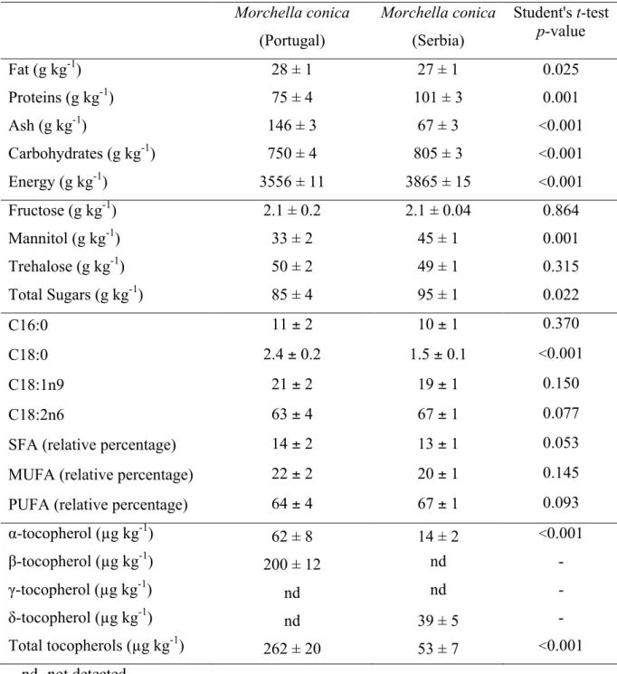
The composition in nutrients of M. conica samples is presented in Table 1. There were
no significant differences concerning fat content. The Serbian sample showed the
highest level of proteins and carbohydrates. However, the Portuguese sample had the
highest levels of ash. The energetic contribution of the Serbian sample was also
superior. Regarding free sugars composition, fructose was detected in both samples and
in the same concentration (Table 1). Mannitol and trehalose, also very common in
mushrooms, were detected in both samples (Figure 1A), and the Serbian one revealed
the highest content in total free sugars. Mannitol, a sugar alcohol, has half calories of
sugar and is half as sweet, and because it is poorly absorbed by the body, it does not
raise insulin levels as much as sugar and also does not promote tooth decay. This sugar
is also important in providing support and expansion of the fruiting body.25
The fatty acids quantified in higher amounts in M. conica (Table 1) were palmitic acid
(C16:0), stearic acid (C18:0), oleic acid (C18:1n9) and linoleic acid (C18:2n6), being
the saturated fatty acids (SFA; palmitic and stearic acids) present in lower amounts.
Portuguese sample; linoleic acid (polyunsaturated fatty acid- PUFA) was the main fatty
acid presented in both samples (Figure 1B). The Portuguese sample was richer in SFA
and MUFA, however Serbian sample presented the highest level of PUFA. PUFA,
especially the n-3 fatty acids family, are claimed to exert a protective effect against the
development of cardiovascular and inflammatory diseases.26,27 Particularly, linoleic acid plays an important role in preventing cardiovascular diseases in adults, promoting the
brain and retina development in infants28-30 or preventing DNA damage.31
α-Tocopherol isoform was detected in both samples and the Portuguese one presented the highest levels (Table 1). On the other hand, β-tocopherol was only present in the
Portuguese sample and δ-tocopherol in the Serbian one. It was the Portuguese sample that revealed the highest content of total tocopherols. The differences found could be
explained by the fact that tocopherols are molecules that are very easily oxidised either
during the fruiting body growth or in the extraction procedures. Therefore, it is
necessary to consider all these aspects, as also perform certain precautions during the
extraction methodologies. Vitamin E is found in large amounts in lipids, and some
evidence suggest that this vitamin prevents or minimizes the damage caused by free
radicals associated with specific diseases, including cancer, arthritis, cataracts and
aging.32,33 Recent evidences suggest that vitamin E reduced Alzheimer’s disease and dementia34,35 and has neuroprotective action against cisplatin peripheral neurotoxicity.36 Vitamin E has the ability to prevent the spread of chain reactions induced by free
radicals in biological membranes.37
Regarding M. conica organic acids profile, it was possible to quantify three different
compounds (Table 2), namely oxalic, quinic, and fumaric acids (Figure 1C). The wild
mushroom from Portugal was the one that revealed the highest concentration in oxalic
Portuguese sample showed the highest content in total organic acids. Concerning
phenolic acids, the studied samples revealed the presence of gallic, protocatechuic, p
-hydroxybenzoic and p-coumaric acids, as also the related compound cinnamic acid
(Table 2). Gallic and p-hydroxybenzoic acids were only present in the Serbian wild
sample. Protocatechuic and p-coumaric acids were found in highest concentrations in
the Portuguese sample. The related compound cinnamic acid was only found in the
Serbian sample. Puttaraju et al.15 did not found p-hydroxybenzoic and cinnamic acids in an Indian sample, but the research group found six other compounds: tannic, gentisic,
vanillic, syringic, caffeic and ferulic acids. Nevertheless, these authors applied a slightly
different extraction methodologies and the method applied to identify the phenolic
compounds also presented differences in the elution procedure, which could result in the
differences found. Analyzing these results we can conclude that the wild sample from
Serbia was richer in phenolic acids comparing with the Portuguese one. Phenolic
compounds, namely phenolic acids, are among the most potent and therapeutically
useful bioactive substances, providing health benefits associated with reduced risk of
chronic and degenerative diseases.38-40
It must be noted that the differences found in the chemical composition among samples
of different origins might be related to a diversity of factors such as, climatic conditions
and soil composition. According to the results it seems that Serbian species grown
under higher stress with an increase in the production of secondary metabolites namely,
phenolic compounds. Furthermore, differences found in individual compounds, can also
be attributed to different extraction and/or analysis methodologies.
Analyzing the results of the antioxidant potential (Table 3), M. conica samples
(methanolic extracts) from Portugal revealed the highest reducing power evaluated
through the Folin-Ciocalteu and Ferricyanide/Prussian blue assays. Total phenolics
content, measured by Folin-Ciocalteu assay, were different from other results from
Turkey (25.38 µg GAE mg-1 extract16 and 20.64 mg GAE g-1 extract17). Both extracts of samples from Turkey had lower amounts of total phenolics when compared to
Portuguese and Serbian wild samples. Nevertheless, Turkoglu et al.14 described the total phenolic amount of M. conica as 41.93 µg mg-1 pyrocatechol equivalents in the ethanolic extract. Puttaraju et al.15 studied water and methanolic extracts of M. conica from India, revealing the total content in phenolics (16.9±0.01 mg g-1 water extract and
4.6±0.01 mg g-1 methanolic extract) and the reducing power ability though
Ferricyanide/Prussian blue assay (3.90±0.25 mg of GAE g-1 of sample water extract and
8.62±0.38 mg of GAE g-1 methanolic extract). Gursoy et al.16, studied the reducing power of methanolic extracts of M. conica presenting their absorbance at 700 nm. The
Portuguese sample also revealed a higher radical scavenging activity, presenting a lower
EC50 value for the DPPH assay. This activity was also previously reported for M. conica
by Turkoglu et al.14 for ethanolic extract (EC50=267 µg mL-1) and Puttaraju et al.15, for water and methanolic extracts (EC50=5.00±0.31 and 1.60±0.33 mg mL-1, respectively)
obtained from Indian samples. Furthermore, Gursoy et al.16 described 43% for DPPH inhibition by a methanolic extract of M. conica at 2 mg mL-1, while Ozturk et al.17 reported 44% of inhibition by ethanolic extract of M. conica at 20 mg mL-1. Therefore,
Indian samples15 showed higher activity than the samples studied herein. However, the sample from Turkey14 gave the highest antioxidant activity of all. The Serbian sample showed highest lipid peroxidation inhibition, once it presented the lowest EC50 values
approximately 40 µg mL-1 promoted 50% of inhibition of oxidation in β-carotene/linoleate assay. Puttaraju et al.15 also studied the inhibition of lipid peroxidation by TBARS assay. Gursoy et al.16 showed that 0.5 mg mL-1 of M. conica extract inhibited 82% of lipid peroxidation measured through β-carotene-linoleate acid method. When comparing with the results of the cited authors, our samples revealed
lower inhibition of lipid peroxidation.
Methanolic extracts of the Portuguese and Serbian wild M. conica samples exhibited
antibacterial activity against all bacteria tested (Table 4). Both extracts showed highest
antibacterial potential against Bacillus cereus; in addiction the Portuguese sample
showed the same results of MIC and MBC against Pseudomonas aeruginosa and
Salmonella typhimurium. The lowest antibacterial activity of the two samples was
achieved against Escherichia coli and Enterobacter cloacae; the Serbian sample also
presented the same results against Micrococcus flavus. Methanolic extract obtained
from the Portuguese sample, in general, showed higher antibacterial potential than
extract prepared from the Serbian sample. Both extracts possessed the same activity
against Listeria monocytogenes, and in the case of Pseudomonas aeruginosa,
Portuguese sample possessed higher activity than the commercial antibiotic ampicillin.
Regarding the antifungal activity (Table 4), the highest antifungal potential was verified
for Trichoderma viride. The lowest antifungal activity was achieved towards
Aspergillus versicolor, Penicillium aurantiogriseum and Aspergillus ochraceus for the
for the both samples. In general, the Portuguese wild sample showed the highest
antifungal activity. Moreover, this sample was more efficient against Trichoderma
viride than ketoconazole (by comparison of MICs). There are few studies about
antimicrobial activity of M. conica, and all of them used agar-well diffusion method.
aeruginosa and Escherichia coli. Ozturk et al.17 also studied Pseudomonas aeruginosa, Escherichia coli and Bacillus cereus inhibition, without presenting any positive results
from M. conica ethanolic extracts. However, both of our samples presented activity
against these three species. Presumably, many factors influence accumulation of
bioactive compounds in mushrooms, but water availability, as well as exogenous
environmental factors, exposure to various light sources and the presence of fungal or
predatory pressures, cause the changes in dynamics of bioactive molecules biosynthesis
and, therefore, cause the different pharmacological potential of the investigated
samples.
As the methanolic extracts displayed antioxidant and antimicrobial activity, it was
important to guarantee their absence of cytotoxicity against liver cells, which are
considered the best in vitro model for studies of human cytotoxicity. The studied
samples revealed no toxicity in liver primary culture PLP2, once the GI50 values
obtained were higher than the highest concentration tested.
Conclusions
Overall, the Serbian sample revealed the highest content in free sugars, PUFA and
phenolic compounds, while the Portuguese sample revealed higher concentrations of
SFA, MUFA, tocopherols and organic acids. The methanolic extract of the Portuguese
sample revealed the highest antioxidant potential (with exception towards lipid
peroxidation inhibition), while the Serbian one showed the highest antimicrobial
activity. Both extracts are considered safe since they did not show toxicity in porcine
liver primary cells. This study provides new data concerning chemical characterization
and Serbian wild samples proved to be a good source of a wide range of nutrients and
bioactive molecules.
Acknowledgements
The authors are grateful to Fundação para a Ciência e Tecnologia (FCT, Portugal) and
COMPETE/QREN/EU for financial support to this work (research project
PTDC/AGR-ALI/110062/2009 and BPD/4609/2008 to L. Barros) and to CIMO (strategic project
PEst-OE/AGR/UI0690/2011). The authors also thank Serbian Ministry of Education,
Science and Technological Development for financial support (grant number 173032).
References
1 Kalac P, Chemical composition and nutritional value of European species of
wild growing mushrooms: A review. Food Chem113:9-16 (2009).
2 Furlani RPZ, Godoy HT, Nutritional value of edible mushrooms: a revision.
Revista do Instituto Adolfo Lutz 64:149-154 (2005).
3 Barros L, Baptista P, Correia DM, Casal S, Oliveira B, Ferreira ICFR, Fatty acid
and sugar compositions, and nutritional value of five wild edible mushrooms
from Northeast Portugal. Food Chem105:140-145 (2007).
4 Ferreira ICFR, Barros L, Abreu RMV, Antioxidants in wild mushrooms. Curr
Med Chem16:1543-1560 (2009).
5 Heleno SA, Barros L, Sousa MJ, Martins A, Ferreira ICFR, Tocopherols
composition of Portuguese wild mushrooms with antioxidant capacity. Food
6 Vaz JA, Barros L, Martins A, Morais JS, Vasconcelos MH, Ferreira ICFR,
Phenolic profile of seventeen Portuguese wild mushrooms. LWT 44:343-346
(2011).
7 Barros L, Pereira C, Ferreira ICFR, Optimized analysis of organic acids in edible
mushrooms from Portugal by Ultra Fast Liquid Chromatography and Photodiode
Array Detection. Food Anal Method6:309-316 (2013).
8 Barros L, Cruz T, Baptista P, Estevinho LM, Ferreira ICFR, Wild and
commercial mushrooms as source of nutrients and nutraceuticals. Food Chem
Toxicol46:2742-2747 (2008).
9 Lindequist U, Niedermeyer THJ, Julich WD, The pharmacological potential of
mushrooms. Evidence-Based Complement Alternative Med2:285-299 (2005).
10 Alves MJ, Ferreira ICFR, Dias J, Teixeira V, Martins A, Pintado M, A review
on antimicrobial activity of mushroom (Basidiomycetes) extracts and isolated
compounds. Planta Medica 78:1707-1718 (2012).
11 Alam N, Bristi NJ, Rafiquzzaman Md, Review on in vivo and in vitro methods
evaluation of antioxidant activity. Saudi Pharm J 21:143-152 (2013).
12 Pittet D, Infection control and quality health care in the new millennium. Am J
Inf Control33:258-267 (2005).
13 Masaphy S, Zabari, L, Observations on post-fire black morel ascocarp
development in an Israeli burnt forest site and their preferred micro-sites.
Fungal Ecology6:316-318 (2013).
14 Turkoglu A, Kivrak I, Mercan N, Duru ME, Gezer K, Turkoglu H, Antioxidant
and antimicrobial activities of Morchella conica Pers. Afri J Biotechn 5
15 Puttaraju NG, Venkateshaiah SU, Dharmesh SM, Urs SMN, Somasundaram R,
Antioxidant activity of indigenous edible mushrooms. J Agric Food Chem
54:9764-9772 (2006).
16 Gursoy N, Sarikurkcu C, Cengiz M, Solak MH, Antioxidant activities, metal
contents, total phenolics and flavonoids of seven Morchella species. Food Chem
Toxicol47:2381-2388 (2009).
17 Ozturk I, Sahan S, Sahin U, Ekici L, Sagdic O, Bioactivity and mineral contents
of wild-grown edible Morchella conica in the Mediterranean Region. J für
Verbraucherschutz Lebensmittelsicherheit 5:453-457 (2010).
18 AOAC, Official methods of analysis. Association of Official Analytical
Chemists, 16th ed., Arlington VA, USA (1995).
19 Heleno SA, Barros L, Sousa MJ, Martins A, Ferreira ICFR, Study and
characterization of selected nutrients in wild mushrooms from Portugal by gas
chromatography and high performance liquid chromatography. Microchem J
93:195-199 (2009).
20 Stojković DS, Barros L, Calhelha RC, Glamočlija J, Ćirić A, Van Griensven LJLD, Soković M, Ferreira ICFR, A detailed comparative study between
chemical and bioactive properties of Ganoderma lucidum from different origins.
Int J Food Sci Nutr65:42-47(2014).
21 Espinel-Ingroff A, Comparation of the E-test with the NCCLS M38-P method
for antifungal susceptibility testing of common and emerging pathogenic
filamentous fungi. J Clin Microbiol39:1360-1367 (2001).
22 CLSI, Clinical and Laboratory Standards Institute Methods for dilution
standard, 8th ed. CLSI publication M07-A8. Clinical and Laboratory Standards
Institute, Wayne, PA (2009).
23 Tsukatani T, Suenaga H, Shiga M, Noguchi K, Ishiyama M, Ezoe T, Matsumoto
K, Comparison of the WST-8 colorimetric method and the CLSI broth
microdilution method for susceptibility testing against drug-resistant bacteria. J
Microbiol Method 90:160-166 (2012).
24 Booth C, Fungal culture media. In J. R. Norris & D. W. Ribbons (Eds.),
Methods in microbiology (pp. 49-94). London and New York: Academic Press
(1971).
25 Dikeman CL, Bauer LL, Flickinger EA, Fahey GCJr, Effects of stage of
maturity and cooking on the chemical composition of select mushroom varieties.
J Agric Food Chem53:1130-1138 (2005).
26 Fürst P. Kuhn KS, Fish oil emulsions: what benefits can they bring? Clin Nutri
19:7-14 (2000).
27 Fang YZ, Yang S, Wu G, Free radicals, antioxidants, and nutrition. Nutrition
18: 872-879 (2002).
28 Simopoulos AP, Omega-3 fatty acids in health and disease and in growth and
development. Am J Clin Nutr54:438-463 (1991).
29 Senter SD, Payne JA, Miller G, Anagnosakis SL, Comparison of total lipids,
fatty acids sugars and non-volatile organic acids in nuts from four Castanea
species. J Sci Food Agric65:223-227(1994).
30 Künsch U, Scharer H, Conedera M, Sassella A, Jermini M, Jelmini G, Quality
assessment of chestnut fruits. Acta Horticulturae494:119-127 (1999).
31 Kok TMCM, Zwingman I, Moonen EJ, Schilderman PAEL, Rhijnsburger E,
human dietary supplementation with linoleic acid. Food Chem Toxicol 41
:351-358 (2003).
32 Morrissey PA, Sheehy PJA, Gaynor P, Vitamin E. Am J Clin Nutr Bethesda
62:260-264 (1994).
33 Heinonen OP, Albanes D, Virtamo J, Tbaylor PR, Huttunen JK, Hartman AM,
Haapakoski J, Malila N, Rautalahti M, Ripatti S, Maenpaa H, Teerenhovi L,
Koss L, Virolainen M, Edwards BK, Prostate cancer and supplementation with
alpha-tocopherol and beta-carotene: incidence and mortality in controlled trial. J
National Cancer Inst90:440-446 (1998).
34 Devore EE, Grodstein F, van Rooij FJA, Hofman A, Stampfer M.J., Witteman
JCM Breteler MMB, Dietary antioxidants and long-term risk of dementia. Arch
Neurology67:819-825 (2010).
35 Mangialasche F, Kivipelto M, Mecocci P, Rizzuto D, Palmer K, Winblad B,
Fratiglioni, L, High plasma levels of vitamin E forms and reduced Alzheimer’s
disease risk in advanced age. J Alzheimer Disease20:1029-1037 (2010).
36 Pace A, Giannarelli D, Galiè E, Savarese A, Carpano S, Giulia MD, Pozzi A,
Silvani A, Gaviani P, Scaioli V, Jandolo B, Bove L, Cognetti F, Vitamin E
neuroprotection for cisplatin neuropathy. Neurology74:762-766 (2010).
37 Traber MG, Packer L, Vitamin E: beyond antioxidant function. Am J Clin Nutr
62:1501S-1509S (1995).
38 Luximon-Ramma A, Bahorun T, Crozier A, Antioxidant action and phenolic and
vitamin C contents of common Mauritian exotic fruits. J Sci Food Agric83
39 Luximon-Ramma A, Bahorun T, Crozier A, Zbarsky V, Datla KK, Dexter DT,
Aruoma OI, Characterization of the antioxidant functions of flavonoids and
proanthocyanidins in Mauritian black teas. Food Res Int38:357-367 2005).
40 Soobrattee MA, Neergheen VS, Luximon-Ramma A, Aruoma OI, Bahorun T,
Phenolics as potential antioxidant therapeutic agents: mechanisms and actions.
Mutation Research/Fundamental and Molecular Mechanisms of Mutagenicity
Table 1. Composition in nutrients expressed in dry weight basis (mean ± SD).
nd- not detected.
Main fatty acids: C16:0 (Palmitic acid), C18:0 (Stearic acid), C18:1n9 (Oleic acid) and C18:2n6 (Linoleic acid); 20 more fatty acids were identified in trace amounts. SFA- Saturated fatty acids; MUFA- Monounsaturated fatty acids; PUFA- Polyunsaturated fatty acids.
Morchella conica
(Portugal)
Morchella conica
(Serbia)
Student's t-test p-value
Fat (g kg-1) 28 ± 1 27 ± 1 0.025
Proteins (g kg-1) 75 ± 4 101 ± 3 0.001
Ash (g kg-1) 146 ± 3 67 ± 3 <0.001
Carbohydrates (g kg-1) 750 ± 4 805 ± 3 <0.001
Energy (g kg-1) 3556 ± 11 3865 ± 15 <0.001
Fructose (g kg-1) 2.1 ± 0.2 2.1 ± 0.04 0.864
Mannitol (g kg-1) 33 ± 2 45 ± 1 0.001
Trehalose (g kg-1) 50 ± 2 49 ± 1 0.315
Total Sugars (g kg-1) 85 ± 4 95 ± 1 0.022
C16:0 11 ± 2 10 ± 1 0.370
C18:0 2.4 ± 0.2 1.5 ± 0.1 <0.001
C18:1n9 21 ± 2 19 ± 1 0.150
C18:2n6 63 ± 4 67 ± 1 0.077
SFA (relative percentage) 14 ± 2 13 ± 1 0.053
MUFA (relative percentage) 22 ± 2 20 ± 1 0.145
PUFA (relative percentage) 64 ± 4 67 ± 1 0.093
α-tocopherol (µg kg-1) 62 ± 8 14 ± 2 <0.001
β-tocopherol (µg kg-1) 200 ± 12 nd -
γ-tocopherol (µg kg-1) nd nd -
δ-tocopherol (µg kg-1) nd 39 ± 5 -
Table 2. Composition in non-nutrients expressed in dry weight basis (mean ± SD).
nd- not detected.
Morchella conica
(Portugal)
Morchella conica
(Serbia)
Student's t-test p-value
Oxalic acid (g kg-1) 1.9 ± 0.1 0.5 ± 0.1 <0.001
Quinic acid (g kg-1) 8.1 ± 0.3 8.8 ± 0.4 0.017
Fumaric acid (g kg-1) 5.61 ± 0.02 1.09 ± 0.02 <0.001
Total organic acids (g kg-1) 15.6 ± 0.3 10.5 ± 0.5 <0.001
Gallic acid (mg kg-1 ) nd 1.83 ± 0.01 -
Protocatechuic acid ( mg kg-1) 20.8 ± 0.1 4.96 ± 0.02 <0.001
p-Hydroxybenzoic acid ( mg kg-1) nd 55.2 ± 0.4 -
p-Coumaric acid (mg kg-1) 2.48 ± 0.01 2.15 ± 0.01 0.001
Total phenolic compounds (mg kg-1) 23.3 ± 0.1 64.1 ± 0.3 <0.001
Table 3. Antioxidant activity of the methanolic extracts (mean ± SD).
Concerning the Folin-Ciocalteu assay, higher values mean higher reducing power; for the other assays, the results are presented in EC50 values,
what means that higher values correspond to lower reducing power or antioxidant potential. EC50: Extract concentration corresponding to 50% of
antioxidant activity or 0.5 of absorbance for the Ferricyanide/Prussian blue assay.
Assay Morchella conica
(Portugal)
Morchella conica
(Serbia)
Student's t-test p-value
Reducing power
Folin-ciocalteu
(mg GAE g-1 extract) 32.8 ± 1.1 26.4 ± 0.4 <0.001 Ferricyanide/Prussian blue
(EC50; mg mL-1)
1.16 ± 0.01 1.88 ± 0.06 <0.001
Radical scavenging activity DPPH scavenging activity (EC50; mg mL-1)
3.56 ± 0.09 9 ± 2 <0.001
Lipid peroxidation inhibition
β-carotene/linoleate (EC50; mg mL-1)
2.5 ± 0.4 0.8 ± 0.1 <0.001
TBARS
(EC50; mg mL-1)
Table 4. Antibacterial and antifungal activities of the methanolic extracts and standard drugs (mg mL-1) (mean ± SD). Bacteria Morchella conica (Portugal) MIC/MBC Morchella conica (Serbia) MIC/MBC Streptomycin MIC/MBC Ampicillin MIC/MBC
Staphylococcus aureus 1.87±0.04c/3.75±0.02b 3.75±0.08d/7.50±0.07c 0.04±0.00a/0.09±0.03a 0.25±0.02b/0.37±0.01a Bacillus cereus 0.70±0.02c/0.95±0.02c 0.95±0.03d/1.87±0.02d 0.09±0.003a/0.17±0.00a 0.25±0.02b/0.37±0.01b Micrococcus flavus 1.87±0.04b/3.75±0.05b 7.5±0.10c/15.0±0.0c 0.17±0.01a/0.34±0.01a 0.25±0.00a/0.37±0.01a Listeria monocytogenes 3.75±0.08c/7.5±0.02b 3.75±0.05c/7.50±0.01b 0.17±0.007a/0.34±0.00a 0.37±0.007b/0.49±0.01a Pseudomonas aeruginosa 0.70±0.02b/0.95±0.02b 7.5±0.007c/15.0±0.00d 0.17±0.01a/0.34±0.01a 0.74±0.003b/1.24±0.09c Salmonella typhimurium 0.70±0.07c/0.95±0.02b 3.75±0.08d 7.50±0.10c 0.17±0.007a/0.34±0.007a 0.37±0.00b/ 0.49±0.01a Escherichia coli 5.62±0.10b/7.50±0.10b 7.5±0.10c/15.0±0.20c 0.17±0.01a/0.34±0.01a 0.25±0.01a/0.49±0.02a Enterobacter cloacae 5.62±0.10b/7.50±0.10b 7.5±0.00c/15.0±0.20c 0.26±0.02a/0.52±0.00a 0.37±0.01a/0.74±0.02a
Fungi Morchella conica (Portugal) MIC/MFC Morchella conica (Serbia) MIC/MFC Bifonazole MIC/MBC Ketoconazole MIC/MBC
Penicillium aurantiogriseum 6.25±0.02b/12.5±0.20b 12.5/±0.20c/25.00±0.30c 0.15±0.02a/0.20±0.00a 0.20±0.00a/0.50±0.02a Aspergillus versicolor 6.25±0.08b/6.25±0.08b 12.5±0.20c/12.5±0.20c 0.10±0.00a/0.20±0.00a 0.20±0.02a/0.50±0.02a Aspergillus ochraceus 3.12±0.04c/6.25±0.08c 12.5±0.20d/12.5±0.00d 0.15±0.02a/0.20±0.02a 1.50±0.00b/2.00±0.10b Aspergillus niger 1.56±0.01b/3.12±0.04c 6.25±0.08c/6.25±0.08d 0.15±0.00a/0.20±0.00a 0.20±0.01a/0.50±0.02b Trichoderma viride 0.78±0.007b/1.56±0.00c 1.56±0.05d/3.12±0.04d 0.15±0.00a/0.20±0.01a 1.00±0.10c/1.00±0.10b
Time (min)
0 5 10
mV
0 20 40 60 80 100
MP
1
2 3
4
A
B
Time (min
0 5 10 15 20 25 30 35
[V]
0.0 0.5 1.0 1.5 2.0 2.5
1 2 3 4 5 6 7
8
C
Figure 1. Individual profile of M. conica from Portugal (----) and Serbia (___) in (A)
sugars: 1- fructose, 2- mannitol, 3- trehalose; (B) fatty acids: 1- caproic acid (C6:0); 2-
caprylic acid (C8:0); 3- capric acid (C10:0); 4- lauric acid (C12:0); 5- myristic acid (C14:0); 6- myristoleic acid (C14:1); 7- pentadecanoic acid (C15:0); 8- palmitic acid (C16:0); 9- palmitoleic acid (C16:1); 10- heptadecanoic acid (C17:0); 11-stearic acid
(C18:0); 12- oleic acid (C18:1n9c); 13- linoleic acid (C18:2n6c); 14- α-linolenic acid
(C18:3n3c); 15-arachidic acid (C20:0); 16-eicosenoic acid (C20:1c); 17- cis-11,14-eicosadienoic acid (C20:2c); 18- cis-11, 14, 17-eicosatrienoic acid and heneicosanoic acid (C20:3n3+C21:0); 19- cis-5,8,11,14,17-eicosapentaenoic acid (C20:5n3); 20- behenic acid (C22:0); 21- Eicosenoic acid (C22:1n9); 22- tricosanoic acid (C23:0);
23-lignoceric acid (C24:0); 24- nervonic acid (C24:1) and (C) organic acids: 1- oxalic acid,
2- quinic acid, 3- fumaric acid. MP- mobile phase.
1 2 3 4 5 6 7 8 9 Time (min)