Clinical, ultrasonographic and histological findings
in varicose vein surgery
Moacir de Mello Porciunculla1 Dafne Braga Diamante Leiderman2 Rodrigo Altenfeder1 Celina Siqueira Barbosa Pereira1 Alexandre Fioranelli 1 Nelson Wolosker 2,3 Valter Castelli Junior1
1. Medical Science Faculty of the Santa Casa of São Paulo, São Paulo/SP, Brasil 2. Hospital Israelita Albert Einstein, São Paulo/SP, Brasil 3. Hospital das Clínicas of the Faculty of Medicine of the University of São Paulo, São Paulo/SP, Brasil
http://dx.doi.org/10.1590/1806-9282.64.08.729
SUMMARY
OBJECTIVE: This study aims to correlate the demographic data, different clinical degrees of chronic venous insufficiency (CEAP), ultra-sound findings of saphenofemoral junction (SFJ) reflux, and anatomopathological findings of the proximal segment of the great saphe-nous vein (GSV) extracted from patients with primary chronic vesaphe-nous insufficiency (CVI) submitted to stripping of the great saphesaphe-nous vein for the treatment of lower limb varicose.
METHOD: This is a prospective study of 84 patients (110 limbs) who were submitted to the stripping of the great saphenous vein for the treatment of varicose veins of the lower limbs, who were evaluated for CEAP clinical classification, the presence of reflux at the SFJ with Doppler ultrasonography, and histopathological changes. We study the relationship between the histopathological findings of the proximal GSV withdrawal of patients with CVI with a normal GSV control group from cadavers.
RESULTS: The mean age of the patients was higher in the advanced CEAPS categories when comparing C2 (46,1 years) with C4 (55,7 years) and C5-6(66 years), as well as C3 patients (50,6 years) with C5-6 patients. The normal GSV wall thickness (mean 839,7 microm-eters) was significantly lower than in the saphenous varicose vein (mean 1609,7 micrommicrom-eters). The correlational analysis of reflux in SFJ with clinical classification or histopathological finding did not show statistically significant findings.
CONCLUSIONS: The greater the age, the greater the clinical severity of the patients. The GSV wall is thicker in patients with lower limb varicose veins, but those histopathological changes are not correlated with the disease’s clinical severity or reflux in the SFJ on a Dop-pler ultrasound.
KEYWORDS: Varicose veins. Ultrasonography. Histology.
DATE OF SUBMISSION: 20-Dec-2017 DATE OF ACCEPTANCE: 24-Dec-2017
CORRESPONDING AUTHOR: Dafne Braga Diamante Leiderman
Rua Dr. Cesário Motta Junior, 61 - Vila Buarque, São Paulo - SP, Brasil – 01221-020 Phone: (11) 21515423 – Fax: (11) 38855361
E-mail: dah.diamante@gmail.com
INTRODUCTION
Amongst chronic vascular diseases, the chronic venous insufficiency (CVI) of the lower limbs (LLS) is the most prevalent.1 In Brazil, the prevalence
rang-es from 35% to 50% of the adult population.2,3As for
socioeconomic impact, according to the Ministry of Health, CVI occupies the 14th position among the 50 top diseases to cause temporary or permanent leave
from work, resulting in the payment of benefits to those ensured by the Brazilian social security system and still of working age.4
Histological performed on varicose veins have demonstrated contradictory findings of the structur-al changes in the smooth muscle of the venous wstructur-all.5
Some researchers have reported an increase in that
muscle’s quantity or activity6, while others have
ob-served reduced amounts of smooth muscle associated to the replacement of connective tissue.7,8 It has also
been suggested that the segregation of muscle cells by fibrous infiltration could interfere on the performance of those cells as a whole, resulting in alterations of the venous wall and, consequently, abnormal dilation.9
However, other studies were not able to demonstrate any differences between varicose veins and normal veins regarding muscle content of the venous wall.8
In the Brazilian national literature, there are no studies on the correlation of histopathological find-ings relating to the trunk of the great saphenous vein (GSV) with anamnesis of physical examination data. There are also no sufficiently conclusive reports re-garding the thickness alterations of the GSV wall on lighter or more advanced classes of the CEAP clinical classification to explain whether there is any differ-ence in the response of the venous wall thickness in the comparison amongst patients who have under-gone surgery multiple times.
OBJECTIVE
The objective of this study is to correlate demo-graphic data and the different clinical levels of chron-ic venous insuffchron-iciency (CEAP) with ultrasound find-ings of reflux in the saphenofemoral junction and anatomopathological findings of the proximal seg-ment of the great saphenous vein extracted from pa-tients with chronic venous insufficiency who under-went stripping of the great saphenous vein to correct lower limb varicose.
METHODOLOGY
A total of 84 consecutive patients were prospec-tively studied with a mean age of 51.2 ± 11.9 years, ranging from 21 to 74 years, out of which 63.6% were females, with CVI who underwent surgical treatment for varicose of 110 lower limbs and total unilateral stripping of the greater saphenous vein (58 patients) or bilateral (26 patients), over a period of three years in the Santa Casa de Misericórdia of São Paulo. The study was approved by the Research Ethics Commit-tee (CEP) (235/06).
We used the CEAP clinical classification to stan-dardize the physical assessment of patients. The clin-ical classification (C) is described below:
C0: No visible or palpable signs of venous disease
C1: Telangiectasies and reticular veins C2: Varicose veins
C3: Edema
C4a: Brown pigmentation (ochre dermatitis) and/ or eczema
C4b: Lipodermatosclerosis or athrophie blanche C5: Healed venous ulcer
C6: Active venous ulcer
The inclusion criteria were: the presence of pri-mary symptomatic varicose veins; absence of ob-structions of the deep venous system (DVT); limbs with GSV at thigh level with reflux in the Doppler ul-trasound (reflux criteria used >0.5 seconds) and with CEAP clinical categories C2 to C6.
All information was collected using an specific form that included anamnesis, the clinical exam-ination using the CEAP categories, ultrasound data (reflux of the great saphenous vein with or without saphenofemoral junction reflux), surgical data, and the anatomopathological study of the proximal por-tion of the great saphenous vein arch removed during surgery, the segment between the surgical ligation of the saphenous vein at the saphenofemoral junction and a second segment 1 cm below it, where the GSV is attached to the vein stripper.
The venous segments were removed for analysis and put in a 10% formalin solution for no longer than 48 hours. Cross-sections were made, which were included in the paraffin and stained with hematox-ylin and eosin and the Masson’s trichrome in order to measure the thickness of the vessel, the only pa-rameter selected for histopathological assessment in this study. The pieces were analyzed in an optical microscope with a magnification of 50x, and the im-ages captured with an attached camera to digitally measure the greater thickness of the vessel, from the tunica intima to the adventitia.
consid-ered the control. Bearing in mind that the thickness is not the uniform around the vessel, we used the big-gest measurement for the analysis.
We analyzed and correlated demographic data, clinical findings with the CEAP classification, the presence of pathological reflux of the SFJ detected by color Doppler ultrasound, and histological chang-es in the thicknchang-ess of the GSV arch wall upon optical microscopy. Lastly, we compared the average of the greatest GSV wall thickness at the arch in patients with CVI who underwent varicose surgical treatment with the healthy great saphenous vein obtained from cadavers (control group).
The statistical study considered data expressed in frequencies and means, averages and standard devi-ations. The chi-square test was applied for frequency comparison; the Student’s t-test was used to compare means and standard deviations; and the Pearson’s r coefficient for correlations between dependent and independent variables. To analyze the null hypothe-sis, a probability of 95% (p<0.05) was adopted.
RESULTS
Analyzing the relationship between the differ-ent CEAP clinical categories and patidiffer-ent gender, we found that most cases (x2 = 79.3089; p<0.0001) were
in the CEAP category C3 (60.0%) for both genders. The relationship between CEAP classification and pa-tient gender and age is presented in Table 1.
The patient age distribution was similar in dif-ferent CEAP clinical classifications. Regarding the different clinical categories and patient age, we ob-served that the older the patient, the greater the se-verity of the varicose veins (r=0.3268; p<0.05). We found a statistical difference when comparing CEAP C2 patients with CEAP categories C4 (p=0.0035) and C5-6 (0.027), as well as when comparing CEAP C3 patients with C5-6 (p=0.0374).
Reflux in the SFJ was found in 81.8% of GSVs, with reflux at the thigh. No statistical difference was found in the analysis comparing the average age of patients with reflux (51.7 years) and without reflux (49 years) at the arch of the GSV, with p=0.3621. The relationship between the CEAP clinical classification and the pres-ence of reflux in the SFJ is shown in Table 2.
No relationship was found between CEAP clinical categories and the presence of reflux in the SFJ (Ta-ble 3). Most cases that presented reflux in the SFJ were in category 3, with a higher frequency of that clinical category in the general sample.
The thickness of the normal GSV (ranging from 401-1.175, mean of 839.7+-243 micrometers) was sig-nificantly lower (p<0.0001) than in the GSV varicose
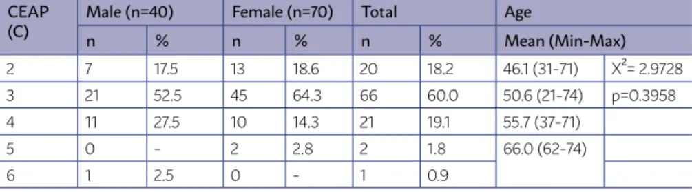
TABLE 1 – RELATIONSHIP BETWEEN CEAP CLINICAL CATEGORIES AND
PATIENT GENDER AND AGE DISTRIBUTION FOR EACH CATEGORY
CEAP
(C) Male (n=40)n % Female (n=70)n % Totaln % AgeMean (Min-Max)
2 7 17.5 13 18.6 20 18.2 46.1 (31-71) X²= 2.9728 3 21 52.5 45 64.3 66 60.0 50.6 (21-74) p=0.3958 4 11 27.5 10 14.3 21 19.1 55.7 (37-71)
5 0 - 2 2.8 2 1.8 66.0 (62-74)
6 1 2.5 0 - 1 0.9
TABLE 2 – RELATIONSHIP BETWEEN CEAP CLINICAL CATEGORIES AND THE
PRESENCE OF REFLUX IN THE SFJ
CEAP Category (C)
Present (n=88) Reflux Absent (n=20)
Total (n=110)
Statistics
n % n % n %
2 13 14.5 7 ^ 35 20 18.2 3 58 64.4 8 40 66 60.0 4 171 18.9 4 20 21 19.1
5 1 1.1 1 5 2 1.8
veins (ranging from 802-2.663, mean of 1,609+,7-376). There was no relationship between gender and age distribution and GSV thickness.
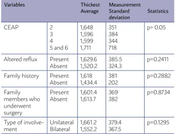
The analysis of the varicose GSV thickness with the CEAP classification, the presence of reflux at the GSV arch, uni- or bilateral involvement, and family history are shown in Table 3.
The relationship was demonstrated between thickness and the CEAP classification, the presence of reflux at the GSV arch, and uni- or bilateral in-volvement. We found that 95.5% of patients reported a family history of CVI, and in 32.7% of cases, there was a previous family history of LLS surgery. Such influence has not been proven during the statistical analysis of the cases.
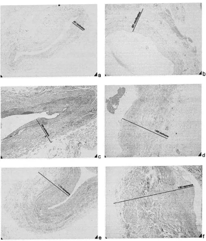
The images of the lowest, average, and highest values recorded during the measuring of GSV wall thickness in the control and study groups are pre-sented in Figure 1.
DISCUSSION
Our study group consists of patients with symp-tomatic varicose veins and GSV insufficiency at the thigh who sought help at a large public hospital in the city of São Paulo and, after the recommendation of conventional surgery, were consecutively included in the study. These patients are mostly aged between 30-50 years (48.8%), with a greater proportion of males (63.6% females and 36.4% males) than in other published studies, 3 to 6 women for 1 man.1,10,11 This
discrepancy with the literature is not explained by
the CVI severity in our study, since, despite previous-ly published works having included less advanced categories of the disease with a bigger aesthetic problem, the advanced clinical categories (C4, C5, and C6) have the same distribution for both genders. Although the evidence of genetic predisposition for varicose veins is still not highly conclusive, a family history of CVI was reported by 95.5% of the patients in this study.
The CEAP clinical classification for assessing the level of patient compromise due to chronic ve-nous disease of the lower limbs was elaborated in the American Venous Forum held in 1994, and its adoption as a universal language was helpful to a more homogeneous understanding of the venous disease, allowing for the use of such classification in scientific studies.12 In this study, 21.8% of mem-bers had advanced CVI, categorized as CEAP C$, 5, or 6, in which there is a greater technical difficulty in surgery and worse outcomes due to lipodermato-fibrosis, eczema, and active or healed ulcers, in ad-dition to higher recurrence rates.13,14 In this study,
all patients were submitted to conventional surgery, during which samples of the GSV arch segment were collected. The effectiveness of conventional varicose surgery and its low recurrence rates are undeniable,15,16 which makes this technique the gold
standard for varicose vein treatment. Despite the in-crease in the use of modern techniques of venous thermal ablation and the fact that its effectiveness is comparable to conventional surgery, Mendes et al.17 demonstrated, in a randomized study, that if
patients are not told the technique used, they can-not differentiate the conventional surgery from the radiofrequency ablation.
We found a clear progression of the clinical classi-fication of the disease with increasing age when com-paring early clinical classifications C2 (average age of 46.1 years) and C3 with the advanced classifications C4 and C5-6, with a statistically significant difference despite the lower number of C5-6 patients (three pa-tients, average age of 66 years). The low number of C5-6 patients is the result of the inclusion of consecu-tive patients who met the research inclusion criteria associated with a lower frequency of the advanced classifications of the disease in the general popula-tion. The clinical deterioration with increasing age reveals the natural history of the disease, illustrating that the complications and severity of CVI increase over the years in patients with no definitive
treat-TABLE 3 – RELATIONSHIP BETWEEN THE VARICOSE
GSV THICKNESS AND THE CEAP CLASSIFICATION, THE PRESENCE OF REFLUX AT THE GSV ARCH, FAMILY HISTORY, AND UNI- OR BILATERAL INVOLVEMENT
Variables Thickest
Average MeasurementStandard deviation
Statistics
CEAP 2 3 4 5 and 6
1,648 1,596 1,599 1,711 351 384 344 718 p> 0.05
Altered reflux Present
Absent 1,629.61,520.2 385.5324.3 p=0.2411 Family history Present
Absent 1,6181,434.4 381202 p=0.2882 Family
members who underwent surgery
Present
Absent 1,601.41,613.7 369382 p=0.8734
Type of
FIGURE 1: (A) Lowest value recorded in the control group: 401 micrometers; (b) Smaller value recorded in the study group: 801 micrometers; (c) Value closest to the average value of the control group: 866 micrometers; (d) Value closest to the average value of the study group: 1,606 micrometers; (e) Highest value recorded in the control group: 1,175 micrometers; (f) Highest value recorded in the study group: 2,663 micrometers.
ment, and that there may be a lack of information among the population regarding the evolution of the chronic venous disease and its complications when there is no early treatment, evidencing the need for awareness campaigns.
direct-ly interferes in the surgical approach and the post-operative results. The abnormal reflux in the sa-phenofemoral junction is considered the cause for 60% to 80% of all primary varicose vein cases.18 In line with some other studies,18,19 we found altered reflux in the saphenofemoral junction in 81.8% of the limbs analyzed with GSV reflux in the thigh. Seidel et al.10 studied the association between CVI
symptoms, visible varicose veins, and GSV reflux, grouping patients in three different groups (asymp-tomatic with varicose veins, symp(asymp-tomatic with no varicose veins, and symptomatic with varicose veins) and concluded that GSV reflux was more frequent in symptomatic patients with visible varicose veins, however they did not study the relevance of SFJ in-sufficiency on symptoms and on the level of varicose veins visible during the physical examination. Even though the presence of altered SFJ reflux is a rele-vant parameter for surgical recommendation20 and
predictor of CVI,19 the presence of such reflux
pre-sented no correlation with the clinical classification of CVI (CEAP) in this study, i.e. Patients with saphe-nofemoral junction reflux did not present a more ad-vanced clinical stage than patients with SFJ and no pathological reflux. Yamaki et al.21 also did not find
an association between the presence of superficial venous reflux or isolated perforator insufficiency and the early (CEAP C1-3) or advanced (CEAP C4 to 6) CVI clinical categories. However, the maximum reflux speed and maximum volume of reflux in the great saphenous vein, saphenofemoral and saphe-nopopliteal junctions, and deep venous system are
higher in patients with advanced CEAP.
Even though alterations in the composition of the venous wall are considered the basic dysfunction for all CVI8 and are related to GSV insufficiency,10 our
study did not show any relationship between an in-creased thickness of the GSV wall and the presence of SFJ reflux.
Amongst possible structural alterations in the varicose GSV, the focus of our study was the thick-ness of the GSV wall in SFJ. Santos Ferreira22,23 found
the thickening of the venous walls, especially in dis-tal positions and in the tunica intima, to be the main and most frequent histological alteration in varicose veins. Silveira24 found thicker walls in varicose veins
than in normal veins in all three tunicas. In our study we also found thicker walls in varicose veins than in normal veins obtained from cadavers with no history of venous disease, but found no correlation between such increased thickness and clinical and ultrasound data, thus confirming the findings of Santos Ferreira and the observations of Garrido et al.22,23
CONCLUSIONS
The findings in this study confirm that the clinical classification of CVI is more severe with increased patient age. However, the thickness of the arch of the great saphenous vein is bigger in patients with vari-cose veins of the lower limbs, and these alterations do not correlate to the clinical classification of the disease or the presence of pathological reflux in the SFJ identified by Doppler ultrasound.
RESUMO
OBJETIVO: Este estudo tem como objetivo correlacionar os dados demográficos, os diferentes graus clínicos da insuficiência venosa crônica (Ceap), com achados ultrassonográficos de refluxo da junção safenofemoral (JSF) e os achados anatomopatológicos do seg-mento proximal da veia safena magna (VSM) extraído de pacientes com insuficiência venosa crônica (IVC) primária submetidos à safenectomia magna para correção de varizes dos membros inferiores.
MÉTODO: Estudo prospectivo de 84 pacientes e 110 membros submetidos à safenectomia magna para o tratamento de varizes de membros inferiores, correlacionando a sua classificação clínica Ceap, presença de refluxo na JSF ao ultrassom Doppler e alterações his-topatológicas. Comparamos ainda os achados histopatológicos da VSM proximal retirada dos pacientes com IVC com grupo controle de VSM normal retirada de cadáveres.
RESULTADOS: Média de idade dos pacientes foi maior nos Ceaps avançados quando comparado Ceap C2 (46,1 anos) com C4 (55,7 anos) e C5-6 (66 anos), e pacientes C3 (50,6 anos) com C5-6. A espessura da parede da VSM normal (média de 839,7 micrômetros) foi sig-nificativamente menor do que das VSM varicosas (média de 1.609,7 micrômetros). As análises de correlação da presença do refluxo em JSF com a classificação clínica ou achado histopatológico não demostraram ser estatisticamente significativas.
CONCLUSÕES: Quanto maior a idade, mais avançada é a classificação clínica da IVC dos pacientes. A espessura da parede da crossa da VSM é maior nos pacientes com IVC e essas alterações não se correlacionam com a classificação clínica da doença ou com a presença de refluxo na JSF ao ultrassom Doppler.
REFERENCES
1. Hobson J. Venous insufficiency at work. Angiology. 1997;48(7):577-82.
2. Cabral ALS. Insuficiência venosa crônica de membros inferiores: prevalên-cia, sintomas e marcadores preditivos [Tese de doutorado]. São Paulo: Universidade Federal de São Paulo, Escola Paulista de Medicina; 2000. p.140.
3. Maffei FH, Magaldi C, Pinho SZ, Lastoria S, Pinho W, Yoshida WB, et al. Varicose veins and chronic venous insufficiency in Brazil: prevalence among 1755 inhabitants of a country town. Int J Epidemiol. 1986;15(2):210-7.
4. Mallick R, Lal BK, Daugherty C. Relationship between patient-reported symptoms, limitations in daily activities, and psychological impact in vari-cose veins. J Vasc Surg Venous Lymphat Disord. 2017;5(2):224-37.
5. Abramson DI. Diseases of the veins: pathology, diagnosis and treatment. JAMA. 1988;260(24):3680.
6. Obitsu Y, Ishimaru S, Furukawa K, Yoshihama I. Histopathological studies of the valves of varicose veins. Phlebology. 1990;5(4):245-54.
7. Rose SS, Ahmed A. Some thoughts on the aetiology of varicose veins. J Cardiovasc Surg (Torino). 1986;27(5):534-43.
8. Jacobs BN, Andraska EA, Obi AT, Wakefield TW. Pathophysiology of vari-cose veins. J Vasc Surg Venous Lymphat Disord. 2017;5(3):460-7.
9. Wali MA, Dewan M, Eid RA. Histopathological changes in the wall of var-icose veins. Int Angiol. 2003;22(2):188-93.
10. Seidel AC, Campos MB, Campos RB, Harada DS, Rossi RM, Cavalari Ju-nior P, et al. Associação entre sintomas, veias varicosas e refluxo na veia safena magna ao eco-Doppler. J Vasc Bras. 2017;16(1):4-10.
11. Seidel AC, Mangolim AS, Rossetti LP, Gomes JR, Miranda Jr F. Prevalência de insuficiência venosa superficial dos membros inferiores em pacientes obesos e não obesos. J Vasc Bras. 2011;10(2):124-30.
12. Venous Forum Annual Meeting, Royal Society of Medicine, London, 14 October 1994. Phlebology. 2016;10(2):79-85.
13. Kokkosis AA, Schanzer H. Anatomical and clinical factors favoring the per-formance of saphenous ablation and microphlebectomy or sclerotherapy as a single-stage procedure. Phlebology. 2015;30(9):627-31.
14. van der Velden SK, Pichot O, van den Bos RR, Nijsten TE, De Maeseneer
MG. Management strategies for patients with varicose veins (C2-C6): re-sults of a worldwide survey. Eur J Vasc Endovasc Surg. 2015;49(2):213-20.
15. Lurie F, Creton D, Eklof B, Kabnick LS, Kistner RL, Pichot O, et al. Prospec-tive randomized study of endovenous radiofrequency obliteration (closure procedure) versus ligation and stripping in a selected patient population (EVOLVeS Study). J Vasc Surg. 2003;38(2):207-14.
16. Siribumrungwong B, Noorit P, Wilasrusmee C, Attia J, Thakkinstian A. A systematic review and meta-analysis of randomised controlled trials comparing endovenous ablation and surgical intervention in patients with varicose vein. Eur J Vasc Endovasc Surg. 2012;44(2):214-23.
17. Mendes CA, Martins AA, Fukuda JM, Parente JB, Munia MA, Fioranelli A, et al. Randomized trial of radiofrequency ablation versus conventional surgery for superficial venous insufficiency: if you don’t tell, they won’t know. Clinics (Sao Paulo). 2016;71(11):650-6.
18. Engelhorn CA, Engelhorn AL, Cassou MF, Salles-Cunha SX. Patterns of saphenous reflux in women with primary varicose veins. J Vasc Surg. 2005;41(4):645-51.
19. Konoeda H, Yamaki T, Hamahata A, Ochi M, Sakurai H. Quantification of superficial venous reflux by duplex ultrasound-role of reflux velocity in the assessment the clinical stage of chronic venous insufficiency. Ann Vasc Dis. 2014;7(4):376-82.
20. Ciostek P, Michalak J, Noszczyk W. Improvement in deep vein haemo-dynamics following surgery for varicose veins. Eur J Vasc Endovasc Surg. 2004;28(5):473-8.
21. Yamaki T, Nozaki M, Fujiwara O, Yoshida E. Comparative evaluation of duplex-derived parameters in patients with chronic venous insufficiency: correlation with clinical manifestations. J Am Coll Surg. 2002;195(6):822-30.
22. Maffei FHA, Lastoria S, Yoshida WB, Rollo HA. Doenças vasculares per-iféricas. 3rd ed. Rio de Janeiro: Medsi; 2002.
23. Garrido M, Santos Ferreira C, Sales EA. Varizes de membros inferiores: Patologia. In: Maffei FHA, Lastória S, Yoshida WB, Rollo HA. Doenças vas-culares periféricas. 3a. ed. Rio de Janeiro; Medsi; 2002. Volume 2. p. 1511-20