RADIOLOGY PAGE
432
Leiomyoma of the urinary bladder: a case report
Margarita Ortiz, Daniel E. Henao, Walter Cardona Maya, Maurizio Massaro Ceballos
Departament of Radiology (MO, MMC) and Reproduction Group (DEH, CM), University of Antioquia, Colombia
_______________________________________________________________________________
ABSTRACT
The case of a 71-year-old woman who pre-sented with one year history of pelvic pain and oc-casional dysuria is reported. Computed tomography and Magnetic Resonance Imaging revealed a well defined intramural bladder mass. The histological findings of the surgical specimen confirmed a leio-myoma of the urinary bladder. The clinical presen-tation, imaging findings and management of this relatively rare benign tumor are discussed.
INTRODUCTION
Leiomyoma of the bladder is a rare benign tumor that occurs mainly in women between the fourth and fifth decades of life. Although no pa-thophysiological mechanisms have been described to explain the occurrence of this tumor, it might be related to an endocrine alteration. The most com-mon clinical characteristics include urinary voiding symptoms (1).
We describe in this report the case of an el-der woman who mainly presented with chronic pel-vic pain. The mass detected within her bladder -by several imaging methods- was later histologically confirmed as leiomyoma.
CASE REPORT
A 71-year-old female presented with a his-tory of chronic pelvic pain radiating to the low back area associated with occasional dysuria. Poor symp-toms relief after several antibiotic treatments for
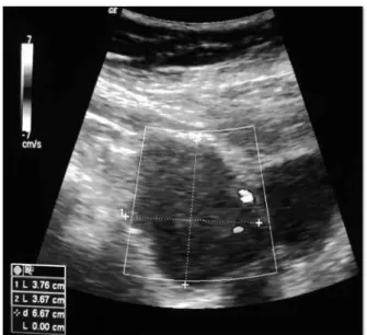
uri-nary tract infections was documented. Initial ab-dominal ultrasound demonstrated normal kidney aspects and a mass of the bladder (Figure-1).
Subsequent CT of the pelvis confirmed a soft tissue mass at the right anterolateral aspect of the bladder’s wall (Figure-2). MR images also demonstrated a mass in the same to location (Fi-gure-3).
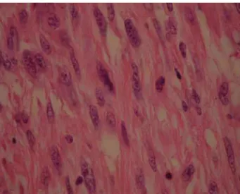
The patient underwent a laparoscopic par-tial cystectomy -after a biopsy of the mass acqui-red by cystoscopy reported a benign leiomyoma- obtaining a surgical specimen of 4 cm of diameter. Finally, histopathology confirms the diagnosis of leiomyoma (Figure-4).
Figure 1 - Abdominal Ultrasound.
Hypoechoic well-defined mass in the vesical wall measuring 3.67 cm and 3.76 cm in their transverse and antero-posterior diameters is shown. Ac-cording to the Doppler it possesses no blood-flow.
Vol. 39 (3): 432-434, May - June, 2013
433
IBJU |RADIOLOGY PAGEDISCUSSION
Leiomyoma of the bladder is a relatively rare tumor: less than 1% of the tumors of this or-gan (1). In a systematic review of the literature carried out by Silva-Ramos et al. (2) the charac-teristics of patients affected by this tumor were: 75.6% were women, the media age of presentation was 45.3 years, 50% of patients presented urinary voiding symptoms.
Ultrasound, CT and MRI are regularly used to diagnose this tumor. It was once proposed that MRI by itself could confirm this diagnosis, no-netheless and considering it cannot differentia-te mesenchymal tumors from the more common transitional cell tumors (TCT) the histopathology study is always necessary to confirm (3,4). Recent findings, nonetheless, indicate that the Apparent Diffusion Coefficient -obtained by diffusion--weighted magnetic resonance- values are signifi-cantly lower in malignant lesions (e.g. TCT) when compared to those obtained from benign tumors (i.e. mesenchymal tumors). The specificity claimed by this technique certainly deserves further empi-rical results and analysis.
The underlying cause of leiomyoma of the bladder is still unclear. No recurrences or ma-Figure 3 - Magnetic Resonance (MR) Imaging of the pelvis.
Figure 2 - Computed Tomography (CT) of the pelvis.
Figure 4 - Piece to specimen.
Coronal portal venous phase CT demonstrating a mass with soft tissue at-tenuation pattern on the right antero-lateral aspect of the bladder’s wall is indicated. The mass possesses smooth boundaries with poor enhancement after contrast medium administration demonstrating a homogeneous lesion.
Histopathology of the removed specimen -composed of spindle-shaped cells without dysplasia, atypia or pleomorphism- confirms the diagnosis of leiomyoma.
Axial fat-suppressed T1-weighted MR image pre-contrast revealed the mass previously described in the CT scan.
434
IBJU |RADIOLOGY PAGElignant degenerations of leiomyomas have been reported. Treatment of leiomyoma of the urinary bladder is mainly surgical. Surgical excision has excellent prognosis after complete resection and should always be offered.
We reported the case of a leiomyoma of the bladder, successfully removed by laparoscopy without compromising the bladder capacity.
ACKNOWLEDGMENTS
Sustainability Strategy 2013-2014 Repro-duction Group
_____________________
Correspondence address:Dr. Margarita Ortiz Carrera 51D No. 62 Medellín, 29, Colombia Fax: + 57 4 219-6476 E-mail: [email protected]
REFERENCES
1. Blasco Casares FJ, Sacristán Sanfelipe J, Ibarz Servio L, Batalla Cadira JL, Ruiz Marcellán FJ. Characteristics of bladder leio-myoma in our setting. Arch Esp Urol. 1995; 48: 987-90. 2. Silva-Ramos M, Massó P, Versos R, Soares J, Pimenta A.
Leio-myoma of the bladder. Analysis of a collection of 90 cases. Ac-tas Urol Esp. 2003; 27: 581-6.
3. Park JW, Jeong BC, Seo SI, Jeon SS, Kwon GY, Lee HM: Leio-myoma of the urinary bladder: a series of nine cases and review of the literature. Urology. 2010; 76: 1425-9.
4. Sundaram CP, Rawal A, Saltzman B: Characteristics of bladder leiomyoma as noted on magnetic resonance imaging. Urology. 1998; 52: 1142-3.
ARTICLE INFO
Int Braz J Urol. 2013; 39: 432-4
__________________
Submitted for publication: June 28, 2012
__________________