J of Evolution of Med and Dent Sci/ eISSN- 2278-4802, pISSN- 2278-4748/ Vol. 4/ Issue 65/ Aug 13, 2015 Page 11308
CLINICAL AND RADIOLOGICAL DIAGNOSIS IN ACUTE ABDOMINAL
EMERGENCIES
Rehan Sabir Momin1, Muhammad Abdul Azhar 2, Sadiq Hussain3
HOW TO CITE THIS ARTICLE:
Rehan Sabir Momin, Muhammad Abdul Azhar, Sadiq Hussain. Clinical and Radiological Diagnosis in Acute Abdominal Emergencies. Journal of Evolution of Medical and Dental Sciences 2015; Vol. 4, Issue 65, August 13; Page: 11308-11315, DOI: 10.14260/jemds/2015/1631
ABSTRACT: BACKGROUND: Acute abdominal pain is a common presentation that requires
immediate management. It includes traumatic and non-traumatic conditions of abdomen and if not diagnosed and treated promptly in the golden hours, it can lead to high mortality rate. Hence, this study was undertaken to correlate different modes of diagnosis which helps in deciding the immediate line of management proved to be lifesaving. OBJECTIVES: To analyse and compare the efficacy of clinical and radiological methods in early diagnosis of acute abdominal conditions.
MATERIALS AND METHODS: This study includes patients admitted in emergency wards of Shadan
General and Teaching Hospital, Hyderabad. Total 100 cases were included in this study who presented with acute abdomen. After thorough clinical examination they were subjected to Plain X-ray Abdomen and Ultrasonography of abdomen and pelvis. RESULTS: All 100 patients were thoroughly evaluated clinically and subjected to Plain X-ray abdomen with Ultrasonography of abdomen. Acute Abdomen was most common in age group between 20-40 years with male predominance. Acute appendicitis was the most common cause of surgical condition, followed by Peritonitis and then Intestinal Obstruction. The diagnostic accuracy rates in male and female patients were 93% and 80% respectively. Ultrasonography had highest sensitivity rate (97.8%) and plain X-ray abdomen had highest specificity rate (88.4%). CONCLUSION: Each of these diagnosing methods in acute abdomen are complementary to each other. With efficient clinical acumen and using ultrasonography and X-ray abdomen as basic diagnostic investigations, one can do early diagnosis with 97% to 99% accuracy and thus can avoid unnecessary operations.
KEYWORDS:
Acute Abdomen, Ultrasonography, Plain X-ray Film.
INTRODUCTION: Without precision there is no perfection this statement holds much truth in the
field of medical science in the diagnosis and management of acute diseases of abdomen. Abdominal pain is a common presentation to emergency department. It is vital that physician has an understanding with the presentations of common diseases of acute abdomen.[1] Diagnosis of acute
abdominal conditions in many instances is challenging and complex.
Acute abdomen forms bulk of emergencies in surgical practice often taxing diagnostic acumen regardless of the experience of the surgeon. Preoperative diagnosis of acute abdomen is crucial to minimize the morbidity and mortality where the diagnostic facilities are limited.[2] Prognosis of acute
Abdomen for both traumatic and non-traumatic origin depends largely on early diagnosis.
J of Evolution of Med and Dent Sci/ eISSN- 2278-4802, pISSN- 2278-4748/ Vol. 4/ Issue 65/ Aug 13, 2015 Page 11309
In the past 20 years, the ability to accurately determine intra-abdominal pathologic process by radiological imaging has allowed earlier and more accurate diagnosis. Previous studies have shown that a considerable volume of diagnostic errors would be reduced by paying more attention to diagnosis before laparotomy.[4] In earlier part of 20th Century, plain X-ray of abdomen was the only
investigation which was introduced as a diagnostic tool in clinical practice even though x-rays are shadows and not true images. Plain X-rays were found to be useful in 40% of acute abdominal cases.
Plain Abdominal films generally are not recommended unless other conditions like perforations of gastrointestinal tract, intestinal obstructions and ureteral calculus are suspected on clinical assessment.[5] Due to tremendous advancements in technology, more diagnostic tools like
ultrasonography, MRI, CT Scan have proved to be more sensitive in its accuracy to arrive at early diagnosis.
So, with this view a study was planned to analyse and evaluate the findings of clinical examination, findings of plain X-ray abdomen and ultrasonography in diagnosing acute abdominal emergencies. Improvement in the surgeon’s power of decision making is the basic pivot of disease diagnosis and therapy, particularly in developing countries with limited diagnostic facilities.[2]
In India and other developing countries where availability of MRI and CT scan in remote areas and affordability of these investigations by poor patients become hindrance to achieve early diagnosis in acute abdomen conditions that is the reason only plain x-ray and ultrasonography was included apart from clinical assessment which is affordable and relatively available across the country. This study can be helpful for those surgeons who are working in developing countries and striving to deliver their best performance with minimal diagnostic tools to manage acute abdomen which is the commonest surgical disease.
MATERIALS AND METHODS:
Source of Data: This study was conducted in Shadan General and Teaching Hospital of Hyderabad,
India. It included patients admitted in emergency wards of all age groups and both the genders who have presented with acute abdomen.
Inclusion Criteria: 100 cases were selected from the study period which includes various acute surgical abdominal conditions including abdominal trauma.
Exclusion Criteria: This study has excluded the patients presenting as acute abdomen having
medical and gynecological causes.
J of Evolution of Med and Dent Sci/ eISSN- 2278-4802, pISSN- 2278-4748/ Vol. 4/ Issue 65/ Aug 13, 2015 Page 11310
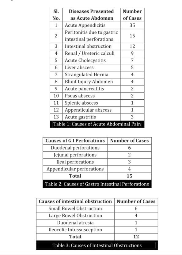
RESULTS: Acute abdomen was most common in the age group 20–40 years with male predominance
(69%). Acute Appendicitis (35%) was the most common cause of acute surgical condition, followed by peritonitis due to Gastro intestinal perforation (15%) and then intestinal obstruction (12%). The most common symptoms with abdominal pain were nausea (69.1%) and vomiting (43.9%). The most common clinical signs were abdominal tenderness (97.1%), voluntary guarding (66.9%) and rebound tenderness (66.2%). The diagnostic accuracy rates in male and female patients were 93% and 80% respectively.
Sl. No.
Diseases Presented as Acute Abdomen
Number of Cases
1 Acute Appendicitis 35
2 Peritonitis due to gastric
intestinal perforations 15 3 Intestinal obstruction 12 4 Renal / Ureteric calculi 9 5 Acute Cholecystitis 7 6 Liver abscess 5 7 Strangulated Hernia 4 8 Blunt Injury Abdomen 4 9 Acute pancreatitis 2 10 Psoas abscess 2 11 Splenic abscess 1 12 Appendicular abscess 1 13 Acute gastritis 3
Table 1: Causes of Acute Abdominal Pain
Causes of G I Perforations Number of Cases
Duodenal perforations 6 Jejunal perforations 2 Ileal perforations 3 Appendicular perforations 4
Total 15
Table 2: Causes of Gastro Intestinal Perforations
Causes of intestinal obstruction Number of Cases
Small Bowel Obstruction 6 Large Bowel Obstruction 4 Duodenal atresia 1 Ileocolic Intussusception 1
Total 12
J of Evolution of Med and Dent Sci/ eISSN- 2278-4802, pISSN- 2278-4748/ Vol. 4/ Issue 65/ Aug 13, 2015 Page 11311
Cause Number of Cases
Acalculous Cholecystitis 5 Calculous Cholecystitis 2
Total 7
Table 4: Acute Cholecystitis
Clinically all 7 cases were diagnosed cholecystitis on basis of history and clinical examination. Plain X-ray was not suggestive. Ultrasonography of abdomen showed Gall bladder wall thickening more than 4mm with sludge in 5 cases of acalculous cholecystitis and gall stones in other 2 cases.
Acute Pancreatitis were 2 in our series. Only one had presented clinically with features of acute pancreatitis. Plain X-ray abdomen erect view showed sentinel loop sign. Ultrasonography showed oedematous pancreas in both cases and calcification noted in one case.
Blunt Injury Abdomen in our series were 4 in number. Of which 2 were splenic injury and other 2 were Liver trauma. In all 4 cases, clinical assessment were suggestive of visceral injury. Plain X-ray Abdomen showed obliteration of psoas shadow and elevation of left diaphragm in splenic injury.
Ultrasonography showed subcapsular haematoma of spleen in one case and intrasplenic haematoma with intraperitoneal collection in other case. In both cases of Liver injuries, echogenic lesion in the liver with intraperitoneal collection was evident. These findings were confirmed on respective laparotomies.
Disease No. of
cases
Diagnosed Clinically
Diagnosed on KUB X ray
Diagnosed on USG Abdomen
Renal Calculi 4 4 3 4
Ureteric Calculi 3 3 2 3
Both Calculus 2 1 1 2
Table 5: Renal and Ureteric calculi
All urolithiasis cases were diagnosed clinically with its typical presentation. Ultrasonography and Plain X ray KUB were complementary to each other in establishing final diagnosis.
Predictive Values USG X ray
Sensitivity (%) 97.8 46.4 Specificity (%) 73 88.4
Table 6: Predictive values of Plain X-ray and Ultrasonography
J of Evolution of Med and Dent Sci/ eISSN- 2278-4802, pISSN- 2278-4748/ Vol. 4/ Issue 65/ Aug 13, 2015 Page 11312
Graph Showing Comparative Study of Percentage of Accuracy in Diagnosing Acute Abdominal Conditions by Clinical Methods, Plain X-Rays and Ultrasonography
DISCUSSION: Patients with acute abdominal pain are a heterogenous group that consumes a great deal of resources.[3] In cases when the diagnosis is suspected, laparotomy has been advised to be
performed,[6] but this policy has increased the rate of negative laparotomies.[7] This study of 100
cases of acute abdomen was conducted in this teaching hospital with the aim of studying the efficacy of various methods of diagnosis which include clinical evaluation, radiological studies and ultrasonographic findings and their correlation with final diagnosis which were done by operative intervention.
In this study, acute abdomen was most commonly seen in age group of 20–40 years, whereas the statistics from other study reported the prevalence of acute abdomen mostly in 20-29 years of age.[2] The causes of acute abdomen are several and their relative incidence varies in different
populations. Several factors are seen to be responsible for these differences.
Socioeconomic factors and diet have mostly been incriminated to be responsible for the observed differences.[8] Acute Appendicitis (35%) was the most commonest cause of acute surgical
condition, followed by peritonitis due to Gastro intestinal perforation (15%) and then intestinal obstruction (12%). Other studies reported acute appendicitis to be leading cause of acute abdomen in 55% cases,[2] visceral perforation and bowel obstruction in 8-12% and 15-24% of cases respectively.
Clinical skills and examinations were found to be more reliable in almost all acute abdomen cases. Plain X-rays were found to be accurate in 87.80% of cases. Chhetri reported sensitivity of 64.8% and specificity 88.8% for plain abdominal X ray.[2] Ultrasonography were found to be accurate
in 94% of cases.
J of Evolution of Med and Dent Sci/ eISSN- 2278-4802, pISSN- 2278-4748/ Vol. 4/ Issue 65/ Aug 13, 2015 Page 11313
Chhetri reported sensitivity and specificity of 69.4% and 81.5% for ultrasonography in the diagnosis of acute abdomen.[2] In diagnosing renal and Ureteral stones, ultrasonography is highly
effective in showing large stones more than 5mm but poor at visualizing stones smaller than 3mm.[9,10] Sonography is reported to have overall sensitivity between 73% to 100% for obstruction of
the collecting system.[10,11] Kidney ureter bladder radiography has been reported to have sensitivity
between 45% and 69% and specificity between 71% and 82%.[12,13] KUB X ray along with
ultrasonography has been reported to achieve clinical accuracy close to that of non-contrast enhanced CT in follow up of renal calculi.[14]
We have found that early diagnosis in acute abdominal conditions can be achieved using one or more of these clinical and radiological investigative tools. Each of these method has been proved to be complementary to the other in early diagnosis. In other studies, diagnosis has been reported 80% by skilled physicians and 50% by young physicians.[15]
Decision to surgically intervene in acute abdominal emergencies is based on the results of a good history, thorough physical examination along with judicial usage of basic radiological imaging tools. Therefore, an accurate and early diagnosis of acute abdomen can avoid unnecessary operations thereby reducing the rate of negative laparotomies. In the study of Chhetri, negative laparotomy rate was 17.6%.[2]
CONCLUSION: The accurate clinical assessment of acute abdominal pain remains one of the more challenging areas of medicine. The variety of conditions that require emergency surgical management vary widely in its clinical presentation and physical examination.[16]
Diagnosis of many acute abdominal conditions relies on a good history and physical examination and the appropriate use of radiological investigations.[17] There is no single radiological
test that is uniformly effective in identifying the cause of acute abdominal pain.[18] In the advent of
new advance diagnostic modalities like MRI, CT scans and PET scans, we have found that the basic radiological investigations along with detailed clinical evaluation still have stood the test of time and have helped in making early and accurate diagnosis in acute abdominal conditions.
Thus the study has strongly suggested that surgeon with efficient clinical skills, good analysis of clinical findings with basic provision of Plain X-ray and Ultrasonography can diagnose acute abdomen diseases with 97% to 99% accuracy.
However, our series is small and the paucity of literature in this field of comparing and evaluating various modalities of arriving at early diagnosis in acute abdomen refrain us from making any dogmatic conclusions.
Hence a further detail study in this regard is suggested to arrive at more concrete conclusions.
BIBLIOGRAPHY:
1. Laurell H, Hanssson LE, Gunnarsson U. Diagnostic pitfalls and accuracy of diagnosis in acute abdominal pain. Scan J Gastroenterol. 2006; 41: 1126-31.
2. Chhetri RK, Shrestha ML. A comparative study of preoperative with operative diagnosis in acute abdomen. Kathmanolu university Med. J. 2005; 3(2):107-110.
3. Saleh M Abbas, Smithers T., Truter E. What clinical and laboratory parameters determine significant intra-abdominal pathology for patients assessed in hospital with acute abdominal
J of Evolution of Med and Dent Sci/ eISSN- 2278-4802, pISSN- 2278-4748/ Vol. 4/ Issue 65/ Aug 13, 2015 Page 11314
4. Gauderer MW. Acute abdomen–when to operate immediately and when to observe. Semin Paediatr Surg. 1997; 6(2):74-80.
5. Silen W. Acute appendicitis. In: Harrison TR, Braunwald E, eds. Harrison’s Principles of Internal Medicine. 15th rd. New York: McGraw-Hill, 2001:1705-7.
6. Scott Hs, Rosin Rd. The influence of diagnostic and therapeutic laparoscopy on patients presenting with acute abdomen, J.R. Soc med. 1993; 86(12):699-701.
7. Tadvrel P, Born MP, Pradel J et al. Acute abdomen of unknown origin: Impact of CT on diagnostic and management. Gastrointestinal Radiol. 1992; 17(4):287-91.
8. Kotiso, Aburahman Z. Pattern of acute abdomen in adult patients in Tikur Anbessa Teaching Hospital, Addis Ababa, Ethopis , East and Central African J Surg. 2006; 12(1):364-52.
9. Fowler K, Locken J, Duchesne JH, Williamson MR. US for detecting renal calculi with nonenhanced CT as a reference standard. Radiology 2002; 222:109-113.
10.Sidhu R, Bhatt S, Dogra VS. Renal colic. Ultrasound Clin 2008; 3:159-170.
11.Ripolles T, Errando J, Agramunt M, Martinez MJ. Ureteral colic: US versus CT. Abdom Imaging 2004; 29:263-266.
12.Gaspari RJ, Horst K. Emergency ultrasound and urinalysis in the evaluation of flank pain. Acad Emerg Med 2005; 12:1180-1184.
13.Rosen CL et al. Ultrasonography by emergency physicians in patients with suspected ureteral colic. J Emerg Med 1998; 16:865-870.
14.Ripolles T et al. Suspected ureteral colic: Plain Film and Sonography vs unenhanced helical CT – A prospective study in 66 patients. Eur Radiol 2004; 1 4:129-136.
15.Paterson-Brown S. Strategies for reducing inappropriate laparotomy rate in the acute
abdomen. BMJ. 1991; 303(6810):1115-8.
16.Andrew B et al. Nontraumatic Acute Abdominal pain: Unenhanced Helical CT compared with three view Acute Abdomen series. Radiol. 2005; 237:114-122.
17.Bohner H, Yang Q and Franke C. Simple data from history and physical examination help to include bowel obstruction and to avoid radiographic studies in patients with acute abdominal pain. Eur J Surg. 1998; 164:777-84.
J of Evolution of Med and Dent Sci/ eISSN- 2278-4802, pISSN- 2278-4748/ Vol. 4/ Issue 65/ Aug 13, 2015 Page 11315
AUTHORS:
1. Rehan Sabir Momin 2. Muhammad Abdul Azhar 3. Sadiq Hussain
PARTICULARS OF CONTRIBUTORS:
1. Associate Professor, Department of General Surgery, Shadan Institute of Medical Sciences, Hyderabad. 2. Assistant Professor, Department of
Radiology, Shadan Institute of Medical Sciences, Hyderabad.
FINANCIAL OR OTHER
COMPETING INTERESTS: None
3. Assistant Professor, Department of General Surgery Shadan Institute of Medical Sciences, Hyderabad.
NAME ADDRESS EMAIL ID OF THE CORRESPONDING AUTHOR:
Dr. Rehan Sabir Momin, 197/A, 4th Nizampur,
Near Baag-e-Firdous Masjid, District: Thane,
Bhiwandi-421302, Maharashtra E-mail: rehan@rediffmail.com
mominrehan@gmail.com