Rev Odontol UNESP. 2017 May-June; 46(3): 158-163 © 2017 - ISSN 1807-2577
ORIGINAL ARTICLE
Doi: http://dx.doi.org/10.1590/1807-2577.00117
he prevalence of mandibular retromolar canals on cone beam
computed tomography and its clinical repercussions
Prevalência de canais retromolares mandibulares em exames de tomograia computadorizada de feixe
cônico e suas repercussões clínicas
George Borja de FREITAS
a*, Alessandra de FREITAS E SILVA
a,
Luiz Roberto Coutinho MANHÃES JÚNIOR
aaFaculdade de Odontologia, Centro de Pesquisas Odontológicas São Leopoldo Mandic, Campinas, SP, Brasil
Resumo
Introdução: O conhecimento da morfologia normal da mandíbula humana e suas possíveis variações anatômicas, que ocorrem, são de fundamental importância na prática odontológica, especialmente nas áreas da cirurgia e implantodontia. A região retromolar é delimitada pela margem anterior do ramo da mandibular, crista temporal e face distal do último molar inferior. Nessa área pode ser observado o canal retromolar que pode emergir pelo forame retromolar. Objetivo: O presente estudo objetiva avaliar a ocorrência de canais retromolares em exames de tomografia computadorizada de feixe cônico e relaciona-la com suas possíveis repercussões clínicas. Material e método: Foram selecionadas 300 imagens de TCFC provenientes do Departamento de Radiologia da Faculdade São Leopoldo Mandic. O presente estudo foi do tipo observacional descritivo e todas as imagens foram processadas e trabalhadas no software XoranCat do próprio equipamento. Resultado: Dos 300 exames de TCFC analisados, os canais mandibulares eram
únicos em 210 (70,0%). Nos demais 90 casos constatou-se a presença alterações anatômicas no canal mandibular, indicando que a taxa de prevalência dessa condição na amostra foi de 30,0%. A ocorrência dos canais retromolares foi observada em 15 pacientes da amostra total (5,0%), sendo 06 pacientes do gênero masculino e 09 pacientes do gênero feminino. Conclusão: Ratifica-se a importância de um minucioso conhecimento da região retromolar devido a grande prevalência de cirurgias realizadas na região posterior da mandíbula, a fim de ter previsibilidade nos planejamentos e consequentemente otimizar os procedimentos anestésicos e cirúrgicos realizados, minimizando as falhas anestésicas e os acidentes cirúrgicos.
Descritores: Cirurgia; anatomia; mandíbula.
Abstract
Introduction: Knowledge on the normal morphology of the human mandible and its possible anatomical variations are of fundamental importance in dental practice, especially in the areas of surgery and implantodontics. The retromolar region is delimited by the anterior margin of the ramus of the mandible, the temporal crest and the distal surface of the last lower molar. In this area, a retromolar canal may be observed emerging through the retromolar foramen. Objective: This study aims to evaluate the prevalence of retromolar canals in cone beam computed tomography (CBCT) images and to correlate it with their possible clinical repercussions. Material and method: 300 CBCT images were selected from the Department of Radiology of the São Leopoldo Mandic Dental School. This was an observational descriptive study and all the images were processed and analyzed on XoranCat. Result: Of the 300 CFCT scans analyzed, a single mandibular canal was observed in 210 (70.0%). In the remaining 90 cases, anatomical changes were observed relating to this canal, indicating that the prevalence of this condition in this sample was 30%. The prevalence of retromolar canals was observed in 15 patients (5.0%), of which 06 were in males and 09 in females. Conclusion: The importance of a full knowledge on the anatomy of the retromolar region is herein reiterated due to the high prevalence of surgical procedures in the posterior region of the mandible, which could optimize predictability at treatment planning as well as anesthetic and surgical outcomes, thus minimizing anesthetic failures and surgical accidents.
implantodontics1. he mandibular canal harbors the neurovascular
bundle and begins in the mandibular foramen on the medial aspect of the ramus of the mandible, where it exists through the mental foramen. he mandibular canal may present some anatomical variations. he retromolar canal (RMC) is an important anatomical variation and should be considered during planning and execution of surgeries in the posterior mandible, as this subject has been neglected in anatomy manuals and, consequently, in the academic training of dental professionals.
here may be several extraosseous branches of the inferior dental nerve prior to penetration through the mandibular canal, and such variations may be associated with the presence of accessory foramens and multiple canals2,3. A signiicant correlation has been
observed between RMC and the accessory mandibular foramen4.
he retromolar region is delimited by the anterior margin of the ramus of the mandible, the temporal crest and the distal aspect of the last lower molar. In this area, a RMC may be present and emerge through the retromolar foramen4,5. he RMC presents
morphological and morphometric variability6, including a posterior
concavity7 as well as straight RMC8.
he presence of RMC has been reported by some authors in diferent populations, showing its increasing incidence2-5. here
have only been a few published studies on this subject and there is no current systematic review on the prevalence of RMC and its clinical implications, namely risk of inferior alveolar nerve block failures, accidents and surgical complications such as paresthesia and hemorrhage9,10.
In this accessory mandibular foramen there may be myelinated nerve ibers and blood vessels that are direct branches of the inferior alveolar neurovascular bundle. hese ramiications may supply the region of the third molar, the mucosa of the retromolar triangle, the buccal mucosa and the lower molars11. hus, accessory canals
in the retromolar region are functionally important for the delivery of the neural and / or vascular components of the mandible.
Panoramic radiography is one of the most suitable radiographic examinations for initial evaluation of dental patients, since it provides an overview of the dental and bone structures of the maxilla and mandible and is cost efective. Many dental surgeons, however, are unaware of anatomical variations of this canal and thus may be unprepared to visualize them on panoramic radiographs. Interpretation of such images is fundamental in planning control of surgical risks and failures in the posterior region of the mandible12.
For Cavalcanti13, conical beam computed tomography
(CBCT) has been shown to be superior to conventional imaging for mandibular canal visualization, though visualization of this landmark may vary signiicantly between individuals and even between diferent mandibular regions within the same individual. he posterior portions of the mandibular canal are better visualized than the anterior aspects and CBCT is superior to conventional or
MATERIAL AND METHOD
he sample was selected from a routine population seen at the Department of Radiology of São Leopoldo Mandic School, Campinas-SP. A non-probabilistic convenience sample was therefore used in this study, which was descriptive and observational.
CBCT images from 500 patients from the archives of the Department of Radiology of São Leopoldo Mandic College, Campinas-SP, were examined and 300 images were selected according to the inclusion and exclusion criteria below. his project was approved by the Research Ethics Committee of the São Leopoldo Mandic Dental School, Campinas-SP, registration nº 811.741.
he sample consisted of tomographic examinations of patients, both male and female, ranging in age from 13 years to 87 years, who underwent radiographic imaging in without controlling for ethnicity, gender, age or type of dentition.
All images had been taken using the Classic I-Cat (Imaging
Sciences International, USA), with voxel standardized at 0.25 mm, Fov (Field of view) of 13 cm and acquisition time of 40 pulsating seconds according to manufacturer’s instructions, with a useful radiation time of 6.6 seconds. he equipment operates at ixed 120 kV (+ or – 5 kV) and 7 mA according to the resolution selected.
All images were processed and analyzed in the XoranCat
sotware (Xoran Technologies, USA). he anatomical planes were irst corrected using the equipment’s own workstation via the multiplanar reconstruction page (MPR).
From an axial slice (0.25 mm thick), a plane was drawn along the alveolar ridge of each patient. A panoramic image was then generated and subsequent cross-sectional slices were performed, being 1.00 mm in thickness and at a distance of 1.00 mm between slices. In this study, only RMC with a diameter greater than 1 mm were included. Images were selected in chronological order of acquisition, using the XoranCAT v. 3.0.34.
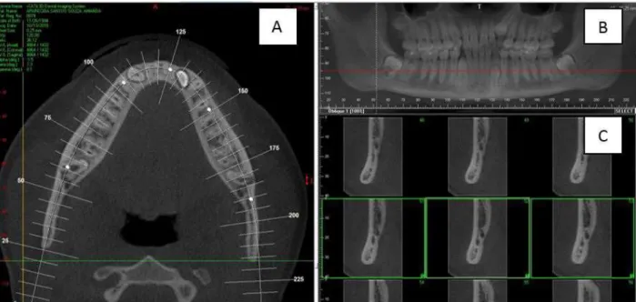
All images were evaluated by a single experienced observer, who was a specialist radiologist. he analysis was performed in a quiet environment with adequate lighting. he images were evaluated in three spatial planes (axial, sagittal and coronal) and in the transaxial or oblique sections of the mandible along the path of the mandibular canal, according to the Figure 1.
In order to optimize identiication of the mandibular canal, small modiications were made in the section plane, such as brightness, contrast and application of image ilters, since the path of the mandibular canal is not linear and should be individualized for each side of the patient. Cases in which the presence of RMC was veriied, oblique sections were also performed, in order to obtain images in the buccal-lingual direction.
satisfactory tomographic quality were included in the sample of patients of both genders who underwent concomitant computed tomography. Patients with a history of mandibular trauma, bone lesions in the lower arch, orthognathic or restorative surgery in the posterior region of the mandible were excluded from the sample. As images were derived from archived scans, the patients were not exposed to additional X-rays. For images with a positive identiication of changes in the mandibular canal, the patient was informed in the original radiographic report. For data collection, an Excel spreadsheet (Microsot, Seattle, USA) was developed to store data such as accession number, afected side, age and gender of the patient, Figure 2 shows a retromolar channel closely related to a horizontally impacted third molar.
Statistical Analysis
he sample was characterized in terms of gender and age in absolute (n) and relative (%) terms.
he indings relating to retromolar canals were described as absolute and relative frequencies, according to the gender of the participants and according to the location (right unilateral, let unilateral and bilateral). In addition, the associations between the mandibular biid canals and gender as well as location were investigated using Fisher’s exact test and chi-square tests, respectively.
Statistical calculations were performed on SPSS 20 (SPSS INC., Chicago, IL, USA) and BioEstat 5.0 (Mamirauá Foundation, Belém, PA, Brazil). he signiicance level was set at 5% (0.05).
RESULT
In this study, 300 concomitant CT scans belonging to the archive of the Department of Radiology of the São Leopoldo Mandic School, located in the city of Campinas - SP, Brazil were evaluated. Of the 300 images, 112 (37.3%) were from males, while 188 (62.7%) were from females.
he CT scans analyzed in this study belonged to individuals aged between 13 to 87 years, and the mean age of the sample was 48.4 years, with a standard deviation of 15.0 years. Among the males, whose ages ranged from 13 to 77 years, the mean age was 46.4 years, with a standard deviation of 16.1 years. Among women, age ranged from 14 to 87 years, with the mean being 49.5 years and a standard deviation of 14.2 years. Using the Student’s t-test for independent samples, no diference in age between male and female subjects was observed (p = 0.077).
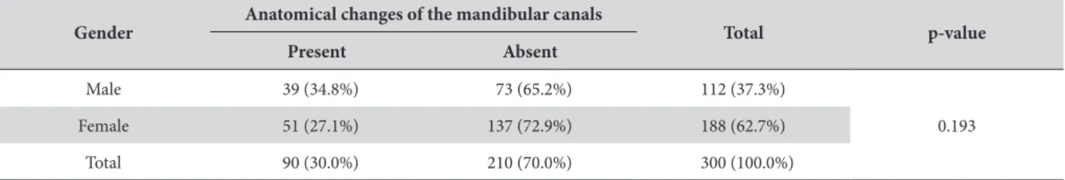
In the 300 CBCT scans analyzed, 210 (70.0%) showed a single canal, whereas the remaining 90 presented anatomical variations in the mandibular canal, indicating that the prevalence of this condition in this study sample was 30.0%, according to Table 1.
In the above-mentioned 90 cases, 39 men and 51 women, anatomical alterations in the mandibular canal were observed. Fiteen patients (5.0%) presented a posteriorly directed trajectory
Figure 1. Illustration of the methods evaluating the tomographic images; (A) Axial image with tracing of the mandibular contour to obtain cross-sectional slices; (B) Panoramic reconstruction; (C) Cross sections.
of the canal, formation 22 retromolar canals, because in 7 patients the retromolar canal occurred bilaterally, while 08 patients had it unilaterally, and an additional retromolar foramina was observed in 7.33% of the cases, as shown in Table 2.
In the 90 patients identiied, 39 males and 51 females, a total of 129 accessory canals were detected. Such canals took diferent directions ater leaving the mandibular canal, e.g. anterior or mesial, lingual, inferior or towards the base of the mandible. he occurrence of RMC was observed in 15 patients of the total sample (5.0%), 06 males and 09 females. In no case was the presence of anatomical alterations of the mandibular canals observed in a buccal, alveolar or upward direction.
DISCUSSION
Full understading of the anatomy of the mandibular canal and its variations, such as biid canals, foramina and RMC have great importance in the planning and execution of surgical and anesthetic procedures. Many anatomy textbooks, however, fail to describe the presence of the retromolar foramen, causing professionals to neglect or ignore its existence.
Chávez-Lomeli et al.15 reported that the mandibular canal
is formed from the fusion of three individual nerve branches at diferent stages of development. In addition to the fusion of the nerve branches, the formation of bone canals around such nerves may also occur. During prenatal growth, bone remodeling takes place via intramembranous ossiication that will give form the mandibular canal. his would explain the occurrence of biid mandibular canals and retromolar canals, in some patients, secondary to incomplete fusion of these three nerves16.
In dental practice, panoramic radiography is one of the most requested diagnostic tests, due to an overview of the components of the maxillary, mandibular, dental complex at relatively low cost. In the present study, the evaluations were made using CBCT and
the anatomical alterations of the mandibular canal were observed in 30% of the cases. Previous studies using panoramic radiographs have reported incidences barely reaching 1%16,17. CBCT-based
studies, however, have shown much higher values with prevalence rates ranging from 15.6% to 65%18-20. herefore, conventional
radiographs cannot be relied upon to detect anatomical variations of the mandibular canal.
Only biid canals with a diameter greater than 1 mm were included as anatomical alteration of the mandibular canal, aiming at standardization and clinical relevance of the results. In addition, entries such as “false mandibular or pseudo-biid canals”, as described by Kim et al.21, were carefully avoided. A similar image to a biid
canals can be produced by the impression of the mylohyoid nerve onto the inner surface of the mandible16. Such images may lead to
misdiagnoses, especially in panoramic reconstructions. herefore, it is important to combine diferent reconstruction approaches in the evaluation of the mandibular canal anatomy.
In the present study, a 5.0% prevalence of retromolar canals was detected. Several other studies have evaluated the presence of RMC, reporting a wide variation in prevalence depending on the method adopted by each study15-18. Regarding the additional
retromolar foramina, the present study found a prevalence of 7.33%, which is corroborated by several studies22-24. Bilecenoglu, Tuncer4
reported a prevalence of 25% for the histologically demonstrated retromolar foramen and that these canals had myelin nerve ibers, an artery and numerous venules, supplying part of the third molar, as well as the retromolar mucosa.
A higher prevalence of retromolar canals occurring bilaterally was observed in this study when compared to other reports in the literature, which have found a higher prevalence of right-sided retromolar canals11,19,22,24, though some authors have not observed
diferences between the right and let sides in dry mandibles2 or
in computed tomography images8. Notwithstanding that, other
studies have demonstrated RMCs more frequently on the let side
0.193
Female 51 (27.1%) 137 (72.9%) 188 (62.7%)
Total 90 (30.0%) 210 (70.0%) 300 (100.0%)
Fisher’s exact test.
Table 2. Absolute (n) and relative (%) RMC frequencies in CBCT, according to location
Gender Male Female No gender distinction
Location Present Absent p-value* Present Absent p-value* Present Absent p-value*
Unilateral R 02 (5.1%) 37 (94.9%) 1.0 01 (1.96%) 50 (98.04%) 0.242 04 (4.4%) 86 (95.6%) 0.529
Unilateral L 02 (5.1%) 37 (94.9%) 1.0 03 (5.8%) 48 (94.2%) 0.242 04 (4.4%) 86 (95.6%) 0.529
Bilateral 02 (5.1%) 37 (94.9%) 1.0 05 (9.8%) 46 (90.2%) 0.242 07 (7.7%) 83 (92.3%) 0.529
than on the right side5,25. In the present study, the presence of RMC
occurred more frequently in females, though the literature have not identiied and gender predilection21,22.
Regarding anesthetic procedures, the presence of alterations in the mandibular canal and RMCs may result in anesthetic failures, since such canals may exit through accessory foramens and contain a neurovascular bundle24. Failure to achieve adequate anesthesia may
therefore be observed when performing the inferior alveolar nerve block for procedures in the region of the last mandibular tooth or in the retromolar area. Also, the solution to this problem would be to select an alternative approach such as the Gow-Gates technique. Another aspect of fundamental importance regarding the occurrence of retromolar canals regards surgical procedures involving the posterior region of the mandible, such as extraction of unerupted/impacted teeth, installation of dental implants, osteotomy for autologous bone grating from the ramus of the mandible or for orthognathic purposes8.
Studies on the incidence of RMC are important to prevent failures in regional anesthesia of the inferior alveolar nerve and buccal nerve ibers4 as well as to minimize the occurrence of accidents
and complications in the posterior region of the mandible. During osteotomies in the posterior mandible for exodontia, paraesthesia
of the mucosa of the retromolar region and of the buccal mucosa on the operated side may occur, secondary to trauma to the nerve that emerges through the retromolar foramen, which is a branch of the nerve inferior alveolar nerve9.
Some authors have identiied that components of the RMC are nerves that provide innervation to the pulp of the third molar, retromolar region and ibers of the temporal and buccinator muscles10,11.
he neurovascular bundle can be injured and cause excessive bleeding during third molar extraction or sagittal osteotomies of the mandible, as well as during procedures involving dissection of tissues, mucoperiosteal detachment and osteotomies in general22.
CONCLUSION
According to the results obtained in this study, a prevalence of 5.0% of retromolar canals was found, occurring more in women and bilaterally. he importance of a thorough knowledge of the retromolar region is herein reiterated based on the high prevalence of surgical procedures performed in the posterior region of the mandible. his in turn would beneit predictability in treatment planning and consequently optimize both anesthetic and surgical procedures, thus minimizing failures and accidents.
REFERENCES
1. Han SS, Hwang YS. Cone beam CT findings of retromolar canals in a korean population. Surg Radiol Anat. 2014 Nov;36(9):871-6. PMid:24504621. http://dx.doi.org/10.1007/s00276-014-1262-1.
2. Muinelo-Lorenzo J, Suárez-Quintanilla JA, Fernández-Alonso A, Marsillas-Rascado S, Suárez-Cunqueiro MM. Descriptive study of the bifid mandibular canals and retromolar foramina: cone beam CT vs panoramic radiography. Dentomaxillofac Radiol. 2014;43(5):20140090. PMid:24785820. http://dx.doi.org/10.1259/dmfr.20140090.
3. Claeys V, Wackens G. Bifid mandibular canal: literature review and case report. Dentomaxillofac Radiol. 2005 Jan;34(1):55-8. PMid:15709108. http://dx.doi.org/10.1259/dmfr/23146121.
4. Bilecenoglu B, Tuncer N. Clinical and anatomical study of retromolar foramen and canal. J Oral Maxillofac Surg. 2006 Oct;64(10):1493-7. PMid:16982307. http://dx.doi.org/10.1016/j.joms.2006.05.043.
5. Rossi AC, Freire AR, Prado GB, Prado FB, Botacin PR, Caria PHF. Incidence of retromolar foramen in human mandibles: ethnic and clinical aspects. Int J Morphol. 2012 Sep;30(3):1074-8. http://dx.doi.org/10.4067/S0717-95022012000300051.
6. Kawai T, Asaumi R, Sato I, Kumazawa Y, Yosue T. Observation of the retromolar foramen and canal of the mandible: a CBCT and macroscopic study. Oral Radiol. 2012 Mar;28(1):10-4. http://dx.doi.org/10.1007/s11282-011-0074-9.
7. Langlais RP, Broadus R, Glass BJ. Bifid mandibular canals in panoramic radiographs. J Am Dent Assoc. 1985 Jun;110(6):923-6. PMid:3860553. http://dx.doi.org/10.14219/jada.archive.1985.0033.
8. Patil S, Matsuda Y, Nakajima K, Araki K, Okano T. Retromolar canals as observed on cone-beam computed tomography: their incidence, course, and characteristics. Oral Surg Oral Med Oral Pathol Oral Radiol. 2013 May;115(5):692-9. PMid:23601225. http://dx.doi.org/10.1016/j. oooo.2013.02.012.
9. Rodella LF, Buffoli B, Labanca M, Rezzani R. A review of the mandibular and maxillary nerve supplies and their clinical relevance. Arch Oral Biol. 2012 Apr;57(4):323-34. PMid:21996489. http://dx.doi.org/10.1016/j.archoralbio.2011.09.007.
10. Sawyer DR, Kiely ML. Retromolar foramen: a mandibular variant important to dentistry. Ann Dent. 1991;50(1):16-8. PMid:1872586. 11. Kodera H, Hashimoto I. A case of mandibular retromolar canal: elements of nerves and arteries in this canal. Kaibogaku Zasshi. 1995
Feb;70(1):23-30. PMid:7785408.
12. Sonick M, Abrahams J, Faiella RA. A comparison of the accuracy of periapical, panoramic, and computerized tomographic radiographs in locating the mandibular canal. Int J Oral Maxillofac Implants [Internet]. 1994;9(4):455-60 [cited 2010 July 6]. Available from: http://www. sonickdmd.com/wp-content/uploads/2012/05/JOMI-1994-A-Comparison-of-the-Accuracy-of-Periapica.pdf
13. Cavalcanti MGP. Tomografia computadorizada por feixe cônico. 2nd ed. São Paulo: Livraria Editora Santos; 2014.
18. Santos O Jr, Pinheiro LR, Umetsubo OS, Sales MA, Cavalcanti MG. Assessment of open source software for CBCT in detecting additional mental foramina. Braz Oral Res. 2013 Apr;27(2):128-35. PMid:23459775. http://dx.doi.org/10.1590/S1806-83242013005000003.
19. Orhan K, Aksoy S, Bilecenoglu B, Sakul BU, Paksoy CS. Evaluation of bifid mandibular canals with cone beam computed tomography in a Turkish adult population: a retrospective study. Surg Radiol Anat. 2011 Aug;33(6):501-7. PMid:21161224. http://dx.doi.org/10.1007/s00276-010-0761-y.
20. Oliveira-Santos C, Souza PH, Azambuja Berti-Couto S, Stinkens L, Moyaert K, Rubira-Bullen IRF, et al. Assessment of variations of the mandibular canal through cone beam computed tomography. Clin Oral Investig. 2012 Apr;16(2):387-93. PMid:21448636. http://dx.doi. org/10.1007/s00784-011-0544-9.
21. Kim MS, Yoon SJ, Park HW, Kang JH, Yang SY, Moon YH, et al. A false presence of bifid mandibular canals in panoramic radiographs. Dentomaxillofac Radiol. 2011 Oct;40(7):434-8. PMid:21960401. http://dx.doi.org/10.1259/dmfr/87414410.
22. Schejtman R, Devoto FC, Arias NH. The origin and distribution of the elements of the human mandibular retromolar canal. Arch Oral Biol. 1967;12(11):1261-8. PMid:5234232. http://dx.doi.org/10.1016/0003-9969(67)90127-6.
23. Lizio G, Pelliccioni GA, Ghigi G, Fanelli A, Marchetti C. Radiographic assessment of the mandibular retromolar canal using cone-beam computed tomography. Acta Odontol Scand. 2013 May-Jul;71(3-4):650-5. PMid:22809124. http://dx.doi.org/10.3109/00016357.2012.7043 93.
24. Orhan AI, Orhan K, Aksoy S, Ozgül O, Horasan S, Arslan A, et al. Evaluation of perimandibular neurovascularization with accessory mental foramina using cone-beam computed tomography in children. J Craniofac Surg. 2013 Jul;24(4):e365-9. PMid:23851871. http://dx.doi. org/10.1097/SCS.0b013e3182902f49.
25. Khan MA, Agarwal S, Mandloi RS. Prevalence of retromolar foramen in dried mandible along with morphometric and analytical study in North India. Natl J Med Dental Res. 2013;2:11-4.
CONFLICTS OF INTERESTS
he authors declare no conlicts of interest.
*CORRESPONDING AUTHOR
George Borja de Freitas, Faculdade de Odontologia, Centro de Pesquisas Odontológicas São Leopoldo Mandic, Rua Dr. José Rocha Junqueira, 13, Ponte Preta, 13045-755 Campinas - SP, Brasil, e-mail: [email protected]