Analysis of Bone Damage during Drilling
Processes
Maria Goreti Antunes Fernandes
Programa Doutoral em Engenharia Mecânica
Processes
by
Maria Goreti Antunes Fernandes
A thesis submitted in conformity with the requirements for the
Doctoral Degree in Mechanical Engineering by the Faculty of
Engineering of the University of Porto
Supervisor:
Renato Manuel Natal Jorge
Associate Professor, Department of Mechanical Engineering, Faculty of Engineering, University of Porto, Porto, Portugal
Co-Supervisor:
Elza Maria Morais Fonseca
Assistant Professor, Department of Applied Mechanics, Polytechnic Institute of Bragança, Bragança, Portugal
“Aprender é a única coisa de que a mente nunca se cansa, nunca tem medo e nunca se arrepende.”
É revigorante ver o resultado final daquilo que foi uma longa caminhada de conhecimentos, desafios, incertezas medos mas sobretudo conquistas. Símbolo daquilo que reúne não só toda uma dedicação e gosto pela investigação mas sobretudo a partilha da sabedoria e o compilar de vários contributos, sem os quais este trabalho simplesmente não existia.
Em primeiro lugar desejo expressar os meus agradecimentos sinceros ao meu orientador Professor Renato Manuel Natal Jorge. Obrigada pela oportunidade, confiança depositada em mim e sobretudo por toda a sua dedicação e esforço em proporcionar as melhores soluções e condições de trabalho essenciais para a realização e conclusão desta tese. De seguida, um reconhecimento especial à minha coorientadora Professora Elza Maria Morais Fonseca. Os meus mais genuínos agradecimentos pela sua constante orientação, total disponibilidade e sobretudo pela motivação e amizade que sempre manteve presente, não só durante este percurso mas também durante grande parte da minha vida académica. Sem dúvida uma referência e símbolo de inspiração para mim.
A eles, orientador e coorientadora, quero exprimir que foi e será sempre uma honra poder trabalhar e sobretudo aprender junto daqueles que considero serem exemplo de dedicação e sucesso.
Referir também o importante apoio de todas as instituições envolvidas, sobretudo o INEGI (Instituto de Engenharia Mecânica e Gestão Industrial), bem como a FEUP (Faculdade de Engenharia da Universidade do Porto) e o IPB (Instituto Politécnico de Bragança). Agradeço as excelentes condições de trabalho e todas as ferramentas fornecidas para o desenvolvimento desta tese de doutoramento.
Tecnologias Agroambientais e Biológicas da Universidade de Trás-os-Montes e Alto Douro) e Maria Cristina Manzanares Céspedes (Unidade de Anatomia e Embriologia Humana da Faculdade de Medicina da Universidade de Barcelona) por proporcionarem todas as condições e ferramentas necessárias à realização dos ensaios experimentais em tecido ósseo de bovino e osso humano cadavérico, respetivamente.
Aos técnicos de laboratório, Jorge Meireles e Luísa Barreira do Instituto Politécnico de Bragança pela colaboração e disponibilidade prestada durante os ensaios experimentais. Para terminar, obrigada a todos os meus amigos, especialmente aqueles que estiveram mais presentes durante esta minha etapa: João Pires, Joana Calejo, Daniela Correia, Ana Lousada e Ana Catarina Lopes. Obrigada pela amizade, carinho, boa disposição e conversas partilhadas.
Aos meus pais, irmãos e David um agradecimento especial pelo apoio, alegria, motivação constante e sobretudo por sempre estimularem a minha evolução profissional e pessoal. São eles que, incondicionalmente, me apoiam e apoiaram e sem eles certamente não seria a pessoa que sou hoje. Serei eternamente grata.
Bone drilling is an essential step in a broad range of clinical interventions. The success of these clinical operations depends of the drilling procedure quality in view to minimize associated injuries to the bone and surrounding tissue. Studies have shown that the bone damage is directly related with the drilling parameters, particularly, drill speed, feed-rate, applied force, drilling depth, drill bit geometry, the use or not a cooling system and also the bone type. Therefore, a thorough understanding of different materials and cutting parameters is essential to reduce the bone damage and help the health professionals in the success of these drilling surgeries.
In the present thesis, a comprehensive research was performed including numerical analysis and experimental tests of the thermal and mechanical aspects of bone tissue, with applications to the drilling situations. The effects of different drilling parameters on the temperature field and generated stresses were investigated taking into account the engineering and biological principles.
A work programme of experimental tests was conducted, using synthetic bone (polyurethane foam materials with different densities) and biological materials (bovine femurs and human cadaveric tibiae). Thermocouples and infrared thermography were used to measure the temperature during the experimental tests, and also linear strain gauges to measure the deformations during the drilling process.
Computational modelling of bone drilling included thermal and mechanical models to analyse the thermomechanical behaviour of bone tissue. The numerical models are able to predict the stresses and temperature distribution, during the perforation for a given material similar to bone characteristics, with a typical drill bit geometry and cutting conditions such as drill speed, feed-rate, drilling depth and use of cooling system. Additionally, novel dynamic models based on real geometries of human bone tibia were
the results validated through the experimental tests. The magnitude and variation tendency of drilling temperatures and stresses obtained by both methodologies are in agreement.
The main outcome of this thesis is a detailed understanding of the bone drilling processes, contributing for the application of optimum drilling parameters that will minimise the bone damage due the invasive procedure. The results contribute to a comprehensive understanding of thermomechanical aspects of bone drilling process and associated predictive models. The developed models can be used to simulate, in a realistic manner, the drilling process under different aspects and to provide an important tool that will enable for the health professionals to plan an accurate and safe surgery with better post-surgery patient recovery.
A furação do tecido ósseo é um procedimento cirúrgico essencial em diversas intervenções clínicas. O sucesso destas cirurgias depende da qualidade do processo de furação, sendo objetivo principal a minimização das lesões associadas ao tecido ósseo e tecidos circundantes. Estudos efetuados têm demonstrado que a lesão óssea está diretamente relacionado com os parâmetros de furação, mais especificamente a velocidade de rotação da broca, velocidade de avanço, força aplicada, profundidade do furo, geometria da broca, o uso ou não de um sistema de arrefecimento e também o tipo de osso. Por conseguinte, a compreensão aprofundada dos diferentes materiais e dos parâmetros de corte envolvidos nestes processos é essencial na redução da lesão óssea e no auxílio aos profissionais de saúde para o sucesso nessas cirurgias.
Na presente tese foi realizada uma pesquisa abrangente que inclui a análise numérica e ensaios experimentais sobre os aspetos térmicos e mecânicos do tecido ósseo, com aplicações nos processos de furação. Os efeitos dos diferentes parâmetros de furação sobre o aumento da temperatura e tensões geradas foram investigados sob o ponto de vista da engenharia e da biologia.
Realizaram-se um conjunto de ensaios experimentais com recurso a osso sintético (materiais em espuma de poliuretano com diferentes massas volúmicas) e materiais biológicos (fémures de bovino e tíbias humanas cadavéricas). Foram utilizados termopares e a termografia para a medição da temperatura durante os ensaios experimentais, e ainda utilizada a extensometria para o registo das deformações durante o processo de furação.
A modelação computacional da furação óssea inclui modelos de materiais térmicos e mecânicos para analisar o comportamento termomecânico do tecido ósseo. Os modelos numéricos são capazes de prever as tensões e a distribuição da temperatura durante a
corte, tais como velocidade de rotação, velocidade de avanço, profundidade do furo e a utilização ou não de um sistema de arrefecimento. Além disso, foram desenvolvidos novos modelos dinâmicos baseados em geometrias obtidas de tíbias humanas. Foram efetuados estudos comparativos entre os modelos numéricos desenvolvidos e os resultados experimentais. A tendência obtida e verificada no campo das temperaturas e das tensões, ao longo da furação, através das diferentes metodologias, estão em concordância.
O principal resultado desta tese é obter uma análise detalhada dos processos de furação óssea, contribuindo para a implementação de parâmetros de furação ideais que minimizem a lesão no tecido ósseo e a agressividade no procedimento. Os resultados contribuem para uma compreensão abrangente dos aspetos termomecânicos do processo de furação e dos modelos preditivos associados. Os modelos desenvolvidos podem ser utilizados para simular, de forma realista, a furação sob diferentes aspetos e fornecer assim, uma ferramenta importante que permitirá aos profissionais de saúde planear uma cirurgia mais precisa e segura, e que possibilite uma melhor recuperação pós-cirúrgica ao paciente.
C
HAPTERI ... 1
General Introduction and Motivations ... 1
1.1 Background and Motivation ... 3
1.2 Thesis Objectives ... 6 1.3 Thesis Structure ... 7 References ... 9
C
HAPTERII ... 13
Literature Overview... 13 2.1 Bone Tissue ... 152.1.1 Mechanical properties of cortical bone ... 17
2.1.2 Mechanical properties of trabecular bone ... 21
2.1.3 Thermal properties of bone tissue ... 22
Specific heat of bone tissue ... 23
Thermal conductivity of bone tissue ... 23
2.2 Bone Drilling in Medicine ... 26
2.2.1 Bone damage induced by drilling ... 26
Mechanical damage in bone drilling ... 27
Thermal osteonecrosis in bone drilling ... 29
2.2.2 Effects of drilling parameters on bone ... 31
Drill bit specifications ... 32
Drill parameters ... 39
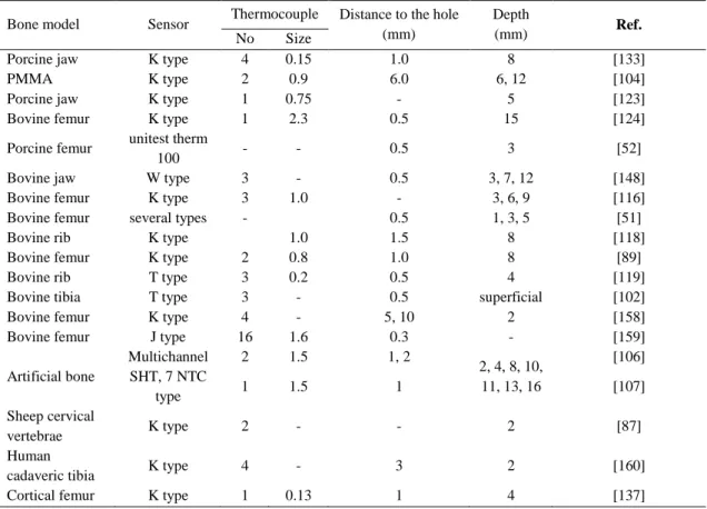
2.2.3 Temperature measurement in bone drilling ... 48
Thermocouples ... 49
Infrared thermography ... 51
Mechanical models ... 54
References ... 56
C
HAPTERIII... 71
Thermal analysis during bone drilling using rigid polyurethane foams: numerical and experimental methodologies* ... 71
Abstract ... 73
1 Introduction ... 75
2 Experimental Methodology ... 76
3 Numerical Analysis ... 80
4 Results and Discussion ... 83
5 Conclusions ... 88
Acknowledgments ... 88
References ... 88
C
HAPTERIV ... 91
Three-dimensional dynamic finite element and experimental models for drilling processes* ... 91
Abstract ... 93
1 Introduction ... 95
2 3D Dynamic FE Model ... 97
2.1 Material properties ... 98
2.2 Element removal and contact interactions ... 99
3 Experimental Setup ... 100
4 Results and Discussion ... 102
5 Conclusions ... 106
Declaration of conflicting interests ... 106
Funding ... 106
References ... 107
C
HAPTERV ... 111
Thermo-mechanical stresses distribution on bone drilling: Numerical and experimental procedures* ... 111
Abstract ... 113
1 Introduction ... 115
2.2 Contact interaction and failure criteria ... 121
3 Experimental Validation ... 121
3.1 Strain measurement ... 122
3.2 Temperature control ... 122
4 Results and Discussion ... 125
4.1 FE results... 125
4.2 Experimental results and FE model validation ... 126
5 Conclusions ... 127
Declaration of conflicting interests ... 128
Funding ... 128
References ... 128
C
HAPTERVI ... 133
Thermal analysis in drilling of ex vivo bovine bones* ... 133
Abstract ... 135
1 Introduction ... 137
2 Temperature Evolution in the Drilling of ex vivo Bovine Bones and Foam Blocks ... 139
2.1 Bovine bones samples preparation ... 139
2.2 Experimental setup in bovine bones samples ... 141
2.3 Experimental setup in polyurethane foam blocks ... 142
2.4 Analysis of the experimental results: bovine bones and foam blocks ... 143
3 Temperature Evolution Inside of the Bone ... 145
3.1 Temperature measurement system ... 145
3.2 Temperature evolution inside of the polyurethane foam blocks ... 146
4 Conclusions ... 149
Acknowledgments ... 150
Conflict of interest statement ... 150
References ... 150
C
HAPTERVII ... 155
Ex vivo experimental and numerical study of stresses distribution in human cadaveric tibiae* ... 155
Abstract ... 157
2.1 Numerical model of human tibia ... 160
2.1.1 Material and constitutive models ... 162
2.1.2 Contact and material failure criteria ... 164
2.2 Experimental validation ... 164
3 Results and Discussion ... 166
3.1 Comparison of the numerical and experimental results ... 167
4 Conclusions ... 168
Acknowledgments ... 169
Conflict of interest statement ... 169
References ... 169
C
HAPTERVIII ... 173
Concluding Remarks and Future Directions ... 173
8.1 Introduction ... 175
8.2 Conclusions and remarks ... 175
8.3 Future directions ... 177
C
HAPTERII:
Figure 1. (a) Classification of bones according to their shape and (b) schematic image of
a long bone with its basics components. (Adapted from [6, 9]) ... 16
Figure 2. Hierarchical structural distribution of bone tissue. (Adapted from [10])... 17
Figure 3. Illustration of load-displacement curve for a bone sample. (Adapted from [11]) ... 18
Figure 4. Illustration of tensile stress-strain curve. (Adapted from [12]) ... 19
Figure 5. The anisotropic behaviour of cortical bone tissue. (Adapted from [13]) ... 19
Figure 6. Comparison of compressive stress-strain curve high and low bone density. (Adapted from [23])... 21
Figure 7. Correlation between density and elastic modulus in tensile (filled circles) and compression (empty circles) of trabecular bone tissue. (Adapted from [32]) ... 22
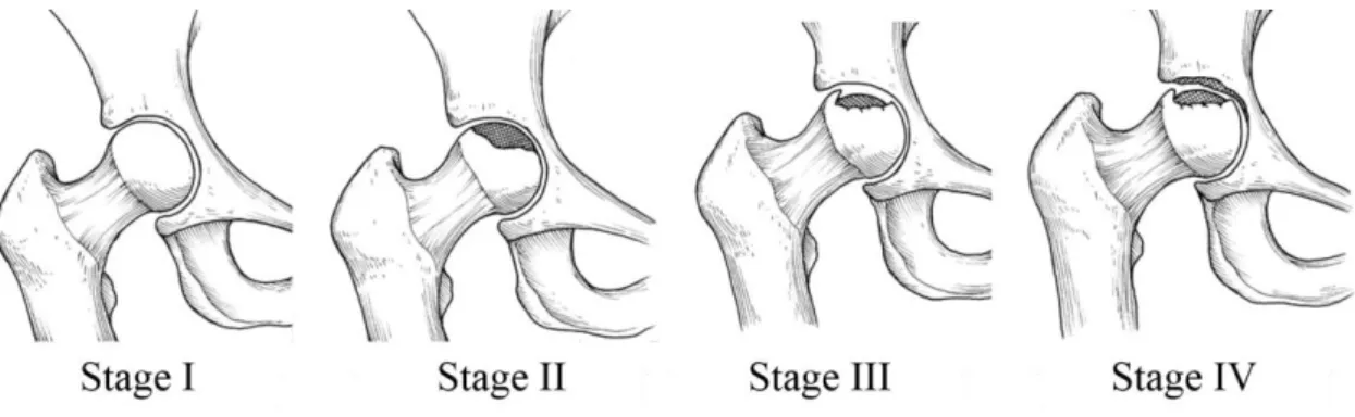
Figure 8. Stages of Osteonecrosis. (Adapted from [93]) ... 30
Figure 9. Geometry of a twist drill bit and its drill bit tip. (Adapted from [98]) ... 32
Figure 10. Cooling systems: (a) external and (b) internal cooling. (Adapted from [144]) ... 44
C
HAPTERIII:
Figure 1. Test Blocks from Sawbones: (a) C+D, (b) C-D, (c) T+D, (d) T-D. ... 77Figure 2. Schematic illustration of the thermocouples positions... 77
Figure 3. Geometrical model of block from Sawbones. ... 79
Figure 4. Drilling process of biomechanical blocks. ... 79
process. ... 83 Figure 7. Time-temperature history in the block C+D: (a) side A, without external cooling, (b) side A, with external cooling, (c) side B, without external cooling, (d) side B, with external cooling. ... 84 Figure 8. Temperature during drilling at 440 s: (a) C+D, (b) C-D, (c) T+D, (d) T-D. .. 85 Figure 9. Thermal images of the drill bit, C+D: (a) before and (b) after drilling; C-D: (c) before and (d) after drilling. ... 87 Figure 10. Thermal images of the drill bit, T+D: (a) before and (b) after drilling; T-D: (c) before and (d) after drilling. ... 87
C
HAPTERIV:
Figure 1. Geometric representation of the drill bit and the biomechanical block. ... 97 Figure 2. FE model of the drill bit, biomechanical block and applied boundary conditions. ... 98 Figure 3. (a) Biomechanical test block from Sawbones and (b) instrumented block. .. 100 Figure 4. Geometrical model of the block and identification of the holes. ... 101 Figure 5. (a) Drilling process and (b) distance between the edge of the drilled hole and the stain gauge. ... 102 Figure 6. Temperature distribution on the block top surface. ... 102 Figure 7. Distribution of von Mises stresses (MPa) at different point-in-times (Vf = 25
mm/min). ... 103 Figure 8. Distribution of von Mises stresses (MPa) at different point-in-times (Vf = 50
mm/min). ... 103 Figure 9. Distribution of von Mises stresses (MPa) at different point-in-times (Vf = 75
mm/min). ... 104 Figure 10. Comparison of normal stress (MPa) in numerical and experimental tests at different times of drilling. ... 104 Figure 11. Variation of normal stresses for different feed-rates at depth = 30 mm. .... 105
C
HAPTERV:
Figure 1. (a) FE model of drilling and (b) boundary conditions. ... 118 Figure 2. Flow stress for the Cowper-Symonds model. ... 120
... 122
Figure 4. (a) Geometrical model and (b) thermocouples position. ... 123
Figure 5. Temperature data applied in the Fe model. ... 124
Figure 6. Experimental drilling process. ... 124
Figure 7. Distribution of von Mises stress (MPa) at different feed-rates and drill speeds (3 mm of depth). ... 125
Figure 8. Normal stresses σzz (MPa) in experimental and FE models with (a) feed-rate and (b) drill speed. ... 126
Figure 9. Maximum normal stresses σzz (MPa) with (a) feed-rate and (b) drill speed. 127
C
HAPTERVI:
Figure 1. Samples preparation for the experimental tests in ex vivo bovine bones. .... 140Figure 2. Fresh bovine femur (a), sample cut from mid-diaphysis (b) and wrapped with gauze swabs in physiological saline solution (c). ... 141
Figure 3. Drilling operations of bovine bones. ... 142
Figure 4. Biomechanical foam blocks: Bl1 and Bl2. ... 142
Figure 5. Drilling process of biomechanical blocks. ... 143
Figure 6. Polyurethane foam block and illustration of the thermocouple positions. .... 145
Figure 7. Temperature evolution at different feed rates and positions of thermocouples. ... 147
Figure 8.Temperature evolution at different drill speeds. ... 148
C
HAPTERVII:
Figure 1. Human cadaveric tibiae and sample dimensions. ... 161Figure 2. (a) Human cadaveric tibia, (b) 3D CAD model and (c) FE model. ... 161
Figure 3. (a) FE model of bone drilling and (b) mesh and boundary conditions. ... 162
Figure 4. Instrumentation of human bone tibiae... 165
Figure 5. Experimental setup on human cadaveric tibiae. ... 166
Figure 6. Comparison of the von Mises stresses (MPa) obtained for drill speeds of 900 and 1370 rpm and 2.5 mm drilling depth. ... 166
Figure 7. Comparisons of normal stress (MPa) from FE model and experiments at different drilling steps (a) and at maximum drilling depth of 3 mm (b). ... 168
C
HAPTERII:
Table 1. Mechanical properties of cortical bone tissue by different tests. (Based from [13])
... 20
Table 2. Mechanical properties (MPa) of trabecular bone from compression tests. (Based from [13]) ... 21
Table 3. Specific heat of bone tissue reported in the literature. ... 23
Table 4. Thermal conductivity of cortical bone reported in literature. ... 24
Table 5. Parameters involved in bone drilling. ... 32
Table 6. Some experimental studies of heat measured using thermocouples. ... 50
Table 7. Some experimental studies of heat measured using infrared thermography. ... 51
C
HAPTERIII:
Table 1. Tagging of thermocouples. ... 78Table 2. Drilling processing parameters. ... 78
Table 3. Parameters used for thermal images acquisition. ... 79
Table 4. Thermal properties of the drill bit and bone. ... 80
C
HAPTERIV:
Table 1. Drilling parameters used in FE simulation. ... 98Table 2. Isotropic material properties and Cowper-Symonds parameters used in drilling simulations. ... 100
C
HAPTERV:
Table 1. Material properties and Cowper-Symonds parameters used in numerical simulations. ... 120 Table 2. Parameters used for thermal images acquisition. ... 123
C
HAPTERVI:
Table 1. Parameters used for the thermal image acquisition. ... 141 Table 2. Parameters used in drilling tests. ... 143 Table 3. Variation of temperature from drill bit, before and after drilling. ... 144 Table 4. Thermocouple labelling. ... 146 Table 5. Parameters used in drilling experiments... 146
C
HAPTERVII:
C
HAPTER
I
1.1 Background and Motivation
Bone is a highly dynamic tissue and the main constituent of the human skeletal system. Its structure differs from the connective tissues in rigidity and hardness [1]. Although the bone is a self-repairing structural material, bone loss or bone fracture continues to be one of the most common public health concerns due to the automobile and motorcycle accidents, sports injuries, falls from a height, ageing population and others diseases. Hard tissue cutting is a common practice in medical and dental fields (e.g. neurosurgery, orthopaedic, trauma and maxillofacial surgeries) to bone repair and fracture reconstruction. The main bone cutting operations are drilling [2], sawing [3] and grinding [4]. Although all of these procedures have an important role in clinical practice, bone drilling is one of the most important surgical operation in the medicine history [5]. Holes perforation into the bone have been used in our societies from the earliest times and they are considered the oldest surgical procedure. The first operation that involved bone drilling was called cranial trepanation, which literally means drilling through the skull. This prehistoric procedure was first identified in 1865 by E. George Squier and became a common and crucial surgery technique throughout many civilizations [6, 7]. Until recently, bone drilling was not considered from technical point of view. The focus was merely to perform the surgical operation by intuitively controlling the parameters for minimal or no damage to the bone tissue without any understanding of the machining process fundamentals [8]. However, the progress of medicine in conjunction with the technology require the development and improvement of the drilling procedures with minimally invasive techniques for the patients. The importance of this subject has motivated the research on bone drilling and the development of recent reviews [9, 10]. Many investigations have been carried out to study the tool materials, mechanical aspects of drilling and their biological and cellular impact on the bone tissue. Dahotre and Joshi [8] indicated an increased from 100 to 10000 published papers over the years of 1940 to 2016 in the field of conventional and laser based bone machining (including drilling, sawing, grinding and milling).
Despite the efforts and the progress, bone drilling is still largely conducted by the surgeon using hand drills, which means a blind operation with unknown hole depth and a feed-rate manually controlled by the surgeon. Such mostly conventional way of surgery is associated with human and tool attributes and hence leaves tremendous room for further
development and improvement of operating tools and techniques. In addition, the reported cases of implant failures and post-operative problems associated to the drilling process continue to be a significant public health problem in the health care system and show the need for improvements in this field [11, 12]. In the year of 1998, Piattelli et al. [13] reported eight cases of failed implants due to suspected thermally induced bone necrosis. Later in 2008, Augustin et al. [14] indicated that the implant failure rate for leg osteosynthesis ranges between 2.1% and 7.1%. They noted that one of the causes is the increase in bone resorption around the implanted screws from thermal osteonecrosis caused by preparative drilling. Abayazid et al. [15] reported that the failure rate of dental implant in the lower jaw is 7.65% in 10 years period and three times higher in the maxilla. More recently, Elangovan and Allareddy [11] have provided detailed information on the nationally representative estimates of hospital based emergency department visits attributed to dental implant failures in the United States. During the period of their studies (years 2008–2010), a total of 1200 emergency department visits were due to dental implant failures. Its conclusions indicate that 31.7% of the visits were related to osseointegration failures and 30.4% were due to post-osseointegration mechanical failures. According to the authors, two etiological factors that can lead to the implant failures are tissue trauma (possibly from overheating the bone during placement) and mechanical overloading.
The clinical problems coupled with the importance to reduce the bone damage risk during drilling procedures were the main motivations for the realization of this thesis. After the research subject has been identified, it was necessary to determine the main research lines and the main challenges and contributions on this domain. For this purpose, a bibliographic review of all relevant studies on bone drilling was conducted, including the different methods used, results and conclusions made by the various researches. Literature review reveals that the main problems associated to the bone damage are the cut effort and the heat generation during drilling. High loads with high speed velocity and tool vibrations may result in drill overrun, damages to the bone microstructure, which can lead to the crack formations and even bone fracture [16]. The heat causes bone cell damage and can result in thermal necrosis [17]. Problems like clogging or built up edge are also associated to bone drilling. These problems are all linked with the drilling parameters [18, 19]. If parameters such as drill speed and feed-rate are not selected properly, the drilling load induces bone damage, which lead to surgical complications.
Therefore, determination of optimal parameters is crucial to the success of this surgeries and further evolution of the patients. In fact, there are a significant number of studies dedicated only to this issue. However, most works available in the literature provide an experimental approach with special attention to thermal effects. The studies on strain and stress distribution during drilling have not been reported so far. An investigation of this effects can contribute significantly for minimizing bone tissue injury during drilling. Even about the experimental works on thermal damage, there still remains a lack of consensus in the literature regarding to critical temperature values and their time durations. The extensive variables number involved and the large discrepancies in the obtained results from different methods complicate the concluding remarks statement. Furthermore, the literature available so far has been concentrating on artificial or animal bones, and relatively few research has been completed on the human bone. Indeed, some animals can resemble the human properties but none of them is equal to the human bone. This inherent disparity in mechanical and thermal properties of specimens taken from different bones results in differences in the results subject to almost identical drilling conditions in repeated experiments [20].
In 2013, Pandey and Panda [10] conducted a literature review of all relevant investigations carried on bone drilling and concluded that there are several significant factors influencing the process on which no general agreement is yet been made, or are not investigated and need an evaluation in the future. They suggested that the further improvement in bone drilling is possible, including the drilling parameters optimization. The advances of technology and computational models in recent decades have contributed to the study and improvement of new drilling techniques. The modelling of drilling process using finite element analysis (FEA) may be considered as a promising and reliable technique for understanding of bone drilling, as well as, in the study and definition of the best drilling conditions. There are studies on literature that describe finite element (FE) models to study the behaviour of the bone under several drilling conditions [21-25]. However, it is complicated to reach a numerical model to simulate the real drilled bone due to the complexity of the material properties inherent to the cutting material separation. Marco et al. [26] performed a review of the main contributions in modelling of bone cutting with special attention to the bone mechanical behavior. They concluded that is difficult to find complete models of bone drilling including chip removal simulation and capable to predict the thermomechanical damage. In the author’s opinion, the available models are still far from clinical application and a strong effort is still needed
in this field. Moreover, the high computational cost of the complete three-dimensional (3D) approach is the major drawback.
Based on the literature review, it has become clear that the accuracy of the developed models to predict the bone damage requires a combination of experimental and numerical methods. It is important to compile different methodologies to achieve real results and extrapolate these results to clinical situations. Developing accurate FE models of bone drilling could be a potential tool of significant improvement of the process and possible substitute for experimental work as it eliminates high costs of the equipment as well as potential health risks associated with biological materials.
In general, the main challenges in this field are the numerical modelling of the bone drilling, that includes the dynamic characteristics of this processes and chip removal; the lack of experimental data on the strain and stress distribution during drilling and the lack of consensus among the available published works; and, lastly, the lack of experimental validation for the developed numerical models. The described challenges are the main motivation for the present research, which provides a comprehensive experimental and numerical study of bone drilling taking into account the engineering and biological principles.
1.2 Thesis Objectives
The main objective of this thesis is to analyse the conventional drilling process with the purpose to reduce the bone damage risk and to contribute for a better knowledge of the involved parameters in the drilling process and their effects. A comprehensive research including experimental tests and numerical modelling for thermal and mechanical analysis of bone drilling is carried out, in order to predict the strain, the stress and temperature distribution for a given drilling conditions, drill bit geometry and material properties of the bone tissue. It is expected that the final findings of this work will be a useful tool on the selection of drilling parameters and an additional support for the health professionals in the decision-making process before drilling.
To achieve the described purpose, the following specific aims were defined in this thesis: 1. Development of experimental methodologies using engineering materials
(synthetic bone with properties similar to the human bone) and biological materials (bovine bones and human cadaveric bones) to study the inherent
variation in mechanical and thermal properties of bones taken from different species;
2. Measurement of temperature distribution in the cutting tool and within the bone tissue during drilling through the use of a thermographic camera and thermocouples;
3. Development of an experimental model to measure the strain and calculate the respective stress distribution during bone drilling through the use of strain gauges; 4. Study the influence of different drilling parameters (feed-rate, drill speed, hole depth, irrigation system and bone density) on bone temperature and stress distribution during bone drilling;
5. Development of a 3D FE model to simulate the thermal behaviour of the bone tissue and to predict the temperature distribution during drilling. The effect of different parameters is analysed through a thermal and transient analysis;
6. Development of a 3D dynamic elasto-plastic FE model with the element removal scheme to simulate the mechanical behaviour of the bone and to predict the strain and the stress distribution during drilling. The FE model aims to incorporate the geometric and dynamic characteristics involved in the drilling processes;
7. Development of a 3D dynamic elasto-plastic FE model with the element removal scheme to simulate the thermo-mechanical behaviour of the bone and to predict the respectively stress distribution during drilling. The FE model aims to incorporate the geometric and dynamic characteristics involved in the drilling processes and the developed temperature inside of material;
8. Development of a new 3D dynamic elasto-plastic FE model, using a real geometry of human cadaveric tibia, captured by 3D handheld scanner. The FE model was optimised to best represent the human bone response of drilling procedures. 9. Validation of the FE models based on a comparison with the experimental results.
1.3 Thesis Structure
The present thesis has been organized into eight chapters and two main parts. First part include Chapter I and II. Chapter I includes the thesis subject with the main issues and concerns on the drilling interventions. The main motivations and overall objectives are presented. Chapter II provides to the reader a bibliographic review on mechanical and
thermal properties of bone tissue, as well as a comprehensive review of the most relevant investigations carried on bone drilling. The main findings are described and compared. The second part is composed for six chapters with the most important published papers (or under review) and a last chapter with the general conclusions and future perspectives. The sequence of papers is organized in the following chapters:
Chapter III. First paper, entitled “Thermal analysis during bone drilling using rigid
polyurethane foams: numerical and experimental methodologies”, describes an experimental and numerical methodology to study the thermal behaviour of the bone tissue during drilling. The effect of some drilling parameters on temperature distribution during drilling is analysed and the 3D thermal FE model is presented.
This paper was published in Journal of the Brazilian Society of Mechanical Sciences and Engineering, by the authors Maria Goreti Fernandes, Elza Fonseca and Renato Natal Jorge.
Chapter IV. Second paper, entitled “Three-dimensional dynamic finite element and
experimental models for drilling processes”, describes an experimental and numerical methodologies to study the mechanical behaviour of the bone tissue during drilling. The effect of some drilling parameters on strain and stress distribution during drilling is analysed and the 3D mechanical FE model is presented.
This paper was published in Proceedings of the Institution of Mechanical Engineers, Part L: Journal of Materials: Design and Applications, by the authors Maria Goreti Fernandes, Elza Fonseca and Renato Natal Jorge.
Chapter V. Third paper, entitled “Thermo-mechanical stresses distribution on bone
drilling: numerical and experimental procedures”, describes an experimental and numerical methodologies to study the thermo-mechanical behaviour of the bone tissue during drilling. The effect of some drilling parameters on temperature, strain and stress distribution during drilling is analysed and the 3D thermo-mechanical FE model is presented.
This paper was published in Proceedings of the Institution of Mechanical Engineers, Part L: Journal of Materials: Design and Applications, by the authors Maria Goreti Fernandes, Elza Fonseca and Renato Natal Jorge.
Chapter VI. Fourth paper, entitled “Thermal analysis in drilling of ex vivo bovine
bones”, describes a set of experimental methodologies to study the temperature generation during drilling of synthetic bone (solid rigid polyurethane foams) and animal bone (bovine origin).The effect of different drilling parameters, such as feed-rate, drill speed and hole depth were evaluated.
This paper was published in Journal of Mechanics in Medicine and Biology, by the authors Maria Goreti Fernandes, Elza Fonseca, Renato Natal Jorge, Mário Vaz and Maria Isabel Dias.
Chapter VII. Fifth paper, entitled “Ex vivo experimental and numerical study of stresses
distribution in human cadaveric tibiae”, describes a new dynamic FE model of bone drilling based on the real geometry of human cadaveric tibia from handheld 3D scanner. The numerical model was validated with experimental tests on human cadaveric tibiae. This paper was submitted to the Computer Methods in Biomechanics and Biomedical Engineering by the authors Maria Goreti Fernandes, Elza Fonseca, Renato Natal Jorge and Maria Cristina Manzanares Céspedes.
Finally, there is the Appendix containing the consent forms signed by all authors.
References
1. Jee WSS (2001) Integrated bone tissue physiology: anatomy and physiology. In: Cowin SC (Eds), Bone Mechanics Handbook. Second Edition, New York: CRC Press, pp. 1-34.
2. Fox MJ, Scarvell JM, Smith PN, Kalyanasundaram S, Stachurski ZH (2013) Lateral drill holes decrease strength of the femur: an observational study using finite element and experimental analyses. J Orthop Surg Res 8-29.
3. Tawy GF, Rowe PJ, Riches PE (2016) Thermal Damage Done to Bone by Burring and Sawing With and Without Irrigation in Knee Arthroplasty. J Arthroplasty 31:1102-1108.
4. Zhang L, Tai BL, Wang G, Zhang K, Sullivan S, Shihb AJ (2013) Thermal model to investigate the temperature in bone grinding for skull base neurosurgery. Med Eng Phys 35(10):1391-1398.
5. Wang W, Shi Y, Yang N, Yuan X (2014) Experimental analysis of drilling process in cortical bone. Med Eng Phys 36(2):261- 266.
6. Andrushko VA, Verano JW (2008) Prehistoric Trepanation in the Cuzco Region of Peru: A View into an Ancient Andean Practice. Am J Phys Anthropol 137(1):4-13. 7. Finger S, Fernando HR (2001) E. George Squier and the discovery of cranial
trepanation: a landmark in the history of surgery and ancient medicine. J Hist Med Allied Sci 56 (4):353-381.
8. Dahotre N, Joshi S (2016) Machining of bone and hard tissues. Springer International Publishing, Switzerland, pag.5. ISBN: 978-3-319-39158-8 (e-Book).
9. Augustin G, Zigman T, Davila S, Udilljak T, Brezak D, Babic S (2012) Cortical bone drilling and thermal osteonecrosis. Clin Biomech 27:3313-325.
10. Pandey RK, Panda SS (2013) Drilling of bone: A comprehensive review. J Clin Orthop Trauma 4:15-30.
11. Elangovan S, Allareddy V (2016) Estimates of Hospital Based Emergency Department Visits due to Dental Implant Failures in the United States. J Evid Based Dent Pract 16(2):81-85.
12. Daokar SS, Agarwal G (2016) Orthodontic Implant Failure: A Systematic review. International Journal of Oral Implantology & Clinical Research 7(1):1-6.
13. Piattelli A, Piattelli M, Mangano C, Scarano A (1998) A histologic evaluation of eight cases of failed dental implants is bone overheating the most probable cause? Biomaterials 19(7-9):683-690.
14. Augustin G, Davila S, Mihoci K, Udiljak T, Vedrina DS, Antabak A (2008) Thermal osteonecrosis and bone drilling parameters revisited. Arch Orthop Trauma Surg 128:71-77.
15. Abayazid M (2010) Modelling heat generation and temperature distribution during dental surgical drilling. M.Sc. Thesis, Delft University of Technology, Netherlands. 16. Li S, Wahab AA, Demirci E, Silberschmidt VV (2014) Penetration of cutting tool into cortical bone: Experimental and numerical investigation of anisotropic mechanical behaviour. J Biomech 47:1117-1126.
17. Hillery MT, Shuaib I (1999) Temperature effects in the drilling of human and bovine bone. J Mater Process Technol 92-93:302-308.
18. Soriano J, Garay A, Aristimuño P, Iriarte LM, Eguren JA, Arrazola PJ (2013) Effects of Rotational Speed, Feed rate and Tool Type on Temperarure and Cutting Forces When Drilling Bovine Cortical Bone. Machine Science and Technology 17(4):611-636.
19. Fonseca EMM, Magalhães K, Fernandes MGA, Barbosa MP, Sousa G (2014) Numerical Model of Thermal Necrosis due a Dental Drilling Process. In: R Natal, et al (eds) Biodental Engineering II, London: Taylor & Francis Group, CRC Press 2013, pp.69-73.
20. Lee J, Ozdoganlar B, Rabin Y (2012) An experimental investigation on thermal exposure during bone drilling. Med Eng Phys 34:1510-1520.
21. Davidson SR, James DF (2003) Drilling in bone: modelling heat generation and temperature distribution. J Biomech Eng 125(3):305-314.
22. Tu YK, Hong Y, Chen YC (2009) Finite element modeling of kirschner pin and bone thermal. Life Science Journal 6(4): 23-27.
23. Sezek S, Aksakal B, Karaca F (2012) Influence of drill parameters on bone temperature and necrosis: A FEM modelling and in vitro experiments. Comp Mater Sci 60:13-18.
24. Lughmani WA, Bouazza-Marouf K, Ashcroft I (2013) Finite element modelling and experimentation of bone drilling forces. J Phys Conf Ser, p. 451.
25. Wang Y, Cao M, Zhao X, Zhu G, McClean C, Zhao Y, Fan Y (2014) Experimental investigations and finite element simulation of cutting heat in vibrational and conventional drilling of cortical bone. Med Eng Phys 36(11):1408-1415.
26. Marco M, Rodríguez-Millán M, Santiuste C, Giner E, Henar Miguélez M (2015) A review on recent advances in numerical modelling of bone cutting. J Mech Behav Biomed Mater 44:179-201.
C
HAPTER
II
2.1 Bone Tissue
The skeletal system is made up of individual bones and connective tissues [1]. Bone, as the main constituent of the human skeleton, is considered a complex biological tissue with a unique capacity to heal and regenerate without the formation of scar tissue [2]. Its excellent characteristics of stiffness and hardness allow the human skeleton to perform important tasks such as locomotion, protection of soft tissues and mineral homeostasis [1].
On a morphological viewpoint, bone tissue is divided in macroscopic and microscopic structures. Macroscopically, bone is classified according to their long shape, short, flat, sesamoid and irregular (Figure 1(a)). Long bones are used as the classical model for the macroscopic structure of bone and characterised for being longer than wider. In these group are included the majority of the bones from lower and upper limbs such as humerus, femur and tibia. Short bones are characterised by their width and length comparable. In these group are included small bones such as carpal and tarsal bones, patellae and sesamoid bones. Its main function is to assist in shock absorption by spreading the load. Flat bones on the other hand are associated to protection functions for body organs. Its thinner and flatted structure provides broad section for protection or muscular attachment. In this group are included the bones from cranial, rib cage and sternum parts. Sesamoid bones also contain functions of protection and are usually found in locations where tendons intersect the ends of long bones such as knee joints. The protection of these tendons from an excessive mechanical load is the main function of sesamoid bones. Lastly, irregular bones include vertebrae, sacrum, coccyx, and hyoid bone. As its name suggests, irregular bones do not have a particular shape characteristic to be classified as long or short or flat [3-6].
Despite these different morphologies, all adult bones are classified into cortical (or compact) bone and trabecular (or cancellous) bone. These two type of bones are very different from each other, especially in their development, architecture, function, blood supply, proximity to the bone marrow and magnitude of age-dependent changes and fractures [1]. Basically, cortical bone is a dense and solid mass with only microscopic channels. They form the outer shell of all bones and represent 80% of the adult human skeleton. Its main functions are associated to their mechanical strength and comprise the protection and support of the skeleton [1].
The rest 20% of the human skeleton comprises of trabecular bone, which is a much softer and porous tissue. Its structure helps in damping of physical shocks and load transmission with the help of joints [7]. However, the distribution of cortical and trabecular bone vary greatly between individual bones, according to site and relate to the need for mechanical support.
As stated above, long bones serve as the classical model for the best visualization of macroscopic bone structure. A typical long bone is composed of three different regions: diaphysis (central cylindrical shaft), two epiphyses (wider and rounded ends) and the metaphysis (conical regions that connect the diaphysis with each epiphysis) (Figure 1 (b)). The diaphysis is composed primarily of cortical bone whereas the epiphysis and metaphysis are composed mostly of trabecular bone with a thin cortical layer.
(a) (b)
Figure 1. (a) Classification of bones according to their shape and (b) schematic image of a long bone with
its basics components. (Adapted from [6, 9])
The bone structure is hierarchical in nature with different features on many levels of scale. Figure 2 demonstrate the length scale distribution of bone structure. Microscopically, there are many additional structures that form the cortical and trabecular bone. Briefly, the main bone microstructures (from 10 to 500 μm) are the osteons, lamellae, canaliculi and Haversian canals. Beyond the microstructure, there is the nanostructure (from a few hundred nanometers to 1 μm) that consists of collagen fibres that make up the osteons.
Finally, there is the sub-nanostructure (below a few hundred nanometers) where molecular structure of constituent elements are identified, such as mineral, collagen, and non-collagenous organic proteins. This hierarchically organized structure has an irregular arrangement and different orientation, making the bone a heterogeneous and anisotropic material [10].
Figure 2. Hierarchical structural distribution of bone tissue. (Adapted from [10])
It has been shown that the various types of the bone described earlier heavily influence the thermophysical properties of the bone tissue [3, 10]. Therefore, it is important to understand the bone structure, and in turn the bone properties in order to get an insight into the bone response towards machining operation. In addition, predictive models for bone drilling require complete and accurate input of bone tissue properties. The wide range of materials and structural features makes the bone properties selection more difficult. Factors such as age, gender, dietary habits and even geographic location also contribute to variety of these properties [3]. According this, the following subsections are dedicated to the thermophysical properties of both cortical and trabecular bone tissues.
2.1.1 Mechanical properties of cortical bone
Mechanical properties of bone tissue have been a topic discussed of several researchers over time. Currently, a wide range of mechanical testing techniques have been defined to measure the bone properties. Depending on the bone type, there are different testing methods that can be implemented.
For cortical bone tissue, uniaxial tensile or compressive tests and three-point or four-point bending are used for measuring its mechanical properties [1]. From these mechanical tests, several properties can be quantified, including the stiffness (S), the ultimate load that corresponds to the load at failure, the energy or work to failure (U) and ultimate displacement (Figure 3).
Figure 3. Illustration of load-displacement curve for a bone sample. (Adapted from [11])
If load is converted to stress and displacement converted to strain, the curve of bone mechanical behaviour will be called the stress-strain curve [1]. In this curve, there are important mechanical properties that can be identified such as elastic modulus or Young's modulus, yield stress and ultimate stress, as shown in Figure 4. In a summarized manner, the stress-strain curve is characterized by an initial linear region before yield (called elastic region) and a post-yield nonlinear region (called plastic region) that contain the ultimate stress and the failure point. The slope of the stress-strain curve within the elastic region represent the Young´s modulus (material stiffness). Yield stress is the transition to nonlinear behaviour, which means that the stress begin to cause permanent damage to the bone structure; and the maximum stress and strain that the bone can sustain are called the ultimate stress (or strength) and ultimate strain, respectively [1, 12]. These properties are strongly dependent on the loading mode (tensile, compression, bending, or shear) and determine the mechanical response of the bone tissue to the drilling operations [3, 12].
Figure 4. Illustration of tensile stress-strain curve. (Adapted from [12])
The properties of human cortical bone from long bones such as tibia, femur and humerus have been found to vary between subjects, although the density was the same [10]. As heterogeneous and anisotropic material, cortical bone exhibits different mechanical properties depending on loading directions of the test method (Figure 5).
Figure 5. The anisotropic behaviour of cortical bone tissue. (Adapted from [13])
According to previous studies, the elastic modulus is different depending on the direction. The longitudinal direction (0º normally the weight direction) elastic modulus is the highest while the transverse (90º lateral directions) elastic modulus is the lowest. The angles between 0º and 90º have intermittent magnitudes [13].
According to the author Guo [14], the orthotropic or transversely isotropic constitutive relation describes cortical bone elastic properties fairly well.
The bone is also considered viscoelastic material, that exhibits both creep and stress relaxation. The ultimate strength, energy to failure and fracture toughness of bone tissue are rate dependent [15]. There are numerous studies in the literature dedicated to this topic. In Table 1, the strength and elastic modulus from cortical bone tissue by compressive, tensile and torsional tests are summarized together.
Table 1. Mechanical properties of cortical bone tissue by different tests. (Based from [13])
Test Bone Type Strength (MPa) Elastic Modulus (GPa) Ref.
Compression Human femur 167-215a 14.7-19.7a [16] Human tibia 183-213a 24.5-34.3a [17] Bovine femur 240-295a 21.9-31.4a [16] Bovine tibia 228 ± 31 20.9 ± 3.26 [16] Tensile Human femur 107-140a 11.4-19.7a [16] Human tibia 145-170a 18.9-29.2a [17] Bovine femur 129-182a 23.1-30.4a [16] Bovine tibia 152 ± 17 21.6 ± 5.3 [16] Torsion Human femur __ 3.1-3.7a [16] Human tibia 66-71a __ [18] Bovine femur 62-67a __ [16]
Bending Human femur 103-238
a 9.82-15.7a [19]
Cattle femur 228 ± 5 19.4 ± 0.7 [20]
a Range of average values from different subjects; Mean ± SD
Material density is another bone property highly variable which makes it difficult to accurately determine them (Figure 6). The density of cortical bone is considered the wet weight divided by the specimen volume and is a function of porosity and mineralization of the bone materials [13]. However, there are two concepts of density: apparent density and material density. The apparent density is known as the ratio of wet mineralised mass to the volume occupied by the tissue. The material density is known as the mineralised mass over the volume occupied by the material itself. According to the author An [13], the apparent density and the material density are basically the same for the cortical bone, because there is no marrow space in compact bone.
Huiskes [21] summarized the density measurements of cortical bone from three investigations. It was found the range values of 1.86 g/cm3 to 2.9 g/cm3 with an average of 2.2 g/cm3. The authors Spatz et al. [22] determined the mechanical properties of cortical bone from mammals, birds and antler bone and found an average apparent density of approximately 1.9 g/cm3.
Figure 6. Comparison of compressive stress-strain curve high and low bone density. (Adapted from [23])
2.1.2 Mechanical properties of trabecular bone
As mentioned earlier, trabecular bone presents a structure more porous than cortical bone. This characteristic makes the trabecular bone lends itself to a mechanical description by both structural and material properties [13]. Measure and test the mechanical properties of trabecular bone tissue is much harder than the cortical bone, due to its extremely small dimension of individual trabeculae [14]. The properties are determined by several major factors such as structural density, trabecular connectivity, location and function [13]. The most common tests to measure the properties of trabecular bone are compression, tensile or bending tests [13]. Previous studies have been shown that the strength and elastic modulus by tensile tests present lower values than compression tests. Kaplan et al. [24] developed methods for tensile tests on the bovine fresh/frozen trabecular bone samples and concluded that the strength by tensile is approximately 60% of the value by compression. Keaveny et al. [25] analysed the elastic modulus of trabecular bone from bovine tibiae and found that the elastic modulus by tensile is approximately 70% of the value by compression test. The ranges of strength and elastic modulus to the trabecular bone tissue found in the literature are 1.5 to 38 MPa and 10 to 1570 MPa, respectively [13]. Table 2 summarizes some mechanical properties of trabecular bone found in the literature.
Table 2. Mechanical properties (MPa) of trabecular bone from compression tests. (Based from [13])
Bone Type Ultimate Strength Elastic Modulus Ref. Human distal femur 5.6 ± 3.8 298 ± 224 [26] Human femoral head 9.3 ± 4.5 900 ± 710 [27] Bovine distal femur 8.5 ± 4.2 117 ± 61 [28] Dog femoral head 12 ± 5.8 435 [29] Pork vertebral body 27.5 ± 3.4 1080 ± 470 [31] Bovine proximal tibia __ 648 ± 430 [32]
As regard to the density of trabecular bone tissue, mechanical tests have shown a strong correlation between the mechanical properties of trabecular bone, both strength and stiffness, and its structural density. Figure 7 shows the correlation between elastic modulus and density of trabecular bone tissue.
Figure 7. Correlation between density and elastic modulus in tensile (filled circles) and compression
(empty circles) of trabecular bone tissue. (Adapted from [32])
2.1.3 Thermal properties of bone tissue
Besides the mechanical properties, during bone drilling the thermal effect also comes into play giving a temperature rise. This effect is critical from bone damage point of view and can result in bone necrosis, which mean irreversible death of bone cells, and in turn can lead to infection and reduced mechanical strength [35, 36]. The bone necrosis occurs when the temperature increases above a threshold and its extent depends on temperature rise and duration [37]. To assess the temperature rise during bone drilling, through experimental and numerical models, it is important to know the geometry, the heat input and the thermal properties of bone tissue. The main properties governing thermal effects during bone drilling are specific heat (a measure of how easily a material heats up), thermal conductivity (the ability of a material to transport heat) and thermal expansion coefficient [34]. Compared with others materials, such as metals, the bone presents a poor thermal heat transport, that makes the properties of specific heat and thermal conductivity have an inverse relationship with the temperature rise.
Unlike the mechanical properties, measurements of thermal bone properties are more difficult to obtain. The thermal behaviour is difficult to study, sensitive to test conditions, specimen preparation and it is an anisotropic material [33, 34].
Although several researchers have calculated the thermal properties of the bone tissue, a search of the literature reveals that these properties are a challenge to find, and are not always in agreement [1, 34].
Specific heat of bone tissue
Specific heat of the material is defined as the energy required to raise the temperature of a system by 1 ºC. The effect of heat on bone tissue during the drilling procedure depends on drilling parameters, temperature and time of exposure. According to the Karmani [34], the maximum temperature reached by a sample depends on balance between heat gain and heat loss. When these two phenomena are equal, the sample is in equilibrium state. This equilibrium state means that the temperature reached by a sample is function of exposure temperature and thermal conductivity [34]. During the drilling procedures on bone tissue is impossible to achieve the temperature equilibrium, since the exposure times are usually too short. For the situations of non-equilibrium state, the heat generated is function of the exposure temperature and time for a constant value of specific heat capacity [38]. Some values for specific heat of bone tissue have been mentioned by several authors in their research. The values reported in the literature are summarized in Table 3.
Table 3. Specific heat of bone tissue reported in the literature.
Bone type Specific heat (J/kg K) Ref. Human cortical bone (fresh) 1.26x10³ [39] reported in [40]
Human cortical bone (fresh) 1.26x10³ [38] reported in [40]
Calf trabecular bone (fresh) 1.15-1.73x10³ [41] reported in [40]
Elephant bone (dry) 1.17x10³
reported in [34] Ox bone (fresh) 1.13x10³
Ox bone (dry) 1.17x10³
Dog bone (fresh) 1.26x10³
Dog bone (dry) 1.09x10³
Most researchers use a specific heat for bone tissue equal to 1.26x10³ J/kgK [42-44]. Thermal conductivity of bone tissue
Thermal conductivity of the material is defined as the amount of heat conducted through a material per unit area, per unit Kelvin and per unit length of heat transfer. Various researchers have reported this parameter for bone tissue (Table 4).
The reported results range from about 0.2 W/mK to around 12.8 W/mK [33, 45]. A wide variation in the quantitative results, test methods and procedures were found in the literature. There are numerous possible reasons for this variability, which includes differences in bone samples, fresh or dry conditions of measurement, directions of heat flow, variation in experimental procedure and equipment [46].
Table 4. Thermal conductivity of cortical bone reported in literature.
Bone Type Conductivity (W/mK) Ref. Human samples (fresh) 0.38 [47] reported in [40] Bovine samples (dry) 0.601
[48] Bovine samples (fresh) 2.27
Bovine and caprine samples 0.888-3.08 [49] Human samples (dry) 3.56-4.9 [38] Human femur (longitudinal direction) 11-12.8
[33] Human femur (radial direction) 9.0-9.75
Human femur (circumferential direction) 8.75-9.75 Human femur (dry) 0.16-0.24
[45] Human femur (fresh) 0.26-0.34
Equine foreleg samples (dry) 0.70
[50] Equine foreleg samples (fresh) 0.80
Bovine femur (longitudinal direction) 0.58
Bovine femur (radial direction) 0.54 [35] Bovine femur (circumferential direction) 0.53
Bovine femur (dry, longitudinal) 0.54 [46]
Vachon et al. [48] and Kirkland [49] were among the first researchers to obtain the thermal conductivity of cortical bone tissue. These authors used a “thermal comparator technique” (device that compares the cooling rates of two heated copper spheres in air) to obtain values of thermal conductivity from cortical bone samples. However, Davidson and James [35] questioned the reliability of these results since the device used for Vachon et al. was sensitive to contact pressure.
According Davidson and James, this problem, together with the problem that the measurement of conductivity is not straightforward with this type of device, makes it difficult to assess the reliability of the data from these authors.
Lundskog [38] investigated the thermal properties of cortical bone from different species. Two thermocouples were used to measure the temperature drop from a known heat flux.
However, according to the authors Zhang et al. [46], the heat flow through the samples was affected due the drilled holes performed to accommodate the thermocouples (including possible microcracks in samples).
Zelenov [33] used experimental methods to measure the thermal conductivity of human femur in different directions, through a hot plate technique. This technique measures the heat input and the resulting temperature drop, for one-dimensional steady-state heat transfer across the bone specimens.
The results presented by Lundskog and Zelenov were also questioned by Davidson and James [35]. According to the authors, the lack of information with regard to description of their apparatuses and to assume that the heat flow through the specimen was equal to the heat generated by a heating element, leads to believe that the results have errors of unknown magnitude [35].
Biyikli et al. [45] obtained the thermal conductivity through the experimental tests at various positions along the length of human femur. This author used insulation and measured the heat flow directly. According to the Davidson and James [35], these are essential requirements for the determination of thermal conductivity and so, can be considered as the most reliable data available.
Moses et al. [50] obtained experimental values for the thermal conductivity of equine cortical bone, through the guarded hot plate apparatus. They used different saturated and dry samples.
Later, Davidson and James [35] improved the experimental tests of Biyikli et al. [45] to measure the thermal conductivity of cortical bone tissue and to determine its variation with heat flow direction. They found that the directional differences were small, concluding that the bovine cortical bone could be treated as thermally isotropic.
Finally, Zhang et al. [46] used an analytic and experimental approach to establish stress and thermal conductivity correlation in bovine cortical bone as a function of nanomechanical compressive stress changes using Raman Thermometry. They found a thermal conductivity in the range from 0.45 to 0.64 W/mK, with an average of 0.54 W/mK. These results are in the range of the values reported by Chato [47], Moses et al. [50] and Davidson and James [35], which vary from 0.38 to 0.70 W/mK.
2.2 Bone Drilling in Medicine
Bone drilling procedure is an essential step in a broad range of clinical interventions including: orthopaedics, neurosurgery, dentistry, plastic and reconstructive surgery, craniomaxillofacial, among others. The success of these surgical operations is dependent upon the quality of the drilling procedure in view to minimize associated injury to the surrounding tissue [51, 52]. In a summarized manner, bone drilling is a machining operation in which a revolving cutting tool is fed along its axis of rotation into the bone producing a hole. The main purpose to create holes in the bone tissue is to accommodate screws or other threaded devices for rigid fixation, which is provided by the integration of bone (cortical and/or trabecular) with the screw threads [53].
Nowadays, bone drilling is a surgical procedure increasingly necessary. Studies have shown an extensive increase in the ageing population and the prevalence of bone diseases is going to increase significantly as a consequence [54]. Due the human aging and associated diseases, improved bone drilling methods are necessary to understand the drilling mechanics effects in its entirety.
At start, when the present work has begun, the majority of the published studies focused on bone drilling, were done in dentistry. Meanwhile, other orthopaedic/traumatological articles have been developed and published, but with numerous contradictions between them [55]. At initial phase of this work, a bibliographical review was made to understand which are the main ambiguities and failures on bone drilling and define the possible contributions to optimise the drilling process. Following subsection covers the essential details about bone drilling, its parameters, limitations and complications that affect the quality and integrity of bone tissue.
2.2.1 Bone damage induced by drilling
Bone drilling is an old medical procedure, nevertheless, the penetration of a sharp tool into bone tissue continues to be a clinical and surgical challenge, as many pertinent questions still remain without solutions.
As a manual method, drilling require a high level of dexterity and an extensive training, due the difficulties arising from vibration and the risk of drill bit breakage during perforation [56]. In addition, drilling operations leads to damaging effects within the bone tissue apart from the material removal. Theses damages can be broadly classified as mechanical and thermal damages.
Improvements are constantly underway and new methods need to be defined to analyse and minimize the damages. The following topics briefly explores the mechanical and thermal effects caused by bone drilling in general.
Mechanical damage in bone drilling
Bone is considered a hard biological material, however, an excessive force generated by a cutting tool can lead to bone physical damage. These damages can occur in the form of cracking, surface roughness, and volumetric changes that, in turn, can produce postoperative complications [57-59]. Some researchers have concluded that the magnitude of the damage is related with the forces generated by movement of the tool and consequently with drilling parameters and the type of bone tissue [60, 61]. As a result of bone composite nature, the mechanical load direction critically determines its response to the drilling operation. Under the action of moving tools the stress begin to accumulate up to reach the limit of ultimate tensile strength supported by bone tissue. Once the limit has been reached, the bone fails. Typically, these failures begin with formation of cracks. These cracks can propagate through the bone cross section and damage the surrounding tissues [3]. In addition, based on drill bit geometry and cutting parameters, the drilling action can produce different surface finish on the bone tissue. Researches have shown that the surface roughness has influence on the mechanical properties of bone as well as the bone healing [62, 63]. Although the volume removal is a necessary action in drilling processes, other secondary effects act as harmful.
Another important factors in these processes is that drilling are implemented by hand drills, which means that the bone drilling force and feed-rate are manually controlled by the surgeon. Thus, the cutting effort sensed by the surgeon is personal and depends on the tool feed-rate, the bone quality and the cutting tool type. Development of methodologies to predict the drilling forces and relating with mechanical damage are essential for improving the drilling processes.
According to the author Lee et al. [64], since the late 1950s, experimental studies on bone drilling have been conducted to determine optimal drilling conditions and drill bit geometries. Various studies examined the effect of drill speed, feed-rate and other drilling parameters on thrust force and torque during bone drilling [64]. The majority of studies have indicated that large forces experienced during bone drilling play significant challenges to effective application of bone drilling [64].
![Table 1. Mechanical properties of cortical bone tissue by different tests. (Based from [13])](https://thumb-eu.123doks.com/thumbv2/123dok_br/19177238.943737/42.892.145.754.315.599/table-mechanical-properties-cortical-tissue-different-tests-based.webp)
![Figure 6. Comparison of compressive stress-strain curve high and low bone density. (Adapted from [23])](https://thumb-eu.123doks.com/thumbv2/123dok_br/19177238.943737/43.892.268.624.112.335/figure-comparison-compressive-stress-strain-curve-density-adapted.webp)

![Figure 9. Geometry of a twist drill bit and its drill bit tip. (Adapted from [98])](https://thumb-eu.123doks.com/thumbv2/123dok_br/19177238.943737/54.892.255.644.727.975/figure-geometry-twist-drill-bit-drill-bit-adapted.webp)