ABSTRACT
Introduction: Therapeutic embolization of head and neck arteries is associated with secondary
retinal branches embolization, a complication that is described unfrequently and whose prevalence is undetermined. We present a case of multiple branch retinal artery occlusion with foveal involvement after embolization of a dural fistula with cyanoacrylate microparticles, presenting particular clinical and fundoscopic aspects.
Methods: Clinical case description.
Results: We report the case of a 45-year-old man with history of hemorrhagic stroke on the left
hemisphere in the previous year secondary to an arteriovenous malformation and para-cavernous arteriovenous fistula. He was submitted to therapeutic embolization of the dural fistula. Twenty-four hours after the procedure he presented sudden loss of visual acuity in the left eye (LE). Best-corrected visual acuity (BCVA) was 10/10 in the right eye (RE) and counting fingers in the LE. A relative afferent pupillary defect in the LE was observed. Biomicroscopy of both eyes was unremarkable and fundoscopy of the LE revealed a cherry red spot with attenuation and segmentation of retinal arterioles of the superior and inferior temporal arcades, exhibiting a unique pattern of multiple branch retinal artery occlusion with foveal involvement. The patient started ocular hypotensive drugs and was immediately referred to a hyperbaric oxygen therapy center, where he completed three therapeutic sessions. Despite evidence of retinal reperfusion after one session, final visual acuity was 0.05 in the affected eye.
Conclusions: Secondary embolization of retinal artery branches is an infrequent complication
of therapeutic embolization procedures, that can result in unique clinical fundoscopic patterns.
INTRODUCTION
Retinal artery occlusive disease generally affects individuals in their sixth decade of life and is associated with atherosclerosis of the retinal arteries related to cardiovascular risk factors in most patients1. Younger individuals should be properly investigated since contributing conditions such as connective tissue disorders, coagulation disorders, paraneoplastic syndromes and migraine, could be present1. Dural intracranial fistulae are rare clinical conditions, with an estimated incidence of 0.29/100.000 adults by some authors,2 and are generally associated with cerebral arteriovenous malformations.3 Therapeutic embolization is indicated upon aggressive neurological deficits, and certain high risk anatomical features, such as tentorial galenic drainage, should be taken into consideration.3 Although the incidence of secondary embolization of retinal artery branches after therapeutic embolization procedures is not established in the literature, case reports of retinal artery occlusion after embolization of head and neck arteries as well as after percutaneous coronary angioplasty procedures have been reported previously, stressing the importance of this possible complication.4-15
CASE REPORT
We report the case of a 45-year-old man with history of hemorrhagic stroke on the left hemisphere in the previous year. Upon clinical investigation, it was determined that it was secondary to an arteriovenous malformation associated with a para-cavernous arteriovenous fistula with tentorial galenic drainage and a vascular net originating in the ophthalmic artery. He was submitted to therapeutic embolization with cyanoacrylate particles of the dural para-cavernous arteriovenous fistula and presented in the ophthalmology emergency room 24 hours later with sudden loss of vision of the LE, preceded by an episode of transient loss of vision just four hours after the procedure. He had no cardiovascular risk factors and pro-thrombotic coagulopathies were excluded.
Best-corrected visual acuity of the RE was 10/10 and counting fingers on the LE. On clinical examination he had a relative afferent pupillary defect on the LE. Biomicroscopy examination of both eyes was normal.
Fundus examination of the LE revealed a cherry red spot with macular sectorial edema, attenuation and segmentation of retinal arterioles of the superior and inferior temporal arcades. Retinal artery occlusion of multiple branches associated with therapeutic embolization was clinically diagnosed. Optical coherence tomography in the subacute phase showed retinal thickening affecting the internal retinal layers compatible with retinal ischemia related to the disease.
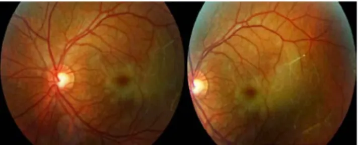
Figure 1 -RETINOGRAPHY ON DAY 1 OF DISEASE –cherry red spot, unperfused
segments in the upper and lower temporal arterioles.
Figure 2 - MACULAR OCT OF THE LEFT EYE – 5 DAYS AFTER: Necrotic, hyperreflective retinal thickening, attenuating the signal of the underlying retinal layers.
The patient started immediately ocular massage, topical timolol 0,5%, oral acetazolamide and high flow oxygen therapy. After exclusion of pulmonary disease (pneumothorax and COPD with pulmonary emphysema), he was referred within a few hours to a hyperbaric oxygen therapy center, where he was treated for three consecutive days. The treatment protocol was interrupted thereafter since no further visual acuity improvement was registered in two consecutive treatments.
Despite some evidence of reperfusion of retinal artery branches after one session, final visual acuity after completing the treatment was 0.05 in the affected eye.
Figure 3 - RETINOGRAPHY OF THE LEFT EYE AFTER THE FIRST SESSION OF HYPERBARIC OXYGEN THERAPY - Decreased area of edema. Partial reperfusion of previously excluded vessels is visible.
Two months later, fundus examination of the LE shows a linear area of atrophy mainly in the temporal macula with foveal involvement. Macular OCT depicts atrophy of the retinal inner layers more visible in the temporal macula, with blunting of the foveal contour. Retinal angiography obtained at the same time shows increase of the foveal avascular area.
Cerebral angiography one month after the procedure confirmed that the dural arteriovenous fistula was eliminated. He has no neurological sequelae related to the procedure.
Figure 4 - RETINOGRAPHY OF THE LEFT EYE 2 MONTHS AFTER THE OCCLUSION–A
linear atrophic lesion compromising the fovea is seen.
Figure 5 - MACULAR OCT OF THE LEFT EYE– 2 MONTHS AFTER THE
OCCLUSION: Decreased retinal thickness and flattening of the foveal depression.
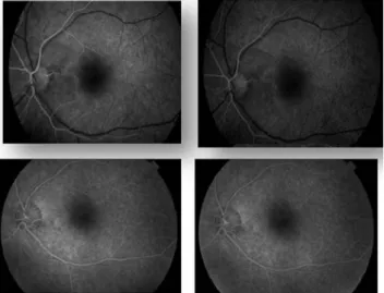
Figure 6 - FLUORESCEINIC ANGIOGRAPHY 2 MONTHS AFTER THE
OCCLUSION.
DISCUSSION
Retinal artery occlusion associated with therapeutic embolization procedures is an infrequent complication, whose clinical incidence, to our best knowledge, is not yet determined in the literature. These procedures are generally safe, however, one can find several case reports describing ocular morbidity4-13 mainly associated with transarterial particulate embolization of structures located in the head or neck, such as maxillary sinus and ethmoidal tumors, as well as embolization of the thyroid artery 4-10,12. Although coil embolization appears even less frequently involved, there is also a case report of branch retinal artery occlusion after coil embolization of an intracranial aneurism.14 Other procedures such as percutaneous coronary angioplasty have also been associated with this complication.15 In our case the para-cavernous fistula was perfused by a vascular net that originated in the ophthalmic artery, an anatomical relation with the retinal arterial system that contributed to this event.
The clinical pattern on fundoscopy was atypical. While we could observe the presence of a cherry red spot, typically evident in central artery occlusion, the area of retinal ischemic edema appeared to be sectorial, involving mainly the fovea and temporal retina and one could observe the segmentation and boxcarring of smaller retinal branches in both the superior and inferior temporal retinal artery divisions. These clinical features associated with the fact that our patient had a relative afferent pupillary defect in the absence of generalized retinal ischemia leads us to believe that he had a transient central retinal artery occlusion with subsequent spreading of the cyanoacrylate particles across the multiple retinal artery branches, creating a pattern of multiple branch retinal artery occlusion with foveal involvement. Obtaining diagnostic confirmation of this pattern of retinal angiography at the time was not possible but we believe the clinical aspects of fundus examination are highly suggestive.
Although the clinical features of the fundoscopy were congruent with a multiple branch retinal artery occlusion, the foveal involvement and the relative afferent pupillary defect led us to manage our patient’s case as a central retinal artery occlusion. Therapeutically there is no gold-standard treatment for patients with central retinal artery occlusion and visual prognosis is generally unfavorable1. Acute management aims at restoring ocular perfusion to the central retinal artery. In our case we started by employing non-invasive strategies such as ocular massage, hypotensive ocular treatment and inhalation of hyperbaric oxygen to increase blood oxygen content and dilate retinal arteries, as described previously in the literature3. Subsequently, we referred our patient to hyperbaric oxygen therapy, even though 24 hours had passed since the embolization procedure, therefore being in the upper limit of the timeline indicated for referral. Our patient´s unique pattern of multiple branch retinal artery occlusion deemed him unsuitable for Nd-YAG embolectomy, which is indicated in cases with a well visible thrombus.19,20
Visual rehabilitation was not possible despite prompt referral. This could be explained by the fact that this therapy is usually indicated for patients with less than 24 hours of visual loss and appears to be most effective in patients with symptoms for 8-12 hours upon referral. Our patient was referred the morning after his therapeutic procedure, possibly exceeding this timeline at the time of
treatment.16,18 The treatment was interrupted after three consecutive days since no visual acuity improvement was registered in two consecutive treatments, an approach previously described in the literature.16
CONCLUSIONS
This case report stresses the importance of considering therapeutic embolization as a possible cause of retinal artery occlusion. More studies regarding the incidence of these events after therapeutic embolization procedures are needed in order to establish the clinical relevance of this entity and appropriately discuss the risk of retinal arterial occlusion with these patients. As seen in our patient’s case, retinal artery occlusion secondary to therapeutic embolization can result in unique fundoscopic patterns. Our patient presented with a multiple branch retinal artery occlusion, with foveal involvement and a relative afferent pupillary defect, that led us to manage this case as a central retinal artery occlusion. Only minimal recovery was achieved.
Current therapeutic strategies for central retinal artery occlusion still have unsatisfactory overall outcomes and results are often unpredictable. Further investigation is therefore crucial to improve the visual prognosis of these patients.
BIBLIOGRAPHY
1. D D Varma, S Cugati, A W Lee, C S Chen. A review of central retinal artery occlusion: clinical presentation and maagement. Eye 2013; 27:688–697
2. Satomi J, Satoh K. Epidemiology and etiology of dural arteriovenous fistula. Brain Nerve 2008; 60(8):883-6.
3. Gupta A, Periakaruppan A. Intracranial dural arteriovenous fistulas: A Review. Indian J Radiol Imaging 2009; 19(1):43-8.
4. Hufendiek K, Hufendiek K, Finkenzeller T, Helbig H, Framme C. Acute visual loss after preoperative embolization of an ethmoidal metastasis. Int Ophthalmol 2012; 32:165–169
5. Finis D, Gumbel H. Central retinal artery occlusion after embolization in juvenile nasopharyngeal
angiofibroma. Klin Monbl Augenheilkd 2009; 226(7):579–580
6. Kunikata H, Tamai M. Cilioretinal artery occlusions following embolization of an artery to an intracranial meningioma. Graefes Arch Clin Exp Ophthalmol 2006; 244(3):401–403
7. Turner T, Trobe JD, Deveikis JP. Sequential branch retinal artery occlusions following embolization of an intracranial meningioma. Arch Ophthalmol 2002; 120:857–860
8. Terada T, Kinoshita Y, Yokote H, Tsuura M, Itakura T,Komai N, Nakamura Y, Tanaka S, Kuriyama T. Preoperative embolization of meningiomas fed by ophthalmic branch arteries. Surg Neurol 1996; 45(2):161–166
9. Roberson GH, Reardon EJ. Angiography and embolization of the internal maxillary artery for posterior epistaxis. Arch Otolaryngol 1979; 105(6):333–337
10. Wen F, Chen X, Liao R. Branch retinal artery occlusion after thyroid artery interventional embolization. Am J Ophthalmol 2000; 129:690–691
11. Stefansson E, Coin JT, Lewis WR III, Belkin RN, Behar VS,Morris JJ Jr, Anderson WB Jr. Central retinal artery occlusion during cardiac catheterization. Am J Ophthalmol 1985; 99:586–589
12. Jamous M, Satoh K, Kageji T, Satomi J, Matsubara S, Nagahiro S, Hayashi M, Nakagawa S. Anterior ischemic optic neuropathy after combined ophthalmic artery embolization and craniofacial surgery–case report. Neurol Med Chir 2001; 41(8):419–422
13. Mames RN, Snady-McCoy L, Guy J. Central retinal and posterior ciliary artery occlusion after particle embolization of the external carotid artery system. Ophthalmology 1991; 98:527–531
14. Ascaso FJ, Cristóbal JA. Partial retinal artery occlusion after coil embolization of an intracerebral aneurysm. Eur J Ophthalmol 1995; 9 (2):142-4
15. Filatov V, Tom D, Alexandrakis G, Skolik SA, Klassen H, Liggett PE. Branch retinal artery occlusion associated with directional coronary atherectomy after percutaneous transluminal coronary angioplasty, Am J Ophthalmol 1995; 120(3):391-3
16. Haddany A et al. Reversibility of retinal ischemia due to central retinal artery occlusion by hyperbaric oxygen, Clin Ophthalmol. 2016; 29(11):115-125
17. Iwama T, Hashimoto N, Takagi Y, Tanaka M, Yamamoto S, Nishi S, et al. Haemodynamic and metabolic disturbances in patients with intracranial dural arteriovenous fistulas: Positron emission tomography evaluation before and after treatment. J Neurosurg 1997; 86:86
18. Menzel-Severing J, Siekmann U, Weinberger A, Roessler G, Walter P, Mazinani B. Early hyperbaric oxygen treatment for nonarteritic central retinal artery obstruction. Am J Ophthalmol 2012; 153(3):454–459
19. Stanca HT, Petrović Z, Munteanu M. Transluminal
Nd:YAG laser embolysis--a reasonable method to reperfuse occluded branch retinal arteries. Vojnosanit Pregl. 2014; 71(11):1072-7.
20. Mason JO, Nixon PA, Albert MA Jr. Trans-luminal nd:YAG laser embolysis for branch retinal artery occlusion. Retina. 2007; 27(5):573-7
CONTACT
Diana Silveira e Silva
Praceta Papa João XXI, número 1, 2º Esquerdo 2685-217 Portela LRS
Portugal
e-mail: diana_silva1@hotmail.com
The authors declare having followed the protocols in use at their working center regarding patients’ data publication.