Cyclic AMP incre ase s the survival o f
ganglio n ce lls in mixe d re tinal ce ll
culture s in the abse nce o f e xo ge no us
ne uro tro phic mo le cule s, an e ffe ct that
invo lve s cho line rgic activity
Programa de Neuroimunologia, Departamento de Neurobiologia, Instituto de Biologia, Universidade Federal Fluminense, Niterói, RJ, Brasil R.C.C. Santos
and E.G. Araujo
Abstract
Natural cell death is a well-known degenerative phenomenon occur-ring duoccur-ring development of the nervous system. The role of trophic molecules produced by target and afferent cells as well as by glial cells has been extensively demonstrated. Literature data demonstrate that cAMP can modulate the survival of neuronal cells. Cultures of mixed retinal cells were treated with forskolin (an activator of the enzyme adenylyl cyclase) for 48 h. The results show that 50 µM forskolin induced a two-fold increase in the survival of retinal ganglion cells (RGCs) in the absence of exogenous trophic factors. This effect was dose dependent and abolished by 1 µM H89 (an inhibitor of protein kinase A), 1.25 µM chelerythrine chloride (an inhibitor of protein kinase C), 50 µM PD 98059 (an inhibitor of MEK), 25 µM Ly 294002 (an inhibitor of phosphatidylinositol-3 kinase), 30 nM brefeldin A (an inhibitor of polypeptide release), and 10 µM genistein or 1 ng/ml herbimycin (inhibitors of tyrosine kinase enzymes). The inhibition of muscarinic receptors by 10 µM atropine or 1 µM telenzepine also blocked the effect of forskolin. When we used 25 µM BAPTA, an intracellular calcium chelator, as well as 20 µM 5-fluoro-2’-deoxy-uridine, an inhibitor of cell proliferation, we also abolished the effect. Our results indicate that cAMP plays an important role controlling the survival of RGCs. This effect is directly dependent on M1 receptor activation indicating that cholinergic activity mediates the increase in RGC survival. We propose a model which involves cholinergic ama-crine cells and glial cells in the increase of RGC survival elicited by forskolin treatment.
Co rre spo nde nce
E.G. Araujo
Departamento de Neurobiologia Instituto de Biologia, UFF Caixa Postal 100.180 24001-970 Niterói, RJ Brasil
Fax: + 55-21-2719-5934 E-mail: adrianno@ openlink.com.br
Research supported by FAPERJ, CAPES and PRO NEX-MCT. R.C.C. Santos is the recipient of a CAPES fellowship.
Received January 26, 2001 Accepted September 3, 2001
Ke y words
·Ganglion cell survival ·cAMP
·Amacrine cholinergic cells ·M1 receptor activity ·Glial cells
Intro ductio n
Apoptosis, or naturally occurring cell death, is an active process of cellular self-destruction with distinctive morphological and biochemical features. During apoptosis
regressive event is essential for normal de-velopment, the molecular mechanisms that carry it out must be properly regulated; when this regulation is disturbed, diseases can ap-pear (2).
During the development of the nervous system, naturally occurring cell death is an important event accompanying synaptogen-esis (3). It has been assumed that an exces-sive number of neuronal cells is generated in order to promote appropriate neuronal con-nections to fit neuronal projection to the size and function of target cells (3,4). Target cells produce and release trophic molecules known as neurotrophins (NT) which play a crucial role in the differentiation, survival and plas-ticity of developing neurons (5,6). Several polypeptides including nerve growth factor (NGF), brain-derived neurotrophic factor (BDNF), NT-3, NT-4/5 and NT-6 belong to a neurotrophic family (7-9). All of these molecules bind to the low affinity receptor p75 (10). However, high affinity receptors (Trk) show a relative selectivity since NGF binds to TrkA, BDNF and NT-4/5 bind to TrkB, and NT-3 binds to TrkC (11,12).
Afferent cells also play an important role controlling natural cell death in the nervous system. Some of the mechanisms involved in afferent control of cell death are not clearly understood but the electrical activity as well as the release of trophic factors by either afferent or glial cells are evident (5).
Intracellular calcium concentration plays an important role in the regulation of apopto-sis. Several lines of evidence show that an increase of cytoplasmic calcium can either block or trigger apoptosis, depending on the cell type studied (13).
Adenosine 3’,5’-cyclic monophosphate (cAMP) is a key second messenger in signal-ing pathways governsignal-ing many cellular pro-cesses. Many signals that regulate the growth, development and metabolism of cells use cAMP as a second messenger. Experimental data show that the second messenger cAMP is also involved in the control of neuronal
cell death (14). This second messenger acti-vates a protein kinase A (PKA) which is a tetrameric holoenzyme consisting of a di-meric regulatory subunit and two monodi-meric catalytic subunits (15). The action of cAMP is interrupted by one or more cyclic nucleo-tide phosphodiesterases (16). Literature data have shown that cAMP can increase the survival of sympathetic, spinal motor and dopaminergic neurons in vitro in the
ab-sence of neurotrophic factors (17). It has also been suggested that cAMP could also modulate the differentiation of brainstem catecholaminergic neurons (18).
The aim of the present investigation was to determine the effect of cAMP on ganglion cell survival kept in mixed retinal cell cul-tures in the absence of exogenous neu-rotrophic molecules. We used 50 µM for-skolin to stimulate an increase in cAMP levels in the cultures. Our results demon-strate that the effect of forskolin is dose dependent and involves an increase in cyto-plasmic calcium levels, activation of protein kinases, release of polypeptides, and cellular proliferation. An important finding is that cholinergic activity mediates the effect of forskolin, indicating an important role for this neurotransmitter in the control of retinal ganglion cell survival.
Mate rial and Me thods
Within the first 24 h after birth, Lister Hooded rats were anesthetized by hypother-mia. One microliter of a solution of 30% horseradish peroxidase (HRP) in 2% di-methyl sulfoxide was injected into each su-perior colliculus. The animals survived for ~16 h before the procedures used for cell culture.
(Wor-thington, Freehold, NJ, USA). The tissue was then resuspended in complete culture medium and triturated by passage through a Pasteur pipette. After complete tissue disso-ciation, 1 ml of the cell suspension was added to glass coverslips previously coated with 50 µg/ml poly-L-ornithine and placed on 35-mm Petri dishes. We used 199 medi-um supplemented with 2 mM glutamine, 100 µg/ml streptomycin, 100 U/ml penicillin and 5% fetal calf serum. After cell plating, cul-tures were usually incubated for 4 h to allow the cells to attach to the coverslips. Then, culture medium alone or culture medium containing the drugs to be tested was added to each Petri dish. Average plating density was 650,000 cells per Petri dish. The cul-tures were maintained in a humidified at-mosphere of 5% CO2 and 95% air at 37ºC.
The presence of the enzyme peroxidase in the cytoplasm of retinal ganglion cells was demonstrated by the protocol of Mesulam (19). Briefly, the monolayers were fixed af-ter 2 days in culture with a mixture of 1% paraformaldehyde and 2% glutaraldehyde in 0.1 M sodium phosphate buffer for 5 min, washed in phosphate buffer, and reacted with tetramethylbenzidine.
Retinal ganglion cells were counted us-ing a Zeiss microscope at a magnification of 400X under a bright field. As an internal control for the percentage of ganglion cells labeled with HRP in distinct experiments, the number of labeled cells after 4 h in culture was taken as 100%. All data are reported as mean ± standard error of the mean of experiments performed at least in duplicate and each experiment was repeated at least three times. Statistical analysis was done using analysis of variance (ANOVA) followed by the Newman-Keuls test.
Re sults
We first analyzed the effect of forskolin on the survival of retinal ganglion cells after 48 h in culture. Figure 1 shows that treatment
with forskolin increased the survival of gan-glion cells in a dose-dependent way. After 48 h the number of retinal ganglion cells in control cultures was reduced by approxi-mately 50%. However, 10, 25 and 50 µM forskolin induced an increase in ganglion cell survival. The strongest effect was ob-tained with 50 µM forskolin (approximately all cells initially plated were alive after 48 h). Based on these results, we used 50 µM forskolin in all subsequent experiments.
To investigate if the forskolin effect was dependent on culture density we used two different plating densities (130 and 65 cells/ mm2). The results presented in Table 1 show
that forskolin enhanced ganglion cell
sur-Figure 1. Dose-dependent effect of forskolin on retinal ganglion cell (RGC) survival after 48 h in culture. Cultures w ere treated w ith forskolin (5, 10, 25 and 50 µM ). CT = control, F = forskolin. The number of RGCs counted in 4-h cultures w as 21,140 cells. Data are reported as the per-centage of RGC survival com-pared w ith 4-h control cultures (100% ) and represent the mean ± SEM of tw o different experi-ments, each performed w ith at least three different Petri dishes (N = 3-4). * P<0.001 compared to the 48-h control (New man-Keuls test).
R
G
C
s
u
rv
iv
a
l
(%
o
f
c
o
n
tr
o
l)
120
100
80
60
40
20
0
12 12 12 12 12
12345 12345 12345 12345 12345 12345 12345 12345 12345 12345 12345 12345 12345 12345 12345 12345
1234 1234 1234 1234 1234 1234 1234 1234 1234 1234 1234
123 123 123 123 123
123 123 123
12 123 123
12 12
CT 4 h F 25
CT 48 h F 10
F 50 F 5
Table 1. The effect of 50 µM forskolin on ganglion cell survival at different plating densities.
Treatment RGC survival (% of control)
65 cells/mm2 130 cells/mm2
48-h Control 57.25 ± 1.05 48.62 ± 1.07
Forskolin 106.30 ± 8.89* 87.18 ± 6.92*
We used tw o different plating densities (130 cells/
mm2 and 65 cells/mm2). Forskolin enhanced the
survival of retinal ganglion cells (RGCs) at both densities studied. The number of RGCs counted
in 4-h cultures w as 25,160 cells (130 cells/mm2)
and 12,670 cells (65 cells/mm2). Data are reported
as the percentage of RGC survival compared w ith 4-h control cultures (100% ) and represent the mean ± SEM of three different experiments, each performed w ith at least three different Petri dishes (N = 3-4). * P<0.001 compared to the 48-h control (New man-Keuls test).
* *
vival at both densities studied. For this rea-son we used the density of 65 cells/mm2 in
all subsequent experiments.
To test if the effect of forskolin was dependent on chronic treatment we exposed the cultures to a 12- or 24-h pulse of this drug. Analyzing the results obtained we con-cluded that the increase in ganglion cell sur-vival was observed only when forskolin was present during the whole period of culture (data not shown).
Literature data suggest that forskolin it-self may induce a neurotrophic effect. To
test this hypothesis we evaluated the effect of different drugs that increase intracellular cAMP levels (Table 2). Initially we began testing a membrane permeable analog of cAMP, 1 mM dibutyryl cAMP. The results demonstrated that this drug mimics the in-crease in ganglion cell survival obtained with forskolin treatment after 48 h in culture. To confirm this result we tested a drug that activates G proteins and indirectly activates adenylyl cyclase, i.e., 100 ng/ml cholera toxin. Again we obtained an increase in gan-glion cell survival after this treatment. To determine if endogenous cAMP would be sufficient to increase the survival of gan-glion cells in culture we used an inhibitor of phosphodiesterase, 200 or 400 µM IBMX. We observed that an increase in survival was obtained following phosphodiesterase inhi-bition.
The next step was to determine if the effect of forskolin was mediated by an in-crease in cytoplasmic calcium levels. Figure 2 demonstrates that when cells were treated with forskolin in the presence of 7.5 µM nifedipine (an inhibitor of voltage-depend-ent L-type calcium channels) or 30 µM dan-trolene (an inhibitor of internal calcium re-lease), there were no changes in the effect of forskolin. On the other hand, when we used an intracellular calcium chelator (25 µM BAPTA-AM) we abolished the effect on
retinal ganglion cell survival.
One major cellular effect of the cAMP cascade activation is the transcriptional stim-ulation after phosphorylation of nuclear fac-tors by the cAMP-dependent kinase, PKA. To investigate the involvement of this en-zyme in the effect of forskolin we used an inhibitor of PKA, 25 µM H89. Figure 3 shows that PKA inhibition abolished the effect on ganglion cell survival. To test the role of other protein kinases on the effect of forskolin we used an inhibitor of protein kinase C (PKC), 1.25 µM chelerythrine chlo-ride. Figure 3 shows that this drug partially abolished the effect of forskolin. Since PKC
Table 2. cAM P and retinal ganglion cell survival.
Treatment RGC survival (% of control)
48-h Control 57.84 ± 0.46
Dibutyryl cAM P 96.26 ± 2.05*
Cholera toxin 83.51 ± 4.88*
IBM X (200 µM ) 82.52 ± 2.81*
IBM X (400 µM ) 96.16 ± 2.23*
We used 1 mM dibutyryl cAM P, 100 ng/ml chol-era toxin and 200 or 400 µM IBM X. All of these drugs induced the same effect as elicited by 50 µM forskolin on retinal ganglion cell (RGC)
sur-vival after 48 h in vitro. The number of RGCs
counted in 4-h cultures w as at least 18,260 cells. Data are reported as the percentage of RGC sur-vival compared w ith 4-h control cultures (100% ) and represent the mean ± SEM of three different experiments, each performed w ith at least tw o different Petri dishes (N = 3-4). * P<0001 com-pared to the 48-h control (New man-Keuls test).
Figure 2. The effect of 50 µM forskolin w as inhibited by 25 µM BAPTA-AM . Cells w ere
main-tained in vitro for 48 h.
Nifedi-pine (7.5 µM ) and 30 µM dantro-lene did not block the effect of forskolin. CT = control, F = for-skolin, Nif = nifedipine (an inhib-itor of voltage dependent L-type calcium channels), Dan = dantro-lene (an inhibitor of internal cal-cium release), BAPTA-AM (an in-tracellular calcium chelator). The number of retinal ganglion cells
(RGCs) counted in 4-h cultures w as 13,350 cells. Data are reported as the percentage of RGC survival compared w ith 4-h control cultures (100% ) and represent the mean ± SEM of three different experiments, each performed w ith at least three different Petri dishes (N = 3-6). * P<0.001 compared to the 48-h control (New man-Keuls test).
R
G
C
s
u
rv
iv
a
l
(%
o
f
c
o
n
tr
o
l)
120 100 80
60 40
20
0
123 123 12
12
12 12 123
123
CT 4 h CT 48 h F Dan Dan/F
Nif
123 123 123 123 123
123 123 123 123 123 123 123 123 123 123
12 12 12 12 12 12 12 12 12
123 123 123 123 123 123 123
Nif/F BAPTA-AM BAPTA-AM /F
was involved in this effect we decided to investigate if the activation of mitogen-acti-vated protein kinase (MAPK) was mediating the effect studied. We used an inhibitor of MAPK enzyme (MEK), 25 µM PD 98059, and observed that this drug completely abol-ished the increase in ganglion cell survival (Figure 3).
To determine if tyrosine kinase enzymes are involved in the effect of forskolin we used 10 µM genistein and 1 ng/ml herbimycin (both inhibitors of these enzymes). Our re-sults demonstrate that in the presence of these drugs the effect of forskolin was totally abolished (Figure 4). Since the activation of the tyrosine kinase pathway is involved in the effect of forskolin, we tested an inhibitor of the phosphatidylinositol (PI)-3 kinase en-zyme, 25 µM Ly 294002. This inhibitor completely inhibited the effect of forskolin on ganglion cell survival (Figure 4). Based on these results, we tested an inhibitor of polypeptide release, 30 nM brefeldin A, which effectively blocked the effect of forskolin on ganglion cell survival (Figure 5). Morphological analysis of retinal cells in culture showed that glial cells keep their proliferative capacity throughout in vitro
culture. To determine if the effect of forskolin was mediated by an increase in glial cell population we tested it in the presence of an inhibitor of cell division, 25 µM fluorode-oxyuridine. Figure 6 shows that treatment with this antimitotic drug abolished the ef-fect of forskolin. This result indicates that ganglion cell survival induced by forskolin is mediated by an increase in the number of glial cells.
In order to verify the cholinergic activity of a subset of amacrine cells in our cultures we tested an antagonist of muscarinic recep-tors, 10 mM atropine, and obtained a com-plete inhibition of the forskolin effect (Fig-ure 7). To investigate which subtype of mus-carinic receptors could be involved in the effect of forskolin we tested M1 and M3 antagonists, 1 µM telenzepine and 200 µM
R G C s u rv iv a l (% o f c o n tr o l) 120 100 80 60 40 20 0 1234 1234 12 12 123 123
CT 4 h CT 48 h F G G/F
Hr Hr/F Ly Ly/F
12 12 12 12 12 123 123 123 123 123 123 123 123 123 123 123 123 123 123 123 123 123 123 123 123 123 123 123
Figure 4. The effect of 50 µM forskolin w as blocked by inhibi-tion of tyrosine kinase activity.
Cells w ere maintained in vitro
for 48 h. CT = control, F = for-skolin, G = genistein and Hr = herbimycin (inhibitors of tyrosine kinase enzymes), Ly = Ly 294002 (an inhibitor of phosphatidylino-sitol-3 kinase). The number of ret inal ganglion cells (RGCs) count ed in 4-h cult ures w as 27,050 cells. Data are reported as the percentage of RGC
sur-vival compared w ith 4-h control cultures (100% ) and represent the mean ± SEM of three different experiments, each performed w ith at least three different Petri dishes (N = 3-6). * P<0.001 compared to the 48-h control (New man-Keuls test).
Figure 3. The effect of 50 µM forskolin w as abolished by inhibi-tion of protein kinase activity.
Cells w ere maintained in vitro for
48 h. CT = control, F = forskolin, H89 (an inhibitor of PKA), PD = PD 98059 (an inhibitor of M EK), CC = chelerythrine chloride (an inhibitor of PKC). The number of ret inal ganglion cells (RGCs) count ed in 4-h cult ures w as 10,000 cells. Data are reported as the percentage of RGC sur-vival compared w ith 4-h control
cultures (100% ) and represent the mean ± SEM of tw o different experiments, each performed w ith at least three different Petri dishes (N = 3-6). * P<0.001 compared to the 48-h control (New man-Keuls test).
R G C s u rv iv a l (% o f c o n tr o l) 120 100 80 60 40 20 0 123 123 12 12 12 12 12 12
CT 4 h CT 48 h F H89 H89/F
PD PD/F CC
12 12 12 12 12 1234 1234 1234 1234 1234 1234 1234 1234 1234 1234 12 12 12 12 12 12 12 12 12 12 12 12 12 * CC/F R G C s u rv iv a l (% o f c o n tr o l) 120 100 80 60 40 20 0 1234 1234 1234 12 12 12 12
CT 4 h
BFA
CT 48 h
BFA/F F 123 123 123 123 123 123 12345 12345 12345 12345 12345 12345 12345 12345 12345 12345 12345 123 123 123 123 123 123
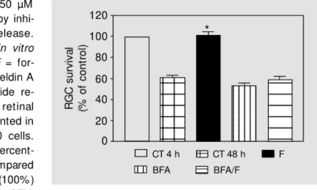
Figure 5. The effect of 50 µM forskolin w as abolished by inhi-bition of polypeptide release.
Cells w ere maintained in vitro
for 48 h. CT = control, F = for-skolin, BFA = 30 nM brefeldin A (an inhibitor of polypeptide re-lease). The number of retinal ganglion cells (RGCs) counted in 4-h cultures w as 10,650 cells. Data are reported as the percent-age of RGC survival compared w ith 4-h control cultures (100% ) and represent the mean ± SEM
of tw o different experiments, each performed w ith at least three different Petri dishes (N = 3-6). * P<0.001 compared to the 48-h control (New man-Keuls test).
*
DAMP, respectively. Figure 7 shows that only telenzepine was able to abolish the effect of forskolin on cultures. This result suggests that the activation of M1 receptors is involved in the survival of ganglion cells in vitro.
D iscussio n
The results presented in this study show that treatment of mixed retinal cell cultures with 50 µM forskolin induced a two-fold increase in ganglion cell survival after 48 h
in vitro. To investigate if these results are directly dependent on cAMP enhancement we used other drugs that increase the cyto-plasmic cAMP concentration. The results presented here show that all drugs tested elicited an increase in ganglion cell survival, suggesting that this effect was mediated by cAMP. These data definitely demonstrate that cAMP signaling is really involved in the control of ganglion cell survival in mixed retinal cell cultures.
In the present study we did not analyze the effect of forskolin on the other cell types
present in our cultures. Further studies will be performed in order to investigate if the effect of forskolin on survival is restricted to the ganglion cell population. However, we have observed that Müller cells proliferate during the time in culture showing a back-ground covering on plating dishes, forming a glial “carpet” for neurons (data not shown). The primary element in cAMP signal transduction is the cAMP-dependent activa-tion of PKA, which mediates many cAMP actions through phosphorylation of serine threonine residues in target proteins (20). Our results demonstrate that the effect of forskolin was abolished when PKA activity was inhibited, indicating that kinase A activ-ity was important for the forskolin effect. The data seem to be true since the major effect of cAMP during development of many tissues is through activation of kinase A anchor protein. This protein, that can be directly activated by PKA, leads to an in-crease in the survival and/or differentiation of cell populations, playing an important role in the amplification of the cAMP signal-ing pathway (21).
Literature data have demonstrated the role of PKC activation in the survival of retinal ganglion cells (22), as well as the crosstalk between the cAMP and PI-3 sig-naling pathway in pancreatic cells (23). In-terestingly, the inhibition of PKC activity completely abolished the effect of forskolin on ganglion cell survival in mixed retinal cultures. These results suggest the possible crosstalk between PKA and PKC pathways in our preparation.
In order to test a possible involvement of tyrosine kinase enzymes in the effect of for-skolin we used inhibitors of these enzymes, 10 µM genistein and 1 ng/ml herbimycin. Our results demonstrate that when the ty-rosine kinase pathway was blocked the ef-fect of forskolin was abolished. One pos-sible explanation for this result involves the participation of trophic factors in this effect. One class of trophic molecules, the
neuro-Figure 7. The effect of 50 µM forskolin w as inhibited by the blockade of M 1 muscarinic re-ceptors. Cells w ere maintained
in vitro for 48 h. CT = control, F = forskolin, A = 10 µM atropine (a muscarinic antagonist), T = 1 µM telezenpine (an M 1 antagonist), DAM P = 200 µM DAM P (an M 3 antagonist). The number of reti-nal ganglion cells (RGCs) count ed in 4-h cult ures w as 27,000 cells. Data are reported as the percentage of RGC
sur-vival compared w ith 4-h control cultures (100% ) and represent the mean ± SEM of three different experiments, each performed w ith at least three different Petri dishes (N = 3-6). * P<0.001 compared to the 48-h control (New man-Keuls test).
R
G
C
s
u
rv
iv
a
l
(%
o
f
c
o
n
tr
o
l)
120
100
80
60
40
20
0
12 12
12 12 12
CT 4 h
FDUR
CT 48 h
FDUR/F F
123 123 123 123 123 123 123
123 123 123 123 123 123
123 123 123 123 123 123 123
Figure 6. The effect of 50 µM forskolin w as blocked by 20 µM
FDUR. Cells w ere maintained in
vitro for 48 h. CT = control, F = forskolin, FDUR = 5-fluoro-2'-de-oxyuridine (inhibitor of cell prolif-eration). The number of retinal ganglion cells (RGCs) counted in 4-h cultures w as 18,000 cells. Data are reported as the percent-age of RGC survival compared w ith 4-h control cultures (100% ) and represent the mean ± SEM of tw o different experiments,
each performed w ith at least three different Petri dishes (N = 3-6). * P<0.001 compared to the 48-h control (New man-Keuls test).
*
R
G
C
s
u
rv
iv
a
l
(%
o
f
c
o
n
tr
o
l)
120
100
80
60
40
20
0
123 123 123 123 123
12 12 12 12 12
12 12 12 12 12
123 123 123 123 123 123 12 12 12
12
12 12 12
12
CT 4 h CT 48 h F A A/F
T T/F DAM P DAM P/F
trophins, binds to several Trk receptors and stimulates several intracellular proteins such as MAPK and PI-3 kinase leading to cell survival (24). Our results also show that inhibitors of these enzymes blocked the ef-fect of forskolin on retinal ganglion cell survival, showing that these enzymes are essential for the forskolin effect, allowing us to assume that forskolin may increase the release of trophic molecules in mixed cul-ture medium. This hypothesis is corrobo-rated by our finding that the inhibitor of polypeptide release brefeldin A blocked the effect of forskolin. Based on these results, we can suggest that an increase in cAMP levels could mediate an enhancement in the release of trophic molecules.
It has been demonstrated that glial cells produce and release trophic molecules after appropriate stimulation (16,18,25). Accord-ingly, glial cells could be a possible source of trophic molecules. Since in our prepara-tion these cells retain their proliferative ca-pacity during culture in vitro we decided to
study the effect of forskolin in the presence of an inhibitor of cell division. Our results clearly demonstrate that fluorodeoxyuridine entirely abolished the effect of forskolin, suggesting that Müller cells could be respon-sible for the major release of trophic mol-ecules in mixed retinal cell cultures.
Literature data have provided evidence that cholinergic neuronal activity is essen-tially involved in the regulation of BDNF and NGF mRNAs in the developing and adult rat hippocampus (26). Since there is a cholinergic subpopulation of amacrine cells in the retina (27), we decided to investigate the possible role of acetylcholine in the ef-fect elicited by forskolin. Our results show that the muscarinic receptor antagonist atro-pine completely blocked the forskolin effect on ganglion cell survival. Based on this re-sult, we decided to test specific muscarinic antagonists, telenzepine and DAMP, in or-der to verify which subtype of muscarinic receptors was involved. We observed that
telenzepine was capable of inhibiting the forskolin effect, showing that the activation of M1 receptors plays an important role in the increase in ganglion cell survival elicited by forskolin. These data are in agreement with the results obtained with BAPTA-AM since M1 stimulation elicits an increase in intracellular calcium levels induced by PI-3 receptor stimulation (28).
In a previous study, Pereira and Araujo (29) demonstrated that 3 µM veratridine in-creased the survival of retinal ganglion cells, an effect also mediated by acetylcholine. These investigators showed that the veratri-dine effect was not mediated by L-type cal-cium channels but was mediated by T-type calcium channels. This result leads us to speculate about a role for T-type calcium channels in the release of acetylcholine from a subpopulation of amacrine cells in our cultures.
them. These very interesting results are in agreement with our data since they indicate that cholinergic activity plays an important role during development by controlling gan-glion cell survival or differentiation.
The data presented here allow us to sug-gest a model for the effect of forskolin on ganglion cell survival in mixed retinal cell cultures. High levels of cAMP following forskolin treatment possibly stimulate cho-linergic amacrine cells to release acetylcho-line (34). This neurotransmitter, by stimulat-ing M1 receptors, could induce an increase in the release or production of trophic mol-ecules from neuronal and glial cells (25,27, 32). Since it was observed that M1 receptors present in astrocytes modulate the release of trophic molecules (35), and Müller cells (the major glial population in the retina) belong to the astrocytic lineage, we may propose the presence of these receptors in this popula-tion (36). Trophic molecules released by Müller cells may be responsible for the in-crease in the survival of ganglion cells through activation of PI-3 kinase and MAPK
pathways, as was extensively demonstrated (37). On the other hand, high levels of cAMP could up-regulate the expression of neuro-trophins and neurotrophin receptors on the plasma membrane of retinal ganglion cells in astroglial cultures and in the rat frontal cortex and hippocampus (38-40), allowing ganglion cells to better respond to trophic molecules released by glial cells in our cul-tures.
Further studies will be performed in or-der to identify which cells express M1 recep-tors in our cultures and which trophic mol-ecules are involved in the increase in retinal ganglion cell survival.
Ackno wle dgm e nts
We would like to thank Alexandre José Fernandes, Bernardino Matheus dos Santos and Jonas Borges da Silva for technical as-sistance. We also thank Dr. Roberto Paes de Carvalho for helpful suggestions and critical comments about the data.
Re fe re nce s
1. Vaux DL & Korsmeyer SJ (1999). Cell
death in development. Cell, 96: 245-254.
2. Stew art BW (1994). M echanisms of ap-optosis: integration of genetic,
biochemi-cal and cellular indicators. Journal of the
National Cancer Institute, 86: 1287-1296. 3. Oppenheim RW (1991). Cell death during
development of the nervous system.
An-nual Review of Neuroscience, 14: 453-501.
4. Hamburger V & Levi-M ontalcini R (1949). Proliferation, differentiation and degen-eration in the spinal ganglia of the chick embryo under normal and experimental
conditions. Journal of Experimental
Zool-ogy, 111: 457-501.
5. Snider WD (1994). Functions of the neu-rotrophins during nervous system devel-opment: w hat the knockouts are teaching
us. Cell, 77: 627-638.
6. Thoenen H (1995). Neurotrophins and
neuronal plasticity. Science, 270: 593-598.
7. Götz R, Köster R, Winkler C, Raulf F,
Lottspelch F, Schartl M & Thoenen H (1994). Neurotrophin-6 is a new member
of the nerve grow th factor family. Nature,
372: 266-269.
8. Hallböök F, Ibáñez CF & Persson H (1991). Evolutionary studies of the nerve grow th factor family reveal a novel member
abun-dantly expressed in Xenopus ovary.
Neu-ron, 6: 845-858.
9. M aisonpierre PC, Belluscio L, Squinto S, Ip NY, Furth M E, Lindsay RM & Yanco-poulos GD (1990). Neurotrophin-3: a neu-rotrophic factor related to NGF and BDNF.
Science, 247: 1446-1451.
10. Carter BD & Lew in GR (1997).
Neurotro-phins live or let die: Does p75NTR decide?
Neuron, 18: 187-190.
11. Barbacid M (1994). The Trk family of
neu-rotrophin receptors. Journal of
Neurobiol-ogy, 25: 1386-1403.
12. Barde YA (1989). Trophic factors and
neu-ronal survival. Neuron, 2: 1525-1534.
13. Franklin JL & Johnson EM (1992).
Sup-pression of programmed neuronal death by sustained elevation of cytoplasmic
cal-cium. Trends in Neurosciences, 15:
501-508.
14. Spiegel AM , Shenker A & Weinstein LS (1992). Receptor-effector coupling by G-proteins: implications for normal and
ab-normal signal transduction. Endocrine
Re-view s, 13: 536-565.
15. Beavo JA & Reifsnyder DH (1990). Pri-mary sequence of cyclic nucleotide phos-phodiesterase isoenzymes and design of
selective inhibitors. Trends in
Physiologi-cal Sciences, 11: 150-155.
16. Hanson Jr M G, Shen S, Wiemelt AP, M cM orris FA & Barres BA (1998). Cyclic AM P elevation is sufficient to promote
the survival of spinal motor neurons in
vitro. Journal of Neuroscience, 18: 7361-7371.
long-term survival of mesencephalic
dopamin-ergic neurons. Journal of
Neurochemis-try, 67: 1633-1642.
18. Edw ards SN, Buckmaster AE & Tolkovsky AM (1991). The death program in cultured sympathetic neurones can be suppressed at a post t ranslat ional level by nerve grow th factor, cyclic AM P and
depolariza-tion. Journal of Neurochemistry, 57:
2140-2143.
19. M esulam M (1978). Tetramethylbenzidine for horseradish peroxidase neurohisto-chemistry. A non-carcinogenic blue reac-tion product w ith superior sensitivity for visualizing neural afferents and efferents.
Journal of Histochemistry and Cytochem-istry, 26: 106-117.
20. Beebe SJ (1994). The cAM P-dependent protein kinases and cAM P signal
trans-duction. Seminars in Cancer Biology, 5:
285-294.
21. Feliciello A, Gottesman M E & Avvedi-mento V (2001). The biological functions
of A-kinase anchor proteins. Journal of
M olecular Biology, 308: 99-114. 22. Santos AA & Araujo EG (2000). The effect
of PKC activation on the survival of rat
retinal ganglion cells in culture. Brain
Re-search, 853: 338-343.
23. Liu YJ, Grapengiesser E, Gylfe E & Hell-man B (1987). Crosstalk betw een the cAM P and inositol triphosphate-signaling
pathw ays in pancreatic cells. Archives of
Biochemistry and Biophysics, 334: 295-302.
24. Kaplan DR & M iller F (2000). Neurotrophin signal transduction in the nervous
sys-tem. Current Opinion in Neurobiology, 10:
381-391.
25. Schmalenbach C & M üller HV (1993). As-troglial-neuron interactions that promote
long-term neuronal survival. Journal of
Chemical Neuroanatomy, 6: 229-237.
26. Berzaghi M P, Cooper J, Castrén E, Zafra F, Sofroniew M , Thoenen H & Lindholm D (1993). Cholinergic regulation of brain-derived neurotrophic factor (BDNF) and nerve grow th factor (NGF) but not neuro-trophin-3 (NT-3) mRNA levels in the
de-veloping rat hippocam pus. Journal of
Neuroscience, 13: 3818-3826.
27. Santos PF, Carvalho AL, Carvalho AP & Duarte CB (1998). Differential acetylcho-line and GABA release from cultured chick
retina cells. European Journal of
Neuro-science, 10: 2723-2730.
28. Wotta DR, Wattenberg EV, Langason RB & El-Fakahany EE (1998). M 1, M 3 and M 5 muscarinic receptors stimulate
mitogen-activated protein kinase. Journal of
Phar-macology, 56: 175-186.
29. Pereira SPF & Araujo EG (1997). Veratri-dine increases the survival of retinal
gan-glion cells in vitro. Brazilian Journal of
M edical and Biological Research, 30: 1467-1470.
30. M eyer-Frank A, Kaplan M R, Pfrieger FW & Barres BA (1995). Characterization of the signaling interactions that promote the survival and grow th of developing
reti-nal ganglion cells in culture. Neuron, 15:
805-819.
31. Shen S, Wiemelt AP, M cM orris FA & Barres B (1999). Retinal ganglion cells lose trophic responsiveness after
axo-tomy. Neuron, 23: 285-295.
32. Stellw agen D, Shatz CJ & Feller M B (1999). Dynamics of retinal w aves are
con-trolled by cyclic AM P. Neuron, 24:
673-685.
33. Reese BE & Collelo RJ (1992). Neurogen-esis in the retinal ganglion cell layer of the
rat. Neuroscience, 46: 419-429.
34. Yao W-D, Rusch J, Poo M -M & Wu C-F (2000). Spontaneous acetylcholine secre-tion from developing grow th cones of
Drosophila central neurons in culture:
ef-fects of cAM P-pathw ay mutations.
Jour-nal of Neuroscience, 20: 2626-2637. 35. Knipper M , da Penha Berzaghi M , Blöchl
A, Breer H, Thoenen H & Lindholm D (1994). Positive feedback betw een ace-tylcholine and the neurotrophins nerve grow th factor and brain-derived neu-rotrophic factor in the rat hippocampus.
European Journal of Neuroscience, 6: 668-671.
36. M urphy S, Pearce B & M orrow C (1986). Astrocytes have both M 1 and M 2
musca-rinic receptor subtypes. Brain Research,
364: 177-180.
37. Dolcet X, Egea J, Soler RM , M artin-Zanca D & Comella JX (1999). Activation of phos-phatidylinositol 3-kinase, but not extracel-lular-regulated kinases, is necessary to mediate brain-derived neurotrophic
factor-induced motoneuron survival. Journal of
Neurochemistry, 73: 521-531.
38. M eyer-Frank A, Wilkinson GA, Kruttgen A, Hu M , M unro E, Hanson Jr M G, Reichardt LF & Barres BA (1998). Depo-larization and cAM P elevation rapidly re-cruit Trk B to the plasma membrane of
CNS neurons. Neuron, 21: 681-693.
39. Condorelli DF, Dell’Albani P, M udo G, Timmusk T & Belluardo N (1994). Expres-sion of neurotrophins and their receptors in primary astroglial cultures: induction by
cyclic AM P-elevating agents. Journal of
Neurochemistry, 63: 509-516.
40. M orinobu S, Fujimaki K, Okuyama N, Takahashi M & Duman RS (1999). Stimu-lation of adenylyl cyclase and induction of brain-derived neurotrophic factor and TrkB mRNA by NKH477, a novel and potent
forskolin derivative. Journal of