INTRODUCTION
Retinal vein occlusion (RVO) is the second most common
vas-cular condition capable of causing irreversible vision loss(1). Reduced
visual acuity (VA) in patients with RVO is usually because of macular
edema (ME) and/or locally impaired capillary perfusion(2-7). Both the
location and total area of capillary non-perfusion greatly affect the
duration of the disease and risk of proliferative complications(6-10).
Post-occlusion ME is typically caused by local inflammation and the effects of increased Vascular Endothelial Growth Factor (VEGF)
levels(2,6-10). VEGF expression is potentiated by tissue pH and the
par-Alternative ways to optimize treatment for retinal vein occlusion with peripheral
capillary non-perfusion: a pilot study
Opções alternativas para otimizar o tratamento da oclusão da veia retiniana sem perfusão capilar
periférica (um estudo piloto)
Svetlana n. tultSeva1, YurY S. aStakhov1, Sergei a. novikov1, Pavel a. nechiPorenko1, alla B. liSochkina1, andranik Y. ovnanYan1, Sergei Y. aStakhov1
Submitted for publication: November 16, 2016 Accepted for publication: February 5, 2017
1 Department of Ophthalmology, First Pavlov State Medical University of St. Petersburg, Saint Petersburg, Russian Federation.
Funding: No specific financial support was available for this study.
Disclosure of potential conflicts of interest: None of the authors have any potential conflict of interest to disclose.
Corresponding author: Svetlana N. Tultseva. Department of Ophthalmology. First Pavlov State Medical University of St. Petersburg ul. Lva Tolstogo, 6-8 - Saint Petersburg - 197022 - Russian Federation - E-mail: [email protected]
Approved by the following research ethics committee: Pavlov First St. Petersburg State Medical University.
ABSTRACT
Purpose: We compared the eicacy and safety of ranibizumab versus ranibizumab plus scatter laser photocoagulation (SLP) in patients with chronic post-central retinal vein occlusion (CRVO) macular edema (ME).
Methods: This prospective non-randomized pilot study included 250 patients with peripheral retinal ischemia and CRVO-related ME. The mean follow-up period was 24.5 ± 6.5 months. The clinical assessments conducted included best corrected visual acuity, optical coherence tomography, and multi-field fluores-cein angiography with measurement of the ischemic area. The study population comprised two comparable patient groups with peripheral retinal ischemia that received diferent treatments for post-CRVO ME: ranibizumab with peripheral SLP of capillary non-perfusion areas (Group 1); and Lucentis® monotherapy (Group 2). Data analyses were performed using Statistica 7 software suite and included the estimation of х ± δ values and their dispersion and covariation coeicients at diferent stages of the study.
Results: Clinically significant retinal ischemia was detected in 175 (70%) patients, occupying an average of 435.12 ± 225.13 mm2, i.e., 167.15 ± 45.16 optic disc areas. Peripheral ischemia was found in 125 patients, representing 50% of all patients with CRVO and 71.4% of all patients with ischemic CRVO. The mean number of ranibizumab injections in patients who underwent SLP was 3.5 ± 1.6. Patients treated with ranibizumab monotherapy for 24 months received 10.6 ± 2.5 injections. Functional and anatomic results were comparable in the two groups.
Conclusions: The combination of ranibizumab injections and peripheral SLP in capillary non-perfusion areas can significantly decrease the number of injections and reduce neovascular complications.
Keywords: Retinal vein occlusion; Ranibizumab; Ischemia; Laser coagulation; Vi sual acuity
RESUMO
Objetivo: A investigação centra-se na terapia de edema macular pós-oclusão da veia retiniana central (OVCR) em casos com isquemia retiniana periférica. O objetivo foi comparar a eficácia e a segurança do tratamento com ranibizumab vs ranibizumab + fotocoagulação com laser de dispersão (SLP) em pacientes com edema macular crônico secundário a oclusão da veia retiniana central isquêmica.
Métodos: O estudo prospectivo não-randomizado incluiu 250 pacientes com isque-mia retiniana periférica e edema macular relacionados a oclusão da veia retiniana central. O tempo médio de seguimento foi de 24,5 ± 6,5 meses. A avaliação clínica incluiu acuidade visual melhor corrigida, tomografia de coerência óptica (OCT ) e angiografia por fluoresceína multi-campo com a medição da área de isquemia. A população estudada foi constituída por dois grupos de pacientes comparáveis com o oclusão da veia retiniana central isquêmica, que receberam tratamento diferente. Em nossa prática anterior, utilizamos ranibizumab (Lucentis®) em monoterapia (de acordo com a licença do medicamento) para edema macular pós-oclusão da veia retiniana central com isquemia retiniana periférica (Grupo 2). Mais recentemente, começamos a combinar ranibizumab com SLP periférica de áreas não perfusão capilar (Grupo 1). As análises de dados foram realizadas com o software Statistica 7 e incluíram a estimação dos valores de х ± δ e seus coeficientes de dispersão e covariân cia em diferentes estágios do estudo.
Resultados: Identiicou-se isquemia retiniana clinicamente signiicativa em 175 (70%) pacientes, atingindo uma média de 435,12 ± 225,13 mm2, ou seja, 167,15 ± 45,16 áreas de disco óptico. Isquemia periférica foi encontrada em 125 casos, representando 50% de todos os pacientes com oclusão da veia retiniana central e 71,4% de todos os pacientes com oclusão da veia retiniana central isquêmica. O número médio de injeções de rani-bizumab em pacientes com SLP foi de 3,5 ± 1,6. Os pacientes tratados com ranibizu mab em monoterapia durante 24 meses receberam 10,6 ± 2,5 injeções. Os resultados funcionais e anatômicos foram comparáveis nos dois grupos.
Conclusões: A combinação de injeções de ranibizumab com SLP periférica em áreas de não-perfusão capilar pode diminuir signiicativamente o número de injeções e reduzir as complicações neovasculares.
tial pressure and concentration of oxygen; hence, hypoxia induces
VEGF synthesis in ischemic RVO(2).
Previously, the relative incidences of ischemic versus
non-is-chemic RVO were reported to be 19% and 81%, respectively(11).
Since the implementation of wide-field fluorescein angiography (FA) in clinical practice, it has been established that 60%-80% of patients with RVO have large areas of capillary non-perfusion in
the mid- and far-peripheral retina, occupying 41-415 mm2 (23-348
optic disc areas)(12,13).
In most of these patients, foveal function is preserved, which explains the rapid improvement in their VA if treated with inhibi-tors of angiogenesis. However, when prescribing ranibizumab for
RVO-related ME, certain limitations specified in its Summary of
Product Characteristics (SPC) must be considered. The SPC for ranibi-zumab states that there are limited data on its use to treat patients with prior episodes of RVO or patients with ischemic branch RVO or central RVO (CRVO), and that its administration is not recommen-ded in patients with RVO presenting clinical signs of irreversible
ischemic loss of visual function(14). In other words, RVO-associated
ischemic maculopathy is a contraindication for the use of ranibizu-mab. However, whether this is true for peripheral ischemic retino-pathy remains unknown. A recent study suggested that prolonged use of anti-VEGF agents decreases the rate of progression of retinal ischemia and reduces neovascularization (without reducing the risk of its occurrence) in patients with CRVO at high risk of iris
neovascula-rization(15). In our opinion, the optimal treatment for patients with
peripheral ischemia and RVO is ranibizumab therapy combined with scatter laser photocoagulation (SLP) of non-perfused areas of the retina. We suggest that this combination shortens the treatment period and lowers the risk of neovascular complications, both of which reduce the cost of rehabilitation(12,16).
Panretinal laser photocoagulation is the gold-standard therapy for ischemic CRVO complicated by iris and/or angle
neovasculariza-tion(1). However, it does not improve vision or resolve ME. CRVO in
-vestigators propose the exclusive use of classical panretinal laser pho tocoagulation (including for early and preventive treatment),
whereas we only use selective SLP of non-perfused areas(1).
Anti-VEGFs are also used to treat post-CRVO ME, although they have no beneicial efect on ischemia and fail to prevent neovascula-rization in some patients. In this study, we sought to identify a
com-bined therapy that caused as little laser injury as possible to improve VA without adverse efects.
METHODS
In this prospective study, we enrolled 250 patients with CRVO (135 women, 115 men; mean age, 62.4 ± 12.5 years) treated at the Ophthalmology Department of the First Pavlov State Medical Uni-versity of St. Petersburg, St. Petersburg, Russia, between 2010 and 2014. The mean interval between disease onset and treatment ini-tiation was 1.5 ± 1.2 months, and the average follow-up period was 24.5 ± 6.5 months. Patients were eligible for inclusion if they had: experienced an episode of CRVO in the preceding 6 months; a VA of ≥0.02 on the Snellen chart; and a central retinal thickness (CRT) of ≥450 µm. Patients were excluded if they: were aged under 18 years; were pregnant; had sufered uncontrolled arterial hypertension, stroke, or myocardial infarction in the preceding 12 months; or had ocular inlammation, media opacity, or unstable primary glaucoma.
Retinal optical coherence tomographywas performed at the irst
visit and repeated monthly during the follow-up period (SPECTRALIS®
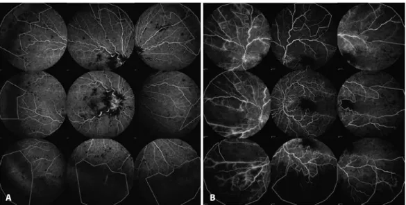
OCT; Heidelberg Engineering GmbH, Heidelberg, Germany). FA was performed at the irst appointment in all patients and repeated at Months 3, 6, 12, and 24-26 in patients with retinal ischemia (Hei delberg Retina Angiograph 2; Heidelberg Engineering). We measured the nonperfused area in every angiographic image of the nine to 13 an -giograms necessary to observe most of the retina with a 55º lens (therefore, the term multi-ield FA is appropriate) and calculated the total non-perfused area (Figure 1). Two FA specialists separately outlined the non-perfused areas. Areas of non-perfusion were
mea-sured in mm2 in unprocessed angiograms using pre-installed Heyex
software v.1.7.0.0 (Heidelberg Engineering). We did not perform any manual adjustments to calculate ischemic indices in contrast to other researchers(17).
Data analyseswere performed using the Statistica 7 software
suite (StatSoft, Tulsa, OK, USA) and included the estimation of х ± δ values and their dispersion and covariation coeicients at diferent stages of the study. Diferences were considered signiicant at p<0.05.
After examination, patients were divided into three groups based on CRVO type: non-ischemic (those who received ranibizumab treat-ment and were not included in further comparisons and analyses);
Figure 1. (A) and (B) Areas of capillary non-perfusion outlined in nine standard angiographic images from two diferent patients. To avoid overstating the total area involved because of the overlap of neighboring images, anatomic structures (such as blood vessels) were used as landmarks.
and two comparable (Table 1) ischemic groups that received dife-rent treatments. Patients with 10 disc areas or more of retinal capillary non-perfusion were considered to have ischemic CRVO.
Non-ischemic patients with CRVO received intravitreal injections of 0.5 mg ranibizumab scheduled as recommended by the
manufac-turer (Lucentis®; Novartis Pharma AG, Basel, Switzerland). This group
was only examined to determine the prevalence of ischemia among patients with CRVO according to wide-ield angiography results, and was not included in further analyses.
Group 1 received combined treatment: intravitreal injections of 0.5 mg ranibizumab every month for a 3-month period and selective peripheral SLP of non-perfused areas of the retina. SLP sessions were performed 30 min before the irst intravitreal injection in all patients and repeated at months 1 and 2 as needed. The following settings were applied: laser wavelength, 514 nm; spot diameter, 400 µm; exposition, 0.15 s, and an energy level suicient to produce a white coagulate. In some patients, we were unable to perform selective pe-ripheral SLP of all the non-perfused areas in one session (for example, if the non-perfused area was too large and the patient experienced too much pain, or if we had to wait until hemorrhages had resolved in the retina). Laser treatment was then repeated at months 1 and 2, according to the opinion of the laser and FA specialists. It should be noted that this approach can leave a considerably large non-perfused area untreated for a relatively long time, which can interfere with VA and CRT. However, the main reason for “deferred” SLP was the presen-ce of retinal hemorrhages, and it was impossible to apply complete SLP before they had resolved (a process that takes up to 3 months). If all non-perfused areas were coagulated in one SLP procedure, SLP was not repeated unless the non-perfused areas were found to have increased at follow-up FA.
Following the administration of the irst three monthly intra vi-treal injections, which was after the patients had completed laser
treatment, ranibizumab therapy was continued pro re nata (PRN) for
another 24 months. An additional injection was considered neces-sary in patients who demonstrated retinal thickening of ≥150 µm and related signiicant vision loss. We deined a “signiicant” VA decrease as a loss of at least two lines on the Snellen chart in patients whose maximum achieved best corrected visual acuity (BCVA) was ≥0.4, and a loss to 0.05 in those whose maximum BCVA was 0.1 on the Snellen chart. In patients with a VA >0.1 and <0.4, reinjection was performed if there was a loss of one line on the Snellen chart.
Patients in Group 2 received monthly ranibizumab monotherapy for the first 3 months and switched to a PRN regimen for the following 24 months.
These treatment regimens and reinjection criteria were selected for economic reasons. In Russia, patients with RVO receive no
reim-bursement for Lucentis® treatment (and use of intravitreal Avastin®
[F. Hofmann-La Roche Ltd, Basel, Switzerland] is prohibited). Therefore, many prefer to wait as long as possible for treatment based on their individual threshold. Thus, patients requested therapy when their vision was very poor. The rationale for the three monthly doses at the start of treatment was that we continued ixed monthly dosing until the improvement in VA was stable on two consecutive visits (in many patients, this occurred at months 2 and 3 or 3 and 4) and started PRN treatment after that. The mean number of injections administered with a ixed regimen was three.
The eye function and edema of patients in this study were worse than those of patients in the CRUISE study, in which no patients with
ischemic RVO were included(18). The conventional criteria of a VA <0.5
and CRT ≥250 μm were therefore not applicable. According to these criteria, we should have used ixed dosing for a longer period, which our patients could not aford.
RESULTS
Multi-field angiography revealed capillary non-perfusion in 175/250 patients with CRVO (70%). Thus, Group 1 comprised 88 pa tients and Group 2 comprised 87 patients. The mean total area
of retinal ischemia was 435.12 mm2 (standard deviation [SD], ±
225.13 mm2), i.e., 167.15 optic disc areas (SD, ± 45.16). We diagnosed
peripheral ischemia in 125 patients, corresponding to 50% of the total number of patients with CRVO and 71.4% of patients with any type of ischemia. The mean total area of peripheral retinal ischemia was
370 mm2 (SD, ± 113.5 mm2), i.e., 142.21 (SD, ± 85.12) optic disc areas.
In Group 1, the mean VA at baseline was 0.25 ± 0.15, and the mean central macular thickness was 524.02 ± 243.85 µm. In the PRN treatment period (24 months), patients in this Group recei-ved on average 3.5 ± 1.6 intravitreal ranibizumab injections. This means that a large proportion of patients in this Group received only the loading dose in the 2-year follow-up period. The average number of laser burns was 1320 ± 245. After the third injection, VA usually reached its maximum (0.52 ± 0.15), and the central macular thickness decreased to 270.7 ± 151.34 µm. In a month, these para-meters had decreased in all patients, reaching 0.41 ± 0.25 µm and 270.5 ± 123.8 µm, respectively, by the end of treatment. There were, however, 12 patients in whom the area of impaired capillary perfusion
increa sed by 25-60 mm2 over the total observation period,
necessi-tating additional laser treatment. There was no correlation between the progression of ischemia and the number of injections.
The average number of injections administered after peripheral SLP sessions was 2.9 ± 1.4. There was no correlation between final VA or final CRT and the total number of injections. As expected, a po sitive statistical relationship was found between the area of ischemia and the number of laser burns, as well as between the area of ischemia and CRT (both initial and final). Importantly, by the end of the study, this Group had reduced in number by three patients. One of them received an insuicient number of peripheral SLP sessions, and presented at Month 15 with neovascular glaucoma and eye pain, necessitating diode laser transscleral cyclophotocoagulation and panretinal photocoagulation (PRP); data from the other two patients were unavailable.
Patients in Group 2 received an average of 10.6 ± 2.5 intravitreal injections during the 24-month period. In this group, the mean VA at baseline was 0.22 ± 0.2 (Snellen chart), and the mean central macular thickness was 626.13 ± 298.06 µm. The highest VA and lowest retinal thickness, 0.45 ± 0.21 and 290.7 ± 214.5 µm, respectively, were regis-tered after the third injection.
Table 1. Baseline characteristics of the patients and data on the study outcome
Characteristic
Ranibizumab and SLP group (#1)
n=88
Ranibizumab alone group
(#2) n=87 p value
Baseline
Mean age, years 062.50 ± 012.90 061.70 ± 011.40 <0.38 Female sex, n (%) 52 (59.1) 53 (60.1) <0.41 Disease duration, months 1.40 ± 1.10 01.60 ± 1.30 <0.34 Retinal ischemia area, mm2 410.20 ± 157.25 425.50 ± 210.14 <0.28 BCVA, Snellen chart 0.25 ± 0.15 00.27 ± 0.09 <0.26 CRT, μm 524.02 ± 243.85 535.04 ± 210.12 <0.32 Outcome
BCVA by month 28, Snellen chart 0.41 ± 0.25 00.40 ± 0.15 <0.29 CRT by Month 19*, μm 270.52 ± 123.81 287.04 ± 130.44 <0.19 Mean number of ranibizumab
injections
3.50 ± 1.60 10.60 ± 2.50 <0.01
Optical coherence tomography was performed in only a few patients between 19 and 28 months; therefore, we did not include these data in the analysis.
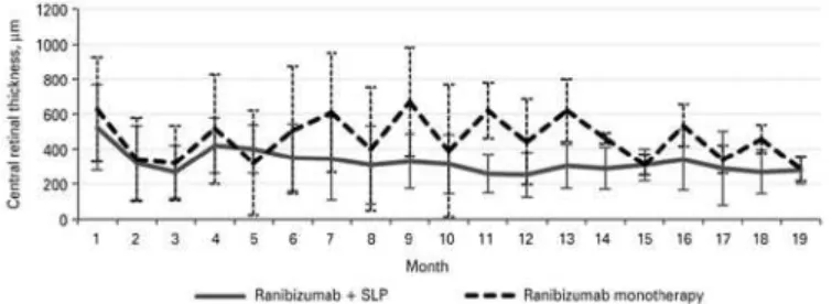
Figure 3. Mean central retinal thickness changes with 95% conidence intervals in groups receiving diferent therapies.
Figure 2. Mean visual acuity changes with 95% conidence intervals in groups receiving diferent therapies.
By the end of the study, 15 patients demonstrated a 45.5 ± 15.6 mm2
increase in the area of ischemia. Iris neovascularization occurred in ive patients an average of 2.5 months after the completion of ra-nibizumab therapy, necessitating PRP (in two patients, diode laser transscleral cyclophotocoagulation was also performed). Two other patients developed posterior segment neovascularization and underwent additional treatment with PRP. Anterior segment neovas-cularization occurred in only one patient in Group 1. All other ins tances of neovascularization occurred in patients treated with ra -nibizumab alone (Group 2).
The dynamics of changes in VA difered between groups 1 and 2. In Group 1, VA was lowest at the 5-month follow-up and then remained generally stable, showing only minor luctuations. In Group 2, VA showed no stabilization, and its luctuations were large (Figure 2). CRT changes correlated well with VA changes (Figure 3). In both ischemic CRVO groups, the initial and inal VA, initial and inal CRT, and number of anti-VEGF injections were positively correlated with the area of capillary non-perfusion.
DISCUSSION
The use of multi-ield angiography for the primary assessment of patients with CRVO enables the detection of retinal ischemia in almost 70% of patients. In most patients, ischemic changes are loca-ted at the periphery of the retina. Their total area is usually large,
occupying on average 370 mm2 (SD ± 113.5 mm2), i.e., 142.21 (SD,
± 85.12) optic disc areas. Patients who fall into this category are at higher risk of proliferative complications; therefore, speciic ischemic RVO management algorithms should be followed.
Notably, foveal function is usually preserved in these patients, ensuring a response to anti-VEGF agents. Ranibizumab monothe-rapy, however, provides only temporary efects. After injections stop, patients’ vision deteriorates again, and ME recurs. For a more stable result, further injections are required. However, further injections confer a higher risk of complications and increased medical costs, making this treatment unfeasible for our patients. In our study, even prolonged (24-month) anti-VEGF therapy failed to reduce the risk of neovascularization. However, we found no correlation between the
number of ranibizumab injections and progression of ischemia in any of our patients. When combined with selective SLP of non-perfused areas of the retina, invasive intravitreal therapy can be minimized. The advantages of this approach include reduced medical costs, a shorter treatment period, and faster stabilization.
In a small randomized study, laser photocoagulation of peri-pheral areas of non-perfusion did not decrease injection frequency
or improve VA in eyes with CRVO treated with ranibizumab(19). The
authors proposed that the complete loss of vascularity of the pe-ripheral retina caused neuroretinal infarction and consequently no long-term VEGF production. Such patients may require less aggressi ve anti-VEGF treatment. However, there are many publications on the positive correlation between the area of non-perfusion and intrao-cular VEGF levels, and in our opinion it is essential to treat all patients with an ischemic retina.
Regarding patients in whom the non-perfused area showed progression over time, we consider the persistently reduced perfu-sion pressure between the arterial and venous vascular retinal network to be one of the main causative factors. Impaired Endo-thelin-1-induced autoregulation and neurovascular coupling may contribute to this process, with retinal venous pressure changes
accompanying CRVO(19,20). Further studies involving more patients
and the measurement of perfusion pressure are needed to provide an answer to this question.
The main strengths of this pilot study are the number of pa-tients observed and long follow-up period. The limitations of this study include the absence of randomization, probable ranibizumab undertreatment for local economic reasons, relative inaccuracy of BCVA measurements using the Snellen chart, and the possible “defer-red laser” efect discussed in the Methods section. More experience in patients with peripheral ischemic CRVO over a longer follow-up period is needed.
REFERENCES
1. Natural history and clinical management of central retinal vein occlusion. The Central Vein Occlusion Study Group. Arch Ophthalmol. 1997;115(4):486-91. Erratum in: Arch Ophthalmol. 1997;115(10):1275.
2. Tultseva SN. Inlammation role in the post-thrombotic macular edema pathogenesis. Modern medical treatment choices. Ophthalmologicheskie Vedomosti. 2012;5(4):45-51. Russian.
3. Tultseva SN. Hyperhomocysteinemia role in the ischemic retinal vein thrombosis pa-thogenesis. Ophthalmologicheskie Vedomosti. 2008;1(3):31-9. Russian.
4. Tultseva SN, Astakhov YS. [Retinal vein occlusion (etiology, pathogenesis, clinical fea-tures, diagnosis, treatment)]. Saint Petersburg: N-L Publishing; 2010. Russian. 5. Tultseva SN. [The role of hereditary and acquired thrombophilia factors in the
patho-genesis of retinal vein occlusion] [dissertation]. Saint Petersburg: Pavlov First Saint Pe tersburg State Medical University; 2014. Russian.
6. Noma H, Funatsu H, Mimura T, Eguchi S. Vitreous inlammatory factors and serous reti-nal detachment in central retireti-nal vein occlusion: a case control series. J Inlamm (Lond). 2011;8:38.
7. Noma H, Mimura T, Yasuda K, Shimura M. Role of soluble vascular endothelial growth factor receptors-1 and -2, their ligands, and other factors in branch retinal vein occlu-sion with macular edema. Invest Ophthalmol Vis Sci. 2014;55(6):3878-85.
8. Noma H, Funtatsu H, Mimura T, Eguchi S, Shimada K. Inlammatory factors in major and macular branch retinal vein occlusion. Ophthalmologica. 2012;227(3):146-52. 9. Noma H, Funatsu H, Mimura T, Tatsugawa M, Shimada K, Eguchi S. Vitreous
inlamma-tory factors and serous macular detachment in branch retinal vein occlusion. Retina. 2012;32(1):86-91.
10. Noma H, Mimura T, Yasuda K, Shimura M. Role of soluble vascular endothelial growth factor receptor signaling and other factors or cytokines in central retinal vein occlusion with macular edema. Invest Ophthalmol Vis Sci. 2015;56(2):1122-8.
11. Hayreh SS, Klugman MR, Beri M, Kimura AE, Podhajsky P. Diferentiation of ischemic from non-ischemic central retinal vein occlusion during the early acute phase. Graefes Arch Clin Exp Ophthalmol. 1990;228(3):201-17.
12. Spaide RF. Peripheral areas of nonperfusion in treated central retinal vein occlusion as imaged by wide-ield luorescein angiography. Retina. 2011;31(5):829-37. 13. Croft DE, van Hemert J, Wykof CC, Clifton D, Verhoek M, Fleming A, et al. Precise
14. Novartis Pharmaceuticals. Lucentis 10mg/ml solution for injection [Internet]. Horsham, United Kingdom: Novartis Europharm; 2015. [cited 2016 Jun 21]. Available at: http:// www.medicines.org.uk/emc/medicine/19409.
15. Brown DM, Wykof CC, Wong TP, Mariani AF, Croft DE, Schuetzle KL; RAVE Study Group. Ranibizumab in pre-proliferative (ischemic) central retinal vein occlusion (CRVO): the rubeosis anti-VEGF (RAVE) trial. Retina. 2014;34(9):1728-35. Comment in: Retina. 2015; 35(10):e59-61.
16. Spaide RF. Prospective study of peripheral panretinal photocoagulation of areas of nonperfusion in central retinal vein occlusion. Retina. 2014;33(1):56-62.
17. Tsui I, Kaines A, Havunjian MA, Hubschman S, Heilweil G, Prasad PS, et al. Ischemic index and neovascularization in central retinal vein occlusion. Retina. 2011;31(1):105-10. 18. Brown DM, Campochiaro PA, Singh RP, Li Z, Gray S, Saroj N, Rundle AC, Rubio RG,
Murahashi WY: CRUISE Investigagors. Ranibizumab for macular edema following cen-tral retinal vein occlusion: six-month primary end point results of a phase III study. Oph thalmology. 2010;117(6):1124-33.
19. Flammer J, Konieczka K, Flammer AJ. The primary vascular dysregulation syndrome: implications for eye diseases. EPMA J. 2013;4(1):14.
20. Flammer J, Konieczka K. Retinal venous pressure: the role of endothelin. EPMA J. 2015; 6:21.