C A SE R EPORT
258 J Vasc Bras. 2015 July-Sept.; 14(3):258-261 http://dx.doi.org/10.1590/1677-5449.0076
Arteriovenous fistula after ultrasound guided foam
sclerotherapy: case report
Fístula arteriovenosa após escleroterapia com espuma guiada por ultrassom:
relato de caso
Felipe Coelho Neto1,2
*, Iruena Moraes Kessler
1, Gilson Roberto de Araújo2
Abstract
Ultrasound-guided foam sclerotherapy has become widespread in the treatment of chronic venous insuiciency secondary to varicose veins. It is a low-cost, outpatient procedure that does not require medical leave. It has demonstrated good clinical results, especially in the more advanced stages of the disease. When employed correctly, it results in low rates of major complications. However, the technique exhibits high recanalization rates over mid and long-term follow up. Additionally, the recanalization mechanism has not yet been explained. his article describes an arteriovenous istula in a recanalized segment of great saphenous vein previously treated with ultrasound-guided foam sclerotherapy that was detected during post-procedure follow-up with ultrasound.
Keywords: varicose veins; arteriovenous istula; sclerotherapy; sclerosing solutions; surgery; ultrasonography interventional.
Resumo
A escleroterapia com espuma guiada por ultrassom tem ganhado espaço no tratamento da insuiciência venosa crônica secundária a varizes dos membros inferiores. Trata-se de procedimento ambulatorial, de baixo custo e sem necessidade de afastamento das atividades habituais. Apresenta bons resultados clínicos, especialmente nos estágios mais avançados da doença. Quando bem aplicada, apresenta baixas taxas de complicações maiores. Porém, a técnica apresenta altas taxas de recanalização no médio e longo prazo, e seu mecanismo ainda não é totalmente compreendido. O presente artigo descreve um caso de fístula arteriovenosa em segmento de recanalização de veia safena magna, após escleroterapia com espuma guiada por ultrassom, e identiicada por exame ultrassonográico de vigilância pós-procedimento.
Palavras-chave: varizes; fístula arteriovenosa; escleroterapia; soluções esclerosantes; cirurgia; ultrassonograia de intervenção.
1Universidade de Brasília – UnB, School of Medicine, Brasília, DF, Brazil. 2Hospital Regional da Asa Norte, Brasília, DF, Brazil.
Financial support: None.
Conlicts of interest: No conlicts of interest declared concerning the publication of this article. Submitted: October 15, 2014. Accepted: March 23, 2015.
259 J Vasc Bras. 2015 July-Sept.; 14(3):258-261
Felipe Coelho Neto, Iruena Moraes Kessler et al.
INTRODUCTION
Chronic venous insuficiency (CVI) is a common disease in clinical practice and its complications, particularly venous stasis ulcers, cause signiicant morbidity.1
One of the treatment options for CVI is ultrasound guided foam sclerotherapy2 (USFS). It offers satisfactory
results, is easy to perform and does not require hospital admission or an operating theater and is generally performed in outpatients settings.3 After sclerotherapy,
the veins are transformed into ibrous cords in a process known as sclerosis.3 However, medium and
long-term recanalization rates are still high.4
This article describes a case in which an arteriovenous fistula was identified with ultrasonography in a recanalized segment of the great saphenous vein that had been treated using ultrasound guided foam sclerotherapy.
CASE DESCRIPTION
An 80-year-old female, white-skinned patient with varicose veins in the lower limbs and a history of arterial hypertension and a mood disorder, complained of pain, feeling of heaviness, tiredness, edema during the afternoon and “darkening” of the distal third of the left leg. Physical examination revealed incipient ochrodermatitis of the ankle and distal third of the left leg and large caliber varicose veins. Arterial clinical examination of the lower limbs revealed no abnormalities. Color Doppler venous ultrasonography ruled out relux or thrombi in the deep vein system and identiied relux at the left saphenofemoral junction and in the left great saphenous vein, transferring the relux into medium and large caliber varicose veins along the medial and lateral surfaces of the leg.
The decision was taken to treat the great saphenous vein trunk relux and the varicose veins using USFS. Polidocanol at 3% was mixed with room air at a proportion of 1:4 to create foam, as described by Tessari et al.5
Three ultrasound guided punctures were performed with no. 23 needles, two into the great saphenous vein in the thigh and one into a varicose tributary, and a total volume of 10 ml of foam was injected. After the injection, the limb was maintained as immobile as possible, without elevation and without compression of the saphenofemoral junction, as recommended by the European Guidelines for Sclerotherapy in Chronic Venous Disorders.6 After 5 minutes in this position,
20-30 mmHg elastic stockings were put on and worn continuously for 1 week, removing them only when performing personal hygiene. The patient was encouraged to walk and to engage in daily activities
without restrictions. She was also instructed to wear the stockings during the daytime only from 7 days after the procedure onwards.
No intercurrent conditions occurred during the procedure and the great saphenous vein, and a proportion of its tributaries, were successfully obliterated after a single session. Control ultrasonography at 7, 30 and 90 days did not detect any low in the great saphenous vein, from the supericial epigastric vein to the point of drainage into the varicose tributaries in the leg. Clinically, there was remission of symptoms and partial resolution of the ochrodermatitis.
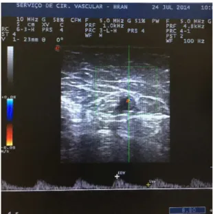
Control ultrasonography examination at 6 months showed recanalization of a segment of the great saphenous vein starting at the mid third of the thigh. There was a continuous, ascending and pulsating low that was not inluenced by respiratory rhythm, the Valsalva maneuver or distal compression. Spectral analysis showed that there was low-resistance arterial low in the venous lumen on a longitudinal scan, compatible with an arteriovenous istula (AVF), characterized by a high diastolic low rate and low resistance index (Figure 1). In segments below the AVF, the great saphenous vein was incompressible and had no luminal low. It was possible to identify the point at which the pulsating low in the vein lumen originated on both longitudinal and transversal scans (Figures 2 and 3). In view of the fact that the patient remained asymptomatic, it was decided to adopt a policy of watchful waiting. Clinical and ultrasonography indings remained unaltered 12 months after treatment and so six-monthly follow-up and conservative treatment were prescribed.
Figure 1. Spectral analysis of luminal low characterized by high
260 J Vasc Bras. 2015 July-Sept.; 14(3):258-261 AVF after ultrasound guided foam sclerotherapy
DISCUSSION
The “minimally invasive” techniques such as endovenous laser, radio frequency and foam sclerotherapy have gained recognition over the last 15 years to the extent that they now play a primary role in treatment of CVI secondary to varicose veins of the lower limbs. These are outpatients procedures performed with tumescent local anesthesia and do not require hospital admission or medical leave and many studies have demonstrated their safety and eficacy for elimination of venous relux.7-10
Ultrasound-guided foam sclerotherapy produces good clinical results, as has been shown in studies conducted in Brazil by Figueiredo et al.,11 Silva et al.2
and Coelho Neto et al.13 However, recanalization rates
varying from 10 to 28% have been observed over varying follow-up periods14 and rates higher than
50% have been observed in studies with the longest follow-up periods.4 When used appropriately, this is a
technique that has low rates of complications15,16 and AVF is not a normal complication of the procedure. Cavezzi and Parsi17 have provided a general panorama of the primary complications of USFS. Severe compilations such as anaphylactic shock are described as rarities and deep venous thrombosis was reported in 1-3% of cases. The same authors state that there are 13 cases of cerebral vascular accident associated with USFS described in publications since 1994. Reported incidences of minor complications, such as thrombophlebitis, edema and neurological damage, are 4.4%, 0.5% and 0.2%, respectively. Esthetic complications such as ‘matting” (15-24%) and pigmentation (10-30%) are more frequent.
Jia et al.18 conducted a large-scale systematic
review covering 9,000 patients and did not list AVF among the complications of USFS.
However, when other minimally invasive techniques are also considered, such as endovenous laser and radiofrequency ablation, for example, cases of AVF are described in the literature as possible physiological mechanism of recanalization.19 There are also reports
of recanalization of thrombi with no apparent cause in the supericial or deep vein systems, with inlammation and neovascularization, causing AVFs.20,21
When AVF is seen in venous thrombosis with no apparent cause in veins previously subjected to ablative treatments, the suspicion is that they were not effectively treated and that thrombosis was induced in the segments treated.20 Future studies employing
animal models and histopathology could aid in understanding the role of istulae.
There are no descriptions in the English-language medical literature of an AVF found by ultrasonography after treatment of saphenous vein trunk relux using USFS. This case description appears to add weight to data reported in publications in the literature on recanalization, in which inflammatory and neovascularization mechanisms act in areas of induced thrombi, rather than in effectively treated veins.18
New techniques have brought with them unexpected post-procedural complications that were not described in relation to the classic vein stripping technique. This case report highlights the need for constant ultrasonographic monitoring of minimally invasive treatments, both for early detection of recanalization and in order to document indings that will contribute to better understanding of the long-term behavior of these treatment methods.
Figure 3. Spectral analysis of transverse scan of saphenous vein.
Figure 2. Point of origin of pulsating low within the saphenous
261 J Vasc Bras. 2015 July-Sept.; 14(3):258-261
Felipe Coelho Neto, Iruena Moraes Kessler et al.
REFERENCES
1. Silva MC. Chronic venous insufficiency of the lower limbs and its socio-economic significance. Int Angiol. 1991;10(3):152-7. PMid:1765717.
2. Wright D, Gobin JP, Bradbury AW, et al. Varisolve® polidocanol mircofoam compared with surgery or sclerotherapy in the management of varicose veins in the presence of trunk vein incompetence: European randomized controlled trial. Phlebology. 2006;21(4):180-90. http://dx.doi.org/10.1258/026835506779115807. 3. Guidelines/Outcomes Committee, Task Force. Guidelines of
care for sclerotherapy treatment of varicose and telangiectatic leg veins. J Am Acad Dermatol. 1996;34(3):523-8. http://dx.doi. org/10.1016/S0190-9622(96)90467-3. PMid:8609276.
4. Belcaro G, Cesarone MR, Di Renzo A, et al. Foam-sclerotherapy, surgery, sclerotherapy, and combined treatment for varicose veins: a 10-year, prospective, randomized, controlled, trial (VEDICO trial). Angiology. 2003;54(3):307-15. http://dx.doi. org/10.1177/000331970305400306. PMid:12785023.
5. Tessari L, Cavezzi A, Frullini A. Preliminary experience with a new sclerosing foam in the treatment of varicose veins. Dermatol Surg. 2001;27(1):58-60. PMid:11231246.
6. Rabe E, Breu FX, Cavezzi A, et al. European guidelines for sclerotherapy in chronic venous disorders. Phlebology. 2014;29(6):338-54. http:// dx.doi.org/10.1177/0268355513483280. PMid:23559590.
7. Darwood RJ, Gough MJ. Endovenous laser treatment for
uncomplicated varicose veins. Phlebology. 2009;24(Supl 1):50-61.
http://dx.doi.org/10.1258/phleb.2009.09s006. PMid:19307441.
8. Gohel MS, Davies AH. Radiofrequency ablation for uncomplicated varicose veins. Phlebology. 2009;24(Supl 1):42-9. http://dx.doi. org/10.1258/phleb.2009.09s005. PMid:19307440.
9. Smith PC. Foam and liquid sclerotherapy for varicose veins.
Phlebology. 2009;24(Supl 1):62-72. http://dx.doi.org/10.1258/
phleb.2009.09s007. PMid:19307442.
10. Proebstle TM, Vago B, Alm J, Göckeritz O, Lebard C, Pichot O. Treatment of the incompetent great saphenous vein by endovenous radiofrequency powered segmental thermal ablation: first clinical experience. J Vasc Surg. 2008;47(1):151-6. http://dx.doi.org/10.1016/j. jvs.2007.08.056. PMid:18178468.
11. Figueiredo M, Araújo SP, Penha-Silva N. Microfoam ultrasound-guided sclerotherapy in primary trunk varicose veins. J Vasc Bras. 2006;5(3):177-83. http://dx.doi.org/10.1590/S1677-54492006000300005.
12. Silva MAM, Burihan MC, Barros OC, Nasser F, Ingrund JC, Neser A. Resultados do tratamento da insuficiência venosa crônica grave
com espuma de polidocanol guiada por ultrassom. J Vasc Bras.
2012;11(3):206-11. http://dx.doi.org/10.1590/S1677-54492012000300007.
13. Coelho Neto F, Araújo GR, Kessler IM, Amorim RF, Falcão DP. Treatment of severe chronic venous insufficiency with ultrasound-guided foam sclerotherapy: a two-year series in a single center in Brazil. Phlebology. 2015;30(2):113-8. http://dx.doi. org/10.1177/0268355513517225. PMid: 24335090.
14. National Institute for Clinical Excellence [site na Internet]. Interventional procedures overview of ultrasound guided foam sclerotherapy for varicose veins. London: NICE; 2004 [citado 2006 set 26]. http://guidance.nice.org.uk/download.aspx?o=ip244overview.
15. Guex JJ, Allaert FA, Gillet JL, Chleir F. Immediate and midterm complications of sclerotherapy: report of a prospective multicenter registry of 12,173 sclerotherapy sessions. Dermatol Surg. 2005;31(2):123-8, discussion 128. http://dx.doi.org/10.1097/00042728-200502000-00001. PMid:15762201.
16. Weiss RA, Weiss MA. Incidence of side effects in the treatment of telangiectasias by compression sclerotherapy: hypertonic saline vs. polidocanol. J Dermatol Surg Oncol. 1990;16(9):800-4. http:// dx.doi.org/10.1111/j.1524-4725.1990.tb01563.x. PMid:2398199.
17. Cavezzi A, Parsi K. Complications of foam sclerotherapy. Phlebology. 2012;27(Supl 1):46-51. http://dx.doi.org/10.1258/phleb.2012.012S09. PMid:22312067.
18. Jia X, Mowatt G, Burr JM, Cassar K, Cook J, Fraser C. Systematic review of foam sclerotherapy for varicose veins. Br J Surg. 2007;94(8):925-36. http://dx.doi.org/10.1002/bjs.5891. PMid:17636511.
19. Labropoulos N, Bhatti A, Leon L, Borge M, Rodriguez H, Kalman P. Neovascularization after great saphenous vein ablation. Eur J Vasc Endovasc Surg. 2006;31(2):219-22. http://dx.doi.org/10.1016/j. ejvs.2005.06.030. PMid:16099695.
20. Labropoulos N, Bhatti AF, Amaral S, et al. Neovascularization in
acute venous thrombosis. J Vasc Surg. 2005;42(3):515-8. http://
dx.doi.org/10.1016/j.jvs.2005.05.036. PMid:16171599.
21. Barros FS, Pontes SM, Silva WP, et al. Arteriovenous fistula in deep venous thrombosis identified by color-flow Doppler ultrasonography. J Vasc Bras. 2006;5(3):224-8.
*
Correspondence
Felipe Coelho Neto Centro Médico Julio Adnet SEPS 709/909, salas 419-422, bloco A CEP 70100-360 - Brasília (DF), Brazil Tel.: +55 (61) 33281940 E-mail: [email protected]
Author information
FCN – Vascular surgeon at the Vascular Surgery Unit, Hospital Regional da Asa Norte; MSc in Medical Sciences from Universidade de Brasília (UnB). IMK – MSc and a PhD in Medical Sciences from Universidade de Brasília (UnB). GRA – Vascular surgeon at the Vascular Surgery Unit, Hospital Regional da Asa Norte.
Author contributions
Conception and design: FCN Analysis and interpretation: FCN, GRA Data collection: FCN Writing the article: FCN, IMK Critical revision of the article: FCN, IMK Final approval of the article*: FCN, GRA, IMK Statistical analysis: N/A Overall responsibility: FCN