Universidade do Minho
Escola de Ciências
Ana Isabel Oliveira da Silva Dias
janeiro de 2018
Effect of simulated microgravity on the cell
cycle in cultured cells of Arabidopsis thaliana
Ana Isabel Oliveira da Silva Dias
Ef
fect of simulated microgra
vity on t
he cell cy
cle in cultured cells of
Arabidopsis thaliana
Universidade do Minho
Escola de Ciências
Ana Isabel Oliveira da Silva Dias
janeiro de 2018
Effect of simulated microgravity on the cell
cycle in cultured cells of Arabidopsis thaliana
Trabalho realizado sob orientação da
Profª Doutora Ana Cunha
e do
Doutor Javier Medina
Dissertação de Mestrado
Mestrado em Biologia Molecular, Biotecnologia
e Bioempreendedorismo em Plantas
iii
ACKNOWLEDGEMENTS
It gives me great pleasure in expressing my gratitude to all those people who have supported me and their contributions in making this thesis possible. First, I want to acknowledge to my supervisors. Dra. Ana Cunha for her constant guidance, support and motivation since the firsts steps of this journey. I also express my profound gratitude to my supervisor and head of our laboratory Dr. Javier Medina for giving me this precious opportunity and untiring help during my stay at CIB.
Furthermore, I would like to acknowledge my colleagues in the laboratory, Gosia and Alicia. I thank them for all learned countless invaluable lessons, all their patience, availability, understanding and friendship. We crossed together for so many moments of difficulties and also success. I would also like to thank my colleague Arantxa for her friendship and help. A piece of my hearth will be always in Madrid.
Finally, my academic studies were only possible with the unwavering support of my family. I am most thankful.
v
ABSTRACT
Effect of simulated microgravity on the cell cycle in cultured cells of Arabidopsis thaliana
Life as we know it on planet Earth always evolved in the presence of the constant gravitational force, both in magnitude and direction. This physical force is responsible for giving the weight to all masses. Altering weight changes many processes in a living being, such fluid regulation, as well as ecological processes on Earth, such rain fall. Thus, it is reasonable to say that gravity shapes life. With this study we pretended to investigate how the gravity modulates cell cycle using the in vitro cellular system of Arabidopsis thaliana cell suspension line MM2d, characterized to be undifferentiated and highly proliferative. In this system cell growth is strictly correlated with the rate of ribosome biogenesis and protein synthesis by the accurate regulation of cell cycle progression. Such coordination is essential for an optimal production of biomass as well as for the viability of daughter cells after division. We investigated the effects of simulated microgravity on cellular functions by growing MM2d cell line suspensions in a 2D clinostat, a device generating simulated microgravity, for a long-term exposure of 24 hours. In order to study this cellular system, the immobilization of cells was optimized by the use of alginate gelling agent, a crucial step for exposure of cell suspension to 2D clinorotation. Under these conditions, the cell proliferation rate showed significant alterations, accompanied by reduction of cell growth. Analysis of cell cycle by flow cytometry showed increase in the proportion of cells in S phase and a decrease in G1 phase, indicating an increase in progression rate of the cell cycle. With respect to cell growth, the rate of ribosome biogenesis was reduced under simulated microgravity, as shown by variations in the abundance of nucleolar proteins nucleolin and fibrillarin, using immunofluorescence microscopy. These results are in agreement with previous observations in root meristems that have shown a decoupling of the meristematic competence, characterized by a strict coordination between cell growth and cell proliferation. Furthermore, it was shown that undifferentiated plant cells also have the ability to respond to changes in the gravity, independently from their integration into plant tissues and organs.
Key words: Simulated microgravity, Arabidopsis thaliana, cell growth, cell proliferation, cultured cells, nucleolin, fibrillarin.
vii
RESUMO
Efeito da microgravidade simulada no ciclo celular de culturas celulares in vitro de Arabidopsis thaliana
A vida como a conhecemos no planeta Terra sempre evoluiu na presença da constante força gravitacional, tanto em magnitude como direção. Esta força física é responsável por dar peso a todas as massas. Mudança no peso altera vários processos nos seres vivos, como a regulação de fluidos, assim como processos ecológicos na Terra, como a queda de chuva. Então, é sensato dizer que a gravidade molda a vida. Com este estudo pretendemos investigar como a gravidade modela o ciclo celular com o uso de sistema celular in vitro de suspensão celular da linha MM2d
de Arabidopsis thaliana, caracterizada como indiferenciada e altamente proliferativa. Neste
sistema o crescimento celular é rigorosamente relacionado com a proporção de biogéneses de ribossomas e com a síntese de proteínas através da regulação precisa na progressão do ciclo celular. Esta coordenação é essencial para uma ideal produção de biomassa para a viabilidade das células filhas após divisão. Os efeitos da microgravidade simulada nas funções celulares pelo crescimento da suspensão celular MM2d foi investigada com o uso do clinostato 2D, um aparelho que gera microgravidade simulada, durante 24 horas. De modo a estudar este sistema celular, a imobilização das células foi otimizada com o uso do agente gelificante alginato, o que é um passo crucial para expor a suspensão celular em clinorrotação neste aparelho. Nestas condições, a proporção da proliferação celular mostrou alterações significativas, acompanhadas pela redução do crescimento celular. Análises do ciclo celular por citometria de fluxo mostraram aumento na proporção das células na fase S e diminuição na fase G1, indicando uma progressão do ciclo celular mais rápida. No que diz respeito ao crescimento celular, a biogénese de ribossomas foi reduzida em microgravidade simulada, como observado pela variação da abundância das proteínas nucleolares nucleolina e fibrilarina, através de microscopia de imunofluorescência. Estes resultados estão em concordância com observações prévias nos meristemas da raiz que mostraram desacoplamento da competência meristemática, caracterizada pela coordenação entre crescimento celular e proliferação celular. Ademais, mostrou-se que células vegetais indiferenciadas também têm aptidão para responder a alterações na gravidade, independentemente da sua integração em tecidos e órgãos vegetais. Palavras chave: Microgravidade simulada, Arabidopsis thaliana, crescimento celular, proliferação celular, culturas celulares, nucleolina, fibrilarina.
ix
ACRONYMS AND ABBREVIATIONS
2D Two dimensional
3D Three dimensional
BSA Bovine Serum Albumin
Ca2+ Calcium ions
CDK Cyclin-dependent kinase
CIB Centro de Investigationes Biológicas
CK2 Casein kinase 2
CYC Cyclins
DAPI 4′,6-diamidino-2-phenylindole
DFC Dense fibrilar componente
DNA Deoxyribonucleic acid
EDTA Ethylenediaminetetraacetic acid
ESA European Space Agency
FCs Fibrillar centres
g Gravity
G Gap
G1 Gap 1 (cell cycle)
G1/S Gap 1 / Synthesis phase
G2 Gap 2 (cell cycle)
G2/M Gap 2 / Mitosis
GBFs Ground based facilities
GC Granular componente
GDL Glucono-δ-lactone
h Hour
HM Homogenization medium
ISS International Space Station
KCl Potassium chloride
kg Kilogram
l Liter
M Molar
min Minute
MM2d A line of Arabidopsis cell suspension culture maintained in dark
mRNA Messenger Ribonucleic acid
MSS Murashige and Skoog (MS) medium supplemented with
Hormones
NAA α-naphtaleneacetic acid
NASA National Aeronautics and Space Administration
NSB Nuclear staining buffer
ºC Celsius degree
PBS Phosphate Buffered Saline
PFA Paraformaldehyde
pH Negative log of hydrogen ion concentration
x
p-value Statistical test provability value
RNA Ribonucleic acid
RPM Random Positioning Machine
rpm Rotation per minute
rRNA Ribosomal Ribonucleic acid
RT Room temperature
s Seconds
T Time of samples
w/v Weight/Volume
xi
LIST OF FIGURES
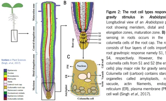
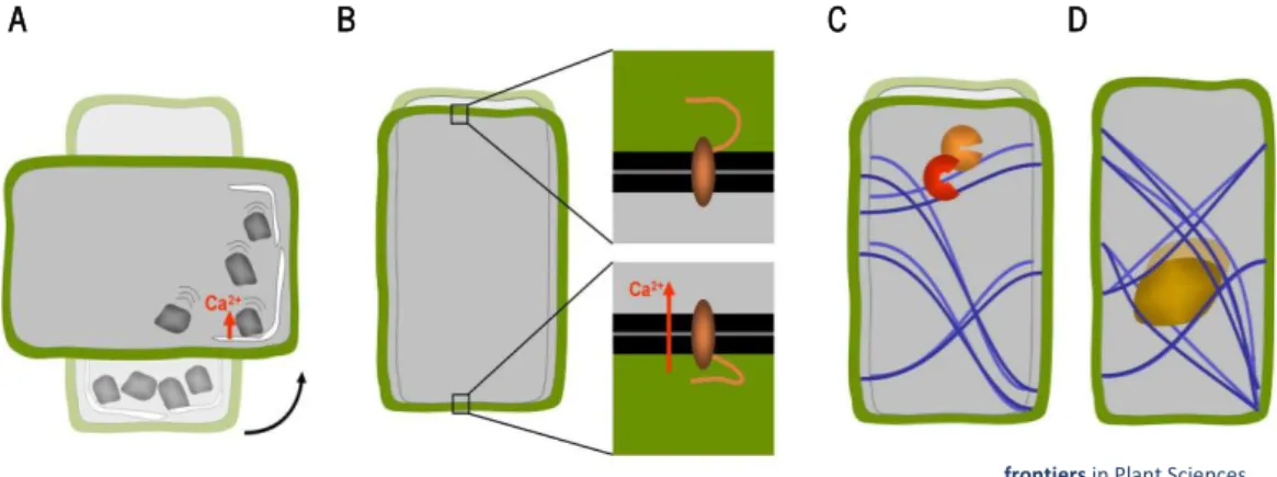
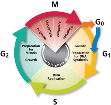
Figure 1: Plant-based Advanced Life support project (Melissa loop), European Space Agency. Melissa (Micro-Ecological Life Support System Alternative) has been conceived as a microorganisms and higher plants based ecosystem intended as a tool to gain understanding of the behaviour of artificial ecosystems, and for the development of the technology for a future regenerative life support system for long term manned space missions. Based on the principle of an "aquatic" ecosystem, Melissa is comprised of 5 compartments colonized respectively by Thermophilic Anaerobic Bacteria (I), Photoheterotrophic Bacteria (Rhodospirillum rubrum) (II), Nitrifying Bacteria (Nitrosomonas/Nitrobacter) (III), Photoautotrophic Bacteria (Arthrospira platensis) (IVA), Higher Plants (IVB), and the crew. Waste products and air pollutants are processed using the natural function of plants which in turn provide food as well as contribute to water purification and supply oxygen for air revitalization. Many other important benefits are being examined for related industrial projects. ... 4 Figure 2: The root cell types responding to gravity stimulus in Arabidopsis. A) Longitudinal view of an Arabidopsis primary root showing meristem, distal and central elongation zones, maturation zone. B) Gravity sensing in roots occurs in the central columella cells of the root cap. The root cap consists of four layers of cells important for root gravitropic response namely S1, S2, S3, S4, respectively. However, the central columella cells from S1 and S2 (the encircled cells) play major role for gravity sensing. C) Columella cell (cartoon) contains starch-filled organelles called amyloplasts, nucleus, vacuole, actin filaments, endoplasmic reticulum (ER), plasma membrane (PM), and cell wall. ... 6 Figure 3: Concepts of cellular gravisensing in plants. A) In statolith-based gravisensing the sedimentation or change of position of intracellular organelles with higher density triggers a signal most likely based on a change in trans-membrane ion fluxes. B) According to the gravitational model the weight of the protoplast causes the forces acting on the membrane-cell wall connections at the upper and lower sides of the cells to be different. C) The tensegrity model predicts that cellular distortion due to a change in g- forces affects the pre-stress in the cytoskeletal array of the cell which in turn changes biochemical activities. D) A variation of the tensegrity model is based on a change in cytoskeletal pre-stress being caused by the weight of heavy organelles that are tethered to the cytoskeletal filaments such as the nucleus. ... 7 Figure 4: Several ground based facilities (GBFs) used in plant biology research. A) 2D clinostat, located at Centro de Investigations Biológicas (CIB), Madrid, Spain. B) Pipette 2D clinostat C) 3D RPM D) Magnetic levitation facility. ... 10 Figure 5: Model of the actions and interactions leading to the maintenance of the meristematic competence (involving the coordination between cell growth and cell proliferation) specifically showing the players involved in the transduction of the gravitropic signal. Under ground 1 g conditions the gravitropic signal is a growth promoter, since it stimulates the growth of primary root according the direction of the gravity. The mechanic stimulus is transduced by the mediator auxin that, in the case of meristematic cells, modulates the expression of a variety of molecules that coordinate the regulation of cell growth and cell proliferation and then keep coupled the processes of protein synthesis (cell growth) and cell cycle progression (cell proliferation)... 12 Figure 6: Schematic of the cell (division) cycle. Cell cycle consists of four essential phases, G1 (Gap 1), S (Synthesis), G2 (Gap 2), and M (Mitosis for cell division). ... 13
xii
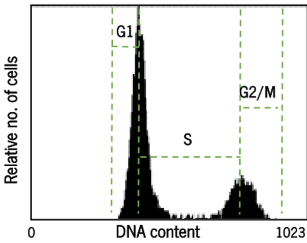
Figure 7: Cyclin-dependent kinase (CDK) activity is regulated at multiple levels. Monomeric CDK lacks activity until it forms a complex with cyclins (CYC) and is activated by phosphorylation by CDK-activating kinase (CAK). In addition, activity can be inhibited by binding of inhibitor proteins such as Kip-related proteins (KRP). Inhibitors may block the assembly of CDK/cyclin complexes or inhibit the kinase activity of assembled dimers. CDK subunit (CKS) proteins scaffold modulate interactions with target substrates. ... 14 Figure 8: Different cyclin-dependent kinase (CDKs) and cyclins regulate G1/S and G2/M transition points through the cell cycle progression. A) At G1/S transition, CDKD/cyclin D phosphorylates CDKA enabling CycD3;1 to bind. A cytokine-induced transcellular induction culminates in CyCD3;1, which activates CDKA that, in turn, phosphorylates Rb1 and 2. This releases the E2F complex, which then induces the expression of S phase-specific genes (e.g. CDK6). B) G2/M transition is regulated by the activation/inactivation of complex CDKA/cyclins (A, B, D) where several theories of control, is identical to that of animals based on complex regulating CDK-CDC25-WEE1 or by CDKB activity. The activity of the CDKA/CYC complexes can be inhibited by their association with CDK inhibitory proteins (ICK/KRP) that respond to stress stimuli or developmental signals. ... 17 Figure 9: Growth characteristics of Arabidopsis cell line MM2d (Menges and Murray, 2002). A) Early stationary phase for 7 days, cells were subculture, and cell growth monitored of cell line MM2d. B) MM2d cell suspension maintains after 7 days in dark conditions. and cell morphology of MM2d through the cultivation time 2, 5, and 7 days, respectively. ... 18 Figure 10: Nucleolar models in the different interphase periods. It is shown the relative nucleolar size, as well as the distribution of the nucleolar structural subcomponents in each period. Morphological and morphometrical features correlate to the rate of nucleolar transcriptional and processing activity. ... 20 Figure 11: Cell cycle phases determination according to the DNA content in each cell. Each panel represents the relative number of cells according to the DNA content in each cell. First peak (2C) corresponds to G1 phase and the second peak (4C) corresponds to G2/M phase, the DNA content between the two peaks corresponds to the cells in S phase (which is manually estimated). ... 36 Figure 12: 2D clinostat used in the project. A) Clinorotation (Sim μg) samples, rotated inside a 9 or 6 cm Petri dishes at 1 rpm; under clinorotation, the cell rotated around an axis passing through its centre. B) 1
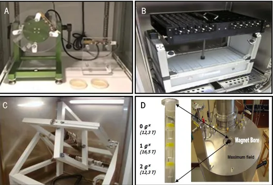
g control; a stationary cell. ... 39 Figure 13: DNA content in A. thaliana cell suspensions after exposure to 63 ºC for 30 min. Each panel represent the relative number of cells according to the DNA content in each cell. A) Cells recovered from the low-melting agarose at 63 ºC. B) Fresh cell culture exposed to 63 ºC. C) Fresh culture without treatment kept at RT. In this panel it is possible to distinguish the typical cell cycle peaks: first peak (2C) that reflects G1 phase, and the second peak (4C) that reflects G2/M phase. In panels A and B, of the cells exposed to 63 ºC, the peaks are not observed. ... 46 Figure 14: Arabidopsis cell cycle phases of cell suspension immobilized in alginate/gelatine and alginate. Flow cytometry analysis, each panel represents the relative number of cells according to the DNA content in each cell. First peak 2C reflects G1 phase and the second peak reflects G2/M phases. ... 47 Figure 15: Extraction of the cell nuclei from MM2d cell suspension cultures. Cell cycle analysis by flow cytometry in a comparison of the two methods. ... 47
xiii
Figure 16: Cell cycle progression of Arabidopsis cell line MM2d for 24 h after clinorotation assessed by flow cytometry (representative essay). Panels A and B represent respectively the relative number of cells according to the DNA content in 1 g control and simulated microgravity. C) Relative proportion of cells in the G1, S and G2/M phases. ... 48 Figure 17: Cells of Arabidopsis in mitosis under simulated microgravity for 24 h experiment. More than 1000 cells were counted A) Confocal images of DAPI staining cells showing metaphase (1) and anaphase (2) phases. B) Cell division bar chart represented by the mitotic index. ... 49 Figure 18: Cell cycle progression of microcalli film Arabidopsis cell line MM2d for 24 h after clinorotation (representative essay). Cell cycle analysis by flow cytometry. A) 1 g control. B) Simulated microgravity. C) Relative proportion of cells in the G1, S and G2/M phases. ... 50 Figure 19: Nucleolar area in 24 h clinorotation experiment in Arabidopsis cells embedded in agarose and alginate. More than 300 nucleolus areas (for each, nucleolin and fibrillarin staining) were measured. Nucleolar nucleolin and fibrillarin staining was observed using the confocal microscope. A) Confocal images from cells recovered from agarose. B) Nucleolar mean areas estimated by nucleolin and fibrillarin fluorescence areas (agarose). C) Confocal images from cells recovered from alginate. D) Nucleolar mean areas estimated by the abundance of nucleolin and fibrillarin using immunofluorescence microscopy (alginate). Significant differences between mean values (p≤0.05) are denoted by *. ... 51 Figure 20: Cell cycle progression for 24 h after aphidicolin block/release of Arabidopsis cell line MM2d. A) Distribution of cell cycle phases by flow cytometry analysis (DAPI) regarding to the DNA content corresponding to the relative number of cells. Samples taken at different times after the release of aphidicolin block in late G1/early S phase (T0). B) Relative proportion of cells in the G1, S and G2/M phases. ... 52 Figure 21: Schematic model of the main factors and functional processes playing a role in the regulation of the coordination between cell growth and cell proliferation in vitro undifferentiated high proliferative cultured cells by environmental gravity. Black arrows represent experimentally supported connections, whereas red arrows indicated suitable processes, compatible with experimental data, but still pending of further investigations for their demonstrations. Sensing of the parameters of the gravity (magnitude, direction) in vitro cultured cells may occurs by receptors of the cell wall. Gravitropic signals are transduced, resulting in alterations of growth coordinators such as protein nucleolin. Mediators of the transduction of gravity mechano-signal sensed in this way are experimentally unknown. The proteins kinase CK2 and CDKA are proposed as candidates to be part of this scheme in view of the experimental findings that put it in close relationship with some cellular processes involved, such as ribosome biogenesis and cell cycle.. ... 66 Figure 22: Gravity levels in potentially life-compatible solar system celestial bodies. Gravity level of some solar system celestial bodies in a comparison with Earth gravity reveals that the importance of the partial gravity studies. ... 70
xiv
LIST OF TABLES
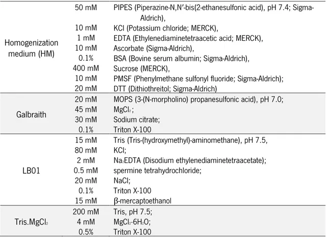
Table 1: Constituents of the extraction buffers. ... 35 Table 2: List of the specific antibodies used in the immunofluorescences bases-analysis. Its references and dilutions in the blocking solutions. ... 37 Table 3: Summary of the results obtained in exposing Arabidopsis thaliana cell suspension to simulated microgravity on 2D clinostat. ... 62
xv
INDEX
ACKNOWLEDGEMENTS ... iii ABSTRACT ... v RESUMO ... vii INDEX ... xvACRONYMS AND ABBREVIATIONS ... ix
LIST OF FIGURES ... xi
LIST OF TABLES ... xiv
INTRODUCTION ... 3
1. Plants in Gravitational Biology ... 3
1.1. The impact of gravity in plant biology ... 4
1.1.1. Plants evolution and physiology: the role of gravity ... 5
1.1.2. Perception of gravity in plant cells ... 5
1.1.3. Gravity alteration induces plant response to biotic and/or abiotic stress ... 8
1.2. Microgravity research in plants ... 9
1.2.1. Definitions used in Gravitational Biology research ... 9
1.2.2. Simulated microgravity on Earth: concept, GBFs and limitations ...10
2. Cell growth and cell proliferation in Arabidopsis thaliana ... 12
2.1. Plant cell cycle ...13
2.1.1. Cell cycle control ...14
2.1.2. Cell cycle transitions and progression ...15
3.1.3. In vitro cell cultures: synchronous cell cycle ...18
2.2. Plant cell growth: the nucleolus and ribosome biogenesis ...19
2.2.1. The plant nucleolus during the cell cycle ...20
2.2.3. Nucleolar dynamics under stress conditions ...22
3. Arabidopsis thaliana cell growth and cell proliferation are affected by gravity alteration ... 23
3.1. Plant cell growth and proliferation in Arabidopsis seedlings were altered by real microgravity 23 3.2. Plant cell growth and proliferation in Arabidopsis seedlings were altered by simulated microgravity ...24
3.3. Plant cell growth and proliferation in Arabidopsis undifferentiated cell cultures were altered by simulated microgravity ...24
xvi
OBJECTIVES ... 27
MATERIAL AND METHODS ... 25
1. Fast-growing Arabidopsis thaliana cell suspension culture (MM2d) ... 31
2. Immobilization and recovery of cell cultures ... 31
2.1. Immobilization and recovery of cell suspension culture by embedding in low melting agarose 31 2.2. Immobilization and recovery of cell suspension culture by embedding in alginate and gelatine .. ...32
2.3. Immobilization and recovery of cell suspension culture by embedding in alginate ...33
2.4. in vitro plated cell culture derived from cell suspension culture ...34
3. Flow cytometry technique ... 34
3.1. Samples preparation ...34
3.1.1. Extraction and staining of the nuclei with Kit Cystain UV precise P ...34
3.1.2. Extraction of the nuclei with optimized homogenization medium ...35
3.2. Determination of individual Cell DNA content (phases and duration) ...36
4. Confocal fluorescence microscopic techniques ... 36
4.1. Confocal immunofluorescence microscopy ...37
4.2. Confocal fluorescence microscopy ...38
5. Clinostat ground-based facility mode of operation, hardware and experimental design ... 39
6. Statistical analysis ... 40
7. Synchronization of cell suspension cultures ... 41
RESULTS ... 45
1. Optimization of a protocol for immobilization/embedding and recovery of plant cell suspensions for clinostat experiments ... 45
1.1. Immobilization and recovery of plant cell suspension culture in low-melting agarose: heat-dependent recovery of the cells affects the quality of DNA as shown in flow cytometry assay (of DAPI-labbeling) ...45
1.2. Immobilization and heat-independent recovery of plant cell suspension culture in alginate/gelatine and in alginate as a suitable technique used for simulated microgravity by clinorotation ...46
2. Effect of simulated microgravity on the cell cycle of cell suspensions ... 47
2.1. Optimized Homogenization medium as the best homogenization buffer for the extraction of nuclei ...47
2.2. Simulated microgravity changes cell cycle phases ...48
2.3. The proliferation of plated plant cell culture (microcalli) is not changed by simulated microgravity ...49
3. Effect of simulated microgravity on the cell growth of Arabidopsis cell cultures ... 50
3.1. Area of localization of proteins involved in ribosome biogenesis is reduced by simulated microgravity ...50
xvii
4. Cell cycle progression ... 52
4.1. Using Arabidopsis cell cycle synchronization with aphidicolin block/release to localize the cell cycle phases ...52
DISCUSSION ... 55
1. Optimized techniques for plant cell suspension immobilization and recovery are suitable for microgravity studies using clinorotation ... 55
1.1. Optimized technique for immobilizing cell suspension to expose to clinorotation: a heat-independent recovery of plant cells suitable for flow cytometry analysis ...55
2. Simulated microgravity causes changes in Arabidopsis thaliana cell proliferation and cell growth ... 57
2.1. Optimized method of plant nuclei extraction from cell cultures for flow cytometry analysis ...57
2.2. Plant cell cycle progression rate is increased under simulated microgravity conditions ...58
2.3. Plant cell growth is reduced by simulated microgravity conditions: nucleolar area measured by labelling of proteins involved in ribosome biogenesis is reduced by simulated microgravity ...61
2.4. Simulated microgravity disrupts the coordination between cell proliferation and ribosome biogenesis in proliferating cell systems in both root meristems and undifferentiated cell culture ...63
3. Implications of alterations in Arabidopsis cell development processes for agriculture on Earth and space exploration ... 67
4. Suggestion of new techniques to be used in future for simulated microgravity experiments using 2D clinostat ... 70
CONCLUSIONS AND FUTURE PERSPECTIVES ... 71
REFERENCES ... 73
1. Plants in Gravitational Biology
2. Cell growth and cell proliferation in
Arabidopsis thaliana
3.
Arabidopsis thaliana
cell growth and cell
proliferation are affected by gravity
alteration
3
INTRODUCTION
1. Plants in Gravitational Biology
Space exploration has started since the humankind looked at the sky and started to question about the shining objects overhead. In our days, we have stepped on the Moon, humans orbit our planet in the International Space Station (ISS) and shuttles explore our solar system and beyond. Next, human being will step on the red planet, Mars (Musk, 2017). Certainly, crossing the frontier of our planet and becoming a multi-planetary species will act as a driver on progress of human civilization. To see this, we can become remembered in the history of society as the Age of Exploration. It was opening of the frontier in “new worlds” that catalysed the progress of the entire worldwide.
Future space exploration depends on the development of long-term habitation on space and planetary surfaces and growing plants on these environments is part of this effort. Plants are integrated in bioregenerative life support systems to food production, atmosphere revitalization, and primary water purification (Finger et al., 2015). They together with microorganisms are an
essential element of these systems (Ferl et al., 2002; Lasseur et al., 2010) (Figure 1). Since
photosynthetic microorganisms, such microalgae Arthrospira platensis, also can provide C02
fixation, O2 generation and food production. Besides, non-edible parts from plants and human
wastes will need to be degraded and recycled, respectively by thermophilic anaerobes bacteria and for both photo-heterotrophic bacteria and nitrifying bacteria, resulting in production of inorganic
nutrients to plants (Lasseur et al., 2010). Plants also can help balance astronauts emotions
providing an earthly element to the artificial environment of spacecraft or orbital platforms (Williams, 2002). A set of specific environmental factors on orbital platforms such as lack of gravity, increased radiation and absence of convection affect plant development and individual cells as it was shown in many plant species (Chebli and Geitmann, 2011). The gravitational force is the only constant factor guiding and affecting the evolution of all organisms in a permanent manner, unlike most biotic and abiotic stresses. As consequence all biological functions and mechanisms of terrestrial organisms have been developed under its influence, and they proceed taking into account the presence of this mechanical force. Thus, microgravity is a novel environment for plants, which can also alter the way in which organisms detect and response to other environmental factors (Herranz and Medina, 2014). Plant microgravity research is particularly difficult on Earth due to
4
ever existing gravity, nevertheless it is crucial to learn about the fundamental processes of plant adaptations to microgravity. Understanding molecular and cellular basis of the plant response to gravity is not only important to grow plants in space, but also to verify the evolutionary value of gravity as an environmental factor and for the general benefit in agriculture on Earth by improving knowledge about plant response to abiotic stresses.
1.1. The impact of gravity in plant biology
Gravity, as any other force, is represented by a vector, meaning that it has magnitude and direction at each point in space. It is responsible for defining the weight of each object which drives many chemical, biological, and ecological processes on Earth. Living organisms have accommodated this force in both their structure and function, developing mechanisms to sense gravity and grow
Figure 1: Plant-based Advanced Life support project (Melissa loop), European Space Agency. Melissa (Micro-Ecological Life Support System Alternative) has been conceived as a microorganisms and higher plants based ecosystem intended as a tool to gain understanding of the behaviour of artificial ecosystems, and for the development of the technology for a future regenerative life support system for long term manned space missions. Based on the principle of an "aquatic" ecosystem, Melissa is comprised of 5 compartments colonized respectively by Thermophilic Anaerobic Bacteria (I), Photoheterotrophic Bacteria (Rhodospirillum rubrum) (II), Nitrifying Bacteria
(Nitrosomonas/Nitrobacter) (III), Photoautotrophic Bacteria (Arthrospira platensis) (IVA), Higher Plants (IVB), and the
crew. Waste products and air pollutants are processed using the natural function of plants which in turn provide food as well as contribute to water purification and supply oxygen for air revitalization. Many other important benefits are
being examined for related industrial projects. (From
5
towards to or against its direction to reach valuable environmental resources such as water and light.
1.1.1. Plants evolution and physiology: the role of gravity
First-ever photosynthetic organisms evolved 3.8 billion years ago in the ancient sea and the first ancestors of land plants evolved from green algae nearly 500 million years ago (Stewart and Mattox, 1975; Zhong et al., 2015). Life on land is under the constant constrain of gravity; the mechanical load on organisms is approximately 1000 times larger on land than in water (Volkmann and Baluška, 2006). Therefore, the development of structural features associated with the support of the mass (a rigid plant body), has been one of the critical evolutionary achievements required for
plants to survive under 1 g conditions. In fact, during evolution plants have acquired specific
organs, tissues, and molecular systems capable of detecting gravity. Root positive gravitropism is essential for water and mineral ion uptake and anchorage of the whole plant. The shoot negative gravitropism, on the other hand, helps the plant to reach sunlight, the energy source for sugar production. Living systems are constantly evolving in response to a wide range of environmental cues. Consequently, all biological functions and mechanisms have developed under the influence of a specific set of environmental conditions. Primitive mechanisms of gravisensing may now coexist with others which arose independently at later stages of evolution (Barlow, 1995).
The study of plants in altered gravitational field is crucial for better understanding the adaptive mechanisms and physiological activities modulated by gravity. Although plants have evolved a set of mechanisms to adapt to extreme environmental conditions, they have never experienced the need to evolve specific mechanisms to respond or adapt to altered gravity.
1.1.2. Perception of gravity in plant cells
Gravity perception in plants plays a key role in their development and acclimation to the environment, from the direction of seed germination to the control of the posture of adult plants. Several models have been proposed to explain plant cellular perception of gravity, based on gravity-sensor organs that drive plant growth according to the direction of the gravity, independently of its magnitude (Perbal et al., 2002; Pouliquen et al., 2017) and on a gravi-resistance mechanism that involves tension on cellular scaffold structures (cytoskeleton, cell wall or membranes), producing an effect that is proportional to the magnitude of the gravity (Soga et al., 2001).
According to the classic starch-statolith hypothesis (Haberlandt, 1900; Nemec, 1900), connecting gravity sensing and gravitropism, the perception of gravity starts in specific cells that act as
6
frontiers in Plant Sciences (Singh, et al., 2017)
statocytes. Statocytes are located in different organs. In shoots, they are found in endodermal cells, in roots, within columns of cells located in the central root cap, called columella (Figure 2); and they have recently been localized in the secondary phloem of mature woody stems (Gerttula et al., 2015). These cells contain starch-filled amyloplasts-called statoliths-characterized by a higher density than the surrounding cytosol. When the orientation of the gravity vector changes relative to the orientation of the organism, the statoliths move toward the new downward direction side of the cell and their sedimentation in this new position exert a force, presumably on the plasma
membrane and on the endoplasmatic reticulum that triggers the activation of ion (Ca2+) channels
(Figure 3A) (Fasano, et al., 2002). The transduction of the gravistimulus leads to the relocation of membrane transport proteins called PIN-FORMED (PIN) proteins (Ottenschläger et al., 2002). Next, the PIN proteins redirect the lateral distribution of the auxin polar transport, the major plant hormone, leading to a differential growth of the organ and ultimately causing bending of the organ
to the desired orientation with respect to the gravity vector direction (the Chodlony-Went hypothesis)
(Cholodny, 1927; Firn and Digby, 1980; Went, 1933). Particularly in roots, this results in a faster growth and/or elongation of the cellular layers on the opposite side, resulting in root curvature in the direction of the gravity vector (Baluška et al., 2010).
At the cellular level, the exact roles of the statoliths, and whether or not they are necessary for a graviperception is still a matter of debate. In experiments using mutants deprived of starch and displaying little if any sedimentation of statoliths, the macroscopic response to a gravistimulation is dramatically diminished compared to the wild-type (Fitzelle and Kiss, 2001; Kiss et al., 1989;
Figure 2: The root cell types responding to
gravity stimulus in Arabidopsis. A)
Longitudinal view of an Arabidopsis primary root showing meristem, distal and central elongation zones, maturation zone. B) Gravity sensing in roots occurs in the central columella cells of the root cap. The root cap consists of four layers of cells important for root gravitropic response namely S1, S2, S3, S4, respectively. However, the central columella cells from S1 and S2 (the encircled cells) play major role for gravity sensing. C) Columella cell (cartoon) contains starch-filled organelles called amyloplasts, nucleus, vacuole, actin filaments, endoplasmic reticulum (ER), plasma membrane (PM), and cell wall (Singh et al., 2017).
A B
7
Pickard and Thimann, 1966). However, the response still exists suggesting that other type of gravity
perception mechanisms take place causing the cell response to the changes in g force. Moreover,
most plant cells are not equipped with statoliths (Chebli and Geitmann, 2011). Evidence for the presence of an alternative mechanism comes from studies on mosses, fungi, and algae which show gravity-dependent growth and differentiation without the presence of statoliths (Staves et al.,
1997). While at times considered a controversy, it became clear that several mechanisms of
gravisensing seem to operate, possibly even in the same cell (Barlow, 1995; Kiss, 2000).
The gravitational pressure model provides a possible but not necessary the only explanation for this phenomenon. The model suggests that the entire mass of the protoplast acts as a gravity sensor that behaves like a water-filled balloon that flattens when placed on a surface due to its own weight (Chebli and Geitmann, 2011). In this model, the role of starch-filled amyloplasts would be that of increasing the overall density of the protoplast. It is postulated that membrane proteins located at the top and bottom of the cell may be activated through the action of differential tensile forces as they interact with the lower and upper cells walls, respectively (Figure 3 B) (Wayne and Staves, 1996).
The cellular tensegrity model is an alternative, but not exclusive view, on how mechanical forces acting on the cells as a whole could be perceived (Ingber, 1997). It proposes that the whole cell is a pre-stresses tensegrity structure, where tensional forces are borne by cytoskeletal array consisting of elements that resist compressive (microtubules) and tensile stresses (actin filaments). The
A B C D
frontiers in Plant Sciences (Chebli and Geitmann, 2011)
Figure 3:Concepts of cellular gravisensing in plants. A) In statolith-based gravisensing the sedimentation or change of position of intracellular organelles with higher density triggers a signal most likely based on a change in trans-membrane ion fluxes. B) According to the gravitational model the weight of the protoplast causes the forces acting on the membrane-cell wall connections at the upper and lower sides of the cells to be different. C) The tensegrity model predicts that cellular distortion due to a change in g- forces affects the pre-stress in the cytoskeletal array of the cell
which in turn changes biochemical activities. D) A variation of the tensegrity model is based on a change in cytoskeletal pre-stress being caused by the weight of heavy organelles that are tethered to the cytoskeletal filaments such as the nucleus (Chebli and Geitmann, 2011).
8
tensional pre-stress that stabilizes the whole cell is generated actively by the contractile actomyosin apparatus and passively from external forces (e.g. gravity, wind, osmotic forces or adhesion to
other cells) (Hamant and Haswell, 2017; Ingber, 2003). The distortion induces a change in
pre-existing force balance and is supposed to affect local thermodynamics or kinetic parameters and in results biochemical activities (Figure 3 C) (Ingber, 2006; Orr et al., 2006). This change in pre-stress cytoskeletal arrays could be generated by the gravity force acting on organelles attached to this network (Figure 3 D) (Yang et al., 2008). Hence, gravity-induced cytoskeleton polymerization and architecture will likely affect many biological properties (Vogel and Sheetz, 2006).
These proposed mechanisms underline how relevant can be a physical force in the general plant development. The plant detects and responds to the physical stimuli and converts the information into a physiological (chemical) signal, which is then transduced and integrated. As stated by van Loon (2007), “There are no biochemical modifications without prior mechanical change”. The fundamental questions concerning the mechanisms of gravity perception, transduction and response remain still unanswered.
1.1.3. Gravity alteration induces plant response to biotic and/or abiotic stress The evolution of all biological functions and mechanisms in living systems was influenced by the presence of the gravity. They include the strategies and mechanisms of perception, response and adaptation to a wide range of abiotic (such as light, temperature, water, wind, magnetic or electric fields) and biotic (interactions with other living beings) environmental stresses. Plants under stressors respond by altering physiological processes and by modulating the expression of specific genes (Timperio et al., 2008; Zupanska et al., 2013).
Studying plants in altered gravity allows us to understand how plants respond to this environmental change, which may also alter detection and response to other environmental factors. Environmental stresses many times have a synergistic effect on plant, in result promoting a complex environmental stress response (Beckingham, 2010; Herranz et al., 2010). Furthermore, changed in the gravity may also influence other environmental conditions; e.g. the distribution, availability or concentration of nutrients in the atmosphere or in the soil.
Recent studies reveal that plant response to gravity alterations has some common and some
unique features compared to other environmental changes (Ferl et al., 2015; Herranz et al., 2013b;
Manzano et al., 2016; Paul et al., 2012). Interestingly, a positive red light-based phototropic
9
In addition, a decrease in response to red light was gradual and correlated with the increase in gravity (Vandenbrink et al., 2016). A curious fact is that ancient plant lineages (moss and fern) shows this red-light phototropism on Earth, but flowering plants have lost this feature during evolution, that is primarily a response to blue light. It is only removal of the gravity factor that unmasks the capacity for directional red-light sensing for phototropism in higher plants
1.2. Microgravity research in plants
Research on microgravity has contributed greatly to disclose the impact of gravity on biological processes. The real microgravity (see below for definition) is only available in the outer space since
the 1 g level cannot be avoided on the surface of our planet. However, access to spaceflights and
to the ISS is limited by the cost and the labour needed to prepare the flights. In addition, it is difficult to distinguish between the effects of microgravity and changes related to the space and spaceflight conditions (radiation, vibration on shuttle, etc.). To overcome these constraints, ground based facilities (GBFs) are available for preparing spaceflight experiments, and also for developing simulated microgravity experiments on the ground thus, providing additional cost-efficient platforms
for gravitational research (Herranz et al., 2013a). In addition, the ground experiments in GBFs
enable testing of biological systems and addressing gravity-related issues prior to space experiments.
1.2.1. Definitions used in Gravitational Biology research
Real microgravity (μg). The term “microgravity” is frequently used as a synonym of
“weightlessness” and “zero-g”, but under microgravity there is a remaining g-force which is not
zero, but just very small. In fact, the term “microgravity” should be applied to g-forces equal or
lower than 10-6 g. Real microgravity can only be achieved in a durable and constant way during
freefall experiments. Those experiments can be performed on board of the spaceflights (sounding rockets, satellites, space stations), parabolic flights (only for times shorter than 20 s combined with hypergravity periods) or in drop towers (for very short time, providing only 5-10 s of microgravity). Mid- and long-term experiments in real microgravity can only be performed in space (Herranz et al., 2013a).
Simulated microgravity (sim μg) conditions. It has been proposed to use the term “simulated
microgravity” to describe the state of acceleration achieved using GBF, as it is perceived by a biological organism (Herranz et al., 2013a). In simulated microgravity experiments the magnitude
10
of the Earth gravity vector cannot be changed, only the way it is perceived (Briegleb, 1992). In consequence, microgravity cannot be achieved with a simulator. Rather, such simulator may generate functional weightlessness from the perspective of the organism or the cell. In effect, the organism or cell does not perceive the gravity since its value is below the sensitivity of the gravity receptors. The choice of the best GBF to be used for a given research and model system should take into account the sensitivity of the researched biological process and organism used (Herranz et al., 2013a).
1.2.2. Simulated microgravity on Earth: concept, GBFs and limitations
Various GBFs with different physical concepts (essentially mechanical and magnetic) have been constructed to simulate microgravity conditions on ground. None of them is absolutely optimal, and consequently, the final choice will depend on the biological material and the experimental analyses to be performed (Figure 4).
One example of GBF is the clinostat. The clinostats are classical mechanical devices designed for simulation of microgravity, dating from the nineteenth century. In this device samples are rotated to prevent the biological system from perceiving the gravitational force direction (Herranz et al., 2013a). The functionality of clinostats is based on redistributing the gravity in a circle, whereas the sample rotate around a single or multiple axis. The low cost of clinostat and its availability makes it one of the most widely used GBFs.
A B
C D
Figure 4:Several ground based facilities (GBFs) used in plant biology research. A) 2D clinostat, located at Centro de Investigations Biológicas (CIB), Madrid, Spain. B) Pipette 2D clinostat C) 3D RPM D) Magnetic levitation facility.
11
2D clinostat or one-axis clinostat has a single rotational axis, which runs perpendicular to the direction of the gravity vector and rotates at speeds that are matched with the particular time of graviresponse for the sample in question (Figure. 4 A) (Briegleb, 1992; Klaus, 2001). This device has a limitation, when a sample is placed at the rim of the rotational axis the force it experiences increases (Brown et al., 1976; Klaus, 2001). However, this constraint can be avoided by positioning the samples close to the centre of the rotational axis. A rotation on a clinostat is often called clinorotation.
Pipette 2D clinostat. This device is used to achieve functional weightlessness for small objects, mainly single cells (Figure 4 B) (Briegleb, 1992). By fast and constant rotation of a small tube, completely filled with liquid, it is assumed that sedimentation of the cells is prevented physically
by a continuous and constant change of the direction of gravity vector (Herranz et al., 2013a;
Klaus, 2001). In this scenario, particles are forced to move on circular paths that depend on rotation adjusted to reach a non-sedimentation effect (Klaus, 2001).
3D clinostat (RPM). Clinostat with two axes are called 3D clinostat. The most commonly used is the Random Positioning Machine (RPM) (Figure 4 C), which operates with different speeds and different directions. The quality of simulation is increased by rotating around two axes and randomized speed compared to a classic 2D clinostat. It is optimal especially for larger objects (Kraft et al., 2000; Van Loon, 2007).
Magnetic levitation technology is another physical concept used to simulate microgravity. It uses the diamagnetic properties of water, which is the major component of biological objects.
Magnetic levitation. A magnetic field applied to biological material can produce a diamagnetic force with the same magnitude as gravity and the opposite direction. This force is capable of compensating the weight of the sample, in result producing the levitation phenomenon (Valles et al., 1997). The advantage of magnetic levitation (Figure 4 D) is the stability of the compensated force, together with the possibility of generating partial gravity and even hypergravity in the same controlled environment. The disadvantage are the secondary effects of the strong magnetic field (Herranz et al., 2013a; Manzano et al., 2012b). Also, this technology in terms of power supply is very high and may require, in some cases, the power supply of a small city (Herranz et al., 2015).
12
E N V I R O N M E N T A L G R A V I T Y (1 g)
Plant Signaling & Behavior (Medina and Herranz, 2010)
Figure 5: Model of the actions and interactions leading to the maintenance of the meristematic competence (involving the coordination between cell growth and cell proliferation) specifically showing the players involved in the transduction of the gravitropic signal. Under ground 1 g conditions the gravitropic signal is a growth promoter, since it stimulates
the growth of primary root according the direction of the gravity. The mechanic stimulus is transduced by the mediator auxin that, in the case of meristematic cells, modulates the expression of a variety of molecules that coordinate the regulation of cell growth and cell proliferation and then keep coupled the processes of protein synthesis (cell growth) and cell cycle progression (cell proliferation). (Scheme is adapted and modified by Medina and Herranz, 2010 from Mizukami, 2001.)
2. Cell growth and cell proliferation in
Arabidopsis thaliana
Arabidopsis thaliana is a model system extensively used for studying plant biology. Its short life cycle, completely sequenced genome, and the existence of a multitude of transgenic plants are just few factors that have contributed to its popularity. These are the reasons why this species has been used to examine fundamental questions on plant growth and proliferation mechanisms under simulated microgravity environment. In meristematic cells, growth and proliferation are strictly coupled and represent so-called “meristematic competence” (Figure 5). Cell growth is required for proliferation which produces daughter cells (Mizukami, 2001).
13
2.1. Plant cell cycle
Plant development consists of initiation and growth of new organs throughout the lifespan of the organism. The sessile nature of plants forces them to respond to the changes in the environment adjusting growth and development. Cell division is one of the major processes that contribute to the plant growth. In effect, the control of the cell cycle is essential for understanding plant growth and development. Cell cycle is an ordered and repetitive process that controls spatially and temporally the replication of genetic material and the segregation of duplicated chromosomes into two daughter cells. This cycle is constituted by four successive phases: G1, S, G2 and M (Figure 6). Lag or gap (G) phases separate the replication of the DNA (S phase) and the segregation of the chromosomes (M phase, mitosis) and allow the revision if the previous phase has been accurately and fully completed.
During G1 phase (the first gap) cell growth occurs by synthesis of proteins and RNA, and energy reservoir is stored in form of ATP. The S phase (phase of DNA synthesis) is the phase when the replication of DNA occurs. The G2 phase (gap) is another phase of growth, distinguished from G1 by the fact cells contain a doubled DNA content and by high protein production. This phase is also engaged in the preparation of the cell for division. Finally, during the M phase the mitosis occurs resulting in two daughter cells. The major regulatory points in the cell cycle are the G1/S and G2/M transitions, these points are of potential arrest that may take place after evaluation of external conditions (Dewitte and Murray, 2003; Van’t Hof, 1985).
Figure 6:Schematic of the cell (division) cycle. Cell cycle consists of four essential phases, G1 (Gap 1), S (Synthesis), G2 (Gap 2), and M (Mitosis for cell division).
14
Differentiating plant cells often display an alternative cycle known as endoreduplication, characterized by an increase in the nuclear ploidy level that results from repeated S phases with no intervening mitosis (Joubès and Chevalier, 2000). In all cases, endoreduplication appears to occur only after cells have ceased normal mitotic cycles (De Veylder et al., 2001; Foucher and Kondorosi, 2000).
2.1.1. Cell cycle control
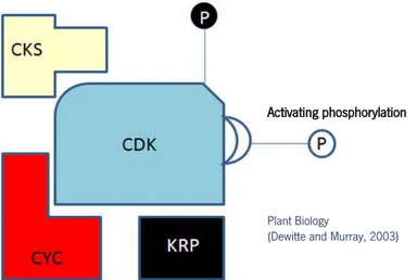
Eukaryotic cell cycle is regulated at multiple points, but all or most of these checkpoints involve the activation of specific class of serine-threonine protein kinases. They require for their activity the binding to regulatory proteins known as cyclins (CYC), and are therefore named cyclin-dependent kinases (CDKs). Cyclins provide the primary mechanisms for the control of CDK activity because the CDK subunit is inactive unless it is bound to an appropriate cyclin (Figure 7) (Dewitte and Murray, 2003; Inzé and De Veylder, 2006).
The cell cycle can be regarded as an oscillator of CDK activity, with low activity in the G1 phase and a peak during mitosis (Coudreuse and Nurse, 2010). This oscillator is driven by highly regulated synthesis and proteolysis of regulatory components through the ubiquitin-mediated selective protein degradation proteasome system at specific points in the cycle (Genschik et al., 2013). Indeed, the exit from mitosis and the return to the ground state in G1 requires the loss of CDK activity through the destruction of cyclins (Scofield et al., 2014).
Inhibitory phosphorylation
Activating phosphorylation
Plant Biology
(Dewitte and Murray, 2003)
Figure 7:Cyclin-dependent kinase (CDK) activity is regulated at multiple levels. Monomeric CDK lacks activity until it forms a complex with cyclins (CYC) and is activated by phosphorylation by CDK-activating kinase (CAK). In addition, activity can be inhibited by binding of inhibitor proteins such as Kip-related proteins (KRP). Inhibitors may block the assembly of CDK/cyclin complexes or inhibit the kinase activity of assembled dimers. CDK subunit (CKS) proteins scaffold modulate interactions with target substrates. (From Dewitte and Murray, 2003)
15
Arabidopsis thaliana genome encodes at least 32 cyclins with a putative role in cell cycle progression; 10 A-type, 11 B-type, 10 D-type and 1 H-type cyclins, in addition to 17 other cyclin-related genes which are classified into types C, P, L, and T (Acosta et al., 2004; Vandepoele et al., 2002; Wang et al., 2004). A-type cyclins generally appear at the beginning of S phase, are involved in S phase progression, and are destroyed around the G2/M transition (Dewitte and Murray, 2003). B-type cyclins appear during G2 phase, control G2/M transition and mitosis, and are destroyed as cells enter anaphase. D-type cyclins control progression through G1 and into S phase and differ from A and B types by generally not displaying a cyclical expression or abundance, their presence appears to depend on extracellular signals that stimulate or maintain division. If such signals are removed, levels of D-type cyclins decline rapidly and cells remain blocked in G1. (Dewitte and Murray, 2003; Renaudin et al., 1996; Van Leene et al., 2010). The levels of cyclins are generally determined by their highly regulated transcription as well as by specific protein-turnover mechanisms (Genschik et al., 1998; King et al., 1996).
2.1.2. Cell cycle transitions and progression
Although the cell cycle is regulated at numerous stages, extracellular growth signals seem to act at two main points, G1/S and G2/M. Therefore, different CDK/CYC complexes phosphorylate target proteins whose inhibitory or activatory post-translational modifications are essential for passing these cell cycle checkpoints (De Veylder et al., 2003; Joubès et al., 2000).
G1/S transition
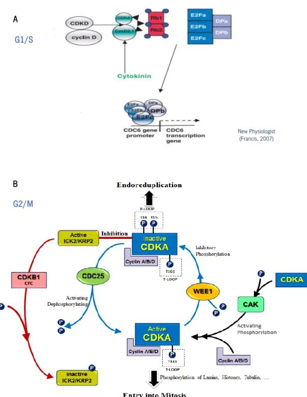
The most outstanding fact in the regulation of the G1/S transition is the induction of the synthesis of D-type cyclins. Plants contain an extensive array of cyclin D genes: genome analysis reveals that A. thaliana has at least ten, as opposed to mammals, with only three. D-type cyclins are primary mediators of the G1/S transition and hence have a major responsibility for stimulating the mitotic cell cycle in the presence of growth factors such sucrose, auxin, cytokinin, and brassinosteroids. Their transcription is activated by extracellular signals, and lead to the formation of active CDKA/CYCD complexes (Dewitte and Murray, 2003; Francis, 2007; Inzé and De Veylder, 2006). The major target of CDKA/CYCD kinase activity complexes in G1 is the retinoblastoma-related
(RBR) protein, which is phosphorylate by these complexes. (Gutierrez et al., 2002). The
retinoblastoma (Rb) protein was identified as a human tumour suppressor protein that controls various stages of cell proliferation through the interaction with members of the E2F family of transcription factors (Desvoyes et al., 2013). It is believed that during the early G1 phase the E2Fs
16
are mainly involved in the repression of several cell cycle-regulated promotors, whereas during the transition from G1 to S phase the release of transcriptionally active E2Fs, resulting from the phosphorylation of the CDKA/CYCD/RBR pocket proteins, is required to drive the expression of S phase genes (Figure 8 A) (Chabouté et al., 2002; De Veylder et al., 2002; Mariconti et al., 2002) G2/M transition
Again, in G2/M, CDKA is the major driver of this transition after its association with D-, A- and, particularly, B-type cyclins (Figure 8 B) (Inzé and De Veylder, 2006). In addition to CDKA,, the G2/M transition requires CDKB activity, the expression of which is dependent on transcriptional control of E2F pathway, properly providing a mechanism by which G1/S and G2/M transitions are regulated (Boudolf et al., 2004). Both CDKA and CDKB-cyclin complexes are activated by a CAK activity (likely CDKD and/or CDKF) before they can phosphorylate a variety of targets that contribute to enter mitosis. The activity of the CDK/CYC complexes can be inhibited by their association with CDK inhibitory proteins (ICK/KRP and SIM) that respond to stress stimuli or developmental signals (Verkest et al., 2005; Churchman et al., 2006). In case of replication stress or DNA damage, there is evidence that CDKA is a WEE1 kinase target mediating G2 arrest (De Schutter et al., 2007). In turn, the CDC25 phosphatase is able to dephosphorylate the residues phosphorylated by WEE1, allowing entry into mitosis (Ferreira et al., 1991; Francis, 2007; Inzé and De Veylder, 2006). Once the CDK/CYC complexes are active, they trigger the G2/M transition through the phosphorylation of a plethora of different substrates. Furthermore, the protein casein kinase CK2 shown discrete activity peaks at G1/S and M in tobacco BY-2 cells, and blocking its activity during G1 abolishes the G2/M checkpoint, resulting in premature entry into prophase; this is an evidence of links between G1 processes and G2 controls (Espunya et al., 1999). Finally, exit from mitosis requires the proteolytic destruction of the cyclin subunits. This destruction is initiated by the activation of the Anaphase-Promoting Complex/Cyclosome (APC/C) through its association with CCS52 protein (Heyman and De Veylder, 2012).
17 A B New Physiologist (Francis, 2007) G1/S G2/M
Figure 8:Different cyclin-dependent kinase (CDKs) and cyclins regulate G1/S and G2/M transition points through the cell cycle progression. A) At G1/S transition, CDKD/cyclin D phosphorylates CDKA enabling CycD3;1 to bind. A cytokine-induced transcellular induction culminates in CyCD3;1, which activates CDKA that, in turn, phosphorylates Rb1 and 2. This releases the E2F complex, which then induces the expression of S phase-specific genes (e.g. CDK6) (From Francis, 2007). B) G2/M transition is regulated by the activation/inactivation of complex CDKA/cyclins (A, B, D) where several theories of control, is identical to that of animals based on complex regulating CDK-CDC25-WEE1 or by CDKB activity. The activity of the CDKA/CYC complexes can be inhibited by their association with CDK inhibitory proteins (ICK/KRP) that respond to stress stimuli or developmental signals (From De Veylder et al., 2007).
18
2.1.3. In vitro cell cultures: synchronous cell cycle
Root meristem is the most commonly used material to investigate the influence of microgravity on plant growth and proliferation, because it plays a key role in sensing the gravity factor. Although the elongation zone is the known target of the transduced gravitropic signal, the meristematic region has been shown to perceive and react to gravitational changes (Medina and Herranz, 2010). In this biological system, the role of auxin is central in connecting the alterations found within meristematic cells in response to gravity changes. However, it does not provide an explanation on how cells lacking specialized gravity-sensing organelles, as statoliths, perceive and alter biological processes in response to changes in gravity.
Cell suspension culture is a perfect model to investigate this topic since it provides a homogeneous population of nearly identical cells, in the absence of developmental processes (Gould, 1984). In
this way, in vitro cell cultures are a powerful tool to study cellular response to environmental
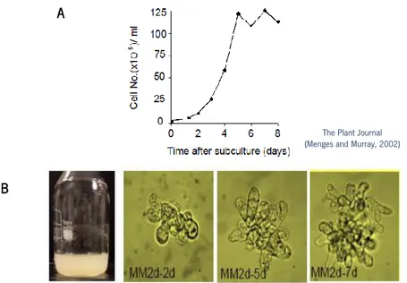
stresses. Also, they are the most suitable systems for synchronisation, allowing detailed analysis of cell cycle studies (Gould, 1984; Menges et al., 2003; Menges and Murray, 2006). The cell line MM2d of Arabidopsis thaliana consists of uniform small clumps of creamy-coloured cells growing fast and at high density, suitable for synchronization. It is composed of a population of undifferentiated actively proliferating cells, similar to the ones in root meristem (Figure 9) (Menges and Murray, 2002).
The Plant Journal (Menges and Murray, 2002)
A
B
Figure 9:Growth characteristics of Arabidopsis cell line MM2d (Menges and Murray, 2002). A) Early stationary phase for 7 days, cells were subculture, and cell growth monitored of cell line MM2d. B) MM2d cell suspension maintains after 7 days in dark conditions. and cell morphology of MM2d through the cultivation time 2, 5, and 7 days, respectively.
19
Ideally in a synchronized cell system, all or most cells progress through the cell cycle at the same rate from the same initial starting point. Various synchronization procedures are based on accumulating cells at a specific point in the cell cycle which is followed by the reactivation of cell cycle progression. The most common approaches use reversible inhibitors of a specific cell cycle
stepor nutrient deficiency to block progression of the cell cycle, followed by the release of the
synchronized cells by the removal of the inhibitor or resupply of specific nutrients (Menges and Murray, 2002). Arabidopsis cell suspension cultures synchronized by aphidicolin block/release is a suitable system for following both the re-entry of cells into the cell cycle and progression from the
S phase. Aphidicolin is a fungal toxin derived from Nigrospora sphaerica that reversibly inhibits
DNA polymerase (Dehghan Nayeri, 2014). Synchronization with aphidicolin produced up to 80% S phase cells, which constitutes a clear separation of different cell cycle phases (Menges and Murray, 2002, Menges and Murray, 2006).
2.2. Plant cell growth: the nucleolus and ribosome biogenesis
Whereas the concept of cell proliferation is unambiguous in reference to progression of the cell cycle, by sustained growth to allow cell division, the concept of cell growth may be caused by different phenomena. Growth is the increase in size, however plant cells may grow either via increase in its cytoplasmic mass or via expansion of intracellular vacuoles. The latter is a feature of cell differentiation and accounts for the growth of differentiated organs. On the contrary, cytoplasmic mass increase is a feature of rapidly dividing cells, such as meristematic cells, in which
vacuoles are extremely small (Magyar et al., 2005).In this case, cell growth essentially is due to
the activity of the protein synthesis (Mizukami, 2001). Thereafter, it is strictly correlated to the rate of biogenesis of ribosomes, since the function of ribosomes is the translation of mRNA into proteins. Ribosome biogenesis occurs in a well-defined nuclear territory called the nucleolus. In actively proliferating cells, nucleolar morphology can serve as a structural marker to state of both cell growth and cell proliferation mechanisms, connecting them in the same framework (Baserga, 2007; Sáez-Vásquez and Medina, 2008). Nucleolar features like size, distribution and accumulation of its components or the level and distribution of certain nucleolar proteins show profound variations throughout cell cycle and are strictly associated to the rate of transcription and processing of ribosomal precursors (Medina and Herranz, 2010).
20
2.2.1. The plant nucleolus during the cell cycle
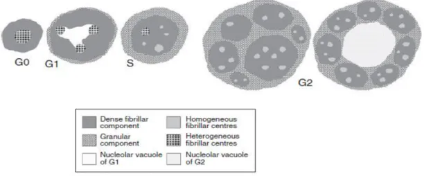
Most eukaryotic cells contain a prominent region within the nucleus called nucleolus. The main function of the nucleolus is linked to ribosome biogenesis and closely associated with other important biological processes, including RNA metabolism, regulation of gene expression, cell cycle regulation, DNA repair and cell aging (Sáez-Vásquez and Medina, 2008; Montacié et al., 2017). As referred above, the nucleolus is the site of ribosome biogenesis, which involves the production of ribosomes subunits by transcription of rRNA genes (in plants 18S, 5.8S and 25S), maturation of the transcripts and their transport to the cytoplasm where they are assembled into functional ribosomes (Sáez-Vásquez and Medina, 2008). Morphological features of the nucleolus can be distinguished for each phase of the ribosome synthesis. In interphase cells, the nucleolus is formed by three basic components: the fibrillar centres (FCs), the dense fibrillar component (DFC) and the granular component (GC), which are sometimes accompanied by other structures, such as vacuoles (Figure 10) (Jordan, 1984). FCs are most likely the sites for anchoring of rDNA where the assembly of transcription complexes takes place. Homogeneous FCs often appear in nucleoli actively producing ribosomes and heterogeneous FCs are associated with low rates of nucleolar transcriptional activity (Risueño and Medina, 1985). In DFC is where the early steps of pre-rRNA processing take place. Finally, in GC, further RNA processing and RNA modification steps occur, together with the formation of pre-ribosomal particles, ready for the export to the cytoplasm after maturation process (Thiry and Lafontaine, 2005).
Figure 10: Nucleolar models in the different interphase periods. It is shown the relative nucleolar size, as well as the distribution of the nucleolar structural subcomponents in each period. Morphological and morphometrical features correlate to the rate of nucleolar transcriptional and processing activity. (From González-Camacho and Medina, 2006).
21
During the cell cycle,the nucleolar size is constant during G1 and S phases and it is doubled in
G2 as a consequence of an enhanced nucleolar activity. Morphological features of the FCs also show a clear association with the cell cycle progression: the number of FCs increases and their size decreases. Structurally, heterogeneous FCs are present in G1 phase whereas in G2 phase they are of the homogenous type (González-Camacho and Medina, 2006; Medina, 1983; Risueño, et al, 1982). In highly proliferating cells GC enlargement occurs in G2 phase (Figure 11) (Hadjiolov,
1985; Smetana and Busch, 1974).Particularly interesting is the behaviour of the nucleolus during
mitosis, when its structure is disorganized and its activity stop, even though the individual transcription and processing complexes are not disassembled. They are carried at the chromosome periphery and then they are organized into discrete entities called prenucleolar bodies, whose fusion, together with the resumption of transcription and processing, originates the new nucleolus (Medina et al., 2000).
2.2.2. Nucleolar proteins and their role in ribosome biogenesis
In Arabidopsis thaliana two proteomic studies of the nucleolus have been performed using cell cultures. The first identified around 217 proteins including several non-ribosomal and even “non-nucleolar” proteins (Pendle et al., 2005). The actualized proteome extended the initial list up to 1602 proteins in the nucleolar fraction (Palm et al., 2016). The nucleolus contains many proteins specific for the nucleolus, but some are also present in other cellular locations. The changes in nucleolus during cell cycle are associated with the changes in the abundance and distribution of certain nucleolar proteins. Two of the most abundant nucleolar proteins are nucleolin and fibrillarin (Shaw, 2005).
Nucleolin is the major non-ribosomal nucleolar protein in all eukaryotic proliferating cells. It plays an important role in growth and development, not only in plants but also in other higher eukaryotic organisms (Tajrishi et al., 2011). It is involved in different steps of ribosome biogenesis, including RNA polymerase I transcription, processing of pre-rRNA, as well as assembly and nucleocytoplasmic transport of ribosome particles (Montacié et al., 2017; Pontvianne et al., 2007; Roger et al., 2002). Additionally, nucleolin has been involved in different cellular aspects that take place in the nucleus and cytoplasm, including chromatin organization and stability, cytokinesis, cell proliferation and stress response (Durut and Sáez-Vásquez, 2014).
In plants, nucleolin can be considered as nucleolin-like protein (Tong et al., 1997). Arabidopsis thaliana encodes two nucleolin genes AtNUC-L1 and AtNUC-L2, in contrast to animals and yeasts,
