Conditioned medium from activated
spleen cells supports the survival of
rat retinal cells
in vitro
1Departamento de Neurobiologia, Instituto de Biologia,
Universidade Federal Fluminense, Niterói, RJ, Brasil
2Instituto de Biofísica Carlos Chagas Filho, Centro de Ciências da Saúde,
Universidade Federal do Rio de Janeiro, Rio de Janeiro, RJ, Brasil A. Sholl-Franco1,2
and E.G. Araujo1
Abstract
Cytokines are a heterogeneous group of molecules that have been associated with several functions in the nervous system, such as survival and differentiation of neuronal and glial cells. In the present study, we demonstrated that conditioned medium from spleen cells activated with concanavalin A increased neuritogenesis and survival of retinal cells, as measured by biochemical and morphological crite-ria. Our data showed that conditioned medium induced a five-fold increase in the amount of protein after 120 h in vitro. This effect was not inhibited by the blockade of voltage-dependent L-type calcium channels with 5.0 µM nifedipine. However, the use of an intracellular calcium chelator (15.0 µM BAPTA-AM) inhibited this effect. Our results support the idea that factors secreted by activated lymphocytes, such as cytokines, can modulate the maintenance and the differentia-tion of rat retinal cells in vitro, indicating a possible role of these molecules in the development of retinal cells, as well as in its protec-tion against pathological condiprotec-tions.
Correspondence
A. Sholl-Franco
Departamento de Neurobiologia Instituto de Biologia, UFF Caixa Postal 100180 24001-970 Niterói, RJ Brasil
Fax: 55 (021) 719-5934 E-mail: [email protected]
Research supported by CAPES, CEG-UFF, FAPERJ and FINEP. A. Sholl-Franco is the recipient of a CAPES fellowship.
Received May 13, 1997 Accepted September 9, 1997
Key words •Cytokines •Growth factor •Neuroimmunology •Retina
•Development
The growth, differentiation, survival, and death of neuronal cells can be modulated by specific molecules produced during the nor-mal development of the nervous system (1). Over the last few years it has been demon-strated that molecules produced by cells of the immune system can affect different neu-ronal populations as well as glial cells (2,3). In addition, glial cells have been shown to produce and release at least some of these molecules (4).
Cytokines are molecules with pleiotropic effects originally described as having immu-noregulatory functions (5,6). However, it is
conditions following neuronal injuries in adult life (2,5,6).
To test the action of cytokines on neu-rons in the developing central nervous sys-tem, we have studied a particularly well-characterized region, the neural retina. In the present study, we investigated the effect of conditioned medium produced by spleen cells activated with concanavalin A (ConA) on retinal cells in culture. When spleen cells are activated with ConA they produce a broad spectrum of growth-promoting factors such as colony-stimulating factors, interleukins, and a nerve growth factor-related molecule (8-10). The present results suggest that these cytokines can promote neuronal survival and neurite outgrowth in dissociated cultures of newborn rat retinal cells, as evaluated by both morphological criteria and protein as-says. Our data indicate that these molecules may have important roles in neuronal and glial development.
Primary cultures were prepared using pro-cedures previously described (11). Briefly, Lister Hooded rats at postnatal day 1 or 2 were killed by decapitation and the retinas were dissected free from scleral tissue and pigmented epithelium in calcium- and mag-nesium-free balanced salt solution (CMF). The retinas were incubated in CMF contain-ing 0.1% trypsin (Worthcontain-ington, Freehold, NJ) for approximately 17 min at 37oC. The cells were mechanically dissociated using a polished Pasteur pipette and added to plastic Petri dishes (35 mm) previously coated with poly-L-ornithine (50 µg/ml; Sigma Chemi-cal Co., St. Louis, MO) at a plating density of 1.0 x 10⁶ cells/dish in complete culture me-dium (199; Gibco, Gaithersburg, MD) con-taining 2 mM glutamine, 100 µg/ml strepto-mycin + 100 U/ml penicillin (Sigma) and 5% fetal calf serum. The cultures were then main-tained at 37oC in an atmosphere of 5% CO
2/ 95% air for several days. The conditioned medium (CM) obtained from ConA-activated spleen cells was prepared by the method of Gozeset al. (9) modified in our laboratory.
Briefly, adult Lister Hooded rats were killed by ether asphyxia and the spleens were dis-sected in CMF solution. The single-cell sus-pension was obtained by passing spleens through a 50-gauge plastic mesh. The cul-ture medium containing the cells was left to rest for 1 h on plastic dishes (35 mm) in order to allow the attachment of macrophages. The supernatant containing the non-adher-ent cells was aspirated, cnon-adher-entrifuged (2000 g
for 5 min), and added at a plating density of 5.0 x 10⁶ cells/ml to complete culture medi-um supplemented with 2-mercaptoethanol (25 µM) and ConA (5.0 µg/ml) for approxi-mately 12 h at 37oC in an atmosphere of 5% CO2/95% air. The supernatant thus obtained was again centrifuged at 2000 g for 10 min and the CM was sterilized by filtration through a 0.2-µm membrane and kept at 4oC. After the maintenance of retinal cells in culture for specific times, the morphology of the culture was analyzed and the total amount of protein in the cultures quantified by the method of Lowry et al. (12).
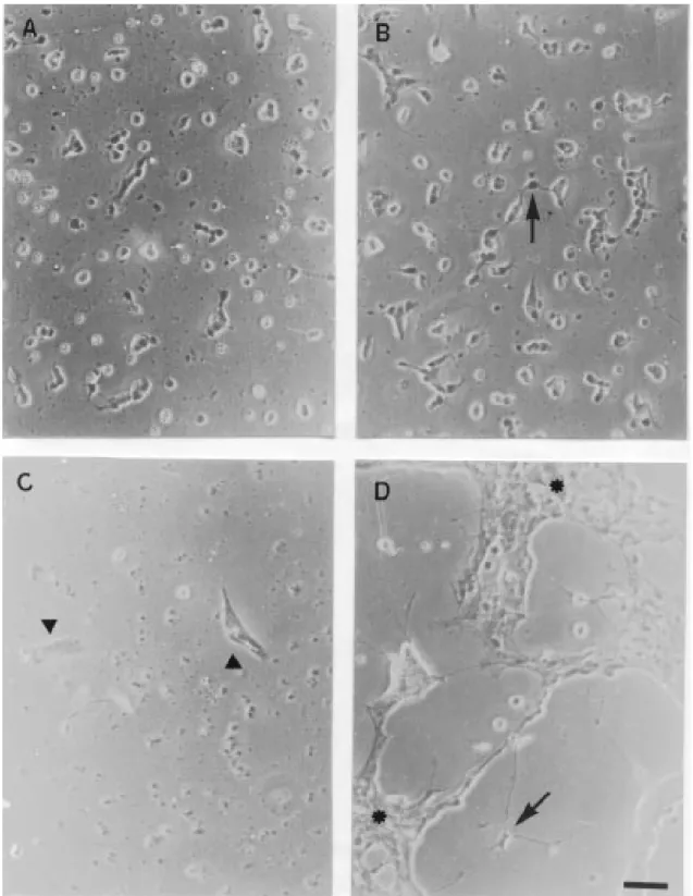
presence of large clusters of neuronal cells supported by glial cells was clearly visible. Neuritogenesis, neuronal survival as well as glial cell proliferation were highly stimulat-ed by CM (Figure 1D). The cells maintainstimulat-ed in the absence of CM died within 120 h as observed by morphological criteria, i.e., shrinkage of the body of neuronal cells and disruption of neuronal processes (Figure 1C). In the control cultures there were only glial cells that took on a flat shape characteristic of this cell type (Figure 1C).
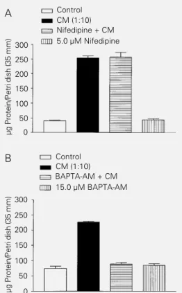
As shown in Figure 2A, CM also in-creased the amount of protein compared with control cultures. The increase in protein in treated cultures reflects not only the prolif-eration of glial cells but also the survival of neuronal cells. In fact, the use of the antimi-totic agent 2-fluorodeoxyuridine (20.0 µM) did not block the survival or neuritogenesis of neuronal cells stimulated with CM (data not shown).
The maintenance of retinal cells in cul-ture by the treatment with CM was
dose-dependent and heat-sensitive, and was ob-served at all plating densities studied (data not shown). Moreover, the blockade of volt-age-dependent L-type calcium channels with nifedipine (5.0 µM) did not inhibit the action of CM on retinal cells (Figure 2A). On the other hand, it appears that the cytoplasmic calcium levels were essential for the effect of CM since BAPTA-AM (15.0 µM), an intracellular calcium chelator, was efficient in blocking the increase in the amount of protein (Figure 2B).
The regulation of cytoplasmic calcium levels has been described in different events during the development, maintenance, and degeneration of neuronal cells. This intra-cellular messenger actively participates in several signaling pathways in the membrane and in the cytoplasm. Indeed, the survival of developing neuronal cells deprived of trophic factors in culture has been related to the control of cytoplasmic levels of free calcium (13). Our results show that this second mes-senger is important for the action of CM since the addition of BAPTA-AM completely abolished this action. Nevertheless, the block-ade of voltage-dependent L-type calcium channels did not alter the effect of CM, even though the influx of calcium through these channels is known to play a key role in the survival of other neuronal cell types (13,14). While most of the reported effects of cytokines are on non-neuronal cells, there is also evidence that they may directly act on neurons (3,4,15,16). In addition, the early time course of the expression of some cyto-kines suggests that these molecules may have important effects on neuronal differentiation (1,7,16,17).
Although the retina has been extensively studied by anatomical, histological and bio-chemical methods, there is still very little information concerning the mechanisms that control its development leading to cellular differentiation. Strong evidence suggests that the development of retinal cells is regulated by cell-cell contacts and by diffusible
mol-µ
g
P
rotein/
P
etri dish (35 mm)
300 250 200 150 100 50 0 Control CM (1:10) Nifedipine + CM 5.0 µM Nifedipine
µ
g
P
rotein/
P
etri dish (35 mm)
300 250 200 150 100 50 0 Control CM (1:10) BAPTA-AM + CM 15.0 µM BAPTA-AM A
ecules (4,11,18).
In general, cytokines produced and se-creted by lymphocytes are related to inflam-matory and degenerative processes (2,5,6). It is well known that lymphoid cells also have the ability to secrete growth-promoting fac-tors that participate in a pleiotropic manner in the development of the nervous system and in its protection against pathological conditions (1,9,15,17). Our results support the idea that molecules secreted by activated
lymphocytes can modulate the maintenance and differentiation of rat retinal cells in vi-tro, indicating a possible role of these mol-ecules in the development of retinal cells.
Acknowledgments
We acknowledge the technical assistance of Alexandre José Fernandes and Bernar-dino Matheus dos Santos.
References
1. Korshing S (1993). The neurotrophic fac-tor concept: a reexamination. Journal of Neuroscience, 13: 2739-2748.
2. Hopkins SJ & Rothwell NJ (1995). Cyto-kines and the nervous system. I. Expres-sion and recognition. Trends in Neuro-sciences, 18: 83-88.
3. Pousset F (1994). Cytokines as mediators in the central nervous system. Biomedi-cine and Pharmacotherapy, 48: 425-431. 4. Puro DG (1995). Growth factors and
Müller cells. Progress in Retinal and Eye Research, 15: 89-101.
5. Holtmann H & Resch K (1995). Cytokines. Naturwissenschaften, 82: 178-187. 6. Rothwell NJ & Hopkins SJ (1995).
Cyto-kines and the nervous system. II. Actions and mechanisms of action. Trends in Neu-rosciences, 18: 130-136.
7. Patterson PH (1993). Cytokines and the function of the mature nervous system. Science de la Vie, 316: 1150-1157. 8. Watson T, Aarden L & Lefkovits I (1979).
The purification and quantitation of helper T cell replacing factors secreted by con-canavalin A activated murine spleen cells. Journal of Immunology, 122: 209-215.
9. Gozes Y, Moskowitz MA, Strom TB & Gozes I (1983). Conditioned media from activated lymphocytes maintain sympa-thetic neurons in culture. Developmental Brain Research, 6: 93-97.
10. Ganea D & Sun L (1993). Vasoactive intes-tinal peptide downregulates the expres-sion of IL-2 but not of INFγ from stimulat-ed murine T lymphocytes. Journal of Neu-roimmunology, 47: 147-158.
11. Araujo EG & Linden R (1993). Trophic fac-tors produced by retinal cells increase the survival of retinal ganglion cells in vitro. European Journal of Neuroscience, 5: 1181-1188.
12. Lowry OH, Rosebrough NJ, Farr AC & Randall RJ (1951). Protein measurement with the Folin phenol reagent. Journal of Biological Chemistry, 193: 265-275. 13. Franklin JL & Johnson Jr EM (1992).
Sup-pression of programmed neuronal death by sustained elevation of cytoplasmic cal-cium. Trends in Neurosciences, 15: 501-508.
14. Collins F & Lile JD (1989). The role of dihydropyridine-sensitive voltage-gated calcium channels in potassium mediated neuronal survival. Brain Research, 502: 99-108.
15. Araujo DM & Cotman CW (1993). Trophic effects of interleukin-4, -7, and -8 on hip-pocampal neuronal cultures: potential in-volvement of glia-derived factors. Brain Research, 600: 49-55.
16. Konishi Y, Kamegai M, Takahashi K, Kunishita T & Tabira T (1994). Production of interleukin-3 by murine central nervous system neurons. Neuroscience Letters, 182: 271-274.
17. Wagner JA (1996). Is IL-6 both a cytokine and a neurotrophic factor? Journal of Ex-perimental Medicine, 183: 2417-2419. 18. Watanabe T & Raff MC (1990). Rod