Hyoid Bone Suspension as a Part of Multilevel
Surgery for Obstructive Sleep Apnea Syndrome
Abd Alzaher Tantawy
1Sherif Mohammad Askar
1Hazem Saeed Amer
1Ali Awad
1Mohammad Waheed El-Anwar
11Department of Otorhinolaryngology and Head and Neck Surgery,
Faculty of Medicine, Zagazig University, Zagazig, Egypt
Int Arch Otorhinolaryngol 2018;22:266–270.
Address for correspondence Mohammad Waheed El-Anwar, MD, Department of Otorhinolaryngology Head and Neck Surgery, Faculty of Medicine, Zagazig University, Shaibet an Nakareyah, Markaz El-Zakazik, Ash Sharqia Governorate 44519, Egypt (e-mail: [email protected]; [email protected]).
Introduction
Patients with obstructive sleep apnea (OSA) syndrome present anatomical and physiological upper airway configurations that lead to repeated airway obstruction. The oropharynx and the hypopharynx are the main locations of this kind of obstruction, although nasal obstructions can further aggravate the symp-toms of OSA. Recently, the hypopharyngeal surgery has re-ceived more attention, and progresses have been made because the oropharyngeal surgery alone is insufficient to cure OSA.1,2 Hyoid suspension represents a common procedure in the
multilevel OSA surgery concept and it is often combined with other procedures, such as palatal surgery or genioglossus advancement.1From the time of thefirst description of hyoid suspension by Riley et al at, in 1986, many investigators utilized it to correct obstruction at the hypopharyngeal airway level. At
first, the hyoid suspension procedure was directed to suspend the hyoid bone to the inferior mandibular border using fascia lata.3In 1993, Riley et al modified the technique to secure the hyoid bone to the thyroid cartilage below.4Thereafter, variable modifications appeared in the literature aiming to optimize the outcome and minimize its complications.1,5,6
Keywords
►
obstructive sleep
apnea
►
hyoid bone
►
snoring
►
palate
Abstract
Introduction
Since oropharyngeal surgery alone is often insuf
fi
cient to treat
ob-structive sleep apnea (OSA), advances have been developed in hypopharyngeal
surgery.
Objective
To assess hyoid suspension surgery as part of a multilevel OSA surgery, also
including palatal surgery.
Methods
The study included patients with OSA symptoms with apnea hypopnea
index (AHI)
>
15. They were scheduled for hyoid suspension after a nasoendoscopy
during Müller maneuver and drug induced sleep endoscopy (DISE). All patients had
body mass index (BMI)
<
35 kg/m2. Hyoidothyroidopexy combined with tonsillectomy
and palatal suspension was performed in all cases.
Results
The mean AHI dropped signi
fi
cantly (
p
<
0.0001) from 68.4
25.3
preo-peratively to 25.6
9.52 postoperatively. The mean lowest oxygen (O
2) saturation
level increased signi
fi
cantly from 66.8
11.3 to 83.2
2.86 (
p
<
0.0001). In
addi-tion, the snoring score signi
fi
cantly decreased (
p
<
0.0001) from a preoperative mean
of 3.4
0.54 to 2
0.7 at 6 months postoperatively. In regard to the Epworth
sleepiness scale (ESS), it showed signi
fi
cant improvements (
p
<
0.0001) as its mean
diminished from 13.8
5.4 preoperatively to 5.2
1.6 postoperatively.
Conclusion
Hyoidothyroidopexy using absorbable suture seems to produce a good
outcome in treating OSA. It could be effectively and safely combined with other palatal
procedures in the multilevel surgery for OSA.
received
June 14, 2017
accepted
September 3, 2017
published online
October 25, 2017
DOI https://doi.org/ 10.1055/s-0037-1607227.
ISSN 1809-9777.
Copyright © 2018 by Thieme Revinter Publicações Ltda, Rio de Janeiro, Brazil Original Research
In this work, the authors assess the hyoid suspension surgery as part of a multilevel surgery, also including palatal work and nasal surgery.
Patients and Methods
This prospective, non-randomized study was conducted on patients who had OSA and were referred to the Otorhinolar-yngology Head and Neck Surgery department of a hospital in Zagazig, Egypt, in the period from February of 2015 to April of 2017. The study included patients with OSA symptoms with apnea hypopnea index (AHI)>15. These patients were sched-uled for hyoid suspension after nasoendoscopy during Müller maneuver and drug induced sleep endoscopy (DISE) showed that they had retropalatal and lateral hypopharyngeal collapse. All patients had body mass index (BMI)<35 kg/m2 and rejected or could not adapt to the positive airway pressure treatment. Exclusion criteria included patients with history of surgical intervention for OSA or for neck pathology. Patients with nasal obstruction and those who showed anteroposterior collapse of the hypopharynx were excluded from the study. Patients who did not obey the scheduled follow-up sessions were also excluded from the study. The study was approved by the International Review Board of our institution.
A written informative consent was obtained from each patient after clarification of the procedure in details. After a detailed patient history taking; a flexible nasoendoscopy during Müller maneuver and a DISE were performed for all patients. The DISE was performed immediately before the surgery. Moreover, all patients had subjective analysis with the Epworth Sleepiness Scale (ESS) as a measure of daytime somnolence.
Six months after the surgery, all patients underwent nasopharyngolaryngoscopy as part of the standard post-surgical protocol using the Müller maneuver. A postopera-tive sleep study was also performed. All patients underwent postoperative assessment of daytime somnolence as well. The primary efficacy endpoints were the mean change in the AHI and ESS total scores. Secondary effectiveness endpoints included the mean change in the lowest oxygen (O2) satura-tion recorded as part of the polysomnographic data1and snoring severity scale (after Morris et al2).
Operative Technique
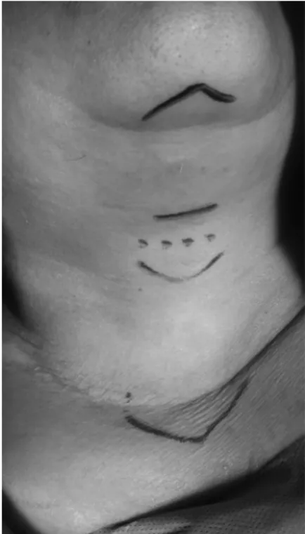
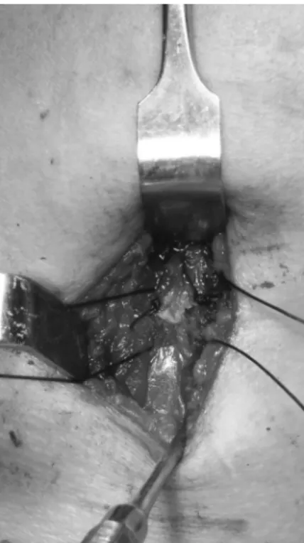
The hyoid suspension procedure was performed under gen-eral anesthesia, with the patient in supine position with the neck extended. Skin marks were lined over the mandibular margin, hyoid bone, thyroid notch and sternal notch (►Fig. 1). The skin incision followed a horizontal skin crease between the body of the hyoid bone and the thyroid notch. The incision was carefully extended through the subcuta-neous tissue and platysma muscle, as the muscle is less defined in the midline. Upper and lower subplatysmalflaps were elevated to expose the strap muscles, which were separated in the midline. The plane of the thyroid cartilage and the surface of the hyoid bone were exposed and the thyrohyoid membrane was clearly defined (►Fig. 2). Vicryl 0 was wrapped around the body of the hyoid bone
on each side of the midline with a sharp needle and then directed to the thyroid lamina of the same side piercing it from the lateral to the medial surface, about ½ cm from the upper border of the cartilage. Two sutures were performed on each side. The sutures were tied steadily and gently, with the neck in neutral position (►Fig. 3). The wound was closed Fig. 1 Skin marks were lined over the mandibular margin, hyoid bone, thyroid notch and sternal notch.
in layers. A suction drainage was placed for 48 hours. After the surgery, pain was easily controlled with usual nonster-oidal anti-inflammatory medications, and all patients were sent home on the following morning.
In all cases, the hyoid suspension procedure was combined with tonsillectomy and palatal suspension, as described by El-Ahl and El-Anwar.7The operative time for the hyoid suspen-sion was counted starting with the neck skin preparation and ending with the last skin suture.
The subjective outcome measures were pre- and post-operative snoring scores and ESS values. The objective changes were presented via polysomnography to determine AHI scores preoperatively and 6 months postoperatively in all patients. Apnea was defined as complete airflow cessation for at least 10 seconds. Hypopnea was defined as a decrease in the airflow30% accompanied by a 4% desaturation.
Preoperative and postoperative evaluations were statis-tically compared using tests from the SPSS program version 17.0 (SPSS Inc., Chicago, IL, USA). A p value0.05 was considered significant.
Results
Thirty-two patients, 14 males and 18 females, were included in this work. Their ages ranged from 29 to 51 years (mean þ464.7). Tonsils were grade 2 in 24 patients (75%) and grade 1 in the remaining 8 patients (25%). The preoperative mean BMI was 33.40.2.01 (range 28.7–34.9). No signifi -cant changes were recorded regarding BMI in the postopera-tive follow-up visits. The mean operapostopera-tive time for the hyoid suspension was 19.13.4 minutes (range 17–23.5). The follow-up period ranged from 6 to 14 months. No early or late postoperative complications were reported.
The mean AHI dropped significantly (p<0.0001) from 68.425.3 preoperatively to 25.69.52 postoperatively. The mean lowest oxygen (O2) saturation level increased significantly from 66.811.3 to 83.22.86 (p<0.0001). In addition, the snoring score significantly decreased (p<0.0001) from a preoperative mean of 3.40.54 to 20.7 at 6 months postoperatively. Regarding the ESS, it showed significant improvements (p<0.0001) as its mean decreased from 13.85.4 preoperatively to 5.21.6 post-operatively (►Table 1).
Discussion
The hypopharyngeal surgery showed rapid advances in recent years because separate oropharyngeal surgery often could not completely treat OSA.1,8In 1981, Fujita et al was thefirst to describe uvulopalatopharyngoplasty (UPPP) as a treatment for patients who had narrowing or collapse of the region. Although UPPP was highly effective for snoring, with a control rate ranging from 75 to 87%, studies showed that the overall response rate with UPPP alone, regardless of the site of obstruction, was only 40.7%. In patients with suspected type 1 narrowing alone, the response rate increased to 52.3%, but for those with type 2 and type 3 patterns, the response rate was only 5.3%.9,10
As a result, authors began to think differently and described the importance of the hypopharyngeal airway widening and stabilization in techniques such as genioglossus advancement, hyoid myotomy and suspension, surgical tongue base reduc-tion and maxillomandibular advancement. These procedures could be performed either alone or in combination.7The role of the genioglossus and medium constrictor muscles is the key point to understand the pathophysiology of OSA patients.11 During inspiration, gravity and negative pressure lead to a passive anteroposterior collapse of the hypopharynx of OSA Fig. 3 The Vicryl hyoidothyroidopexy sutures were tied steadily and
gently.
Table 1 Preoperative and postoperative results
Preoperative Postoperative t test p
AHI 68.425.3 25.69.52 8.3781 <0.0001 HS
ESS 13.85.4 5.21.6 8.08 <0.0001 HS
Lowest O2saturation 66.811.3 83.22.86 7.4449 <0.0001 HS
Snoring score 3.40.54 20.7 8.3794 <0.0001 HS
patients with diminished genioglossus muscle tone.12On the contrary, the decreased tone of the medium constrictor mus-cles in such patients results in a transversal collapse of the hypopharynx.8Thus, hyoid suspension is a hypopharyngeal procedure that is indicated in patients with a retrolingual obstruction.13
The era of progress in hypopharyngeal surgery began in 1986, when Riley et al presented the procedure of cephalic advancement andfixation of the hyoid bone to the mand-ible using harvested fascia lata.3 Later in 1993, the same group revised their technique and secured the hyoid bone to the thyroid cartilage. This mobilization of the hyoid bone requires a limited myotomy of part of the suprahyoid musculature with stylohyoid ligament division.4
The concept of this revised procedure was that the hyoid bone is part of the hypopharyngeal area; hence anteroinfer-ior movement of the hyoid could widen the posteranteroinfer-ior airway space and might avoid obstruction at the tongue base level. The effectiveness of this procedure, in combination with other palatal procedures, has been proved.8,14–17The revised technique has a lower morbidity. It entailed no needs to harvest fascia lata and avoided more radical surgery, such as maxillomandibular advancement or glossectomy.
In 2003, Neruntarat performed an inferior myotomy under local anesthesia. During this procedure, the exposure of the hyoid bone was narrow.1,18 In 2004, Hörmann and Baisch described a technique that utilized a single wire suture instead of four permanent ones, with stylohyoid ligaments preservation.5Although the procedure was effec-tive, unfortunately, complications were noted, for example, perforation of the pharyngeal mucosa.6Tschopp, in 2007, suggested a modification by threading a steel wire through a hole in the hyoid bone.6Later, in 2014, Piccin et al experi-enced a thyroid cartilage fracture by steel wire traction.1
In this work, we followed the same principle of hyoi-dothyroidopexy by caudal movement of the hyoid bone to be secured to the thyroid lamina. Also, we support the idea that hyoid suspension surgery is supposed to restore ten-sion to the lateral walls of the pharynx, hence preventing lateral collapse. The greater horn of the hyoid bone provides an insertion to the middle constrictor muscle, which forms the lateral wall of the hypopharynx.8We reported a highly significant improvement (p<0.0001) in all registered cri-teria postoperatively, reflecting how effective the used procedures were.
However, no metal wires were used, avoiding traumatic traction complications to the thyroid cartilage and to the hyoid bone. Also, no holes were drilled in the hyoid bone, preserving its integrity. Moreover, in contrast with the technique described by Piccin et al in 2014, we did not use thyroid lamina miniplates (and screws) nor metal wire. Only resorbable Vicryl sutures were used, which were wrapped around the hyoid bone (without drilling it) and then passed through the thyroid lamina, piercing it in separate points (on each side). We support the idea that hyoid suspension results in functional effects because of changes in muscle tone and a reduction in soft tissue collapsibility rather than widening of the airway.19
The operative time was limited, and the intraoperative blood loss was minimal; moreover, we did not face soft tissue trauma, such as pharyngeal perforation. Another important point is that the procedure is more economical, since it requires no special materials or special instrumentation. This feature assumes a fundamental role in less developed countries.
Hyoidothyroidopexy was combined with tonsillectomy and El-Ahl and El-Anwar suspension sutures7 as a multilevel single-stage procedure. As both El-Ahl and El-Anwar palatal suspension sutures7 and hyoidothyroidopexy are mucosa preserving procedures with less post-procedures edema, the recovery from anesthesia was uneventful in all cases and no case required postoperative stay in the ICU. Obviously, this allowed discharge from the hospital on the second postopera-tive day. The learning curve of the used procedures appeared rapidly progressing. Moreover, in currently used multilevel procedures there are fewer concerns about bleeding and dysphagia than in submucosal minimally invasive lingual excision (SMILR) and there is no need for the surgery to be robot-assisted.
Finally, this is the first study of multilevel surgery that includes the conventional hyoid suspension and the new El-Ahl and El-Anwar palatal suspension sutures7that has proven its efficacy in the treatment of OSA. The used hyoid suspension technique produces good results and eliminates the risks of serious complications, such as thyroid cartilage fracture and pharyngeal perforations.
Conclusion
Hyoid suspension could be combined with palatal suspen-sion sutures and tonsillectomy in an effective multilevel OSA surgery. The procedure is well-tolerated by patients and has a rapidly progressing learning curve. It is an economic and less traumatic maneuver that does not make use of metal wires, miniplates or screws.
Acknowledgment
Authors thank Prof Ashraf Shora, MD, and members of the sleep studies center and of the Zagazig University Hospi-tals for their efforts during the study.
References
1 Piccin O, Scaramuzzino G, Martone C, Marra F, Gobbi R, Sorrenti G. Modified hyoid suspension technique in the treatment of multi-level related obstructive sleep apnea. Otolaryngol Head Neck Surg 2014;150(02):321–324
2 Morris LG, Kleinberger A, Lee KC, Liberatore LA, Burschtin O. Rapid risk stratification for obstructive sleep apnea, based on snoring severity and body mass index. Otolaryngol Head Neck Surg 2008;139(05):615–618
3 Riley RW, Powell NB, Guilleminault C. Inferior sagittal osteotomy of the mandible with hyoid myotomy-suspension: a new proce-dure for obstructive sleep apnea. Otolaryngol Head Neck Surg 1986;94(05):589–593
reconstruction. J Oral Maxillofac Surg 1993;51(07):742–747,
discussion 748–749
5 Hörmann K, Baisch A. The hyoid suspension. Laryngoscope 2004; 114(09):1677–1679
6 Tschopp KP. Modification of the Hörmann technique of hyoid suspension in obstructive sleep apnoea. J Laryngol Otol 2007;121 (05):491–493
7 El-Ahl MA, El-Anwar MW. Expansion pharyngoplasty by new simple suspension sutures without tonsillectomy. Otolaryngol Head Neck Surg 2016;155(06):1065–1068
8 Benazzo M, Pagella F, Matti E, et al. Hyoidthyroidpexia as a treatment in multilevel surgery for obstructive sleep apnea. Acta Otolaryngol 2008;128(06):680–684
9 Sher AE, Schechtman KB, Piccirillo JF. The efficacy of surgical modifications of the upper airway in adults with obstructive sleep apnea syndrome. Sleep 1996;19(02):156–177
10 Hessel NS, de Vries N. Results of uvulopalatopharyngoplasty after diagnostic workup with polysomnography and sleep endoscopy: a report of 136 snoring patients. Eur Arch Otorhinolaryngol 2003; 260(02):91–95
11 White DP. Sleep-related breathing disorder.2. Pathophysiology of obstructive sleep apnoea. Thorax 1995;50(07):797–804 12 Silverstein K, Costello BJ, Giannakpoulos H, Hendler B.
Genioglos-sus muscle attachments: an anatomic analysis and the
implica-tions for genioglossus advancement. Oral Surg Oral Med Oral Pathol Oral Radiol Endod 2000;90(06):686–688
13 Bowden MT, Kezirian EJ, Utley D, Goode RL. Outcomes of hyoid suspension for the treatment of obstructive sleep apnea. Arch Otolaryngol Head Neck Surg 2005;131(05):440–445
14 Kezirian EJ, Goldberg AN. Hypopharyngeal surgery in obstructive sleep apnea: an evidence-based medicine review. Arch Otolar-yngol Head Neck Surg 2006;132(02):206–213
15 den Herder C, van Tinteren H, de Vries N. Hyoidthyroidpexia: a surgical treatment for sleep apnea syndrome. Laryngoscope 2005;115(04):740–745
16 Lin HC, Friedman M, Chang HW, Gurpinar B. The efficacy of multi-level surgery of the upper airway in adults with obstructive sleep apnea/hypopnea syndrome. Laryngoscope 2008;118(05):902–908 17 Askar SM, El-Anwar MW, Amer HS, Awad A. Single triangular suture: A modified technique for hyoid suspension as a treatment for obstructive sleep apnea: Our experience with 24 patients. Clin Otolaryngol 2017; [Epub ahead of print]
18 Neruntarat C. Hyoid myotomy with suspension under local an-esthesia for obstructive sleep apnea syndrome. Eur Arch Otorhi-nolaryngol 2003;260(05):286–290