de História e Filosofia das Ciências
! ! ! ! ! !!
!
Towards an Epistemology
of Medical Imaging
Margherita Silvia Di Marco
Doutoramento em História e Filosofia das Ciências
Dottorato di Ricerca in Filosofia
2015
! !
de História e Filosofia das Ciências
! ! ! ! ! !!
Towards an Epistemology of Medical Imaging
Margherita Silvia Di Marco
Tese orientada pela Prof.ª Dr.ª Olga Maria Pombo Martins e pelo Prof.
Dr. Andrea Pinotti, especialmente elaborada para a obtenção do grau de
doutor em História e Filosofia das Ciências e em Filosofia, em cotutela
entre a Universidade de Lisboa e a Università degli Studi di Milano
Tesi orientata dalla Prof. Olga Maria Pombo Martins e dal Prof. Andrea
Pinotti, redatta specificamente per l’ottenimento del titolo di dottore di
ricerca in Storia e Filosofia della Scienza e in Filosofia, in cotutela tra
Universidade de Lisboa e Università degli Studi di Milano
2015
! !
natural senses. H. Arendt – The Life of the Mind
Then he flung himself into his chair, and drew out his keepsake, his treasure, that consisted, this time, not of a few reddish-brown shavings, but a thin glass plate, which must be held toward the light to see anything on it. It was Clavdia’s X-ray portrait, showing not her face, but the delicate bony structure of the upper half of her body, and the organs of the thoracic cavity, surrounded by the pale, ghostlike envelop of flesh. T. Mann – The Magic Mountain
What is the use of a book thought Alice, without pictures or conversations? L. Carroll – Alice’s Adventures in Wonderland
I am grateful to the Fundação para a Ciência e a Tecnologia for giving financial support to this work through the doctoral grant SFRH/BD/64050/2009.
I also thank the Universidade de Lisboa and the Università degli Studi di Milano for providing the institutional framework necessary to the realization of this doctorate in co-supervision.
This dissertation was really an adventure, and this adventure would never have started without Olga Pombo. She offered me the opportunity to be granted a fellowship within the research project “Image in Science and Art” (PTDC/EAT/64201/2006), where the early ideas for this dissertation were in-cubated. She gave invaluable support in writing the doctoral proposal and accompanied my work all throughout. I thank her for teaching me so much during all these years.
Andrea Pinotti also provided enormous support. I am particularly grateful to him for having brought to my attention the importance of Peirce’s semiotics to the theory of photography, and for introducing me to Benjamin’s concept of the optical unconscious. His careful critiques and his encouragement, together with his good humor, were much appreciated.
I am indebted to the librarians of the central library of the Faculdade de Ciências for helping me in getting books from other libraries in Portugal and abroad. In particular, I am grateful to the chief librarian, Ana Fraga, for her helpfulness and for answering my emails even when she was on holidays.
Thanks also to Chiara Tartarini, in Bologna, Pietro Conte and Rosanna Feroldi, in Milan, Lorenza Moronetti and Cecilia Cotta-Ramusino, in Boston, for being so kind to take time to search the archives of their university libraries and send me copies of articles and book chapters that were too old to be found online, or too expensive to be bought.
The Centro de Filosofia das Ciências da Universidade de Lisboa offered me excellent opportunities for study, research, and intellectual growth, and I met some extraordinary people there. I am grateful to Zbigniew Kotowicz for read-ing this dissertation with a rigorous but sympathetic eye. His remarks and encouragement were precious. I thank Elena Casetta, for being the funniest
and smartest “desk-mate” ever; Marco Pina and Nathalie Gonthier, for their friendly support, especially in the early phases of my research; Nuno Melim, for his kindness and black humor; Filipe Varela and Nuno Jerónimo, for being such good friends and such special guys.
I am particularly grateful to María de Paz, for letting me explain to her many of my ideas before I wrote them down, for her uncanny ability to understand better than me what I was thinking and wanted to say, for reading what I wrote and giving suggestions, for having such a charming and contagious passion for philosophy, and for being such a generous friend: thank you.
For making me feel at home since the first time I met them, and for mak-ing the Pensão Estrelinha such a welcommak-ing and lively place, I wish to thank Rosário, Manuel, and Tó Maneira.
Thanks to all my friends in Italy, for still being there after all these years, and through all these changes. Thanks to Fabio, for being so unrestful and for stopping over in Lisbon each time he flies to Africa; to Elena, for always finding time to see me; to Anna, who made a very special little book; and to Milena, who magically appears when she is more needed.
My debt is immeasurable to my stubborn father, Antonio. I suspect I took from him the passion for asking questions and questioning the answers, a love for reasoning and arguing, and a sometimes irritating inclination to check the meaning of words in the dictionary, or the internet, during mealtime.
I am forever grateful to Viviana, my sister, for her unconditional love and support, for being at my side in the hardest days, for her surreal sense of humor and for her smile. Thanks to Martina and Maia, for being Martina and Maia. And thanks to Stefano, for his patience, good heart, and for helping so much.
Finally, I want to thank José. For being the reason I came to Portugal in the first place, for believing in my work even on those moments in which I was at odds with it myself, and because his curiosity and intellectual honesty are inspiring, everyday.
The objective of this dissertation is to contribute to the development of an epis-temology of medical imaging. My central thesis is that medical imaging does not merely produce more or less accurate pictures of the inner organs, it rather transforms the living body into a scientific object by changing its very visibility. The imaging apparatus turns the body into a visual object that can be observed under experimental conditions: unlike the real body, it can be filed, retrieved, shared, measured and manipulated in several ways. Alongside this main thesis there are two others: firstly diagnostic images like all scientific images -are actual cognitive instruments, epistemic objects inscribed within theoretical contexts and experimental practices. Secondly, an image of the inner body has diagnostic meaning and value only in the scope of a specific conceptualization of the body and its ailments. Accordingly, if we are to develop an epistemology of medical imaging, we cannot limit our analysis to diagnostic images qua images, we also have to understand them qua diagnostic instruments.
This is the reason why I take into examination the historical and conceptual conditions of possibility of radiography – the first medical imaging technology, invented in 1895 – in the first chapter of the dissertation. My aim is to under-stand which medical theories and practices had to be at work in the nineteenth century for those shadow-images produced by the X-ray apparatus, to be per-ceived and employed as diagnostic devices. I argue that the diagnostic relevance of radiography is rooted in the conceptualization of body, disease and diagnosis put forward by clinical anatomy, as early as the end of the eighteenth century. I also defend the idea that the stethoscope, developed in 1816, was the ma-terial and intellectual predecessor of medical imaging, because it introduced a primitive form of mediated perception in medical diagnosis, and allowed the clinician to explore the inner body of the living patient from the outside, ex-tracting from it signs of illness. The stethoscope was only the first of a vast array of instruments invented in the nineteenth century to visualize different aspects of the inner morphology and physiology of the living body. Each of these instruments fulfilled specific diagnostic aims and posed distinct epistemo-logical problems, but all of them shared some commonalities: they were meant
to replace the subjective sensations of patients and doctors with objective in-dices of health and disease; they created visual records of the inner body that could be filed, retrieved and shared among physicians; they required the devel-opment of a specialized language agreed upon by a community of experts; they created a progressive physical separation between the body of the patient and the body of the physician. It was in this complex scenario of medical practices, objects, images and ideas that radiography appeared and progressively acquired its diagnostic function.
In the second chapter, I take into account the early developments of medical photography in order to understand how the early technology for the production of mechanical images entered and influenced the domain of medicine. The main theoretical references in this chapter are Charles Sanders Peirce’s semiotics (in particular his classification of signs in indices, icons and symbols) and Walter Benjamin’s reflections on the photographic series (mechanical production and reproduction of an image and of the body it represents), on the intrinsic analytic and dissecting potential of photography (the photographer as a surgeon), and on the optical unconscious (photography as a prosthesis that enriches and trans-forms our sensorial experience). Drawing on these authors, and analyzing the works of early physicians-photographers in psychiatry, dermatology, neurology and physiology, I show that the photographic series collected in medical jour-nals, manuals and hospital archives produced a clinical gaze in the Foucauldian sense. I also argue that the photographic series was part of a larger experimental apparatus, which encompassed the patient, the camera and the observer, and whose aim was to turn the body and disease into a visual object available for scientific analysis.
In the third chapter, I discuss the problem of the invisible referent, that is, I analyze the processes whereby photographs that reveal invisible phenomena are endowed with meaning. This is likely to be the fundamental problem of all scientific imaging. When the referent of a picture is invisible, the iconic mode of signification fails, because in this case the image produced by the mechanical or electronic apparatus does not look like anything we already know, it resem-bles nothing. So, how do we know that the object we see in the photograph – e.g., a cell or a tubercular lesion – is really there and does really look like that? Drawing on the theoretical analysis developed in the previous chapter, I maintain that the visualization of the invisible entails a peculiar combination of the indexical, iconic and symbolic modes of signification. My reasoning opposes Lorraine Daston and Peter Galison’s idea of mechanical objectivity, and demon-strates that their notion of mechanical objectivity as the moralizing suppression of subjectivity is a caricature of the actual ideas and practices developed by the
scientists of the nineteenth century to deal with the problem of visualizing the invisible. The argument is articulated in three moments, corresponding to the analysis of the problem of objectivity and image signification in microphotogra-phy, chronophotogramicrophotogra-phy, and radiography.
In the fourth chapter, I argue that images are cognitive tools and that repre-sentation and observation are never an act of automated repetition, they always entail a creative component. As in the previous chapter, part of my discourse is built in contrast with Daston and Galison, challenging their claims about the passive nature of representation. For these authors, up until the development of digital technologies for image manipulation, scientific images were mere re-presentations of the world, focused on copying nature. Computer images, on the contrary, are presentations, because the observer can virtually manipulate them so that they show the object in ever changing ways. I criticize this classi-fication of scientific images with historical and theoretical arguments. From the historical point of view, I show that at least since the sixteenth century there have been attempts to create images that can be actually manipulated by the observer. From the theoretical perspective, I draw on a variety of literature spanning from art theory to neuroscience, to demonstrate that the very notion of a passive representation is unsustainable, because images always engage the observer in an embodied act of perception, which elicits not only visual, but also tactile sensations and motor reactions. Moreover, I argue that Daston and Gali-son’s emphasis on nanoimaging as the only technology that allows manipulating the object of study during the process of image production is misleading. In fact, even when they do not reach the peaks of technological sophistication that characterizes nanoimages, scientific images are the result of some manipulation of the natural object they represent. A scientific image cannot be a passive copy of nature, because it is part of an experimental praxis, whose goal is to un-derstand natural phenomena, not just to reproduce them. To corroborate this idea I explore actual scientific practices of image signification, taking into ac-count written documents (semiotic analysis of a radiology article) and material practices (laboratory ethnography describing the interpretation of electrophore-sis images in a molecular biology laboratory, and description of an example of signification of electron microscopy pictures). From this analysis three remarks can be put forward: (1) the process of signification of scientific images has a distributed character, because it can involve different persons, objects and ac-tivities; (2) scientific images can be considered experimental tools, in the sense that scientists and physicians handle them in several forms in order to explore different aspects of their object of study; (3) scientific images are to be
under-stood as controlled, artificial phenomena produced with the aim of redefining the visibility of natural objects.
In order to clarify this latter idea, in the final chapter I introduce Gas-ton Bachelard’s concept of phenomenotechnique. Although the idea of phe-nomenotechnique cannot be directly applied to medical imaging, there are two characterizing elements of this concept that provide important insights for con-ceptualizing medical imaging. The first is the idea that in order to study a natural phenomenon, scientists must previously transform it into a scientific ob-ject. The second, closely related to the former, is that scientific experience is by necessity mediated, and such mediation has both an intellectual and material character. This means that the development of instruments and new technolo-gies is not a second-order product of science, it is part and parcel of the scientific process. Technology is embedded into science, because our scientific grasping of the world is necessarily mediated by instruments; scientific instruments, in turn, are materializations of a vast body of scientific knowledge and practices (in the case of digital imaging this knowledge has an eminently mathematical character). Thus, science and technology are reciprocally constituted. On these grounds, I propose a description of medical imaging in terms of phenomenotech-nique, using this concept as a key word around which to reorganize the ideas previously discussed. Firstly, I resort to the concept of phenomenotechnique to gain insight into how diagnostic images mediate the physician’s sensory and intellectual experience. Secondly, I give an account of diagnostic images as ar-tificial phenomena (visual reconfigurations of non-visual signals) that work as simulations of the patient’s body, and that reify different domains of knowledge (from medicine to physics and engineering). Finally, I argue that the proper and efficient signification of a diagnostic image requires a phenomenotechnique of the observer. To recognize the signs of disease in an image of the inner body, one has to master the explicit and implicit rules necessary to make sense of the novel sensory domain produced by the technological apparatus. This implies abandoning spontaneous modes of perception and signification to engage in a process of educated perception. The expert viewer goes through a formal and informal training that deeply transforms natural vision, by placing the act of watching within a wide epistemic network that encompasses both theoretical and practical knowledge.
Key-words: epistemology; history of medicine; image theory; medical
O objetivo deste trabalho é o de contribuir para o desenvolvimento de uma epistemologia da imagiologia médica. A minha tese central é que a imagiologia médica não produz meramente imagens mais ou menos precisas dos órgãos in-ternos, antes torna o corpo vivo num objeto científico ao modificar a sua própria visibilidade. As tecnologias de imagiologia tornam o corpo num objeto visual que pode ser observado em condições experimentais: ao contrário do corpo real, pode ser arquivado, recuperado, partilhado, medido e manipulado de variadas formas. Esta tese é acompanhada por duas outras: em primeiro lugar, as im-agens diagnósticas, como todas as imim-agens científicas, são efetivamente instru-mentos cognitivos, objetos epistémicos inscritos em contextos teórico-práticos específicos. Em segundo lugar, uma imagem do interior do organismo tem sig-nificado e valor diagnóstico apenas no âmbito de uma dada contextualização do corpo e da doença. Por conseguinte, se queremos desenvolver uma episte-mologia da imagiologia médica, não podemos analisar as imagens de diagnóstico simplesmente como imagens, mas também como instrumentos médicos.
É por isso que no primeiro capítulo da dissertação tento compreender quais foram as condições de possibilidade históricas e conceptuais da radiografia – a primeira tecnologia de imagiologia médica, inventada em 1895. O meu objetivo é o de entender quais as teorias e práticas médicas que estavam em jogo no século XIX, que permitiram que umas imagens que mostravam sombras do interior do corpo fossem percecionadas e usadas como um instrumento clínico. Defendo que a relevância diagnóstica da radiografia está enraizada na conceptualização de corpo, doença e diagnóstico estabelecida pela anatomia clínica já em finais do século XVIII. Defendo também que o estetoscópio, desenvolvido em 1816, foi o precursor material e intelectual da radiografia, pois introduziu uma forma prim-itiva de perceção mediada no diagnóstico médico, e permitiu ao médico explorar a partir do exterior, o interior de um corpo vivo, extraindo sinais de doença. O estetoscópio foi apenas o primeiro de um vasto conjunto de instrumentos in-ventados no século XIX para visualizar diferentes aspetos tanto da morfologia como da fisiologia do corpo humano. Cada um desses instrumentos respondia a objetivos de diagnóstico específicos e punha problemas epistemológicos
tos, mas todos partilhavam alguns aspetos comuns: eles deveriam substituir as sensações subjetivas de pacientes e médicos por indicadores objetivos de saúde e doença; criavam registos visuais do interior de um corpo vivo; necessitavam do desenvolvimento de uma linguagem especializada fruto de um acordo da co-munidade médico-científica; criaram uma progressiva separação física entre o corpo do paciente e o corpo do médico. Foi neste cenário complexo de práti-cas, instrumentos, representações e ideias médipráti-cas, que a radiografia apareceu e progressivamente adquiriu a sua função diagnóstica.
No segundo capítulo examino o nascimento da fotografia de modo a entender como a primeira tecnologia de produção de imagens mecânicas entrou nas teorias e práticas médicas. A principal referência teórica neste capítulo é a semiótica de Charles Sanders Pierce, em particular a sua classificação dos signos em índices, ícones e símbolos, e as reflexões de Walter Benjamin sobre as séries fotográficas (produção e reprodução mecânica de uma imagem e do corpo que ela repre-senta), sobre o intrínseco potencial analítico e de “dissecção” da fotografia (o fotógrafo como cirurgião), e sobre o inconsciente ótico (a fotografia como uma prótese que enriquece e transforma a nossa experiência sensorial). Baseando-me nestes autores e analisando os trabalhos dos priBaseando-meiros médicos-fotógrafos em psiquiatria, dermatologia, neurologia e fisiologia, mostro que as séries fo-tográficas colecionadas em revistas médicas, manuais e arquivos de hospitais, produziram um “olhar clínico” no sentido Foucauldiano. Defendo também que a série fotográfica era parte de um dispositivo experimental mais vasto, que abrangia o paciente, a câmara fotográfica e o observador, e cujo objetivo era tornar o corpo e a doença num objeto visual, disponível para análise científica. No terceiro capítulo discuto o problema do referente invisível, isto é, analiso os processos através dos quais é atribuído significado às fotografias que rev-elam fenómenos invisíveis. Este é provavelmente o problema fundamental de toda a imagiologia científica. Quando o referente de uma imagem é invisível, a modalidade de significação icónica falha, porque neste caso a imagem pro-duzida pelos instrumentos (sejam eles mecânicos ou eletrónicos) não se parece com nada que conheçamos já. De facto, podemos dizer que não se parece com nada. Então, como podemos saber que o objeto que vemos na fotografia – por exemplo, uma célula ou uma lesão tubercular – está realmente lá e tem
real-mente o aspeto do que vemos? Baseando-me na análise teórica desenvolvida no
capítulo anterior, defendo a ideia de que a visualização do invisível comporta uma combinação peculiar das modalidades de significação de índice, ícone e sím-bolo. A minha argumentação é construída em oposição à ideia de objetividade mecânica de Lorraine Daston e Peter Galison, e demonstra que a noção de ob-jetividade mecânica como a supressão moralizante da subob-jetividade, defendida
por estes historiadores, é uma caricatura das ideias e práticas desenvolvidas pe-los cientistas do século XIX para lidar com o problema de visualizar o invisível. A argumentação é articulada em três momentos, correspondendo à análise do problema da objetividade e significação das imagens na área da micro-fotografia, crono-fotografia e radiografia.
No quarto capítulo defendo que as imagens são instrumentos cognitivos (no sentido forte, não metafórico da palavra instrumento) e que representação e observação nunca podem ser atos de repetição automática, porque comportam sempre uma componente criativa. Como no capítulo precedente, parte da argu-mentação é construída em contraste com Daston e Galison, desafiando as suas posições acerca da natureza passiva da representação. Para estes autores, até ao desenvolvimento das tecnologias digitais, as imagens científicas eram meras “re-apresentações” do mundo focadas em copiar a natureza. Com o desenvolvi-mento das tecnologias digitais, porém, as imagens passaram a ser “apresen-tações”, porque através dessas imagens o observador pode visualizar o objeto representado de muitas maneiras, e manipulá-lo virtualmente. A minha crítica a esta posição é baseada em argumentos históricos e teóricos. Do ponto de vista histórico, mostro que pelo menos desde o século XVI houve tentativas de criar imagens que podem ser de facto manipuladas pelo observador. Do ponto de vista teórico, apoio-me numa vasta literatura que vai desde a teoria da arte às neurociências, para demonstrar que a própria noção de representação passiva é insustentável, porque as imagens envolvem sempre o observador num ato de perceção corpórea, que provoca sensações não só visuais, mas também táteis, bem como reações motoras. Além disso, mostro que é enganadora a ênfase posta por Daston e Galison no nanoimaging como a única tecnologia que permite a manipulação do objeto de estudo durante o processo de produção da imagem. De facto, mesmo quando não atingem os picos de sofisticação tecnológica que caracteriza as nano-imagens, as imagens científicas são o resultado de alguma manipulação do objeto natural que representam. Uma imagem científica não pode ser uma cópia passiva da natureza, porque é parte de uma praxis experi-mental, cujo objetivo é o de aprender algo acerca dos fenómenos naturais, não apenas reproduzi-los. A fim de corroborar esta ideia, analiso algumas práticas concretas de significação de imagens científicas, tomando em conta documen-tos escridocumen-tos (análise semiótica de um artigo de radiologia) e práticas materiais (etnografia de laboratório sobre a interpretação de imagens de eletroforese em biologia molecular, e descrição de um caso de significação de imagens de micro-scopia eletrónica). Esta análise permite fazer três observações: (1) O processo de significação das imagens científicas é um processo distribuído, e pode incluir várias pessoas, ações e instrumentos; (2) As imagens científicas podem ser
con-sideradas instrumentos de investigação, no sentido em que cientistas e médicos as manipulam de várias formas, para explorar aspetos diferentes dos seus obje-tos de estudo; (3) As imagens científicas devem ser entendidas como fenómenos artificiais controlados, produzidos com o intuito de redefinir a visibilidade dos objetos naturais.
Para esclarecer e aprofundar esta última ideia, no capítulo final introduzo o conceito de fenomenotécnica de Gaston Bachelard. A ideia de fenomenotéc-nica não pode ser aplicada diretamente à imagiologia médica. Não obstante, há dois elementos caracterizantes deste conceito que fornecem ensinamentos importantes para uma filosofia da tecnologia e, consequentemente, para uma epistemologia da imagiologia médica. O primeiro é a ideia de que, para se estu-dar um fenómeno natural, os cientistas devem previamente transformá-lo num objeto científico. O segundo, estreitamente relacionado com o anterior, é o de que a experiência científica é necessariamente mediada, e que essa mediação tem um caráter tanto intelectual como material. Isto significa que a construção de instrumentos e o desenvolvimento de novas tecnologias não é um produto se-cundário da ciência, mas sim parte integrante do próprio processo científico. A tecnologia está integrada na ciência, porque o nosso entendimento científico do mundo é necessariamente mediado por instrumentos; por outro lado, os instru-mentos científicos são materializações de um vasto conjunto de conheciinstru-mentos e práticas científicas (no caso da imagiologia médica este conhecimento tem um caráter eminentemente matemático). Portanto, ciência e tecnologia são stituídas reciprocamente. Com bases nessas considerações apresento uma con-ceptualização da imagiologia médica em termos de fenomenotécnica, utilizando este conceito como palavra-chave que permite reorganizar as ideias desenvolvi-das anteriormente. Em primeiro lugar, recorro ao conceito de fenomenotécnica para explicar como as imagens de diagnóstico medeiam a experiência sensorial e intelectual do médico. Em segundo lugar, descrevo as imagens de diagnós-tico como fenómenos artificiais (reconfiguração visual de sinais não visuais) que funcionam como simulações do corpo do paciente e que incorporam diferentes áreas do conhecimento (da medicina à física e engenharia). Finalmente, defendo que a significação correta e eficiente de uma imagem de diagnóstico requer uma fenomenotécnica do observador. Para reconhecer os sinais de doença numa im-agem do interior do corpo, o médico tem de dominar as regras implícitas e ex-plícitas necessárias para extrair um sentido do novo domínio sensório produzido pelo dispositivo tecnológico. Isto implica abandonar a perceção espontânea para entrar num processo de educação da perceção-significação que molda as capaci-dades sensoriais do observador. O observador especializado é um observador que
tem feito um percurso formativo que transforma profundamente a visão natural, colocando o ato de olhar dentro de uma vasta rede epistémica.
Palavras chave: epistemologia; história da medicina; imagiologia médica;
L’obiettivo di questo lavoro è quello di contribuire allo sviluppo di un’epistemolo-gia dell’imaging medico, intendendo con questo termine sia le immagini utiliz-zate a fini diagnostici, sia le tecnologie che le producono. La mia tesi principale è che le tecnologie di imaging medico non si limitano a produrre immagini più o meno accurate degli organi interni e di alcuni processi fisiologici, ma piut-tosto trasformano il corpo in un oggetto scientifico, operando un cambiamento profondo della sua visibilità. Gli strumenti di imaging mutano il corpo in un oggetto visivo che può essere osservato in condizioni sperimentali. A differenza del corpo reale, tale oggetto può essere archiviato, consultato, condiviso, mis-urato e manipolato in varie maniere. Questa tesi di fondo è accompagnata da al-tre due: (1) Le immagini diagnostiche, come tutte le immagini scientifiche, sono veri e propri strumenti cognitivi, strumenti epistemici integrati in un quadro teorico-pratico specifico; (2) Un’immagine che rivela l’interno dell’organismo ha significato e valore diagnostico solo nell’ambito di una specifica concettualiz-zazione del corpo e della malattia, di conseguenza uno studio sull’epistemologia dell’imaging medico non si potrà limitare a esaminare le immagini diagnostiche in quanto immagini, ma dovrà analizzarle anche nella loro veste di strumenti di diagnosi medica.
Per questo motivo nel primo capitolo della dissertazione traccio le linee gen-erali delle condizioni di possibilità storiche e concettuali della radiografia – la prima tecnologia di imaging medico – inventata nel 1895. Lo scopo è quello di comprendere quali teorie e pratiche mediche dovessero essere vigenti alla fine del XIX secolo, affinché immagini che parevano ombre del corpo interno potessero essere considerate strumenti diagnostici. La spiegazione da me proposta è che la rilevanza diagnostica della radiografia si fonda sulla concettualizzazione di corpo, malattia e diagnosi resa operativa dall’anatomia clinica già alla fine del XVIII secolo. Seguendo e supportando questa linea di ragionamento mostro che lo stetoscopio, inventato nel 1816, può essere considerato il predecessore materiale e intellettuale dell’imaging medico perché introdusse una primitiva forma di mediazione sensoriale nel campo della diagnostica e permise al medico di esplorare dall’esterno le profondità del corpo del paziente, estraendone segni
di malattia. Lo stetoscopio è solo il primo di una vasta famiglia di strumenti inventati nel XIX secolo per visualizzare diversi aspetti della morfologia interna e della fisiologia del vivente. Sebbene ciascuno di questi strumenti rispondesse a specifiche necessità diagnostiche e ponesse specifici problemi epistemologici, si possono identificare alcune caratteristiche comuni: tutti avevano come obbi-ettivo quello di sostituire le sensazioni soggettive dei pazienti e dei medici con indici oggettivi di salute e malattia; tutti creavano registri visivi dell’interno del corpo umano che potevano essere archiviati, recuperati e condivisi da diversi medici; tutti richiedevano la creazione di un linguaggio specializzato, condiviso da una comunità medico-scientifica; tutti creavano una progressiva separazione tra il corpo del paziente e il corpo del medico. È in questo complesso scenario di pratiche, oggetti, raffigurazioni e idee che la radiografia fece la sua comparsa e acquisì la sua funzione diagnostica.
Nel secondo capitolo prendo in esame la nascita della fotografia, al fine di comprendere in che modo la prima tecnologia di produzione meccanica di im-magini influenzò la medicina. I principali riferimenti teorici di questo capitolo sono dati dalla semiotica di Charles Sanders Peirce, in particolare la sua classifi-cazione dei segni in indici, icone e simboli, e dalla riflessione di Walter Benjamin sulla serie fotografica (produzione e riproduzione meccanica di un’immagine e del corpo in essa rappresentato), sull’intrinseco potenziale analitico e di dissezione della fotografia (il fotografo come chirurgo), e sull’inconscio ottico (fotografia come protesi che arricchisce e trasforma l’esperienza sensibile). Basandomi su questi autori e esaminando i lavori dei primi medici-fotografi nell’ambito della psichiatria, dermatologia, fisiologia e neurologia, mostro che le serie fotografiche raccolte in riviste mediche, manuali di studio e archivi ospedalieri produssero uno sguardo clinico in senso foucauldiano. Sostengo, inoltre, che la serie fo-tografica faceva parte di un più ampio apparato sperimentale che includeva il paziente, la macchina fotografica e l’osservatore il cui scopo era trasformare il corpo e la malattia in oggetti visivi che potessero essere sottoposti ad analisi scientifica.
Nel terzo capitolo discuto il problema del referente invisibile, ossia anal-izzo i processi attraverso cui le immagini fotografiche di oggetti invisibili ven-gono dotate di significato. Probabilmente questo è il problema fondamentale di qualunque tipo di imaging scientifico. Quando il referente di una fotografia è invisibile, la modalità iconica di significazione non può essere messa in atto, perché nell’immagine prodotta dallo strumento (sia esso meccanico o elettron-ico) non possiamo riconoscere nessuna similitudine con l’oggetto rappresentato. Di fatto, potremmo dire che in questi casi l’immagine non assomiglia a nulla. Come sappiamo, dunque, se l’oggetto che vediamo nella fotografia – per esempio
una cellula o una lesione tubercolare – è davvero là, e possiede davvero l’aspetto mostrato dall’immagine? Sulla scorta dell’analisi teorica sviluppata nel capitolo precedente, difendo l’idea che la visualizzazione dell’invisibile richieda una pecu-liare combinazione delle modalità di significazione indicale, iconica e simbolica. La mia argomentazione è costruita in opposizione al concetto di oggettività meccanica proposto da Lorraine Daston e Peter Galison. In particolare, di-mostro che l’idea di oggettività meccanica come soppressione moralizzante del soggetto proposta dai due storici è una caricatura delle idee e pratiche svilup-pate dagli scienziati del XIX secolo per risolvere il problema della visualizzazione dell’invisibile. La mia argomentazione si articola in tre momenti, corrispondenti all’analisi del problema dell’oggettività e della significazione delle immagini in tre diversi ambiti: microfotografia, cronofotografia e radiografia.
Nel quarto capitolo affronto il problema del valore cognitivo delle immag-ini, sostenendo che le immagini sono strumenti epistemici (nel senso forte, non metaforico della parola strumento) e che rappresentazione e osservazione non sono mai atti puramente automatici, perché richiedono sempre una componente creativa. Come nel capitolo precedente, parte del mio discorso è una refutazione della posizione di Daston e Galison, in particolare per quanto riguarda le loro affermazioni sulla natura passiva di certe rappresentazioni visive. Secondo Das-ton e Galison, infatti, fino allo sviluppo delle tecnologie digitali, le immagini scientifiche erano mere ri-presentazioni [re-presentations] del mondo, miranti a copiare la natura. Con la comparsa del digitale, invece, si è passati a un’epoca in cui le immagini sono presentazioni [presentations], perché attraverso di esse l’osservatore può visualizzare l’oggetto in mutevoli forme, manipolandolo vir-tualmente. La mia critica a questa posizione è basata su argomenti storici e teorici. Sul piano storico mostro che i primi tentativi di creare immagini mediche manipolabili risalgono almeno al XVI secolo. Sul piano teorico, ricorrendo alla letteratura prodotta in campi così diversi come la teoria dell’arte e le neuro-scienze, dimostro che la nozione di ricezione passiva di un’immagine è insosteni-bile, perché le immagini coinvolgono sempre l’osservatore in un atto corporeo di percezione che sollecita non solo sensazioni visive, ma anche sensazioni tattili e reazioni motorie. Inoltre, sostengo che l’enfasi posta da Daston e Galison sul
nanoimaging come l’unica tecnologia che permette di manipolare l’oggetto
du-rante la fase di produzione di un’immagine è fuorviante. Infatti, anche nei casi in cui non raggiungono le vette di sofisticazione tecnologica proprie delle nano-immagini, le immagini scientifiche sono sempre il risultato di una manipolazione dell’oggetto naturale rappresentato. Un’immagine scientifica non può essere una mera copia della natura, perché è sempre parte di una praxis sperimentale il cui obiettivo è comprendere un fenomeno naturale, non solo riprodurlo. Per
corrob-orare questa idea analizzo alcune pratiche concrete di significazione di immagini scientifiche, prendendo in esame documenti scritti (analisi semiotica di un ar-ticolo di radiologia) e pratiche materiali (etnografia di laboratorio riguardante l’interpretazione di immagini di elettroforesi in biologia molecolare e descrizione di un caso di significazione di immagini di microscopia elettronica). Questa analisi permette di fare tre osservazioni: (1) Il processo di significazione delle immagini scientifiche è un processo distribuito; (2) Le immagini scientifiche pos-sono essere considerate strumenti di ricerca, nel senso che scienziati e medici le manipolano in varie forme al fine di esplorare aspetti diversi del loro oggetto di studio; (3) Le immagini scientifiche vanno comprese come fenomeni artificiali controllati prodotti allo scopo di ridefinire la visibilità degli oggetti naturali.
Per approfondire meglio quest’ultima idea, nel capitolo finale introduco il concetto di fenomenotecnica sviluppato da Gaston Bachelard. La nozione di fenomenotecnica non può essere applicata direttamente all’imaging medico, ma alcuni degli elementi che caratterizzano il concetto bachelardiano offrono spunti importanti per pensare l’imaging medico. Il primo di questi elementi è l’idea che per studiare un fenomeno naturale, lo scienziato deve innanzitutto trasformarlo in un oggetto scientifico. Il secondo elemento, strettamente legato al primo, è che l’esperienza scientifica è necessariamente mediata, e tale mediazione ha un carattere intellettuale e materiale. Questo significa che la costruzione di stru-menti e lo sviluppo di tecnologie non sono un prodotto della scienza, ma piut-tosto un elemento interno al processo scientifico. La tecnologia è integrata nella scienza, perché la nostra apprensione? scientifica del mondo è necessariamente mediata da strumenti. Gli strumenti, a loro volta, sono materializzazioni di un vasto corpo di conoscenze e pratiche scientifiche (nel caso dell’imaging digitale tale sapere ha un carattere eminentemente matematico). Scienza e tecnologia, dunque, si costituiscono reciprocamente. A partire da queste considerazioni propongo un descrizione dell’imaging medico in termini di fenomenotecnica, us-ando tale concetto come parola chiave attorno alla quale riorganizzare le idee discusse in precedenza. In primo luogo ricorro al concetto di fenomenotecnica per spiegare come le immagini diagnostiche mediano l’esperienza sensoriale e intellettuale del medico. Successivamente descrivo le immagini diagnostiche in termini di fenomeni artificiali (riconfigurazione visiva di segnali non visivi) che funzionano come simulazioni del corpo del paziente e che materializzano ambiti della conoscenza differenti (dalla medicina alla fisica, passando per l’ingegneria). Infine, mostro che la significazione corretta ed efficace di un’immagine diagnos-tica richiede una fenomenotecnica dell’osservatore. Per riconoscere i segni di malattia in un’immagine dell’interno del corpo è necessario padroneggiare le regole implicite ed esplicite che permettono di dare senso al nuovo dominio
sensoriale prodotto dalla tecnologia. Ciò implica un abbandono dei modi spon-tanei di percezione-significazione e il passaggio attraverso un processo educativo che modula le capacità percettive. L’osservatore specializzato è un osservatore che ha preso parte a un processo di formazione che trasforma profondamente la visione naturale, inserendo l’atto del guardare all’interno di una vasta rete epistemica che include conoscenze teoriche e pratiche concrete.
Parole chiave: epistemologia; imaging medico; fotografia; radiografia;
Acknowledgements v
Abstract ix
Resumo xiii
Riassunto xix
Table of Contents xxv
List of Figures xxix
Introduction 1
1 Disease and diagnosis in the 19th century 13
1.1 From symptoms to lesions . . . 15 1.2 In search of hidden signs of disease . . . 22 1.2.1 Laennec’s stethoscope: mechanical mediation begins . . 23 1.3 Endoscopic visions . . . 31 1.3.1 The case of the ophtalmoscope . . . 33 1.4 Looking into the microscopic body . . . 35 1.4.1 Cellular pathology . . . 35 1.4.2 Microbiology . . . 38 1.5 Functional anatomy: disease in numbers and graphs . . . 39 1.5.1 The spirometer . . . 41 1.5.2 The thermometer . . . 44 1.5.3 The sphygmometer and the sphygmograph . . . 46 1.5.4 Objectivity and the physiological gaze . . . 50
2 The beginnings of medical photography: visualizing illness 53
2.1 Londe: medical photography in practice . . . 54 2.2 Photographs as indices and icons . . . . 57 2.3 A new optical truth . . . 61
2.3.1 Pictures from the optical unconscious . . . 63 2.3.2 The photographer as anatomist . . . 67 2.3.3 Image series and the aura of the body . . . . 69 2.4 Bodies, archives, and the photograph as symbol . . . 72 2.5 Physiognomic laws and therapeutic portraits . . . 79 2.5.1 Clinical cases: articulating pictures and words . . . 82 2.5.2 Ambiguous, intimate portrayals . . . 84 2.6 Dermatology: the struggle for realism . . . 88 2.7 Duchenne de Boulogne: electricity meets photography . . . 95 2.8 Charcot: the aesthetics of hysteria . . . 100
3 Photography, radiography, and the visualization of the invisible107
3.1 Truth-to-nature, mechanical objectivity, and trained judgment . 108 3.2 Microphotography: intersubjectivity vs. mechanical objectivity . 111 3.2.1 The myth of the faithful record . . . 111 3.2.2 The problem of the invisible referent . . . 115 3.2.3 Depictions and detections . . . 119 3.3 Chronophotography: revealing the optical unconscious . . . 122 3.3.1 Subjectivity superseded . . . 123 3.3.2 Mechanical sensibility . . . 130 3.4 X-rays and the transparent body . . . 134 3.4.1 X-ray vision . . . 135 3.4.2 Oculis subjecta fidelibus . . . 137 3.4.3 Radiography and the challenges of internal medicine . . . 140 3.4.4 On the interpretation of X-ray images . . . 142 3.4.5 Mechanical images and trained judgment . . . 145 3.4.6 Making sense of shadows . . . 151 3.4.7 Radiography and the legacy of clinical anatomy . . . 156
4 Scientific images are tools 161
4.1 Representation as presentation . . . 162 4.1.1 Traditional, virtual, and haptic images . . . 163 4.1.2 Image manipulation before the digital era . . . 167 4.1.3 Aesthesiological considerations . . . 169 4.1.4 Manipulating images and objects . . . 175 4.2 From vision to meaning . . . 180 4.2.1 Six general features of scientific representations . . . 181 4.2.2 Images as part of semiotic networks . . . 184 4.2.3 Images as part of experiments . . . 195
5 Medical imaging as “phenomenotechnique” 203
5.1 Diagnostic images and phenomenotechnique . . . 208 5.1.1 Diagnostic images as a medium of the inner body . . . . 212 5.1.2 Diagnostic images as visual reconfigurations of non-visual
signals . . . 214 5.1.3 Diagnostic images as reified knowledge . . . 221 5.1.4 Diagnostic images and the “phenomenotechnique of the
observer” . . . 228
Conclusions 233
1.1 Figure representing different forms of tubercular matter and some of its effects. T. Laennec, Traité de l’auscultation mediate, 2nd Edition, 1826, Paris, Plate II, Fig. 2. . . 25 1.2 Illustration of Virchow’s cell theory. Archiv fur Pathologische
Anatomie und Physiologie (now Virchows Archive), 1847, first
issue. Wikimedia Commons. . . 37 1.3 Silhouette illustration of how to position the patient’s body in
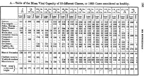
relation to the spirometer in order to perform the respiratory test. From J. Hutchinson, 1846, 236. . . 42 1.4 An example of Hutchinson’s tables. Here vital capacity is
pre-sented in relation to professional activity and stature. From J. Hutchinson, 1846, 156. . . 43 1.5 An example of Hutchinson’s charts. From J. Hutchinson, 1846,
155. . . 43 1.6 A direct sphygmograph designed by Marey. E.-J. Marey, La
cir-culation du sang à l’état physiologique et dans les maladies, Paris,
1881, 214. . . 49 1.7 Example of sphygmograms. E.-J. Marey, La circulation du sang
à l’état physiologique et dans les maladies, Paris, 1881, 216. . . . 50 2.1 Adiantum pedatum. Maidenhair fern, young rolled-up fronds
en-larged 8 times. Photogravure after K. Blossfeld, Urformen der
Kunst, Berlin, 1928, Plate 55. . . . 64 2.2 A man walking. Photogravure after Eadweard Muybridge, 1887.
Wellcome Library, London. . . 65 2.3 Example of signaletic photograph, A. Bertillon, Instructions
sig-nalétiques. Album, Melun, 1893. Wellcome Library, London. . . 74 2.4 Specimens of composite portraiture, F. Galton, Inquiries into
Hu-man Faculty and It’s Development, London, 1883, Frontispiece
illustration. Wellcome Library, London. . . 76
2.5 Patient at Surrey Asylum, case identified as “religious melan-choly.” Calotype by H.W. Diamond, 1852. . . 81 2.6 Patient at Surrey Asylum, case identified as “melancholia passing
on to mania.” Calotype by H.W. Diamond, 1852. . . 83 2.7 Patients at Surrey Asylum. Calotypes by H.W. Diamond, 1852. 86 2.8 Patient at Surrey Asylum. Calotype by H.W. Diamond, 1852. . 87 2.9 Female patients affected by impetigo (left) and pemphigus
fo-liaceus (right). A. Hardy and A. de Montméja, Clinique
pho-tographique de l’Hôpital Saint-Louis, Paris, 1868. . . . 92 2.10 Syphilitic alopecia (left). Pigmentation probably caused by
photo-sensitization due to a cosmetic product (right), F. Mehéux, ca. 1884-1893. . . 94 2.11 Muscular atrophy, Duchenne de Boulogne, Album de
photogra-phies pathologiques, Paris, 1862, Figure 9. Public domain. . . . 97 2.12 The facial expression of profound attention on the human face
being induced by electrical current. Duchenne de Boulogne, Le
Mécanisme de la physionomie humaine, Paris, 1862, Plate 1,
fig-ure 9. Wellcome Library, London. . . 98 2.13 Passional attitudes (ecstasy). Photograph by Bourneville and
P. Regnard, Iconographie photographique de la Salpêtrière, Paris, 1878, Plate 23. . . 103 2.14 Hystero-epileptic seizure. Photograph by Bourneville and P.
Reg-nard, Iconographie photographique de la Salpêtrière, Paris, 1879-1880, Plate 3. . . 103 2.15 Hysterical woman yawning. Photograph by A. Londe, Nouvelle
iconographie de la Salpêtrière, Paris, 1890. Wellcome Library,
London. . . 105 2.16 Episode of male hysteria. Photograph by A. Londe, Nouvelle
iconographie de la Salpêtrière, Paris, 1888. . . 106
3.1 Blood corpuscles. Etchings produced after the original daguerreo-types. A. Donné and L. Foucault, 1845. . . 112 3.2 Examples of chronophotographs by E.-J. Marey. . . 124 3.3 Images of a runner reduced to a system of bright lines for
rep-resenting the positions of the limbs (geometrical chronophoto-graph). Photograph by E-J. Marey, 1886. . . 125 3.4 Man with experimental shoe. E.-J. Marey, Le Movement, Paris,
3.5 Diagram of the sequence and duration of the footfalls during four different forms of walking. E.-J. Marey, 1894. . . 128 3.6 Diagram of the sequence and duration of the footfalls of a horse
at a full trot. E.-J. Marey, 1894. . . 128 3.7 Chronophotograph of a semicircular arc of polished brass rotating
around a vertical axis. E.-J. Marey, 1894. . . 132 3.8 The bones of a hand with a ring on one finger, viewed through
X-rays. Roentgen, W.K., 1895. Photoprint from radiograph. Wellcome Library, London. . . 136 3.9 J.M.W. Turner (1775-1851), Rain, Steam and Speed – The Great
Western Railway, 1844. National Gallery, London. . . 143
4.1 Examples of images produced using the Visible Human Project dataset. . . 164 4.2 Anatomical fugitive sheet bound at the end of Valverde,
Vi-vae imagines partium corporis humani aereis formis expressae.
Antwerp, 1566. Wellcome Library, London. . . 168 4.3 Table of correspondences between X-ray imaging and physical
examination of the thorax. From J.F. Halls Dally, 1903. . . 185 4.4 Table showing the range of mobility of the of the diaphragm.
From J.F. Halls Dally, 1903. . . 188 4.5 Examples of Halls Dally’s diagrammatic drawings representing
physical and radioscopic (fluoroscopic) examinations. From J.F. Halls Dally, 1903, 1805. . . 190 4.6 Example of gel electrophoresis autoradiograph (DNA fingerprint).
Credit: Alec Jeffreys, Wellcome Images. . . 197 5.1 Radiograph showing air-filled trachea and lungs, diaphragmatic
domes, mediastinal structures, and vascular markings. Arrows indicate costophrenic angles. Source: US Army medical training course. Wikimedia Commons. . . 216 5.2 Computed tomography scan of the brain, axial view. . . 217 5.1 Cluster heat map of lung tumor image features extracted from
Since its initial proposal, this dissertation has gone through a deep reformula-tion of both the object of study and the method of investigareformula-tion. Developed within the context of an interdisciplinary research project called Image in Sci-ence and Art,1 my early ideas for a doctoral thesis revolved around the artistic
use of medical imaging technologies. The objective was to understand how med-ical imaging has transformed our perception and first-person experience of the inner body, both at an individual and social level. The main question was to understand if our experience of the body changes, once we can look inside it without having to cut it open. Do we relate differently to our own body, and to the bodies of other people, if we perceive them as potentially transparent rather than ineluctably opaque? The working hypothesis was that the study of artworks related to medical imaging technologies could provide valuable insights into such problems. The rationale underlying this approach was that, as expres-sion of an individual, artworks can help disentangling questions related to body visualization and personal identity. At the same time, as cultural objects, they can be used as instruments of analysis to explore social notions of body and technology.2 Thus, the initial research plan did not focus on medical images per
se, but rather on the meanings these images acquire outside the medical setting.
It concerned the first-person perception of the inner body mediated by images, rather than the epistemology of medical imaging.3
1The FCT research project Image in Science and Art (PTDC/EAT/64201/2006), directed
by Prof. Olga Pombo, began in 2007 and ended in 2011 with the exhibition “CorpoImagem. Representações do Corpo na Ciência e na Arte”, held in February and March 2011 in Lisbon, at the Pavilhão do Conhecimento-Ciência Viva.
2See Coulombe, 2006.
3In the first year or so of my doctoral research, I attempted a comprehensive survey of the
artists who work with medical imaging. The task proved unattainable, but nevertheless it was possible to outline a general view of the multiple ways whereby visual artists encapsulate medical imaging in their work, both at the conceptual and formal level. The results of this early phase of the research were published in the article “Inside the body: Medical imaging and visual arts” (Di Marco, 2012a), and in a chapter of the book Representações do Corpo na
Ciência e na Arte (Azevedo Tavares, 2012) published within the context of the aforementioned
FCT research project. Both texts are in the Appendix attached in electronic format to this dissertation.
The change of conceptual focus derived from the need to extricate and clarify the multiple meanings that tend to grow around medical images. In fact, if an image arises not only different interpretations, but also different emotions,4 it
is because it is inherently ambiguous. In order to unravel the origins of this ambiguity, it was necessary to understand the material and conceptual genesis of medical imaging. That is, it was necessary to understand how such images are produced and endowed with meaning in their native context, clinical medicine. In the course of my research this historical, conceptual, and epistemological analysis became the central object of the dissertation. In other words, an inquiry into the extra-medical meaning of medical imaging morphed into a study on its epistemology. More precisely, it morphed into a study that aims at clarifying the relationship between the images produced by medical imaging technologies and the body they make visible.
Before saying more about the questions approached in this dissertation and the methodology employed, it is important to clarify what is meant by medical imaging. The term medical imaging refers to a vast array of images and imag-ing technologies developed since the end of the nineteenth century to provide indirect visual access to the inner organs (morphological imaging), and to some physiological or molecular processes (functional imaging). The first of such tech-nologies was radiography, invented by the German physicist Wilhelm Roentgen in 1895. Since then, a number of different imaging modalities have emerged, and they have completely redefined the visibility of the interior of the living body. Nowadays Computed Tomography (CT), Magnetic Resonance Imaging (MRI) and ultrasound scanning allow visualizing inner structures with astonishing de-tail, while Positron Emission Tomography (PET) and functional MRI (fMRI) allow visualizing the metabolic activity of different biological tissues, including the brain. Medical imaging is generally used for diagnostic aims. However, it can also serve research purposes, as in the case of neuroimaging in cognitive science, or rather mundane goals, as in the case of fetal ultrasound, which is routinely performed not only to check the health conditions of the fetus, but also to let parents enjoy the experience of seeing their prospective baby on the screen.5 In my study I focus on the diagnostic use of medical imaging. This is
4The art historian and image theorist James Elkins remarks that, unlike other scientific
images, which can be strictly informational, medical images tend to show vestiges of expressive meaning, because they can evoke questions of life and death, gender and sexuality, pleasure and pain. See Elkins, 1995, 556.
5See Chudleigh, 1999; Mitchell, 2001; van Dijck, 2005, Ch. 6. Fetal sonograms are
partic-ularly interesting images, from both an epistemological and sociological perspective. Before the introduction of ultrasound scanning for pregnancy monitoring, the presence of the unborn could be felt only by the pregnant woman. Since it has become visible, however, the fetus has acquired a new status. On the one hand, it has become an object of study, as well as a medical subject (fetal medicine). On the other hand, it has acquired a new identity and individuality,
why I will often use the expression diagnostic images. I also employ the des-ignations mechanical images, radiological images, and medical images as loose synonyms, even though, strictly speaking, the term medical images encompasses a much larger domain of images, including, for instance, anatomical drawings.
A peculiarity of medical imaging is that the images it creates seem to ren-der the body transparent. Intuitively, we think that the medical relevance of diagnostic images depends on the fact that they are faithful representations of the inner body, but, as soon as we set out to study them in terms of diagnostic instruments and, more generally, in terms of cognitive objects, we come across a number of problems: What do these images represent, exactly? And how do they represent? Do they work by mimesis? If we do not know how their refer-ent looks like, how can we trust their represrefer-entational value? Are these images portraits of the inner body? Or are they maps? If they are portraits, are they the portrait of someone (a person) or of something (a disease)?
These questions, present already in the first version of my doctoral project, turned out to be much more difficult to answer than I had initially assumed. In fact, although there are several works on medical imaging from the per-spective of cultural studies6 and anthropology,7 very little has been published
about medical imaging epistemology. Indeed, unlike historians and sociologists, philosophers of science have traditionally neglected images and, until relatively recently, in both continental and analytic philosophy the reflection on visual rep-resentation has been a preserve of aesthetics and art theory.8 For what concerns
the specific domain of medical imaging, the philosophers of science who have paid attention to this topic have typically focused on the use of neuroimaging in cognitive science and, to a lesser extent, psychiatry.9 However, to the best of my
entering as a silent actor into a set of relationships between medical practitioners, parents and society. The effects of this transformation on the creation of a new social and juridical subject has been widely investigated by sociological and anthropological literature. Feminist authors, in particular, have repeatedly analyzed the role of fetal scans in the increased medicalization and commercialization of pregnancy, in the strengthening of ideologies of good motherhood, in the creation of the fetus as an autonomous individual independent from the mother’s body, and in the debates surrounding abortion. See, for instance, Petchesky, 1987; Duden, 1993; Taylor, 1998 and 2008; Morgan and Michaels, 1999; Morgan, 2009; Roberts, 2012.
6See Zwijnenberger and van de Vall, 2009; van Dijck, 2005; Natale, 2008, 2011, 2012;
Stephens, 2012.
7See 2004; Radstake, 2009; Müller-Rostock, 2009; Estival, 2009, 2010.
8This situation has been changing over the last decade. See, for instance, Pombo and
Di Marco, 2010; Pombo and Gerner, 2010; Carusi, 2011, 2012. In the analytic tradition see French, 2003; Perini, 2005ab, 2006, 2012.
9See Kosslyn, 1999; Bogen, 2002; Taraborelli, 2003; Roskies, 2007; Huber, 2009; Huber
and Huber, 2009, Mole and Klein, 2010; Klein, 2010. Even in the book Medical Imaging and
Philosophy. Challenges, Reflections and Actions (Fangerau et al., 2012), all the articles that
knowledge, virtually nothing has been published on the philosophy of medical imaging as a diagnostic instrument.10
Objectives and methodology The main objective of this dissertation is
to contribute to the development of an epistemology of medical imaging. My central thesis is that medical imaging does not merely produce more or less accurate pictures of the inner body, it rather turns the body into a scientific object by transforming its very visibility. Simultaneously it aims at reducing illness to a visual entity. Medical imaging does not simply make visible the inner organs, it actually presents the body in such a way that it becomes possible to extract relevant diagnostic information from it. This is why I maintain that the images produced by medical imaging technologies are more akin to simulations than to portraits. The imaging apparatus turns the body into a visual object that can be observed under experimental conditions: unlike the real body, it can be filed, retrieved, shared, measured and manipulated in several ways.
This thesis is accompanied by two others: first, diagnostic images, as all scientific images, are actual cognitive instruments. They are epistemic objects inscribed within theoretical contexts and experimental practices. Second, an image of the inner body has diagnostic meaning and value only in the scope of a specific conceptualization of the body and disease. This means that, in order to put forward an epistemology of medical imaging, to develop a the-ory of technology-mediated images and technology-mediated perception is not enough. One must also clarify the medical concepts and practices that pro-vide the substratum for the whole process of production and signification of diagnostic images.
Medical imaging is to be understood both as an imaging technology and as a diagnostic practice, hence, as an object of philosophical analysis it must be approached from different perspectives. For this reason in my research I resorted to literature from a variety of disciplinary fields, such as image theory, semiotics, history and philosophy of medicine, and history and philosophy of science. The aim was to put forward an epistemology of medical imaging that accounts for the poietic rather than mimetic nature of visual representation, as well as for the fact that through medical imaging the human body is embedded
10An exception is the doctoral dissertation Signal into Vision: Medical Imaging as
In-strumentally Aided Perception, defended by Nola Semczyszyn at the University of British
Columbia in 2010. Drawing mostly on analytic philosophy of art and perception, Semczyszyn appeals to theories of pictorial representation to explain how medical imaging represents and how we access its content. I refer to her work in Chapter 5, Section 5.2.2. Overton et al., 2011, focuses on the problem of the relation between diagnostic images and verbal language, and the creation of ontologies associated to software tools to improve communication among radiologists, other health professionals, and patients.
into an empirical apparatus, which encompasses medical instruments, medical knowledge, and clinical practices.
For what concerns the methodology, I relied chiefly on historical and con-ceptual analysis. On the one hand, I studied the genesis, development and transformation of concepts such as disease and diagnosis; on the other hand, I critically applied philosophical concepts developed by different authors to prob-lematize and examine my objects of research (e.g., the production of mechanical images and the visualization of the invisible). The study of original scientific documents, such as medical treatises and medical journals’ articles was indis-pensable for understanding how medical imaging has progressively imposed itself as a fundamental diagnostic method, and how illness has become a visual object. Thus, I relied on documents’ analysis to unravel how the diagnostic meaning of images was generated in the scope of actual scientific and clinical practices and debates. The emphasis on material scientific practices was reinforced, whenever possible, by resorting to literature from laboratory ethnology and anthropol-ogy.11 Also, since this work is about images, I devoted much attention to the
iconographic apparatus of the documents I examined, employing iconographic and semiotic analysis.
As mentioned above, given the multifaceted nature of the epistemological problems posed by medical imaging, I had to draw on concepts and ideas devel-oped by a variety of thinkers. In particular, I follow Charles Sanders Peirce’s truth-value theory of photography to better understand how mechanically pro-duced images were conceptualized in the nineteenth and early-twentieth cen-turies.12 Moreover, I resort to his semiotics, in particular his classification of
signs into indices, icons and symbols, to develop a tool for semiotic analysis that I employ at various points of the dissertation to investigate the processes of signification of different kinds of instrument-generated images that visualize invisible referents (from microphotographs to radiographs and PET scans).
I explore at length the idea that optical media such as photography and cin-ema are prostheses that enrich and transform our sensorial experience through Walter Benjamin’s notion of optical unconscious. Benjamin remarked that pho-tography and cinema allow for manipulations of space and time that are pre-cluded to natural perception. Consequently, these imaging technologies endow us with enhanced mechanical senses that allow exploring completely new facets
11See Chapter 4, Section 4.2.3, and Chapter 5, Section 5.2.3.
12In my study I payed much attention to photography because when radiography appeared,
at the turn of the nineteenth century, it was perceived as a particular kind of photography. Hence, to understand how radiography was conceptualized, it is necessary to look at the photographic practices and theories of the time.
of nature.13 I combine the concept of optical unconscious with Peirce’s
semi-otics to discuss the problem of the invisible referent, that is, the question of how we can make sense of images that are meant to visualize invisible phenom-ena. Besides the idea of optical unconscious, two other aspects of Benjamin’s reflection on photography and cinema proved fruitful for my investigation on medical imaging. One is his seminal discussion of the cognitive effects of the mechanical reproduction of images; the second is his account of the objectify-ing nature of photography, which he compares to the dissectobjectify-ing activity of the surgeon. I draw on his considerations on these subjects to elaborate an account of the role played by photographic series in creating a new clinical gaze, and to reflect upon the role played by photography and radiography in turning the human body into a scientific object.
The idea of mechanical objectivity, developed by the historians Lorraine Das-ton and Peter Galison,14 offers me the opportunity to engage with the problems
of image realism and objectivity through polemical reasoning. These authors developed an articulated taxonomy of scientific images aimed at demonstrating the historical character of objectivity, which should be considered one of the many epistemic ideals that can drive scientific work in different historical pe-riods. Mechanical objectivity, they argue, has been the main epistemic ideal between the 1830s and the 1920s. It prescribed the strenuous suppression of the subjectivity of the image makers, and put harsh limitations on image interpre-tation, too. In order to refute this account of what it means for an image to be objective, and to clarify what sort of photographic realism was embraced by the scientists of the late-nineteenth and early-twentieth centuries, I develop a number of arguments concerning the relationship between objectivity and inter-subjectivity, as well as between mechanically produced images and imagination. Similarly, Daston and Galison’s characterization of virtual and haptic images gives me the opportunity to critically delve into the problem of the embodied and material dimension of scientific images’ production and fruition.
A concept I came across in a late phase of my research is Gaston Bachelard’s “phenomenotechnique.”15 Bachelard developed the idea of phenomenotechnique
to account for the fact that, in advanced physics and chemistry, scientific en-tities (e.g., the Zeeman effect, perfect crystals, atomic isotopes) are not found
13See Benjamin, 1939, 266.
14The historiographic work of Daston and Galison is guided by strong epistemological
as-sumptions. Indeed, together with the philosopher Ian Hacking, they are considered among the most prominent exponent of so-called historical epistemology, an approach to the study of scientific knowledge that emphasized the historical development of scientific concepts and objects, as well as of scientific disciplines and styles of reasoning. See Hacking; 2002, Kusch, 2011; Sturm, 2011.
in nature ready made. They must be produced in the laboratory as technical phenomena. They are the materialization of mathematical rationality or, as Bachelard put it, of a mathematical noumenon. In other words, through phe-nomenotechnique, science creates its own phenomena in a process of progressive rationalization of the real. Through this process science leaves behind the world as it is understood by commonsensical experience and received mental habits, and sets free “a surrationalism that will multiply the occasions for thinking.”16
If rigorously interpreted, the notion of phenomenotechnique cannot be applied to medical imaging and diagnostic images, for reasons I discuss in detail in the dissertation. Yet, the analysis of this concept offered me an additional stimulus for thinking medical imaging. In fact, not only it provides specific insights for reflecting on contemporary medical imaging, wherein mathematical algorithms for image acquisition and reconstruction play a fundamental role; more impor-tantly, it works as an organizing concept, which helps bring together and refine the different ideas and intuitions developed in the various steps of my research.
Plan of the dissertation The dissertation begins with an analysis of the
his-torical and conceptual conditions of possibility of medical imaging. By outlining an archaeology of radiography, I will try to understand what medical theories and practices had to be at work in the nineteenth century, for a technology that produced shadow-images of the inner body to be perceived and employed as a di-agnostic instrument. I will suggest that when radiography appeared, at the turn of the twentieth century, it engendered less a theoretical than a technological revolution, because its diagnostic relevance was grounded in the conceptual-ization of body, disease and diagnosis put forward by clinical anatomy at the end of the eighteenth century. Following Michel Foucault’s The Birth of the
Clinic, I will maintain that it was with the work of the French clinicians Xavier
Bichat and Jean-Nicolas Corvisart that diagnosis became a matter of eliciting signs from the inner body and that visibility became a fundamental epistemo-logical and perceptual category of medicine.17 I will also defend the idea that
the stethoscope, invented by Théophile Laennec in 1816, was the material and intellectual predecessor of radiography, because it introduced a primitive form of mediated perception in medical diagnosis, and allowed the clinician to ex-plore from the outside the inner body of the living patient, extracting signs of disease.18 In the course of the nineteenth century, several ways of understanding
16Bachelard, 1936, 7, my translation. 17See Foucault, 1963, 166.
18The stethoscope is used to auscultate the patient. Auscultation is a basic clinical
ex-amination. It consists of listening to the sounds produced by the heart and by the air that circulates in the respiratory organs.