R E S U M O Introdução: A grelina, hormona descoberta
em 1999, é o ligando endógeno do receptor dos secretagogos da hormona de crescimento (GHS-R1a). Estudos recentes sugerem que possa regular de forma importante a função do coração normal e insuficiente. No entanto, tem sido difícil distinguir os seus efeitos miocárdicos directos dos indirectos, secundários à libertação de hormona de crescimento e à acção vasomotora. Neste trabalho, estudámos os efeitos contrácteis da grelina e a expressão do seu receptor GHS-R1a no miocárdio normal e hipertrófico.
Métodos: Ratos Wistar adultos foram
submetidos aleatoriamente a uma injecção sc de monocrotalina (MCT; n = 9; 60 mg/kg) ou veículo (n = 7; 1 ml/kg). Três semanas depois, após avaliação hemodinâmica do ventrículo direito (VD), procedeu-se à colheita de amostras da parede livre do VD e à dissecção dos músculos papilares do mesmo ventrículo. Os efeitos miocárdicos da grelina foram avaliados em músculos papilares do VD de ratos normais (n = 7) e injectados com MCT (n = 9), pela adição de um fragmento activo da grelina (fG, 10-6M) à solução superfusora.
A expressão de ARNm do GHS-R1a foi determinada em amostras da parede livre do VD de ratos normais (n = 7) e injectados com MCT (n = 9) por real time RT-PCR.
Resultados: No grupo Normal, o fG provocou
uma redução de 27,9 ± 4,0 % da tensão activa (TA), de 28,5 ± 6,7 % da velocidade máxima de contracção (dT/dtmax) e de 21,4 ± 4,2 % da velocidade máxima de relaxamento (dT/dtmin). No grupo MCT, o fG provocou uma redução
Efeitos Contrácteis da Grelina e Expressão
do seu Receptor GHS-R1a
no Miocárdio Normal e Hipertrófico
[88]
JOÃO-BRUNOSOARES,AMÂNDIOROCHA-SOUSA,PAULOCASTRO-CHAVES,TIAGOHENRIQUES-COELHO,
ANDRÉPEDROLOURENÇO,ROBERTORONCON-ALBUQUERQUE JR.,ADELINOF. LEITE-MOREIRA
Serviço de Fisiologia da Faculdade de Medicina da Universidade do Porto, Porto, Portugal
1235
A B S T R A C T
Contractile Effects of Ghrelin and Expression of its Receptor GHS-R1a in Normal and Hypertrophic Myocardium Introduction: Ghrelin, isolated in 1999, is an
endogenous ligand for the growth hormone secretagogue receptor (GHS-R1a). Recent studies suggest that it may influence the function of normal and failing hearts. Nonetheless, it has been difficult to differentiate its effects on the intrinsic properties of the myocardium from the secondary effects resulting from growth hormone release and vasomotor action. This study investigated the contractile effects of ghrelin and expression of its receptor GHS-R1a in normal and hypertrophic myocardium.
Methods: Adult Wistar rats randomly received
monocrotaline (MCT; n = 9; 60 mg/kg, sc) or vehicle (n = 7; 1 ml/kg). Three weeks later, after right ventricular (RV) hemodynamic evaluation, the effects of 10-6M of a
pentapeptide active fragment of ghrelin (fG) were tested on contractile parameters of RV papillary muscles (Normal, n = 7; MCT, n = 9). GHS-R1a mRNA expression was estimated in RV transmural free-wall samples (Normal, n = 7; MCT, n = 9), using real-time RT-PCR.
Results: In the Normal group, fG reduced
active tension (AT), maximum velocity of tension rise (dT/dtmax) and maximum velocity of tension decline (dT/dtmin), by 27.9 ± 4.0 %, 28.5 ± 6.7 % and 21.4 ± 4.2% respectively. In the MCT group, fG reduced AT, dT/dtmaxand dT/dtmin, by 24.1 ± 6.3 %, 24.3 ± 6.5 % and 24.5 ± 6.1 % respectively. GHS-R1a mRNA Recebido para publicação: Agosto de 2005 • Aceite para publicação: Setembro de 2005
Received for publication: August 2005 • Accepted for publication: September 2005
INTRODUÇÃO
A
grelina é um peptídeo formado por 28 ami-noácidos (aa), com um grupo acilo ligado ao terceiro a.a (Ser), e produzido fundamental-mente pelo estômago(1). Foi, recentemente, identi-ficada como o ligando endógeno do receptor dos se-cretagogos da hormona de crescimento (GHS-R1a)(1). Este receptor é expresso essencialmente na hi-pófise e hipotálamo e é responsável pela secre-ção da hormona de crescimento (HC) induzida pela grelina(1, 2).Estudos recentes demonstraram a expressão de ARNm de grelina e GHS-R1a a nível do co-ração, sugerindo que a grelina possa influenciar a função cardíaca, independentemente da liber-tação de HC(2). Essa hipótese foi já confirmada em estudos subsequentes. Em voluntários sau-dáveis e doentes com insuficiência cardíaca (IC), a administração de grelina diminuiu a re-sistência vascular periférica e aumentou o dé-bito cardíaco, sem alteração significativa da frequência cardíaca(3). Em doentes com IC, a administração repetida de grelina melhorou a função do ventrículo esquerdo e a capacidade de exercício e diminuiu a atrofia muscular(4). A administração repetida de grelina atenuou tam-bém a hipertensão pulmonar e a hipertrofia do ventrículo direito (VD) em ratos com hiperten-são pulmonar induzida pela monocrotalina (MCT)(5).
Relativamente à modulação da função mio-cárdica pela grelina, tem sido difícil distinguir 1236
INTRODUCTION
G
hrelin is a peptide consisting of 28 amino acids (aa), with an acyl group attached to the third aa (serine), and produced mainly in the stomach(1). It was recently identified as the endogenous ligand for the growth hormone se-cretagogue receptor (GHS-R1a)(1). This receptor is mainly expressed in the pituitary gland and hypothalamus and is responsible for secretion of growth hormone (GH) induced by ghrelin(1, 2).Recent studies have shown expression of mRNA of ghrelin and GHS-R1a in the heart, suggesting that ghrelin may influence cardiac function, independently of GH release(2), a hypothesis that has been confirmed in subse-quent studies. In healthy volunteers and pa-tients with heart failure (HF), administration of ghrelin reduced peripheral vascular resistance and increased cardiac output, with no signifi-cant change in heart rate(3). In HF patients, re-peated administration of ghrelin improved left ventricular function and exercise capacity and reduced muscular atrophy(4). Repeated adminis-tration has also been shown to reduce pulmo-nary hypertension and right ventricular (RV) hypertrophy in rats with pulmonary hyperten-sion induced by monocrotaline (MCT)(5).
With regard to the modulating effect of ghre-lin on myocardial function, it has been difficult to differentiate its effects on the intrinsic prop-erties of the myocardium from the secondary effects resulting from GH release and vasomo-de 24,1 ± 6,3 % da TA, vasomo-de 24,3 ± 6,5 % da
dT/dtmaxe de 24,5 ± 6,1 % da dT/dtmin. A expressão do receptor da grelina foi
semelhante nos dois grupos (Normal: 2,3*105± 5,4*104; MCT: 3,0*105± 1,1*105; p > 0,05).
Conclusão: Demonstrámos que a grelina tem
efeitos inotrópico e lusitrópico negativos no miocárdio normal, que se encontram preservados, à semelhança da expressão do seu receptor, na hipertrofia ventricular direita, sugerindo a sua potencial utilização como novo alvo terapêutico no contexto da progressão para a insuficiência cardíaca.
expression was similar in the two groups (Normal: 2.3*105± 5.4*104; MCT: 3.0*105± 1.1*105; p > 0.05).
Conclusion: This study shows that ghrelin has
negative inotropic and lusitropic effects. These effects and expression of its receptor are preserved in RV hypertrophy, suggesting that ghrelin may be a new target in
progression to heart failure.
Palavras-Chave Grelina; GHS-R1a; Miocárdio; Hipertrofia
Key words
os seus efeitos directos dos indirectos, secundá-rios à libertação de HC e à acção vasomotora (vasodilatação sistémica e vasoconstrição coro-nária)(1, 6, 7). Recentemente, um estudo atribuiu à grelina uma acção inotrópica negativa sobre o miocárdio normal(8). Quanto aos efeitos contrác-teis da grelina sobre o miocárdio hipertrófico, não existe qualquer estudo.
No presente trabalho, estudámos os efeitos contrácteis directos da grelina e a expressão do seu receptor no miocárdio normal e hipertró-fico.
MATERIAL E MÉTODOS
A experimentação animal foi realizada de acordo com a Lei Portuguesa para o bem-estar animal. Foram utilizados machos de ratos Wistar adultos.
Modelo experimental de hipertrofia ventricular direita (VD)
Para obtermos hipertrofia VD utilizámos o modelo da hipertensão pulmonar induzida pela MCT(9).
Os animais receberam aleatoriamente uma injecção sc de MCT (n = 9, 60 mg/kg) ou vo-lume idêntico de veículo (n = 7, 1 ml/kg). Três semanas depois, foram sujeitos a uma avaliação hemodinâmica do VD.
Resumidamente, os animais foram anestesia-dos com pentobarbital, traqueostomizaanestesia-dos para ventilação mecânica e cateterizados na veia ju-gular direita, sob visualização microscópica. Procedeu-se depois a uma esternotomia media-na e pericardiotomia para exposição do coração. De seguida introduziu-se um micromanómetro de pressões na cavidade VD para avaliação he-modinâmica. Foram analisados os seguintes pa-râmetros: pressão telediastólica (PTD), pressão sistólica máxima (Pmax), velocidades máximas de subida (dP/dtmax) e de queda (dP/dtmin) de pres-são e constante de tempo do relaxamento isovo-lumétrico tau (τ).
Após a colecção dos dados hemodinâmicos, procedeu-se a cardiectomia para colheita de amostras da parede livre do VD e dissecção dos músculos papilares do mesmo ventrículo.
Efeitos miocárdicos da grelina
Os músculos papilares (Grupo Normal, n = 7; Grupo MCT, n = 9) foram colocados num banho
de 10 mL contendo uma solução de Tyrodes 1237
tor action (systemic vasodilation and coronary vasoconstriction)(1, 6, 7). In a recent study, ghrelin was shown to have a negative inotropic action on normal myocardium(8). There have been no studies of its contractile effects on hypertrophic myocardium.
The present study investigated the direct contractile effects of ghrelin and expression of its receptor in normal and hypertrophic myocar-dium.
METHODS
The animal experiment was conducted in accordance with Portuguese animal welfare law and used adult male Wistar rats.
Experimental model of right ventricular hypertrophy (RV)
Monocrotaline-induced pulmonary hyper-tension was used to model RV hypertrophy(9). The animals randomly received a subcuta-neous injection of MCT (n = 9, 60 mg/kg) or a similar volume of vehicle (n = 7, 1 ml/kg). Three weeks later, they underwent RV hemodynamic evaluation.
The animals were anesthetized with pento-barbital, tracheotomized for mechanical ventila-tion and the right jugular vein catheterized under microscopic visualization. Median ster-notomy and pericardiotomy were performed to expose the heart, and a pressure micromanome-ter was then inserted into the RV for hemodyna-mic evaluation. The following parameters were determined: end-diastolic pressure (PED), maxi-mum systolic pressure (Pmax), maximaxi-mum velocity of pressure rise (dP/dtmax) and decline (dP/dtmin) and isovolumetric relaxation time (τ).
Following collection of hemodynamic data, a cardiectomy was performed to collect samples of the RV free wall, as well as dissection of the RV papillary muscles.
Myocardial effects of ghrelin
The papillary muscles (Normal group, n = 7; MCT group, n = 9) were placed in a 10 mL bath containing a modified Tyrode solution (in mM: NaCl 120, KCl 5.4, MgCl21.05, NaH2PO4 0.42, CaCl2 1.25, NaHCO3 22.6, glucose 5, ascorbic acid 0.28, EDTA 0.05 and atenolol 0.02), at 35 ºC, together with a cardioplegic agent (BDM, 3 %) and bovine serum (5 %), in equilibrium with 95 % CO2 and 5 % O2to maintain pH be-tween 7.36 and 7.42. After connecting the
mus-modificada (em mM: NaCl 120, KCl 5.4, MgCl2 1.05, NaH2PO40.42, CaCl21.25, NaHCO322.6, Glucose 5, Ascorbic acid 0.28, EDTA 0.05 e Atenolol 0.02), a 35 ºC, com uma substância car-dioplégica (BDM, 3 %) e soro de vitelo (5 %) e em equilíbrio com 95 % de CO2e 5 % de O2de modo a manter o pH entre 7.36 e 7.42. Após conecção dos músculos a um transdutor isomé-trico/isotónico, foi determinada a pré-carga com base nas dimensões dos músculos e fixada a frequência de estimulação em 1Hz. Vinte minu-tos depois, a solução superfusora foi substituída por uma solução semelhante, sem BDM. Uma hora após, a solução foi novamente substituída, agora por uma sem soro de vitelo. Após a deter-minação do Lmax e a obtenção de 2 contracções isométricas e isotónicas sobreponíveis, foi ini-ciado o protocolo experimental. Este consistiu na adição, à solução superfusora, de um fragmento activo da grelina (fG, Peptides International), de modo a obter no banho uma concentração de 10-6M, com registo subsequente da perfomance muscular. Os parâmetros analisados incluíram a tensão activa (TA) e as velocidades máximas de subida (dT/dtmax) e de queda (dT/dtmin) da tensão muscular. Os dois primeiros permitiram-nos avaliar os efeitos inotrópicos e o último os efeitos lusitrópicos (sobre o relaxamento) da grelina.
Expressão de ARNm do GHS-R1a
Os níveis de ARNm de GHS-R1a foram quantificados em amostras da parede livre do VD de ratos dos grupos Normal (n = 7) e MCT (n = 9) por Real-Time RT-PCR. Esta metodolo-gia foi descrita de forma mais pormenorizada em estudo prévio(5). Os primers utilizados são apresentados no Quadro I.
Análise estatística
Os valores são apresentados como média ± erro padrão em termos absolutos ou como per-centagem de variação em relação ao controlo
1238
cles to an isometric/isotonic transducer, preload was determined based on their size and the sti-mulation frequency fixed at 1 Hz. Twenty minu-tes later, the superfusion solution was replaced with one of a similar composition but without BDM. After an hour, the solution was again re-placed, this time with one without bovine se-rum. After determining Lmax and obtaining two similar isometric and isotonic contractions, the experimental protocol was begun. This consis-ted of adding an active fragment of ghrelin (fG, Peptides International) to the solution, to obtain a concentration of 10-6M in the bath, and sub-sequent recording of muscle performance. The parameters analyzed included active tension (AT) and maximum velocity of tension rise (dT/dtmax) and tension decline (dT/dtmin). The first two parameters were used to assess the inotropic effects of ghrelin, and the third its lu-sitropic (relaxation) effects.
GHS-R1a mRNA expression
Levels of GHS-R1a mRNA were estimated in RV transmural free-wall samples in the Nor-mal group (n = 7) and MCT group (n = 9) by real-time RT-PCR. The methodology has been described in detail in a previous study(5). The primers used are given in Table I.
Statistical analysis
Values are presented as means ± standard error in absolute terms or as the percentage of variation from the controls (when indicated). The groups were compared using the unpaired Student’s t test. Differences were considered significant when p < 0.05.
RESULTS
Experimental model of RV hypertrophy
The results of right ventricular morphome-tric and hemodynamic evaluation are shown in
Table II. The MCT group presented higher RV
Quadro I
Sequência nucleotídica dos primers usados no Real Time RT-PCR
Primer Sequência
Reverse 5’-AGG AAA GTA CAG GGA CCA G-3’
Forward 5’-CAC CCA GAG GAC AGA GGA CAA-3’
Forward-standard 5’-GCG AAT TAA CCC TCA CTA AAG GGA CAC CCA GAG GAC AGA GGA CAA GGC ATC AAG CTG TCA GGA-3’
Table I
Nucleotide sequence of primers used in real-time RT-PCR
Primer Sequence
Reverse 5’-AGG AAA GTA CAG GGA CCA G-3’
Forward 5’-CAC CCA GAG GAC AGA GGA
CAA-3’
Forward-standard 5’-GCG AAT TAA CCC TCA CTA AAG
GGA CAC CCA GAG GAC AGA GGA CAA GGC ATC AAG CTG TCA GGA-3’
(quando indicado). A comparação dos grupos foi feita com um teste t não emparelhado. As diferenças foram consideradas significativas quando p < 0,05.
RESULTADOS
Modelo experimental de hipertrofia VD
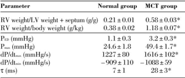
Os resultados referentes à avaliação morfo-métrica e hemodinâmica do VD são apresenta-dos no Quadro II. O grupo MCT apresentou maiores relações Peso VD/Peso VE + Septo e Peso VD/Peso Corporal. Quando comparado com o grupo Normal, o grupo MCT apresentou também maiores PTD, Pmax, dP/dtmax e constante de tempo de relaxamento isovolumétrico tau (τ). Não se registaram diferenças significativas rela-tivamente à dP/dtmin.
Efeitos miocárdicos da grelina
A função miocárdica basal e a caracteriza-ção morfológica dos músculos papilares utiliza-dos são apresentadas no Quadro III. Apesar das diferenças não serem significativas,
observou-1239 weight/LV weight + septum and RV weight/body weight ratios, as well as higher PED, Pmax, dP/dtmax and isovolumetric relaxation time (τ). There were no significant differences with regard to dP/dtmin.
Myocardial effects of ghrelin
Baseline myocardial function and morpholo-gical characteristics of the papillary muscles used are presented in Table III. Although the differences were not significant, there was a tendency for increased AT, dT/dtmaxand dT/dtmin in the MCT group compared to the Normal group.
In the papillary muscles of the Normal group, fG induced negative inotropic and lusi-tropic effects (Fig. 1), reducing AT, dT/dtmaxand dT/dtmin, by 27.9 ± 4.0 %, 28.5 ± 6.7 % and 21.4 ± 4.2 % respectively compared to the con-trols (Fig. 1).
In the MCT group, the fG-induced reduction in AT, dT/dtmax and dT/dtmin (respectively 24.1 ± 6.3 %, 24.3 ± 6.5 % and 24.5 ± 6.1 %) was not statistically different from that found in the Normal group (Fig. 1).
Quadro II
Avaliação morfométrica e hemodinâmica do ventrículo direito nos grupos Normal e MCT
Parâmetro Grupo Normal Grupo MCT
Peso VD/Peso VE + Septo (g/g) 20,21 ± 0,01 20,58 ± 0,03*
Peso VD/Peso Corporal (g/kg) 20,38 ± 0,02 21,18 ± 0,07*
PTD(mmHg) 221,1 ± 0,32 223,2 ± 0,3*2
Pmax(mmHg) 224,6 ± 1,82 249,4 ± 1,7*2
dP/dtmax(mmHg/s) ,1227 ± 80,2 ,1616 ± 102*,
dP/dtmin(mmHg/s) ,– 909 ± 110, ,– 1088 ± 59,222
τ (ms) 222,7 ± 1,22 22,28 ± 3*,22
VD: Ventrículo Direito; VE: Ventrículo Esquerdo; MCT: Monocrotalina; PTD: Pressão Telediastólica; Pmax: Pressão Sistólica Máxima; dP/dtmax: Veloci-dade Máxima de Subida da Pressão; dP/dtmin: VelociVeloci-dade Máxima de Queda da Pressão; e τ: Constante de Tempo de Relaxamento Isovolumétrico Tau. Valores apresentados como Média ± Erro Padrão.* p < 0,05: MCT vs Nor-mal.
Table II
Right ventricular morphometric and hemodynamic evaluation in Normal and MCT groups
Parameter Normal group MCT group
RV weight/LV weight + septum (g/g)20.21 ± 0.01 20.58 ± 0.03* RV weight/body weight (g/kg) 20.38 ± 0.02 21.18 ± 0.07* PED(mmHg) 221.1 ± 0.32 223.2 ± 0.3*2 Pmax(mmHg) 224.6 ± 1.82 249.4 ± 1.7*2 dP/dtmax(mmHg/s) ,1227 ± 80,2 ,1616 ± 102*, dP/dtmin(mmHg/s) ,– 909 ± 110, ,– 1088 ± 59,222 τ (ms) 222,7 ± 1,22 22,28 ± 3*,22
RV: Right ventricle; LV: Left ventricle; MCT: Monocrotaline; PED: End-diastolic pressure; Pmax: Maximum systolic pressure; dP/dtmax: Maximum velocity of pressure rise; dP/dtmin: Maximum velocity of pressure decline; τ: Isovolumetric relaxation time. Values presented as means ± standard error. * p < 0.05: MCT vs. Normal.
Quadro III
Caracterização morfológica e contráctil
dos músculos papilares dos grupos Normal e MCT
Parâmetro Grupo Normal Grupo MCT
n 7 9 Comprimento (mm) 222,2 ± 0,22 222,3 ± 0,32 Peso (mg) 220,6 ± 0,32 220,7 ± 0,42 Pré-carga (mN) 223,0 ± 1,12 223,3 ± 0,72 TA (mN/mm2) 211,2 ± 1,52 218,7 ± 3,92 dT/dtmax(mN/mm2/s) 179,7± 25,0 292,7 ± 54,2 dT/dtmin(mN/mm2/s) – 142,2 ± 22,52 –206,3 ± 37,82
MCT: Monocrotalina; TA: Tensão Activa; dT/dtmax, dT/dtmin: Velocidades Máximas de Subida e de Queda da Tensão, respectivamente. Valores apre-sentados como Média ± Erro Padrão.
Table III
Morphological and contractile characteristics of papillary muscles in Normal and MCT groups
Parameter Normal group MCT group
n 7 9 Length (mm) 222.2 ± 0.22 222,.3 ± 0.32 Weight (mg) 220.6 ± 0.32 220.7 ± 0.42 Preload (mN) 223.0 ± 1.12 223.3 ± 0.72 AT (mN/mm2) 211.2 ± 1.52 218.7 ± 3.92 dT/dtmax(mN/mm2/s) 179.7± 25.0 292.7 ± 54.2 dT/dtmin(mN/mm2/s) – 142.2 ± 22.52 –206.3 ± 37.82
MCT: Monocrotaline; AT: Active tension; dT/dtmax, dT/dtmin: Maximum velo-cities of tension rise and decline respectively. Values presented as means ± standard error.
-se uma tendência para o aumento da TA, dT/dtmax e dT/dtmin no grupo MCT, comparativamente ao grupo Normal.
Nos músculos papilares do grupo Normal, o fG induziu efeitos inotrópico e lusitrópico nega-tivos (Fig. 1). A adição de fG produziu uma di-minuição da TA, dT/dtmax e dT/dtmin, respectiva-mente, de 27,9 ± 4,0 %, 28,5 ± 6,7% e 21,4 ± 4,2 %, relativamente ao controlo (Fig. 1).
Nos músculos papilares do grupo MCT, a di-minuição da TA, dT/dtmax e dT/dtmin (respectiva-mente, 24,1 ± 6.3 %, 24,3 ± 6,5 % e 24,5 ± 6,1 %) induzida pelo fG não foi estatisticamente dife-rente da observada no grupo Normal (Fig. 1).
Expressão de ARNm do GHS-R1a
Foi detectada a expressão de ARNm do GHS-R1a nos grupos Normal e MCT. O nível dessa expressão não diferiu significativamente entre os grupos Normal (2,3*105± 5,4*104) e MCT (3,0*105± 1,1*105).
DISCUSSÃO
A fracção activa da grelina é formada ape-nas pelos primeiros quatros a.a. Uma vez que o fragmento que utilizámos é formado pelos cinco primeiros a.a, os resultados obtidos serão idên-ticos aos obtidos com o peptídeo completo(5, 10). Com o presente trabalho demonstrámos que a grelina é capaz de regular de forma directa (isto é, independente da libertação de HC e da acção vasomotora) e aguda a função sistólica e diastólica do miocárdio, exercendo sobre este 1240
GHS-R1a mRNA expression
GHS-R1a mRNA expression was found in both groups, its level not differing significantly between the Normal group (2.3*105± 5.4*104) and the MCT group (3.0*105± 1.1*105).
DISCUSSION
The active fragment of ghrelin consists of the first four aa only. Since the fragment we used was formed of the first five aa, the results were the same as would be obtained with the complete peptide(5, 10).
The present study has shown that ghrelin is able to regulate myocardial systolic and diastolic function directly (i.e. independently of GH release and vasomotor action) and acutely, producing negative inotropic and lusitropic effects. This conclusion is possible since the study was performed in isolated cardiac muscle preparations, which removed any influence on myocardial function of ghrelin-induced GH re-lease and vasomotor action (systemic vasodila-tion and coronary vasoconstricvasodila-tion)(1, 6, 7). These results confirm the negative inotropic and lusi-tropic effects suggested by a previous study(8), but appear to be in disagreement with in vivo studies, in which administration of ghrelin in-creased cardiac index(3, 4). However, in those studies, this effect may have been the result of ghrelin-induced GH release and systemic vaso-dilation (with consequent reduction in after-load) rather than a direct effect of ghrelin on the myocardium.
Fig. 1 Efeitos miocárdicos do fG
(10-6M) nos grupos Normal e MCT.
fG: Fragmento Activo da Grelina; MCT: Monocrotalina; TA, Tensão Activa; dT/dtmax, dT/dtmin, Velocidades
Máximas de Subida e de Queda da Tensão, respectivamente. Valores apresentados como Média ± Erro Padrão da percentagem de varia-ção em relavaria-ção ao controlo. p < 0,05: * vs controlo.
Fig. 1 Myocardial effects of fG
(10-6M) in Normal and MCT groups.
fG: Active fragment of ghrelin; MCT: Monocrotaline; AT: Active tension; dT/dtmax, dT/dtmin:
Maxi-mum velocities of tension rise and decline respectively. Values pre-sented as means ± standard error of the percentage variation compa-red to the controls. p < 0.05: MCT * vs. controls. p > 0,5 * * * * * *
efeitos inotrópico e lusitrópico negativos. Tal conclusão é possível porque o estudo foi levado a cabo em preparações de músculo cardíaco isolado, o que permite retirar a influência, so-bre a função miocárdica, da libertação de HC e efeitos vasomotores (vasodilatação sistémica e vasoconstrição coronária) provocados pela gre-lina(1, 6, 7). Estes resultados confirmam os efeitos inotrópico e lusitrópicos negativos sugeridos por um estudo prévio(8), mas está em aparente desa-cordo com estudos in vivo, nos quais a adminis-tração de grelina produziu um aumento do índice cardíaco(3, 4). No entanto, nestes estudos, este efeito poderá ser consequência da libertação de HC e da vasodilatação sistémica (com conse-quente diminuição da pós-carga) provocadas pela grelina e não um efeito directo da grelina sobre o miocárdio.
Se em relação aos efeitos da grelina no mio-cárdio normal havia já um estudo que sugeria os efeitos por nós confirmados, relativamente aos efeitos da grelina no miocárdio hipertrófico, o nosso trabalho foi o primeiro a demonstrar que os efeitos inotrópico e lusitrópico negativos da grelina estão preservados no miocárdio hiper-trófico. A avaliação dos efeitos contrácteis da grelina no miocárdio hipertrófico foi feita utili-zando músculos papilares de ratos injectados com MCT. O modelo experimental de hiperten-são pulmonar induzida pela MCT é um modelo reprodutível de hipertrofia VD(9). Os resultados da análise morfométrica e hemodinâmica do VD confirmaram a presença de hipertrofia VD no grupo MCT.
A quantificação do ARNm do receptor da grelina GHS-R1a, permitiu-nos confirmar a ex-pressão de ARNm desse receptor no miocárdio normal(2) e, pela primeira vez, demonstrar que essa expressão se mantém no miocárdio hiper-trófico, isto é, que uma das vias de sinalização da grelina está preservada na progressão para a insuficiência cardíaca.
Os resultados deste estudo demonstram assim que quer os efeitos miocárdicos da gre-lina, quer uma das suas vias de sinalização (GHS-R1a) estão preservados no miocárdio hi-pertrófico, sugerindo o seu potencial interesse como novos alvos terapêuticos no contexto da progressão para a insuficiência cardíaca.
AGRADECIMENTOS
Trabalho subsidiado pelos Projectos n.º POCTI/CBO/47519/02 e n.º POCTI/SAU-FCF/ 60803/2004, através da Unidade I&D
Cardio-vascular n.º 51/94 da FCT. 1241
While there had been one previous study on the effects of ghrelin on normal myocardium that suggested the results obtained by us, ours was the first study to demonstrate that the nega-tive inotropic and lusitropic effects of ghrelin are preserved in hypertrophic myocardium. As-sessment of the contractile effects of ghrelin in hypertrophic myocardium was carried out using the papillary muscles of rats injected with MCT, MCT-induced pulmonary hypertension being a reproducible model of RV hypertrophy(9). The results of right ventricular morphometric and hemodynamic analysis confirmed the presence of RV hypertrophy in the MCT group.
Quantification of the mRNA of the ghrelin receptor GHS-R1a confirmed its expression in normal myocardium(2)and demonstrated, for the first time, that this expression is maintained in hypertrophic myocardium; this means that one of the signaling pathways of ghrelin is preser-ved in progression to heart failure.
The study’s results thus show that the myo-cardial effects of ghrelin and one of its signa-ling pathways (GHS-R1a) are preserved in hypertrophic myocardium, suggesting that ghre-lin may be a new therapeutic target in progres-sion to heart failure.
ACKNOWLEDGEMENTS
This work was supported by projects no. POCTI/CBO/47519/02 and no. POCTI/SAU-FCF/60803/2004, through the Cardiovascular R&D Unit (51/94) of the Foundation for Science and Technology.
Pedidos de separatas para: Address for reprints:
ADELINO F. LEITE-MOREIRA
Serviço de Fisiologia da Faculdade de Medicina do Porto Alameda Prof. Hernâni Monteiro
4200-319 PORTO, PORTUGAL e-mail: [email protected]
1. Kojima M, Hosoda H, Date Y, Nakazato M, Matsuo H, Kangawa K. Ghrelin is a growth-hormone releasing acylated peptide from stomach. Nature 1999;402:656-60.
2. Gnanapavan S, Kola B, Bustin SA, Morris DG, McGee P, Fairclough P et al. The tissue distribution of the mRNA of ghrelin and subtypes of its receptor, GHS-R, in humans. J Clin Endocrinol Metab 2002;87:2988.
3. Nagaya N, Kojima M, Uematsu M, Yamagighi M, Hosoda H, Oya H et al. Hemodynamic and hormonal effects of human ghrelin in healthy volunteers. J Physiol Regul Integr Comp Physiol 2001;280:1483-7.
4. Nagaya N, Miyatake K, Uematsu M, Oya H, Shimizu W, Hosoda H et al. Hemodynamic, renal and hormonal effects of ghrelin infusion in patients with chronic heart failure. J Clin Endocrinol Metab 2001;186:5854-9.
5. Henriques-Coelho T, Correia-Pinto J, Roncon-Albuquer-que R Jr, Baptista MJ, Lourenço AP, Oliveira SM et al. Endo-genous production of ghrelin and beneficial effects of its exo-genous administration in monocrotaline-induced pulmonary hypertension. Am J Physiol Heart Circ Physiol 2004;287: H2885-90.
1242
6. Wiley KE, Davenport AP. Comparison of vasodilators in human internal mammary artery: ghrelin is a potent physiolo-gical antagonist of endothelin-1. Br J Pharmacol 2002;136: 1146-52.
7. Pemberton CJ, Tokola H, Bagi Z, Koller A, Pontinen J, Ola A et al. Ghrelin induces vasoconstriction in the rat coronary vasculature without altering cardiac peptide secretion. Am J Physiol Heart Circ Physiol 2004;287:H1522-9.
8. Bedendi I, Alloatti G, Marcantoni A, Malan D, Catapano F, Ghé C et al. Cardiac effects of ghrelin and its endogenous de-rivatives des-octanoyl ghrelin and des-Gln14-ghrelin. Eur J Pharmacol 2003;476:87-95.
9. Werchan PM, Summer WR, Gerdes AM, McDonough KH. Right ventricular performance after monocrotaline-induced pulmonary hypertension. Am J Physiol 1989;256:H1328-36. 10. Bednarek M, Feighener S, Pong S, McKee K, Hreniuk D, Silva M et al. Structure-function studies on the new growth hormone-releasing peptide, ghrelin: minimal sequence of ghrelin necessary for activation of growth hormone secretago-gue receptor 1a. J Med Chem 2001;43:4370-6.