Vol.59: e16150312, January-December 2016 http://dx.doi.org/10.1590/1678-4324-2016150312
ISSN 1678-4324 Online Edition
BRAZILIAN ARCHIVES OF BIOLOGY AND TECHNOLOGY
A N I N T E R N A T I O N A L J O U R N A L
Expression of Ghrelin and GHSR-1a in Long Term Diabetic
Rat’s Kidney
Aysegul Burcin Yildirim
1, Derya Karabulut
1, Munis Dundar
2, Hasan Basri Ulusoy
3and
Mehmet Fatih Sonmez
1*1Department of Histology and Embryology; Faculty of Medicine; Erciyes University; Kayseri - Turkey. 2Department of
Medical Genetics; Faculty of Medicine; Erciyes University; Kayseri - Turkey. 3Department of Pharmacology; Faculty of
Medicine; Erciyes University; Kayseri - Turkey
ABSTRACT
The aim of this work was to study the relative ghrelin and growth hormone secretagogue receptor (GHS-R)1a gene expression in the kidney of long-term diabetic rats. Forty male Wistar albino rats were divided into four groups: C- control group, DI- one month diabetic rats group, DII- two months diabetic rats group, and DIII- three months diabetic rats group. Diabetes was induced by streptozotocin STZ (40mg/kg i.p). The rats were decapitated under ketamine anesthesia and their kidney tissues were removed. Tissue GHS-R mRNA levels, ghrelin expression, and histopathological damage scores were compared. Dilatation in the distal tubules, epithelial desquamation into the lumen of the tubules and transparent tubules showing glycogen vacuolation were observed in all the diabetic groups. Ghrelin immunoreactivity was significantly higher in group DI compared to group C, whereas in groups DII and DIII, ghrelin immunoreactivity was similar with group C. GHSR-1a mRNA level in group DIII was significantly lower than in group C. As a result, ghrelin immunoreactivity increased at the beginning of diabetes; however, with increase in the duration of diabetes ghrelin immunoreactivity approached to the control values. The expression of GHSR-1a mRNA decreased with increase in diabetes duration. It seemed that down-regulation of GHSR-1a contributed to the renal damage induced by long-term diabetes.
Key words: Diabetes mellitus, Ghrelin, GHSR-1a, Kidney, Immunohistochemistry, RT-PCR
INTRODUCTION
Ghrelin is known to be an endogenous natural ligand and it binds the growth hormone secretagogue receptor (GHS-R). It was recently isolated first from the rat and then from the human stomach (Kojima et al. 1999). Ghrelin is mainly produced by the X/A-like endocrine cells of the gastric oxyntic mucosa. It is also produced in the hypothalamic arcuate nucleus (Kojima et al. 1999), brain (Nakazato et al. 2001), uterus (Unsal and Sonmez 2014), placenta (Gualillo et al. 2001), and kidney (Mori et al. 2000b). Ghrelin has been reported to function in the regulation of energy metabolism, food intake and body weight. It has
*Author for correspondence: [email protected]
also role in GH and gastric acid secretion, cell proliferation and in the protection of neuronal and cardiovascular cells (Hewson and Dickson 2000; Wren et al. 2000; Asakawa et al. 2001; Nakazato et al. 2001; Yin et al. 2014). Ghrelin has 28 amino acids, and the Ser3 residue is known to be n-octanoylated (Inui 2001; Kojima et al. 2001). Acylation is important for binding to the GHSR, crossing the blood–brain barrier and its GH-releasing and other endocrine activities (Muccioli et al. 2002).
(Howard et al. 1996; Kageyama et al. 2005). GHSR-1a is expressed mostly in the hypophysis. It is also expressed in less density, in the kidney, myocardium, pancreas, spleen, thyroid and adrenal gland (Gnanapavan et al. 2002; Venables et al. 2011). It regulates apoptosis, cell proliferation and signal transmission related with inflammation (Yuan et al. 2014). Expression of ghrelin has been detected in the glomerulus and in the distal and collecting tubules (Mori et al. 2000b; Yabuki et al. 2006; Venables et al. 2011). Also, it was shown that pre-proghrelin mRNA was expressed in the glomerulus and in mesangial cell and podocyte cultures. On the other hand, it was reported that GHSR-1a was expressed in the straight parts of the distal tubules and the thin limbs of the loops of henle in the kidney (Venables et al. 2011).
Streptozotocin (STZ) is an antibiotic, which is toxic to the β cells of the pancreas. It has been used to produce a type 1 diabetic animal model. Diabetic nephropathy is one of the important complications of diabetes (Rahimi et al. 2005). All diabetic patients have a risk for developing nephropathy (Ozbek 2012). Total and active ghrelin levels were significantly increased in STZ-induced diabetic rats (Masaoka et al. 2003). It has been suggested that increased ghrelin levels in diabetes contributed to the pathogenesis of nephropathy by increasing GH levels (Kuloglu and Dabak 2009).
However, there are unsatisfactory data on ghrelin and ghrelin receptor (GHSR-1a) mRNA expression levels in the diabetic kidney. Furthermore, there has been no study as yet showing ghrelin expression in STZ-induced long-term diabetic rat kidney. This study aimed to show ghrelin and ghrelin receptor (GHSR-1a) regulation in STZ-induced long-term diabetic rats using RT-PCR and immunochemical methods.
MATERIAL AND METHODS
Animals
Forty sexually mature male Wistar rats, which were obtained from the Hakan Cetinsaya Experimental and Clinical Research Center at Erciyes University, Kayseri, Turkey, were used for this study. They were housed in plastic cages in a well-ventilated rat house and allowed ad libitum access to food and water and kept at a 12-h light : dark cycle. All the animals received humane care according to the standard guidelines. Ethical approval for the study was obtained from the Erciyes University Animal
Research Ethics Committee. Ethical regulations were followed in accordance with the national and institutional guidelines. The rats were randomly assigned to four groups. There were ten rats in each group. Group C (control): served as control; Group DI: one-month diabetic rats; Group DII: two months diabetic rats; Group DIII: three months diabetic rats.
Diabetes was produced in male Wistar rats by intraperitoneal injection of STZ (40 mg/kg) (Sc-200719, Santa Cruz Biotechnology, CA, USA) (n=27); a physiological saline injection was administered to the control rats (n =9). Rats were confirmed for hyperglycemia by assessing glucose levels seventy-two hours after the streptozotocin injection. Animals with average blood glucose levels higher than 250 mg/dL were considered as diabetic. Glycemia was also checked at sacrifice at 4, 8, or 12 weeks after STZ injection. At the end of the experimental period, animals were killed by decapitation under intraperitoneal ketamine (75 mg ⁄ kg) + xylazine (10 mg ⁄ kg) anesthesia. After decapitation, the kidney tissues were quickly removed. Some of the kidney tissues were used for RT-PCR analyses and the other tissues were used in histological procedures.
The kidney tissues were washed with saline and were fixed with 4% formaldehyde solution for histopathological examination. The kidney tissues were incubated for 48 h in fixing solution, followed by overnight washing in flowing tap water. Samples were then dehydrated by incubation with increasing concentrations of alcohol. Xylene washes were used to increase the transparency of the samples, and then the tissue samples were blocked by embedding in paraffin. Sections (5-6 μm) obtained from the paraffin blocks were spread on polylysine-coated slides. In order to evaluate the general histological structure, the sections were stained with periodic acid schiff and were examined under the Olympus BX51 light microscope.
Immunohistochemistry
deionized water and antigen retrieval was carried out by microwave treatment in 0.01 M sodium citrate buffer (pH 6.0) at 95°C for 5min; then the slides were cooled rapidly to room temperature for 20 min. After washing the sections with phosphate-buffered saline (PBS), endogenous peroxidase activity was inhibited by 3% H2O2 in methanol for
10 min. A 5% blocking solution was used for the nonspecific staining. Afterwards, rabbit polyclonal antibody against ghrelin in a dilution of 5 µg/mL was incubated overnight at 4 °C. After serial washes with PBS, the sections were incubated using secondary antibodies for 30 min. The sections were washed 3x 5 times with PBS and then incubated in HRP complex for 30 min. The visualization was performed by DAB and the sections were lightly counter-stained with hematoxylin. Negative controls, where incubation with the primary antisera was omitted, were completely unlabeled. In order to measure the density of ghrelin immunopositivity, 10 sections were taken from each group. From each section, five different areas from the cortex and five different areas from the medulla were randomly selected. The densities of the areas were evaluated using the image J program. The statistical evaluations were made using the SPSS program.
qRT qRTqRT qRT----PCRPCRPCRPCR
Total RNA was isolated from 20-30 mg kidney tissues using an MO Bio kit (MO Bio, Solana Beach, CA, USA cat.:15000-50), following the manufacturer’s protocol. Reverse transcription was
performed using hexamer and oligo-dT primers and Transcriptor High Fidelity Reverse Transcriptase (Roche, REF: 05081955001; Version 6.0), following the manufacturer’s protocol (1.0 μg of isolated RNA per reaction). The acquired cDNA was subjected to quantitative RT-PCR reaction using a LightCycler® 480 Probes Master (Roche, REF: 04707494001; Version 09) and detected with Universal ProbeLibrary Probes (Roche, Applied Sciences, Manheim, Germany) for the ghrelin receptor and housekeeping genes (GAPDH) (Table 1). UPL probes were labeled at the 5' end with fluorescein (FAM) and at the 3' end with a dark quencher dye. UPL assays were compatible with all real-time PCR instruments capable of detecting fluorescein (FAM) or SYBR Green I. The probes for ghrelin receptor were labeled with the FAM dye. Amplification of rat glyceraldehyde-3-phosphate dehydrogenase (GAPDH) was used as the housekeeping/reference gene for the quality and endogenous normalization of the RNA samples investigated. The GAPDH was labeled with FAM dye, so that amplifications were carried out in the same reactions as GHSR amplification. The reaction was conducted on a LightCycler® 480 II (Roche) in 45 cycles and reaction results were analyzed via the efficiency corrected advanced relative quantification algorithm on LightCycler® 480 SW 1.5 software. The conditions of the reaction were as follows: denaturation 950C for 5 min,
annealing of primers 500C for 15 s, elongation 720C
for 10 s. All samples were tested in duplicate.
Table 1 - Sequences of primers and ID numbers of UPL probes used for qRT-PCR.
Organism Gene
Symbol Gene Description
Assay ID
Forward Primer Sequence
Reverse Primer Sequence
UPL Probe No
R. norvegicus GapdH glyceraldehyde-3-phosphate
dehydrogenase 502303
AAAGCTGTGGCGT GATGG
TTCAGCTCTGGG
ATGACCTT 26
R. norvegicus Ghrl
Appetite-regulating hormone Precursor (Growth hormone secretagogue)
503935 GGAGGAGCTGGAA
ATCAGGT
GCTGGTACTGA
GCTCCTGACA 148
R. norvegicus Ghsr Growth hormone secretagogue
receptor type 1 (GHS-R) 503831
CCAGAACCACAAG CAGACAG
CGAAGGACTTG
GAAAAGAGGT 71
Statistical analysis
The statistical analyses were conducted using ANOVA and post hoc Tukey tests. All results are given as mean ± SEM. Values were considered
statistically significant if p< 0.05. The SPSS/PC program (Version 15.0; SPSS, Chicago, IL) was used for the statistical analysis.
RESULTS
DI, DII and DIII), the average body weights were significantly decreased when compared to the control group. In a similar way, in the rats belonging to the diabetes group’s blood sugar levels had increased significantly compared to group C. The weights of kidneys were decreased in group DII compared to the controls (Table 2).
Histological Results
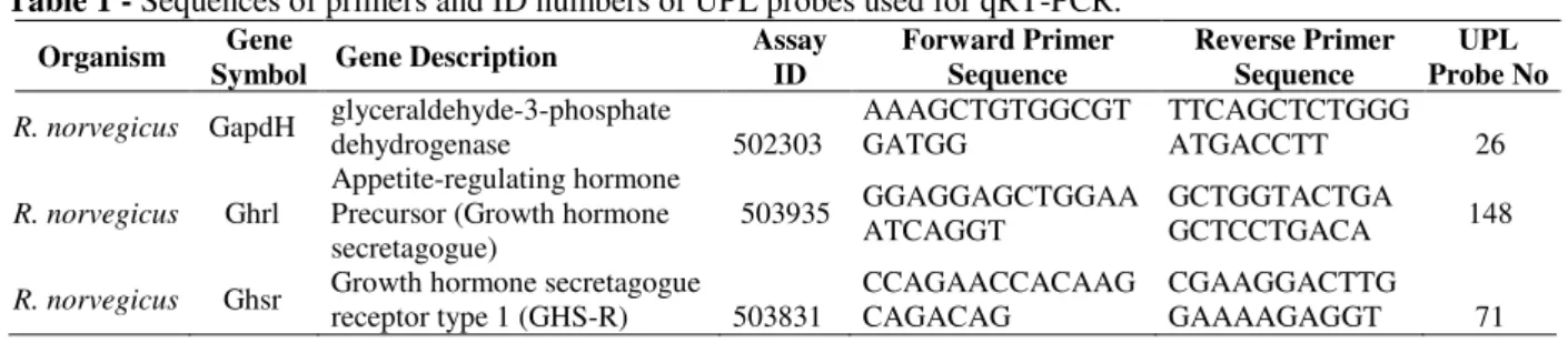
The microscopic examination of kidney tissue belonging to the control group subjects exhibited normal glomerulus and tubules (Fig. 1A). In the kidney tissues of group DI, epithelial desquamation into the lumen of the tubules and transparent tubules showing glycogen vacuolation (Armanni– Ebstein Lesions) were significantly detected (Fig.
1B). The findings were more prominent in group DII (Fig. 1C) and group DIII (Fig. 1D).
Table 2 - Body weight, Kidney weight and Blood glucose
levels of control and streptozotocin (STZ)-induced diabetic rats.
Body weight (g)
Kidney weight (g)
Blood glucose(mg/dl) Group C
(Control) 340.75±12.43 1.20±0.04 112.33±2.73 Group DI
(One Month) 280.88±16.41
* 1.06±0.03 504.55±34.52 **
Gorup DII
(Two Months) 167.00±17.12
** 1.00±0.03* 515.88±35.94 **
Group DIII (Three Months)
209.83±14.30** 1.19±0.03 450.83±36.06 **
Values are expressed as mean ± SE. *P < 0.05 compared to control group. ** P < 0.001 compared to Group C.
Figure 1 - Light microscopy of kidney tissue in different groups (A) In controls, normal kidney architecture were observed. In kidney tissues, transparent tubules showing glycogen vacuolation (*) were detected in diabetic groups. (B) One month diabetes, (C) Two months diabetes, (D) Three months diabetes. Kidney cross sections were stained with PAS.
Immunohistochemical Results
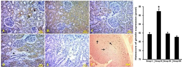
Expression of ghrelin was observed in the distal tubules and collecting ducts in group C (Fig. 2A), which was also observed in the proximal tubules in addition to the distal and collecting tubules in diabetes groups. Ghrelin immunoreactivity was significantly higher in group DI (Figure 2B) compared to group C, whereas in group DII (Fig. 2C) and group DIII (Fig. 2D), ghrelin immunoreactivity was similar to group C.
RT-PCR results
The GHSR-1a mRNA levels in groups DI and DII
were similar to that in group C. On the other hand,
the GHSR-1a mRNA level in group DIII was
significantly lower than in group C (Fig. 3).
Figure 2 - Ghrelin-immunopositive cells (arrow) in the kidney and histoscore of ghrelin-immunopositive cells can be seen. (A) Control, (B) One month diabetes, (C) Two months diabetes, (D) Three months diabetes. (E) Negative controls (Three months diabetes) (F) Positive control: ghrelin immunopositive cells in stomach tissue.
DISCUSSION
Ghrelin, a hormone with 28 amino acids is produced by endocrine cells in the gastric fundus. It is an endogenous ligand of GHSR. It has important functions in GH secretion, modulation of glucose metabolism and regulation of appetite and body weight (Wren et al. 2000; Kojima et al. 1999). In many studies, expression of ghrelin has been shown in the glomerulus and in the distal and collecting tubules (Mori et al. 2000a; Yabuki et al. 2006; Venables et al. 2011). In addition, ghrelin immunoreactivity was detected in the proximal tubules. It was shown that the density of the staining was less in the proximal tubules than in the distal tubules (Dagli et al. 2009). In the end stage renal disease, 2.8 fold increase in serum ghrelin level was detected. This increase was thought to be due to decreased elimination of ghrelin by non-functional kidneys (Yoshimoto et al. 2002). It was reported that ghrelin attenuated renal fibrosis in rats with unilateral ureteral obstruction (Sun et al. 2014). In another study, intrarenal ghrelin infusion stimulated distal nephron-dependent sodium reabsorption in the rats (Kemp et al. 2011). It was shown that ghrelin had an important effect against endotoxemia-induced acute kidney injury by inhibiting proinflammatory cytokines (Wang et al. 2009).
Diabetes and diabetic kidney disease continue to increase worldwide (Ozbek 2012). Diabetic nephropathy is the most common microvascular complication of diabetes and a major cause of
end-stage kidney disease (Gohda et al. 2014). Nephropathy develops in about one-third of type 1 diabetic patients (Rahimi et al. 2005). In diabetic nephropathy, histopathologic changes such as thickening in the glomerular and tubular basal membranes, glomerular and tubular hypertrophy, tubular vacuolation, mesangial cell proliferation and mesangial matrix increase were shown (Donder et al. 2013). In the present study, in concordance with the literature, transparent tubules showing glycogen vacuolation and dilatation in the distal tubules with detachment and degeneration in the tubular epithelium were significantly observed in-group DI. In addition, these findings increased with progression of diabetes
Ghrelin and GHSR are expressed in many tissues. Diabetes affects the ghrelin levels in different ways. Yoshimoto et al. (Yoshimoto et al. 2002) showed increased levels of ghrelin and des-acyl ghrelin in patients with renal damage. Masaoka et al. (Masaoka et al. 2003) showed increased ghrelin levels in the plasma and pre-proghrelin mRNA expression in the stomach in STZ-induced diabetic rats. However, they did not find any change in the duodenum and the colon. Adeghate and Ponery (Adeghate and Ponery 2002) found that ghrelin stimulated insulin secretion in normal and diabetic rats. They also detected increased ghrelin immunoreactive cell number in the islet of Langerhans in diabetic rats. It was shown that des-acyl ghrelin prevented cardiac dysfunction by
2014). In a study investigating the effect of enalapril and losartan on ghrelin immunoreactivity, it was shown that the immunoreactivity was significantly increased in the kidneys of diabetic rats (Donder et al. 2013). Other studies showed that ghrelin immunoreactivity in the distal and collecting tubules was gradually increased with duration of diabetes (Yabuki et al. 2006; Kuloglu and Dabak 2009). In the present study, ghrelin immunoreactivity was observed in the distal and collecting tubules in the control group, whereas it was also observed in the proximal tubules in the diabetes groups. In-group DI, ghrelin immunoreactivity was significantly increased compared to the control group. However, in groups DII and DIII ghrelin immunoreactivity was similar to that in the control group. In 2, 4 and 6-week diabetic subjects, increased expression of ghrelin were found with the increased duration of diabetes (Yabuki et al. 2006). In the present study, diabetes was produced for longer periods of 1, 2 or 3-months. There was no increase in ghrelin immunoreactivity with increased duration of diabetes unlike in other studies. At the beginning of diabets, there was increased ghrelin immunoreactivity. However, ghrelin immunoreactivity was similar to that in the controls in long-term diabetes.
Most of the effects of ghrelin are generated by the interaction of ghrelin with GHSR located in the cell membrane (Howard et al. 1996). GHSR-1a was shown to be expressed in the stomach, testis, heart, kidney and pancreas (Kojima et al. 1999; Date et al. 2002; Tena-Sempere et al. 2002; Venables et al. 2011). Limited localization of GHSR in the mouse kidney has been reported. GHSR was expressed in the straight parts of the distal tubules and the thin limbs of the loops of henle. However, its expression was not detected in the glomerulus, proximal tubules, convoluted distal tubules, or collecting ducts (Venables et al. 2011). It was shown that blockade of GHSR caused an increase in urinary sodium excretion (Kemp et al. 2011). It was observed that in GHSR null mice, renal tubular damage and renal fibrotic changes induced by angiotensin II were increased compared to wild-type mice (Fujimura et al. 2014). These studies showed that GHSR-1a contributed to the renal physiology. In the present study, the GHSR-1a mRNA level was parallel to ghrelin expression in the groups DI and DII. However it was significantly decreased in the group DIII. This decrease could be due to down-regulation of GHSR-1a. In the present
study, it was found that histological damage in the groups DII and DIII were higher than that in group DI. Considering other studies in the literature, a decrease in GHSR-1a density could contribute to this damage.
CONCLUSION
In conclusion, ghrelin immunoreactivity was increased at the beginning of diabetes. However, with increase in the duration of diabetes, ghrelin immunoreactivity approached to the control values. In addition, expression of GHSR-1a mRNA was decreased with increase in the duration of diabetes. It seemed that down-regulation of GHSR-1a contributed to the renal damage induced by long-term diabetes.
ACKNOWLEDGMENT
This work was supported by a research grant from the Erciyes University Scientific Research Projects Unit (EUBAP, TSA-11-3657).
REFERENCES
Adeghate E, Ponery AS. Ghrelin stimulates insulin secretion from the pancreas of normal and diabetic rats. J Neuroendocrinol. 2002; 14(7): 555-560. Asakawa A, Inui A, Kaga T, Yuzuriha H, Nagata T,
Ueno N, et al. Ghrelin is an appetite-stimulatory signal from stomach with structural resemblance to motilin. Gastroenterology. 2001; 120(2): 337-345.
Dagli AF, Aydin S, Karaoglu A, Akpolat N, Ozercan İH, Ozercan MR. Ghrelin expression in normal kidney tissue and renal carcinomas. Pathology - Research and Practice. 2009; 205(3): 165-173.
Date Y, Nakazato M, Hashiguchi S, Dezaki K, Mondal MS, Hosoda H, et al. Ghrelin is present in pancreatic alpha-cells of humans and rats and stimulates insulin secretion. Diabetes. 2002; 51(1): 124-129.
Donder E, Dogan MM, Kuloglu T, Dabak ÖD, Kocaman N, Ozkan Y. The Investigation of the Effects of Enalapril and Losartan on Ghrelin Immunoreactivity in Kidney of Streptozotocin-Induced Diabetic Rats. Fırat Tıp Dergisi. 2013; 18(1): 1-6.
Fujimura K, Wakino S, Minakuchi H, Hasegawa K, Hosoya K, Komatsu M, et al. Ghrelin protects against renal damages induced by angiotensin-II via an antioxidative stress mechanism in mice. PLoS One. 2014; 9(4): 94373.
GHS-R, in humans. J Clin Endocrinol Metab. 2002; 87(6): 2988
Gohda T, Mima A, Moon J-Y, Kanasaki K. Combat Diabetic Nephropathy: From Pathogenesis to Treatment. Journal of Diabetes Research. 2014; 2014(1).
Gualillo O, Caminos J, Blanco M, Garcia-Caballero T, Kojima M, Kangawa K, et al. Ghrelin, a novel placental-derived hormone. Endocrinology. 2001; 142(2): 788-794.
Hewson AK, Dickson SL. Systemic administration of ghrelin induces Fos and Egr-1 proteins in the hypothalamic arcuate nucleus of fasted and fed rats. J Neuroendocrinol. 2000; 12(11): 1047-1049.
Howard AD, Feighner SD, Cully DF, Arena JP, Liberator PA, Rosenblum CI, et al. A receptor in pituitary and hypothalamus that functions in growth hormone release. Science. 1996; 273(5277): 974-977. Inui A. Ghrelin: an orexigenic and somatotrophic signal from the stomach. Nat Rev Neurosci. 2001; 2(8): 551-60.
Kageyama H, Funahashi H, Hirayama M, Takenoya F, Kita T, Kato S, et al. Morphological analysis of ghrelin and its receptor distribution in the rat pancreas. Regulatory Peptides. 2005; 126(1-2): 67-71.
Kemp BA, Howell NL, Gray JT, Keller SR, Nass RM, Padia SH. Intrarenal ghrelin infusion stimulates distal nephron-dependent sodium reabsorption in normal rats. Hypertension. 2011; 57(3): 633-639.
Kojima M, Hosoda H, Date Y, Nakazato M, Matsuo H, Kangawa K. Ghrelin is a growth-hormone-releasing acylated peptide from stomach. Nature. 1999; 402(6762): 656-660.
Kojima M, Hosoda H, Matsuo H, Kangawa K. Ghrelin: discovery of the natural endogenous ligand for the growth hormone secretagogue receptor. Trends Endocrinol Metab. 2001; 12(3): 118-122.
Kuloglu T, Dabak DO. Determination of Ghrelin Immunoreactivity in Kidney Tissues of Diabetic Rats. Renal Failure. 2009; 31(7): 562-566.
Masaoka T, Suzuki H, Hosoda H, Ota T, Minegishi Y, Nagata H, et al. Enhanced plasma ghrelin levels in rats with streptozotocin-induced diabetes. FEBS Lett. 2003; 541(1-3): 64-68.
Mori K, Yoshimoto A, Takaya K, Hosoda K, Ariyasu H, Yahata K, et al. Kidney produces a novel acylated peptide, ghrelin. FEBS Lett. 2000; 486(3): 213-216. Muccioli G, Tschop M, Papotti M, Deghenghi R,
Heiman M, Ghigo E. Neuroendocrine and peripheral activities of ghrelin: implications in metabolism and obesity. Eur J Pharmacol. 2002; 440(2-3): 235-54. Nakazato M, Murakami N, Date Y, Kojima M, Matsuo
H, Kangawa K, et al. A role for ghrelin in the central regulation of feeding. Nature. 2001; 409(6817):194-198.
Ozbek E. Induction of oxidative stress in kidney. Int J Nephrol. 2012; 2012: 465897.
Pei XM, Yung BY, Yip SP, Chan LW, Wong CS, Ying M, et al. Protective effects of desacyl ghrelin on diabetic cardiomyopathy. Acta Diabetol. 2014; 52(2): 293-306.
Rahimi R, Nikfar S, Larijani B, Abdollahi M. A review on the role of antioxidants in the management of diabetes and its complications. Biomedicine & Pharmacotherapy. 2005; 59(7): 365-373.
Sonmez MF, Ozan E. Determination of ghrelin immunoreactivity in the rat stomach after fasting and refeeding. Acta Histochem. 2007; 109(3): 193-199. Sun GX, Ding R, Li M, Guo Y, Fan LP, Yue LS, et al.
Ghrelin attenuates renal fibrosis and inflammation of obstructive nephropathy. J Urol. 2014; 193(6): 2107-2115.
Tena-Sempere M, Barreiro ML, Gonzalez LC, Gaytan F, Zhang FP, Caminos JE, et al. Novel expression and functional role of ghrelin in rat testis. Endocrinology. 2002;143(2): 717-725.
Unsal F, Sonmez MF. The effects of ovariectomy on ghrelin expression in the rat uterus. Adv Clin Exp Med. 2014; 23(3): 363-370.
Venables G, Hunne B, Bron R, Cho H-J, Brock JA, Furness JB. Ghrelin receptors are expressed by distal tubules of the mouse kidney. Cell and Tissue Research. 2011; 346(1): 135-139.
Wang W, Bansal S, Falk S, Ljubanovic D, Schrier R. Ghrelin protects mice against endotoxemia-induced acute kidney injury. Am J Physiol Renal Physiol. 2009; 297(4): 1032-1037.
Wren AM, Small CJ, Ward HL, Murphy KG, Dakin CL, Taheri S, et al. The novel hypothalamic peptide ghrelin stimulates food intake and growth hormone secretion. Endocrinology. 2000; 141(11): 4325-4328.
Yabuki A, Taharaguchi S, Ichii O, Kojima M, Nishi Y, Mifune H, et al. Immunohistochemical localization of ghrelin in rodent kidneys. Histochem Cell Biol. 2006;126(2): 231-238.
Yin Y, Li Y, Zhang W. The Growth Hormone Secretagogue Receptor: Its Intracellular Signaling and Regulation. International Journal of Molecular Sciences. 2014;15(3): 4837-4855.
Yoshimoto A, Mori K, Sugawara A, Mukoyama M, Yahata K, Suganami T, et al. Plasma Ghrelin and Desacyl Ghrelin Concentrations in Renal Failure. Journal of the American Society of Nephrology. 2002; 13(11): 2748-2752.
Yuan MJ, Huang H, Huang CX. Potential new role of the GHSR‑1a‑mediated signaling pathway in cardiac remodeling after myocardial infarction (Review). Oncology Letters. 2014; 8(3): 969-971.