NMR and molecular recognition of compounds
related to the immune system
Helena Maria Nobre Coelho
Doctoral Thesis
2019
Doctoral Thesis Co-Tutelage between the University of the Basque Country UPV/EHU and the New University of Lisbon.
Supervisors:
Dr. Jesús Jiménez Barbero (CIC bioGUNE) Dr. Filipa Marcelo (UCIBIO)
University Tutor: Dr. Esther Lete (UPV/EHU)
This doctoral thesis was performed at Center for Cooperative Research in Biosciences (CICbioGUNE) and at Applied Molecular Biosciences Unit (UCIBIO).
AUTORIZACION DEL/LA DIRECTOR/A DE TESIS
PARA SU PRESENTACION
Dr. Jesús Jimenéz-Barbero con N.I.F. 01107969J como Director de la Tesis Doctoral: NMR and molecular recognition of compounds related to the immune system, realizada en el Programa de Doctorado Química Sintética y Industrial por el Doctorando Dña. Helena Maria Nobre Coelho, autorizo la presentación de la citada Tesis Doctoral, dado que reúne las condiciones necesarias para su defensa.
En Bilbao a 23 de Enero de 2019
EL DIRECTOR DE LA TESIS
AUTORIZACION DEL/LA DIRECTOR/A DE TESIS
PARA SU PRESENTACION
Dra. Filipa Margarida Barradas de Morais Marcelo con N.I.F. 210790806 (Portugal) como Directora de la Tesis Doctoral: NMR and molecular recognition of compounds related to the immune system, realizada en el Programa de Doctorado Química Sintética y Industrial por el Doctorando Dña. Helena Maria Nobre Coelho, autorizo la presentación de la citada Tesis Doctoral, dado que reúne las condiciones necesarias para su defensa.
En Caparica a 23 de Enero de 2019
LA DIRECTORA DE LA TESIS
PARA SU PRESENTACION
Dra. Esther Lete como Tutora de la Tesis Doctoral: NMR and molecular recognition of compounds related to the immune system realizada en el Programa de Doctorado Química Sintética y Industrial por el Doctorando Dña. Helena Maria Nobre Coelho, y dirigida por los Drs Jesús Jimenéz-Barbero y Filipa Marcelo, autorizo la presentación de la citada Tesis Doctoral, dado que reúne las condiciones necesarias para su defensa.
En Leioa a 23 de Enero de 2019
LA TUTORA DE LA TESIS
DE DOCTORADO
La Comisión Académica del Programa de Doctorado en Química Sintética y Industrial en reunión celebrada el día 24 de Enero de 2019 ha acordado dar la conformidad a la presentación de la Tesis Doctoral titulada: NMR and molecular recognition of compounds related to the immune system dirigida por los Drs. Jesús Jimenéz-Barbero y Filipa Marcelo y presentada por Dña. Helena Maria Nobre Coelho adscrita al Departamento Química Orgánica II.
En Leioa, a 24 de Enero de 2019
LA RESPONSABLE DEL PROGRAMA DE DOCTORADO
El Consejo del Departamento de Química Orgánica II en reunión celebrada el día 24 de Enero de 2019 ha acordado dar la conformidad a la admisión a trámite de presentación de la Tesis Doctoral titulada: NMR and molecular recognition of compounds related to the immune system dirigida por los Drs. Jesús Jimenéz-Barbero y Filipa Marcelo y presentada por Dña. Helena Maria Nobre Coelho ante este Departamento.
En Leioa, a 24 de Enero de 2019
VºBº DIRECTOR/A DEL DEPARTAMENTO SECRETARIO/A DEL DEPARTAMENTO
ACTA DE DEFENSA DE TESIS DOCTORAL
DOCTORANDA DÑA. Helena Maria Nobre Coelho
TITULO DE LA TESIS: NMR and molecular recognition of compounds related to the immune system
El Tribunal designado por la Comisión de Postgrado de la UPV/EHU para calificar la Tesis Doctoral arriba indicada y reunido en el día de la fecha, una vez efectuada la defensa por el/la doctorando/a y contestadas las objeciones y/o sugerencias que se le han formulado, ha otorgado por___________________la calificación de:
unanimidad ó mayoría
SOBRESALIENTE / NOTABLE / APROBADO / NO APTO
Idioma/s de defensa (en caso de más de un idioma, especificar porcentaje defendido en cada idioma):
Castellano ___________________________________________________ Euskera ____________________________________________________ Otros Idiomas (especificar cuál/cuales y porcentaje) ___________________
En a de de
EL/LA PRESIDENTE/A, EL/LA SECRETARIO/A,
Fdo.: Fdo.:
Dr/a: ____________________ Dr/a: ______________________
VOCAL 1º, VOCAL 2º, VOCAL 3º, Fdo.: Fdo.: Fdo.:
Dr/a: Dr/a: Dr/a:
The work herein described was performed at the Center for Cooperative Research in Biosciences (CICbioGUNE) and at Applied Molecular Biosciences Unit (UCIBIO), under the supervision of Prof. Dr. Jesús Jiménez-Barbero and Dr. Filipa Marcelo.
Firstly, I would like to thank my PhD supervisors: Prof. Dr. Jesús Jiménez-Barbero and Dr. Filipa Marcelo for giving me the opportunity to participate in this research project to develop my doctoral thesis. I am extremely thankful for their continuing support and availability during these years of thesis, that helped me to grow as a person and professional.
During these years I also completed three secondment periods abroad also go my acknowledgements: 1) Center for Biological Research of the Spanish National Research Council (CIB-CSIC) (Madrid, Spain), under the supervision of Dr. Sonsoles Martín Santamaría and 2) Università degli Studi di Milano-Bicocca (UNIMIB) (Milan, Italy), under the supervision of Prof. Francesco Peri and 3) Lofarma, S.p.A, under supervision Dr. Gianni Mistrello.
In terms of collaborations, I would like to thank to all who participated to the works presented in this Thesis.
For the financial support, I am grateful to the European Union’s Horizon 2020 research and innovation programme that financed this project (ETN TOLLerant project, Marie Skłodowska-Curie grant agreement No 642157) and the FCT-Portugal to IF project (IF/00780/2015) that supported the recent times.
To my laboratory colleagues from Chemical Glycobiology Lab (CIC bioGUNE) and (Bio)molecular Structure and Interactions by NMR Lab (UCIBIO).
To my family and friends.
Contents
Abbreviations i
Resumen iii
Resumo ix
Abstract xiii
Chapter 1 - General Introduction 1 1.1 General Introduction 1
1.1.1 Principles of NMR Spectroscopy 1 1.1.1.1 Chemical shift 3 1.1.1.2 Spin-spin couplings 3
1.1.1.3 Spin relaxation 4 1.1.2 The Nuclear Overhauser Effect (NOE). 6
1.1.3 Diffusion ordered spectroscopy 8
1.1.4 NMR methods for the study of Protein-Ligand interactions: 8 1.1.4.1 Ligand-detected methods 10
1.1.4.2 Receptor-detected methods: Chemical Shift Perturbations 16
1.1.4.3 Specific isotopic labeling to study protein by NMR 19
1.1.5 Carbohydrates and Carbohydrate-Protein Interactions 21
1.1.5.1 Structure and Conformation 22
1.1.5.2 Carbohydrate-protein interactions 25
1.1.6 References 28
1.2 Goals 33
Chapter 2 - Deciphering GalNAc O-glycosylation: From structure to function in human health & disease 35
2.1 Introduction 37
2.1.1 GalNAc-Transferases 38
2.1.1.2 Preferences and Specificity 43
2.2 GalNAc-Ts glycosylation follow an induced-fit catalytic mechanism. 47
2.2.1 Introduction 49
2.2.2 Results and Discussion 51
2.2.2.1 Stability and Binding assays 51
2.2.2.2 X-Ray Crystallography & MD Simulations 53
2.2.2.3 19F labelling of the WT GalNAc-T2 and F104S mutant for NMR experiments. 57
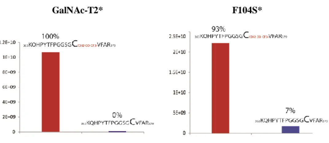
2.2.2.4 19F-NMR experiments 60
2.2.3 Conclusions 64
2.2.4 Supporting Information 66
2.2.4.1 19F-NMR spectra 66
2.3 Deciphering the Mechanism of Long and Short Distance-Glycosylation of GalNAc-Ts 69
2.3.1 Introduction 71
2.3.1 Results and Discussion 73
2.3.1.1 Long distance-glycosylation 73
2.3.1.1 Short distance-glycosylation 83
2.3.2 Conclusion 95
2.3.3 Supporting Information 96
2.4 O-glycosylation of mucin MUC1 by GalNAc-Ts 103
2.4.1 Introduction 105
2.4.1 Results and Discussion 109
2.4.1.1 Design, Expression, Purification and NMR characterization of isotopic labeled MUC1 with four TR domains 109
2.4.1.2 Monitoring MUC1-4TR glycosylation by GalNAc-Ts using NMR spectroscopy. The role of the lectin domain. 110
2.4.1.3 Conformation of the glycosylated MUC1-4TR products modulates glycosylation preferences of GalNAc-Ts 137
2.4.3 Supporting information 151
2.5 Methods 161
2.5.1 NMR experiments 161
2.5.1.1 Assignment of (glyco)peptide 161
2.5.1.2 Saturation Transfer Difference (STD) 161
2.5.1.3 19F-NMR experiments 162
2.5.1.4 NMR Glycosylation Assay 163
2.5.1.5 Percentage of glycosylation of individual residues 164
2.5.1.6 Combined chemical shift perturbation (CSP) 165
2.5.2 Overexpression and Purification of 15N-MUC1-4TR 165
2.5.2.1 Purification of the glycosylated products 168
2.5.3 Mass spectrometry: MALDI-TOF/TOF 168
2.5.3.1 Sample preparation 168
2.6 References 170
Chapter 3 - The conformational and interaction features of glycolipid with TOLL-like receptors and accessory proteins 177
3.1 Introduction 179
3.2 The interaction of Lipid A mimics with antimicrobial peptides 183
3.2.1 AMPs enhances FP7 antagonist activity in HEK-Blue hTLR4 cells 183
3.2.2 LL-37 (AMP6) enhances FP7 antagonist activity in human PBMC 187
3.2.3 NMR and TEM analysis of glycolipid/peptide interaction 188
3.2.3.1 NMR analysis from the AMP point-of-view 188
3.2.3.2 NMR analysis from the FP7 point-of-view 193
3.3 Design of new TLR4 antagonists based on FP7 200
3.4 Characterization of natural LPSs/MD-2 interaction 203
3.4.1 LPS from Bradyrhizobium BTAi-1 Δshc 204
3.4.1.2 Cryo-Electron Microscopy 207 3.4.2 LPS from Acetobacter pasteurianus 207
3.4.2.1 NMR experiments 208
3.4.2.2 Cryo-Electron Microscopy 211
3.5 Conclusions 212
3.6 Methods 214
3.6.1 NMR experiments 214
3.6.2 Expression and Purification of MD-2 protein 215 3.6.3 Transmission Electron Microscopy 216
3.7 References 216
Chapter 4 – Final Remarks 221
4.1 Scientific publications during this dissertation 225 4.2 Contribution to congresses during this dissertation 226
AMP Anti-Microbial Peptides
CD14 Cluster Differentiation antigen 14 CHO-K1 Chinese hamster ovary - K1 CLD Carbohydrate lectin domain CMC Critical Micellar Concentration
Cryo-TEM Cryogenic Transmission Electron Microscopy CSI Chemical shift index
CSP Chemical Shift Perturbation
DCs Dendritic Cells
DOSY Diffusion Order Spectroscopy E. Coli Escherichia coli
ER Endoplasmic Reticulum
GalNAc-Ts GalNAc-Transferases GTs Glycosyltransferases
HDL-C High-density lipoprotein cholesterol HSQC Heteronuclear Single-Quantum Coherence IRF3 Interferon Regulatory Factor 3
Kd Dissociation constant
Km Michaelis constant
LBP Lipopolysaccharide Binding Protein LOS Lipooligosaccharide
LPS Lipopolysaccharide LRR Leucine-Rich Repeat
MD Molecular Dynamics
MD-2 Myeloid Differentiation protein
MUC1 Mucin-1
MUC1-4TR Mucin-1 with four tandem repeats NF-κB Nuclear Factor Kappa B
NOESY Nuclear Overhauser effect spectroscopy PAMPs Pathogen-associated molecular patterns PBS Phosphate Buffered Saline
PHA Plant Lectin Phytohemagglutinin PLTP Phospholipid transfer protein PRRs Pattern Recognition Receptors RMSD Root-mean-square-deviation SAXS Small-angle X-ray scattering STD Saturation Transfer Difference TEM Transmission Electron Microscopy
TEV Tobacco Etch Virus
TLR4 Toll-Like Receptor 4 TLRs Toll-like receptors
TOCSY Total correlation spectroscopy
TR Tandem repeat
TSP 2,2,3,3-tetradeutero-3-trimethylsilylpropionic acid UDP Uridine diphosphate
UDP-GalNAc Uridine diphosphate N-acetylgalactosamine
Vmax Maximum rate
WT Wild-type
Las macromoléculas biológicas: las proteínas, los ácidos nucleicos y los hidratos de carbono son los motores principales de la célula viva. La esencia de la vida está regulada por las interacciones que se dan entre estas entidades, con distintos niveles de complejidad. El conocimiento de su estructura tridimensional, su dinámica y la manera en que se relacionan entre ellas y con otras moléculas más pequeñas es imprescindible para entender en profundidad el funcionamiento de la compleja maquinaria celular.
Los hidratos de carbono son de las moléculas más variables y complejas de los sistemas biológicos. La glicosilación de proteínas y lípidos tiene la capacidad inigualable de generar una amplia gama de estructuras diferentes. El conocimiento de la comunicación intermolecular, a escala atómica y molecular, entre los hidratos de carbono y sus receptores debería llevar al entendimiento y modulación de las señales biológicas en eventos fisiológicos y patológicos.
Existen varias herramientas para obtener información a escala atómica de la comunicación de este tipo de moléculas: la espectroscopía de Resonancia Magnética Nuclear (RMN), la difracción de rayos X y la crio-microscopía electrónica (cryo-EM). También coexisten una legión de técnicas que proporcionan información estructural menos detallada, como otras espectroscopías (IR, UV, Raman), la microscopía electrónica (EM), o los métodos basados en transferencia de energía de resonancia (FRET).
En este trabajo, empleamos la espectroscopia de Resonancia Magnética Nuclear (RMN) para obtener información, a distintas escalas, sobre diferentes procesos de reconocimiento molecular entre hidratos de carbono y proteínas con interés biomédico. En particular, nos hemos centrado en cáncer (mucina-1 (MUC1)) estudiando el mecanismo de glicosilación de mucinas por GalNAc-transferasas (GalNAc-Ts) y en infecciones bacterianas (receptor Toll-like 4 (TLR4)).
Capítulo 1
En la introducción de esta tesis he proporcionado una visión general de las bases de RMN, centrada en la descripción de los métodos de RMN que se usan para seguir eventos de reconocimiento molecular.
El reconocimiento molecular por RMN puede hacerse del punto de vista del ligando y/o del receptor. Los experimentos de RMN enfocados en la observación de las señales del ligando revelan información clave sobre el epítopo de unión del ligando. Uno de los experimentos de RMN más relevantes dentro de esta clase es el experimento de diferencia de transferencia de saturación (STD-NMR) que se basa en la transferencia de magnetización entre el receptor, que es objeto de irradiación selectiva, y aquellos ligandos que, en exceso y en régimen de intercambio rápido, se unen a él.
Entre los métodos basados en la observación de las señales del receptor, regularmente proteínas, podemos destacar los experimentos basados en la correlación heteronuclear de cuanto simple 1H/15N (1H/15N-HSQC). El HSQC proporciona información a nivel atómico para definir con precisión los aminos ácidos del sitio de unión.
Objetivos
El objetivo general de la tesis es aplicar métodos de RMN basados en ligandos y receptores para estudiar la interacción de una variedad de hidratos de carbono con receptores de interés biológico y / o biomédico. Por lo tanto, un objetivo específico ha sido avanzar en la comprensión de las características estructurales que gobiernan las interacciones de los hidratos de carbono así como determinar de sus epítopos de reconocimiento.
- Comprender el mecanismo de acción de GalNAc-Ts.
- Evaluar las interacciones de diferentes moléculas naturales y sintéticas con péptidos antimicrobianos y la proteína MD-2, en el contexto de la comprensión de la inmunidad innata relacionada con TLR4.
Capítulo 2
La gran familia de GalNAc-Ts es responsable de una modificación postraduccional de muchas proteínas de la superficie celular. Existen cambios de expresión de GalNAc-Ts en las células en el caso de procesos tumorales. Así, el ajuste fino de la expresión de GalNAc-Ts regula la glicosilación con O-GalNAc en ciertas proteínas. Desde esta perspectiva, y usando una combinación de métodos de RMN y de modelado molecular, hemos analizado la especificidad y dinámica de diversas GalNAc-Ts en el proceso de O-glicosilación para desentrañar el mecanismo de acción de estas enzimas y definir los determinantes moleculares para el reconocimiento de sustrato.
Así, hemos descubierto que el reconocimiento de GalNAc-Ts sigue un mecanismo de ajuste inducido en el que el UDP-GalNAc es absolutamente necesario. También hemos determinado las bases moleculares de las preferencias de GalNAc-glicosilación a largo y corto rango para la GalNAc-T4.
Además, hemos demostrado que las enzimas GalNAc-T2 -T3 y -T4 tienen un proceso de glicosilación altamente ordenado, en que todas las repeticiones en tándem se glicosilan de una manera gradual. La acción combinada del dominio de la lectina y el dominio catalítico de esas enzimas es esencial para conseguir modificar todos los lugares de glicosilación de MUC1.
de inhibidores que sirvan para regular la expresión de GalNAc-Ts en enfermedades, especialmente en cáncer.
Capítulo 3
El otro apartado importante de esta tesis se ha centrado en el receptor Toll-like 4 (TLR4). TLR4 se expresa en la superficie de las células inmunitarias y reconoce específicamente endotoxinas de las bacterias, o sea, lipopolisacáridos (LPS) o lipooligosacáridos (LOS), el núcleo molecular de las paredes de las bacterias gram-negativas.
La modulación de receptores de inmunidad innata por agonistas y/o antagonistas con pequeñas moléculas sintéticas permiten controlar la actividad biológica del TLR4. Estas moléculas representan una herramienta poderosa para estudiar TLR4 y son de gran interés farmacológico como agentes antisépticos y antiinflamatorios (antagonistas) o como adyuvantes de vacunas (agonistas). El conocimiento de los aspectos moleculares del reconocimiento de LPS por CD14 (cluster de diferenciación 14) y por el complejo TLR4/MD-2 (proteína de diferenciación mieloide 2) es esencial para comprender las diferentes respuestas mediadas por TLR4 a diferentes variantes de LPS o a moléculas sintéticas.
En esta tesis nos involucramos en el estudio de estos eventos de reconocimiento molecular y caracterización de las estructuras supramoleculares de estas moléculas naturales y sintéticas mayoritariamente por métodos RMN y de microscopía electrónica. En particular, caracterizamos la interacción entre FP7 (antagonista sintético de TLR4) y péptidos antimicrobianos (AMPs) así como la agregación de diferentes glicolípidos, naturales y miméticos, por técnicas de RMN y TEM (microscopía electrónica de transmisión).
experimentos de RMN para diseccionar los detalles moleculares de estos procesos de reconocimiento biomolecular. Así, se han empleado métodos de RMN basados en el ligando y en el receptor, en combinación con una variedad de técnicas experimentales y teóricas. Se han usado multitud de herramientas biofísicas, bioquímicas, de biología molecular, así como diversas metodologías computacionales para complementar los datos de RMN y así obtener una descripción más completa de los eventos de interacción.
Os hidratos de carbono estão entre as moléculas mais variadas e complexas dos sistemas biológicos. O processo de glicosilação é a modificação mais complexa de proteínas e lípidos, com uma capacidade inigualável de gerar uma ampla gama de estruturas diferentes. O reconhecimento molecular de hidratos de carbono por recetores específicos traduz-se em sinais biológicos em eventos fisiológicos e patológicos.
Neste trabalho, aplicamos a espectroscopia de Ressonância Magnética Nuclear (RMN) para obter informações estruturais de diferentes processos de reconhecimento molecular entre hidratos de carbono e diversas proteínas com relevância biomédica. Com principal destaque nos principais aspetos do mecanismo de glicosilação da proteína MUC1 pela família de enzimas GalNAc-Transferases (GalNAc-Ts). Os alvos estudados ao longo desta tese têm impacto no cancro (mucina-1 (MUC1)) e infeções bacterianas (recetor Toll-like 4 (TLR4)).
Capítulo 1
A introdução desta tese contém uma visão geral das bases de RMN, no entanto, focou-se sobretudo em descrever as metodologias de RMN aplicadas ao estudo de eventos de reconhecimento molecular.
Objetivo
O objetivo global desta tese foi avançar na compreensão das características estruturais que conduzem as interações de hidratos de carbono e sobretudo na determinação dos seus epítopos de reconhecimento.
Ts.
- Avaliar as interações de diferentes moléculas naturais e sintéticas com peptídeos antimicrobianos e a proteína MD-2, no contexto da compreensão da imunidade inata relacionada ao TLR4.
Capítulo 2
A grande família das enzimas GalNAc-Ts é responsável por iniciar a modificação de pós-tradução de muitas proteínas da superfície celular. Alterações na expressão das GalNAc-Ts ocorrem em eventos de neoplasia celular, conduzindo a uma glicosilação aberrante das proteínas. Assim, o ajuste fino da expressão das GalNAc-Ts poderá permitir regular o processo de O-glicosilação. Nesta perspetiva, estudámos as especificidades e a dinâmica do mecanismo das GalNAc-Ts aquando a O-glicosilação por métodos de RMN, em conjunto com estudos de modelação molecular, para desvendar o mecanismo de ação de GalNAc-Ts e definir os determinantes moleculares necessários ao reconhecimento do substrato por essas enzimas.
Neste trabalho, fornecemos evidências de que o processo de reconhecimento de GalNAc-Ts segue um mecanismo de ajuste induzido para o qual o UDP-GalNAc é absolutamente necessário.
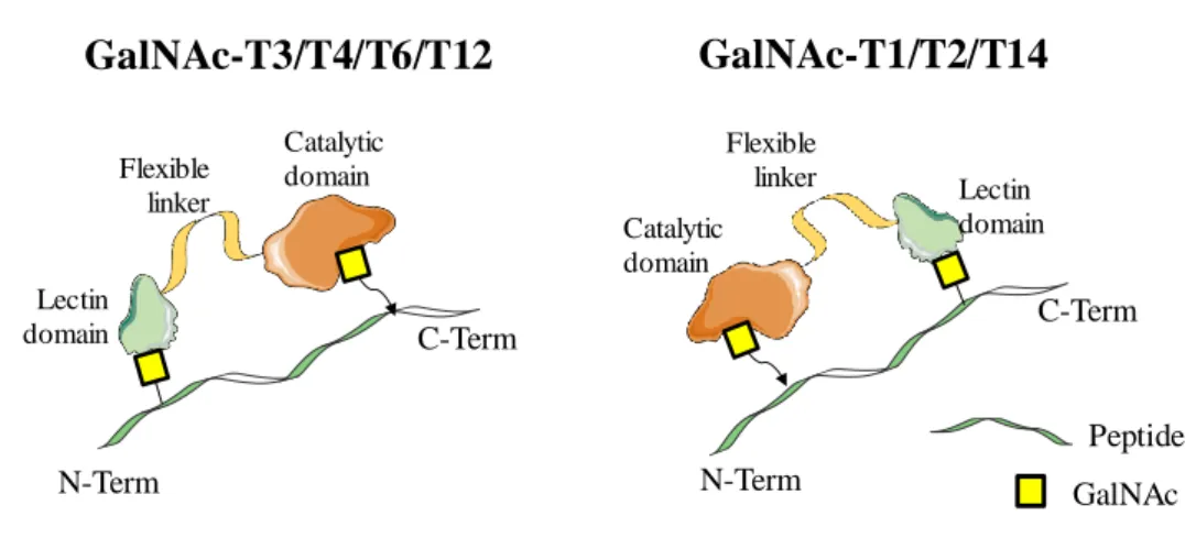
Além do mais a base molecular para as preferências de glicosilação a longa e curta distância foram identificadas. O processo de glicosilação a longa distância é orientado por um linker flexível que fornece capacidade de rotação ao domínio de lectina; enquanto para a glicosilação a curta distância existe no domínio catalítico um sítio de ligação que é responsável pelo reconhecimento do resíduo de GalNAc adjacente ao sítio a glicosilar.
T3 e -T4 tem um processo de glicosilação altamente ordenado, em que todas as repetições em tandem da proteína MUC1 são totalmente O-glicosiladas, de um modo gradual. Dentro deste trabalho, demonstramos evidências de que é muito importante a ação concertada entre o domínio de lectina e o domínio catalítico destas enzimas para uma catálise eficiente de todos os locais de glicosilação da proteína MUC1.
Capítulo 3
O outro tópico desta tese é o recetor Toll-like 4 (TLR4), o qual é expresso na superfície das células imunes inatas e reconhece especificamente endotoxinas bacterianas. Em particular este recetor reconhece o lipopolissacarídeo (LPS) ou seu lipoligossacarídeo de versão incompleta (LOS), os principais componentes moleculares das paredes celulares de bactérias gram-negativas. A modulação dos recetores da imunidade inata por agonistas e/ou antagonistas, por exemplo através de pequenas moléculas sintéticas, permite controlar a atividade biológica do recetor TLR4. Sendo assim estas pequenas moléculas sintéticas representam uma ferramenta poderosa para estudar o sistema de TLR4 e são de grande interesse farmacológico como agentes antissépticos e anti-inflamatórios (antagonistas) ou como adjuvantes de vacinas (agonistas). O conhecimento dos aspetos moleculares do reconhecimento de LPS por CD14 (cluster de diferenciação 14) e pelo complexo TLR4/MD-2 (recetor Toll-like 4/proteína de diferenciação mieloide 2) é essencial para compreender as diferentes respostas mediadas pelo TLR4 a diferentes variantes de LPS / lípido A ou pequenas moléculas sintéticas.
Nesta tese dedicamo-nos ao estudo destes eventos de reconhecimento molecular e à caracterização das estruturas supramoleculares destas moléculas naturais e sintéticas maioritariamente usando técnicas de RMN e de microscopia eletrónica. Em particular, caracterizámos a interação entre FP7 (antagonista sintético de TLR4) e peptídeos antimicrobianos (AMPs) bem como
RMN e TEM (microscopia eletrônica de transmissão).
Ao longo desta Tese, a RMN foi a técnica central para elucidar os detalhes moleculares, estruturais e mecanísticos, dos processos de reconhecimento biomolecular em estudo.
Glycans are among the most varied and complex molecules in biological systems. The glycosylation process is the most complex and widespread modification of proteins and lipids, with an unsurpassed capacity to generate a wide array of different structures. Recognition of glycans by specific receptors translates the glycome into unprecedented biological signals in physiological and pathological events.
In this work, we applied Nuclear Magnetic Resonance (NMR) spectroscopy to gain structural insights of different molecular recognition processes between glycans and diverse proteins with biomedical relevance. As key highlight, key aspects of the glycosylation mechanism of MUC1 by GalNAc-Transferases (GalNAc-Ts) have been unraveled, paying special attention to the site-specificity of the process. Furthermore, our targets have an important impact in disease, particularly, in cancer (mucin-1) and in bacterial infections (Toll-like receptor 4).
Throughout this Thesis, state-of-the-art NMR experiments have been applied to dissect the molecular details of these biomolecular recognition processes. Thus, ligand-based and receptor-based methods have been used, in combination with a variety of additional experimental and theoretical techniques. Thus, biophysical, biochemical, molecular biology and computational methodologies have complemented the NMR data in order to attain a complete description of the interaction events.
Chapter
1
1.1 General Introduction
Nuclear magnetic resonance spectroscopy (NMR spectroscopy), based on the discovery and development of Nuclear Magnetic Resonance by the end of 1930s-beginning of 1940s,1,2 consists in a powerful and widely recognized research
technique that makes use of the interaction between nuclear spins (I ≠ 0) and electromagnetic radio frequency (RF) pulses to permit the “visualization” and consequent study of molecules at atomic scale.3 This development has been
awarded with three Nobel prizes in physics (one in 1944 for Rabi, and two in 1952 for Bloch and Purcell)4 in the last century. In this way, as the research progressed,
it became possible to determine physicochemical properties of the atoms and molecules, as well as detailed information about structure, dynamics, reactions state, and chemical environment, making use of different NMR parameters and using techniques governed by quantum theory.3
Nowadays, NMR spectroscopy has seen an unprecedented growth and is now one of the most powerful and versatile tools, with increasing applications in chemistry, biology, medicine and materials science. The fundamentals and methods of NMR form a vast, complex and evolving discipline.
1.1.1 Principles of NMR Spectroscopy3,4
Nuclear Magnetic Resonance (NMR) spectroscopy detects nuclear-spin reorientation in an applied external magnetic field. For each kind of observed nuclei, the information provided by NMR depends on the electron environment in which the nuclei are immersed and on the positions of nuclei within molecules. Thus, NMR is a powerful tool for probing molecular structure and dynamics. The nuclei observable by NMR are those with a spin quantum number is different to zero (I≠ 0). In an external magnetic field (B0), a spin will prefer an alignment along with the external field rather than oppose it. This is due to an energy difference
(ΔE) between the two states that depends on the strength of B0 and the isotope-specific gyromagnetic ratio (γ) (Figure 1.1) (equation 1).
∆𝐸 = 𝛾ℏ𝐵0
Equation 1
Where ℏ is the reduced Planck constant.
Figure 1.1 - Representation of the Zeeman effect in the energy states separation in NMR
spectroscopy.
The radio-frequency of the electromagnetic radiation that is emitted from transitions between the two states is called the Larmor frequency, which is equal to the ratio of ΔE and the Planck constant (ℏ) and can be given in units of angular frequency (ω0) or Hertz (ʋ0) (Equation 2). NMR spectroscopy is the technique that allows measuring the Larmor frequency of spins in a magnetic field, thus enabling investigation of molecular properties.
𝜔0= ∆𝐸 ℏ = 𝛾𝐵0 Equation 2 ΔE ms= -1/2 ms= +1/2 En e r gy B0 0 0
To extract chemical information from a molecule, there are three fundamental concepts of solution NMR spectroscopy that are helpful, chemical
shifts (i.e., the specific Larmor frequencies of spins), spin-spin couplings (i.e., spin
interactions) and spin relaxation (i.e., the time dependency of an NMR signal).
1.1.1.1 Chemical shift
The magnetic field slightly differs for different spins, due to shielding effects from electron density around the specific nuclei. Therefore, spins will have shifted frequencies depending on their chemical environment (neighboring atoms and type of chemical bonding). Thus, each site experiences a slightly different magnetic field and has a different position in the NMR spectrum. Also, the chemical shifts will vary depending on the molecular orientation with respect to B0, due to the shielding
of B0 caused by anisotropic electron density of the atomic orbitals, a phenomenon
called chemical shift anisotropy (CSA). In solution, this effect is averaged to zero, so that only one frequency is observed for each chemically distinct site.
1.1.1.2 Spin-spin couplings
The NMR signal of a spin does not always appear as a single peak at a given chemical shift in an NMR spectrum. Instead, it is often observed as a split signal centered at the chemical shift. This phenomenon, the spin-spin coupling, arises from interactions between neighboring spins. The magnitude of the peak splitting is referred to as the coupling constant (J) and is equal for both spins involved in the interaction. The most obvious spin-spin coupling in solution NMR spectroscopy is the scalar coupling, which is mediated by electron interactions through covalent bonds. The strength of the interaction is measured by the scalar coupling constant,
n
JIS, in which n is the number of covalent bonds between the nuclei I and S and its
The J-coupling constant is also a source of information regarding the molecular shape, because of its dependence on bond geometry. The relationship can be described by Karplus-type equations (i.e., Equation 3), which differ depending on the nuclei involved in the bonds.
𝐽(𝜃) = 𝐴𝑐𝑜𝑠2𝜃 + 𝐵𝑐𝑜𝑠2𝜃 + 𝑐
Equation 3
Where J is the 3J coupling constant, and A, B, and C are constants that depend on
the specific coupled nuclei. θ is the dihedral angle.
1.1.1.3 Spin relaxation
An RF pulse applied onto a sample at equilibrium causes a perturbation on the nuclear spins removing them from the thermal stationary state. As consequence of this pulse, the system will try to return to the equilibrium, losing the excess energy. However, due to the low transition energies associated with magnetic resonance, the lifetime of the excited states may be extremely long (few seconds to minutes for small molecules). These long lifetimes are fundamental for NMR spectroscopy as they result in sharp lines (as a consequence of the Heisenberg uncertainty principle).
In NMR, two relaxation parameters are defined:
- T1, the spin-lattice or longitudinal relaxation time (R1 for spin-lattice
relaxation rate, R1 = 1/T1);
- T2, the spin-spin or transverse relaxation time (R2 for the spin-spin
T1 measures the efficiency with which the excited nuclear spins return to
their ground state by exchanging energy with their surroundings. T2 is a
measurement of the efficiency with which spins exchange energy with each other. The more efficient this exchange, the shorter the relaxation time. In different experiments, these relaxation times can be studied to understand protein’s dynamics, transverse (T2) or longitudinal (T1), in the timescale of milliseconds (ms)
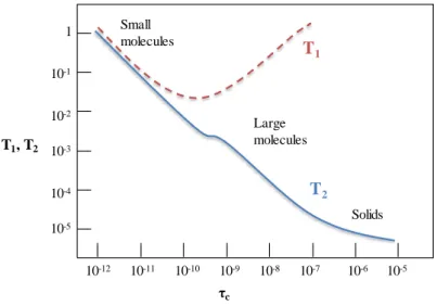
to seconds (s), respectively (Figure 1.2). In solution, the resonance line widths are inversely proportional to the T2 relaxation time, which decreases with the increase in molecular size and tumbling time.
Figure 1.2. Behavior of T1 and T2 as a function of the correlation time for spin ½ nuclei
relaxing by the Dipole-Dipole mechanism.
Dipole-dipole interaction is probably the most important mechanism of relaxation pathway for protons in molecules containing contiguous protons and for carbons with directly attached protons. This is also the source of the Nuclear Overhauser Effect (NOE). Dipolar coupling occurs when the magnetic field of one nuclear dipole affects the magnetic field at another nucleus. It depends on the distance between the nuclei and takes place through-space. Dipole-dipole relaxation is also dependent on the correlation time, τc. Small molecules tumble
10-12 10-11 10-10 10-9 10-8 10-7 10-6 10-5 10-5 10-4 10-3 10-2 10-1 1 T1 T2 Small molecules Large molecules Solids τc T1, T2
very fast and have short τc, usually in the order of picoseconds. Large molecules, such as proteins, usually move slowly and have long τc, usually in the order of nanoseconds. In summary, several dynamic processes can be studied by NMR. The different time scales of NMR observable phenomena are graphically represented in Figure 1.3.
Figure 1.3 - Time scales of some important molecular dynamic processes and
multidimensional NMR methods available to study them.
1.1.2 The Nuclear Overhauser Effect (NOE).
The nuclear Overhauser effect (NOE) defines the correlation between protons close in space (up to 5 - 6 Ǻ distance) and is a consequence of dipole-dipole cross-relaxation in nuclear spin systems. Intra-proton distances can be estimated.5
The NOE represents the change in intensity of a signal when the spin transitions of another nucleus cause a perturbation of its equilibrium populations. The two nuclei do not share a scalar, through bond, coupling; instead, they are sufficiently close in space to share a dipolar coupling. Thus, the NOE originates from dipolar cross-relaxation between proton pairs and depends on the proton-proton distance and on the molecular motion of the inter-proton vector (Equation 4).
10
-1210
-910
-610
-310
010
3Molecular tumbling, Local fluctuations
Domain motions, Enzyme kinetics, Protein folding, Signal transdution
Spin relaxation Relaxation-dispersion
Residual dipolar coupling
SOFAST Real-time 2D-NMR
𝐼𝑁𝑂𝐸 ≈ (
1
𝑟6) 𝑓(𝜏𝑐) Equation 4
Where INOE is the NOE intensity, r is the proton-proton distance, and f is a
function that depends, among others factors, on the correlation time (τc) that
describes the motion of the inter-proton vector.
τc is the correlation time (decay time of the correlation function). When
considering isotropic molecular tumbling, τc is related with the time taken for the
molecule to rotate by 1 radian about any axis. Therefore, rapidly tumbling molecules will have short correlation times, while slowly tumbling molecules will have long correlation times. Thus, the correlation time of a molecule is related with its molecular weight.
Cross-relaxation rates for small molecules are positive while for large molecules, as proteins, are negative (Figure 1.4). For medium size molecules, such as small peptides, cross-relaxation rates can be positive or negative, which means very small (often not measurable) NOEs. This problem can be overcome by using the ROE technique (rotating frame NOE), in which cross-relaxation occurs in the transverse instead of in the longitudinal plane (Figure 1.4).
Figure 1.4 - Maximum NOE and ROE in function with ωτc.
N O E m a x ωτc 0.01 0 -0.5 0.5 -1.0 NOESY ROESY 1 100
1.1.3 Diffusion ordered spectroscopy
Diffusion ordered spectroscopy (DOSY) identifies the molecular components of a mixture and obtaines, at the same time, information on their size. This information may be accessed by measuring the self-diffusion coefficient, which measures the random translational motion of molecules, driven by their internal kinetic energy. Self-diffusion coefficients are related to the structural properties of a molecule by their dependence on the physical properties of the molecule (e.g. size, charge and shape). Furthermore, self-diffusion coefficients also depend on the characteristics of the surrounding medium (e.g. temperature and viscosity).
For a spherical molecule moving in an unconstrained environment, the Stokes–Einstein law predicts a correlation between the hydrodynamic radius r and the self-diffusion coefficient (D) (Equation 5)
𝐷 = 𝑘𝑇 6𝜋𝜂𝑟
Equation 5
Where k is the Boltzmann constant, T is the temperature and η is the medium viscosity.
The diffusion of molecules is measured by evaluating the attenuation of a spin echo signal using pulsed-field gradients (PFG).6
1.1.4 NMR methods for the study of Protein-Ligand interactions:
The binding of small ligands to large proteins usually follows a bimolecular association reaction, which can be described by one-site binding model:
Equation 6
The Equation 6 represents a dynamic equilibrium involving three species: the free protein P, the free ligand L, and the receptor-ligand complex PL, with kon
and koff being the on (association) and off (dissociation) rate constants. The
unimolecular rate constant koff is inversely proportional to the mean lifetime B of
the protein-ligand complex. The bimolecular rate constant kon measures the
probability of encounter between free receptor and ligand.
The binding affinity can be quantified by the temperature-dependent equilibrium dissociation constant (Equation 7):
Equation 7
Where [P], [L] and [PL] are the equilibrium concentrations of protein, ligand and the complex, respectively. [P] and [L] are also referred to as the free state and [PL] is varyingly referred to as ‘bound ligand’. KD has the units of concentration.
When the receptor and ligand molecules are free, they retain their intrinsic NMR parameters (e.g. chemical shifts, relaxation rates, translational diffusion coefficients). However, in each other’s presence, their mutual binding affinity drives a two-state exchange process that can toggle both sets of molecules between the free and bound states (Figure 1.5). Besides providing structural information, NMR methods can supply a wide variety of transient and dynamic information on the complex. NMR methods can be divided in those detecting the ligand signals
(ligand-based experiments) and those detecting the receptor signals (receptor-based experiments).7,8
1.1.4.1 Ligand-detected methods
Generally, ligand-based NMR experiments assay for binding are employed by:
i. Exploiting the differential mobility of the ligand in the free versus bound state: bound ligands will transiently experience the much slower rotational and translational mobility of the large receptor, leading to altered relaxation parameters and diffusion coefficients, respectively;
ii. exploiting transfer of magnetization processes.9
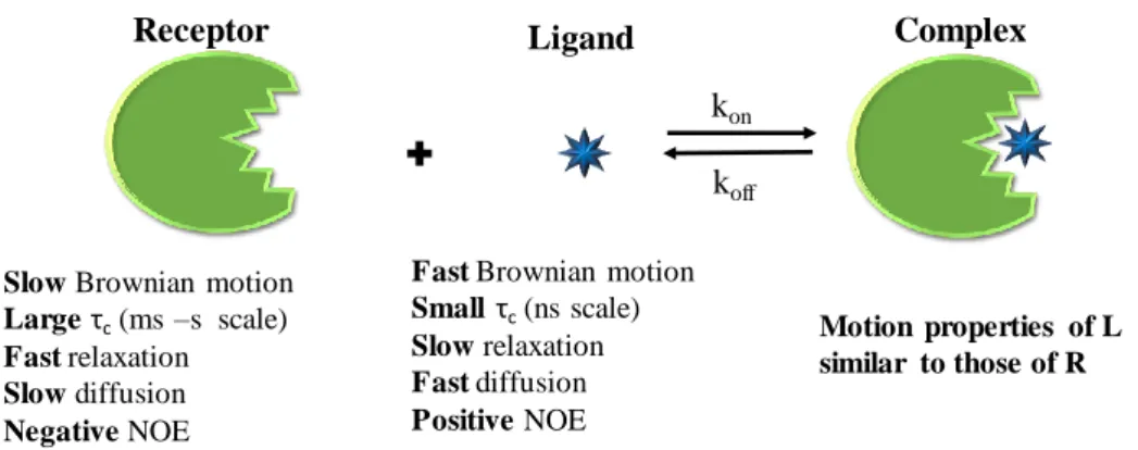
Ligands, small molecules, are characterized by small relaxation rates, positive 2D-NOESY cross-peaks, and large translational diffusion coefficients, D. In contrast, bound ligands share the NMR relaxation properties of the receptor, usually large molecules, with fast relaxation; negative 2D-NOESY cross-peaks, highly efficient spin-diffusion, and smaller molecular diffusion coefficients (D). These apparent differences are the basis of ligand-based methods, where changes in the ligand NMR parameters upon binding to the receptor can be exploited to detect and characterize the interaction (Figure 1.5).
Figure 1.5 - Illustration of the changes in different physical properties when a small
ligand interacts with a large protein.
1.1.4.1.1 Line Broadening in NMR
The primary NMR methods for detection of ligand-protein interactions rely on measurement of T1/T2 relaxation rates of ligand resonances. Line broadening in the presence of the protein is often an indicator of protein–ligand interactions. The degreeof line broadening depends on many factors, including the transverse relaxation rate, the exchange rate, and the fractions of the ligand in the free and bound states. Ligands that bind to a macromolecular receptor experience an enhancement of their T2 relaxation: the molecular motion of the ligand dramatically changes becoming similar to that of the receptor with the selective shortening of the T2 relaxation times, which is translated into a broadening of certain protons of the ligand, which can even disappear.10
1.1.4.1.2 Saturation Transfer Difference (STD) NMR
The STD experiment is a popular and highly versatile screening method for the identification and characterization of protein-ligand interactions.7,9,11 The
success of this technique is a consequence of its robustness and the fact that it is focused on the signals of the ligand, without any need of processing NMR
Motion properties of L similar to those of R
Complex
Receptor Ligand
Fast Brownian motion Smallτc(ns scale)
Slow relaxation Fast diffusion Positive NOE Slow Brownian motion
Large τc(ms –s scale) Fast relaxation Slow diffusion Negative NOE koff kon
information about the receptor, only using small amounts of non-labeled macromolecule.
The STD NMR experiment is the difference between two different 1H-NMR
spectra performed on the same ligand-receptor sample and relies on the magnetization exchange from the protein-bound state to the free state of the ligand. Basically, the first one (on-resonance spectrum) is a spectrum in which the protein is selectively saturated, by irradiating at a region of the spectrum that contains only resonances of the receptor (from 0 ppm to -2 ppm). The saturation is efficiently propagated across the entire protein through spin-diffusion and transferred to the binding compounds via intermolecular 1H-1H cross relaxation at the ligand-receptor
interface. The second spectrum (off-resonance spectrum) is recorded as a reference or blank spectrum. Here, the chosen saturation region is free from ligand and protein signals (e.g. 100 ppm).
The protein-to-ligand saturation transfer will affect the intensity of the ligand resonance signals in the spectrum obtained with selective receptor saturation (ISAT),
and when compared to the spectrum acquired without saturation transfer (I0). Thus,
the difference in intensity due to saturation transfer can be quantified (ISTD = I0 – ISAT) and constitutes an indication of binding (Figure 1.6). STD is ideally suited to
receptors with large masses (>30 kDa). Receptors with large molecular masses display large rotational correlation time, τc that enhance spin diffusion and, consequently, saturation transfer within the receptor and to the ligand.
Figure 1.6 - Schematic representation of the STD experiment. When a protein becomes saturated, the intermolecular NOE is only transferred to those ligands, with shape and size that interact with the protein. The transference of saturation is detected by subtraction of on to off-resonance spectrum in order to obtain an NMR spectrum where only the signals from molecules that bind to the protein are present. Resonance signals from non-binders do not show up in the difference spectrum. Adapted from 12.
Furthermore, for a given compound, the building blocks of the ligand in closest proximity to the receptor molecule receive higher amounts of magnetization, which are translated into stronger signals in the STD spectrum. Thus, the method can be also used to obtain detailed structural information on the binding epitope of the ligand (epitope mapping) (Figure 1.6).13
Regarding group epitope mapping analysis, computing methods have been developed to predict and/or interpret STD data of reversibly forming ligand-receptor complexes. In particular, CORCEMA (Complete relaxation and conformational exchange matrix) package14,15 predicts the expected STD intensities
for a given model of a ligand-protein complex, and compares them quantitatively with the experimental STD data. This version is very useful for rapidly determining if a model for a given ligand-protein complex is compatible with the STD-NMR data obtained in solution.
Selective saturation
off resonance spectrum
koff kon STD spectrum on resonance spectrum 100 Aromatic or aliphatic region H H H H H H H H H H H H H Increasing saturation
Remarkably, it is also possible to measure KD if STD signals are recorded in
the presence of a competitor inhibitor.14,16 Competition experiments can be
performed for designed mixtures of both ligands by evaluation of the gradual decay of the STD signals from the reference in the presence of increasing concentrations of the inhibitor. Assuming a simple bimolecular association reaction for ligand L and a competitive inhibitor I to a receptor protein, the KD value of the ligand L can
be determined from a known value of KI. However, more complicated situations
such us those arising from binding of ligands to secondary binding sites should be taken into account. STD competition experiments have been also used for the detection of high-affinity ligands.14
STD methods can be combined with any NMR pulse sequence generating a whole suite of concatenated STD NMR experiments such as TOCSY or STD-HSQC.15 The additional deconvolution of signals in a second dimension can be very
helpful for mapping the binding epitope of ligands with complex 1D proton spectra.
The major drawbacks associated with STD experiments are related to the potential self-association of the ligands, especially with aromatic molecules, and to the possibility of non-specific binding with the receptor. These potential problems can be circumvented by employing different ligand/receptor molar ratios, different saturation frequencies, and employing competitive binders (or inhibitors) of the interaction process, if they are available.
1.1.4.1.3 WaterLOGSY
The waterLOGSY (Water-Ligand Observed via Gradient SpectroscopY) technique, like STD, relies on excitation of the receptor-ligand complex through a selective RF pulse scheme. However, waterLOGSY experiment achieves this effect indirectly, by selective perturbation of the bulk water magnetization.17 The intended
transfer of magnetization is therefore water →receptor → ligand. The magnetization of the water molecules is selectively saturated or inverted, and
during a long mixing time of up to several seconds, the magnetization is transferred, via 1H-1H cross-relaxation, to the ligand spins at the protein-ligand interface.
There are essentially two pathways by which this transfer of the magnetization occurs:
i. Direct cross-relaxation from the water molecules tightly bound at the protein-ligand interface,
ii. Chemical exchange between the water protons and the labile protons of amine and hydroxyl groups in the protein which in turn cross-relax with the protons of the bound ligand.
In either case, the perturbation of the bulk water magnetization is transferred to the binding compounds while residing in the receptor binding site. Distinguishing binding from nonbinding compounds in the waterLOGSY experiment is achieved by observation of the differential cross-relaxation properties of these ligands with water. Bound ligands interact directly or indirectly with inverted water spins with motional and relaxation properties of the receptor, with a slow tumbling rate and therefore, a long correlation time. This yields negative cross-relaxation rates, and inversion of the NOE signs for the bound ligand. For their part, non-interacting ligands receive the magnetization only in the free state, via water molecules involved in their solvation sphere, leading to positive cross-relaxation rates and the sign of the NOE remains unaltered. As a consequence, binders and non-binders display waterLOGSY peak intensities of opposite sign, as in tr-NOESY (see below).
1.1.4.1.4 Methods based on transferred NOE effects
Transferred NOE methods are based on the changes in the rotational motion properties of a ligand upon binding to a large macromolecular receptor. For small molecules, characterized by a short correlation time, NOEs are positive. However,
for large receptors, the correlation time is in the ns time scale and the associated NOEs are negative. Thus, this technique has become a classical method for studying the binding of ligands to large receptors and to deduce the conformation of the binding ligand in the protein site.9
In trNOESY, low protein:ligand ratios are typically employed (from 1:5 to 1:50, depending on the affinity and kinetic parameters). This implies that the NOEs observed for the ligand (tr-NOEs) will keep the information of its bound state, provided that the off-rate is fast in the relaxation time scale. Therefore, the signals of small ligands will experience a NOE sign change in the presence of the receptor, from positive (free state, small molecule, short correlation time) to negative (large complex, large correlation time), which will reflect the conformation of the ligand in the binding site. Technically, the tr-NOESY experiment consists of acquiring an ordinary NOESY spectrum for the ligand in presence of the protein. Normally, with lower mixing times is in the range of 50 to 100 ms, whereas for nonbinding molecules it is four- to ten-times longer.
1.1.4.2 Receptor-detected methods: Chemical Shift Perturbations & HSQC
For small proteins (~10 kDa) and peptides structure determination, 2D experiments, as COSY (COrrelation SpectroscopY) and TOCSY (TOtal Correlation SpectroscopY), are used to give information between protons, due to the scalar coupling through covalent bonds. NOESY experiments (Nuclear Overhauser Effect SpectroscopY) are the most important multidimensional experiments in structure determination since they correlate protons through space, allowing the determination of the distances between close protons.18 These
methodologies can be used to determine the structure of proteins up to 10-15 kDa.18
For large molecules, the number of hydrogen atoms is extremely high. The rotational correlation times of globular proteins and, therefore, the line widths of
the NMR resonances also increase linearly with the molecular mass. For these reasons, conventional assignment procedures based on sequential NOE correlations become very difficult, if not impossible. Heteronuclear experiments are then necessary and can solve these problems for proteins with molecular masses less than 30 kDa.18 However, isotopic labeling of the samples is required with NMR
active isotopes 13C, 15N, and sometimes 2H.19
In protein NMR, the Heteronuclear Single-Quantum Coherence (HSQC) experiment19 correlates the 15N or 13C nuclei with the attached 1H via the one-bond
scalar coupling JN-H or JC-H, respectively. Thus, in the 1H/15N-HSQC one signal is
expected for each amino acid residue with the exception of proline, which has no amide-hydrogen due to the cyclic nature of its backbone. In this sense, 1H/15
N-HSQC spectrum is the ‘fingerprint’ of the protein.
One of the most frequently used receptor-based methods relies on monitoring the perturbation of the receptor 1H/15N-HSQC chemical shifts upon binding of the
ligand.
1.1.4.2.1 Chemical Shift Perturbations
The chemical shift is an extremely sensitive parameter to environmental changes (pH, temperature, and folding states) and is able to reveal interaction processes. Moreover, nuclei located in the protein binding pocket usually show the largest effects. Thus, mapping chemical shift perturbations via heteronuclear shift-correlation experiments is a straightforward NMR technique to detect binding and also to define the protein residues that are involved in the interaction.
The 1H/15N-HSQC spectrum of the protein, considered as the NMR
fingerprint, is acquired as a reference spectrum. Then, sequential 1H/15N-HSQC
spectra are acquired for different amounts of ligand. The proximity of the ligand will modify the environment of the nuclei that are at the interface of the
protein-ligand complex. Therefore, the residues involved in the binding will have a different chemical shift than in the unbound form. When the exchange rate between free and bound states is fast on the chemical shift time scale, the 1H/15N cross-peaks
will move smoothly from their position in the free spectrum to those in the bound spectrum, with the frequency of the signal at any titration point being the weighted average of free and bound chemical shifts. When the exchange rate is slow on the chemical shift time scale, the free will signal gradually disappear and the bound signal will appear, as the intensities of the two peaks reflect the concentrations of the free and bound protein.20 The chemical shift perturbations allow identifying
binding and to localize the binding site on the protein structure. Mapping 1H/15N
chemical shift perturbations along the structure of the protein is usually applied to detect residues with larger chemical shift perturbations, which can be considered as part of the binding site (Figure 1.7).
Figure 1.7 - Schematic representation of the chemical shift perturbation induced by the
interaction of an unlabelled small ligand with a 15N-labelled protein as detected in the 1H/15N-HSQC spectrum. Slow exchange (strong binding) after the addition of a
sub-stoichiometric quantity of ligand; two signals are observed one for the free (blue) and another for the bound (orange) protein. Fast exchange (weak binding), the scheme represents the superposition of 1H/15N-HSQC sequence obtained for increasing quantities
of ligand in the order blue, dark red, green and orange; only one signal is observed in each spectrum with the shift (arrow) depending on the structural changes induced by the ligand at the specific residue.
Slow exchange Fast exchange
15N
1H 1H
Fast relaxation of the protein 1H/15N signals becomes an issue when dealing
with high molecular weight macromolecules (> 35 kDa). Fortunately, the implementation of transverse relaxation optimized spectroscopy (TROSY) HSQC variant allows targeting larger protein receptors (up to 100 kDa) by selectively recording the most slowly relaxing signal component of the 15N-1H correlation.21
Regarding specific labeling schemes, selective labeling methods have been also developed to improve or simplify NMR spectra of large proteins.
1.1.4.3 Specific isotopic labeling to study protein by NMR
1.1.4.3.1 13C-methyl labeled proteins
A frequent, although expensive, approach is to use 13C-methyl labeled
proteins, which contain 13C probes in the side chain of the protein, such as alanine,
valine, isoleucine, leucine and methionine. For 13C-methyl labeled proteins, 2D 1H/13C-HMQC experiments are typically used. These experiments have some
advantages in comparison to 1H/15N-HSQC experiments. Specifically, methyl
groups have three protons with a three-fold degeneracy and give rise to stronger signals. They also tend to resonate in a sparsely populated region of the 1H/13C
correlation spectrum, reducing spectral overlap.22
Several labeling schemes for amino acids, including alanine, isoleucine, leucine, methionine, and threonine, have been developed.22 These labeling
strategies commonly involve metabolic pathways, metabolic precursors, and the amino acid’s propensity for isotopic scrambling at other sites in the protein. For example, in the case of leucine and valine, which share the same metabolic pathway, using metabolic precursors common to both amino acids, such as α-ketoisovalerate, labeling techniques often result in the incorporation of isotopes into
metabolic precursor.23 For alanine and methionine, residue specific labeling can be
obtain by via of supplementation of minimal expression medium with the appropriate isotopically labeled amino acid.24
1.1.4.3.2 19F labeled molecules
Although classical spectroscopic methods, 1H, 15N, and 13C, have been used
to study the behavior of biomolecules in solution, 19F-NMR is also becoming very
popular although its implementation requires manipulation of the system. The 19F
nucleus displays different properties that render it ideal for NMR studies. In fact,
19F is an NMR active isotope with spin ½, and it is 100% naturally abundant. It also
possesses a high gyromagnetic ratio that results in excellent sensitivity (83% of 1H).
In addition, the fluorine chemical shifts are extremely sensitive to changes in local environment (the chemical shift is +/- 100-fold larger than that of 1H).25
The 19F atom has been used in biological NMR, like a molecular probe into
proteins, peptides, and carbohydrates26–29 to provide information on structure and
dynamics,30–32 protein-ligand interactions,25,33 and protein (un)folding,34,35
demonstrating unequivocally its versatility as probe for NMR.
In the protein field, several methods to prepare 19F-modified proteins have
been described.25,30–35 These methods fall into three main categories:
i. Post-translational covalent attachment of 19F-containing moieties to
the protein by conjugation of the 19F-containing moiety to a reactive
group with an -SH group on a solvent accessible cysteine.25,30,31,33
The great advantage of this method is the ability to incorporate the label into proteins for which biosynthetic labeling is expensive, like a mammalian cells expression.
ii. Specific incorporation of the type of biosynthetic amino acids modified with 19F.25,33–35 This approach is carried out by expression
in a defined growth media, supplemented with the 19F-modified
amino acid
iii. Site-specific incorporation of the 19F-modified amino acid using
recombinant expressed orthogonal tRNA / tRNA-tRNA pairs.25,33
This system is based on an extension of the genetic code to beyond the natural 20 amino acids.
1.1.5 Carbohydrates and Carbohydrate-Protein Interactions
Carbohydrates (glycans, sugars, saccharides) are the most abundant biomolecules in nature and they are estimated to account for 70% of the total biomass on earth.36 As a product of the photosynthesis process, carbohydrates
function as energy storage in organisms and their ability to form polymers make them important for the structure of the cell. Cell membranes themselves contain sugars (as well as proteins and lipids.) These glycans play a fundamental role in a variety of biological processes, including cell adhesion and communication. Therefore, molecular recognition events between sugars and proteins constitute the first interaction process in many molecular mechanisms related to health and disease.
In the initial part of this chapter, different NMR concepts have already been introduced. Although the particular systems of interest that have been studied in this Thesis will be explicitly described below, I will briefly point out now the different types of information that can be extracted from NMR experiments in this particular topic, along with the description of the basic features that are behind the recognition of glycans by protein receptors.
In brief, key features of the structural, conformational, dynamic and interaction properties of carbohydrates will be explored in the following chapters:
i. Molecular shape (structure and conformation), which can be
ii. Molecular motion, which can be investigated by NMR relaxation
and chemical exchange, depending on the timescale of motion.
iii. Molecular interactions, which can be assessed by chemical shift,
relaxation and NOE data.
1.1.5.1 Structure and Conformation
Carbohydrates are extremely diverse, as consequence of the large number of different monosaccharide units that can be combined by employing different types of glycosidic linkages to form oligo- or polysaccharides.37 These complex
glycosylation patterns result in a large variation in shapes and geometries of carbohydrates.
Monosaccharides, the simplest forms of carbohydrates, are polyhydroxylated carbon chains carrying a aldehyde functionality (aldoses) or a keto functionality (ketoses). The carbon chains contain at least three carbons (trioses), although molecules with five (pentoses) and six (hexoses) carbon atoms are the most common ones.
Monosaccharides exist in many different diastereomeric forms because of the high number of stereogenic centers. Each diastereomer has its own name (e.g., glucose, galactose). Enantiomers are named according to the D and L notation, which is determined by the stereochemistry at the highest numbered stereogenic carbon in the saccharide chain (Figure 1.8). The typical cyclization process results in the formation of a new stereogenic center at the hemiacetal carbon (anomeric carbon), which now displays either α or β configuration (Figure 1.8).
Figure 1.8 - Cyclic forms of D-galactose (Gal). The hashtag marks the highest numbered
stereogenic carbon that defines the enantiomeric notation.
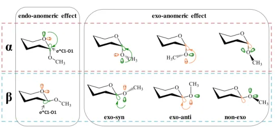
1.1.5.1.1 The anomeric effect
The anomeric effect was first described in the 1950s by J. T. Edward and R. U. Lemieux.38 The anomeric effect phenomenologically describes the stabilization
of the axial (versus equatorial) alkoxy groups at Cl of a pyranose ring. Nowadays, it is accepted that the reason is due to orbital and electronic factors produced by the simultaenous presence of two oxygens attached to one single carbon atom.
In principle, the tendency of the anomeric hydroxyl group or electronegative substituent to adopt axial orientation (α-anomer) rather than equatorial (β-anomer) is against to what would be expected based on steric factors. However, the favorable interaction between one lone electron pair located at a molecular orbital (n) on either glycoside oxygen atoms and the vicinal anti-bonding molecular orbital (σ*) of the contiguous C-O bond overcomes the possible steric hindrance provided by the axisal orientation. The anomeric effect (endo and exo) are now reasonably explained by computational approaches, including ab initio calculations (Figure 1.9).39
β-D-Galactofuranose
α-D-Galactofuranose α-D-Galactopyranose β -D-Galactopyranose