www.journalpulmonology.org
ORIGINAL
ARTICLE
Connective
tissue
disease-associated
interstitial
lung
disease
R.P.
Oliveira
a,∗,
R.
Ribeiro
a,b,
L.
Melo
a,
B.
Grima
a,
S.
Oliveira
a,
J.D.
Alves
a,baUnidadedeDoenc¸asImunomediadasSistémicas(UDIMS),Servic¸odeMedicinaIV,HospitalProf.DoutorFernandoFonseca,
Amadora,Portugal
bCEDOC/NOVAMedicalSchool,Lisboa,Portugal
Received23August2019;accepted10January2020
KEYWORDS Connectivetissue disease; Interstitiallung disease; Rheumatoidarthritis; Systemicsclerosis; Tocilizumab; Nintedanib Abstract
Background: Connectivetissuediseases(CTD)arefrequentlyassociatedwithinterstitiallung
disease(ILD),significantlyimpactingtheirmorbidityandmortality.
Aim: AnalyzetheexperienceofanautoimmunespecializedunitontreatingCTD-ILDand char-acterizethepopulationbasedonmostfrequentdiseases,imagingpatterns,lungfunctiontests results,serologyandtreatment.Assessmortalityandmortalitypredictorsinthesepatients.
Methods:Retrospective, descriptiveandstatisticalanalysisoftheCTD-ILDpatientsfollowed
upatanautoimmunediseasesunitduringa6-yearperiod.
Results:Overthestudyperiod,75patientswithCTD-ILDweretreatedwithameanfollow-up
of49±31months.
ThemostfrequentCTDweresystemicsclerosisandrheumatoidarthritis.ILDwasdiagnosed prior toCTDin8%ofpatients andconcomitantlyin35%.Nonspecificinterstitial pneumonia was theCTpatternin60%and35%hadanisolateddiminishedDLCO onlungfunctiontests. Pulmonary hypertensionwaspresent in12% anditwasthe singlemostimportantmortality predictor(OR14.41,p=0.006).Corticosteroidsarethemainstayoftreatment butbiologics wereprescribedin39%ofthepatients(mostlytocilizumabandrituximab).Twoscleroderma patientswererecentlytreatedwithnintedanib.
Conclusions: ILDisapotentialcomplicationofeveryCTDandcanimposeadramaticburdenon
thesepatients.TheclinicalrelevanceofILDtogetherwiththeirearlyexpressioninthecourse ofthediseaseunderlinestheimportanceofthepresenceofchestphysiciansintheseunits. ©2020SociedadePortuguesadePneumologia.PublishedbyElsevierEspa˜na,S.L.U.Thisisan openaccessarticleundertheCCBY-NC-NDlicense( http://creativecommons.org/licenses/by-nc-nd/4.0/).
∗Correspondingauthorat:HospitalProf.DoutorFernandoFonseca,IC19,2720-276,Amadora,Portugal.
E-mailaddress:[email protected](R.P.Oliveira).
https://doi.org/10.1016/j.pulmoe.2020.01.004
2531-0437/©2020SociedadePortuguesadePneumologia.PublishedbyElsevierEspa˜na,S.L.U.ThisisanopenaccessarticleundertheCC
Introduction
Connectivetissuediseases(CTD)areagroupofdiseaseswith heterogeneous systemic features and immune-mediated multi-organ disfunction. The respiratory tract can be targetedinvirtuallyeveryCTDandwithamultitudeof man-ifestations,bringingimportantimplicationstothediagnosis, follow-up, treatment and prognosis of these patients.1,2
Lunginvolvementisadecisivecontributortothemortality inCTD,andisnowtheleadingcauseof deathinsystemic sclerosis(SSc)3andanincreasingcauseofdeathin
rheuma-toidarthritis(RA),evenastheoverall mortalityrates are falling.4
Interstitial lung disease (ILD) is frequently present in patientswithautoimmune myopathies,SSc,Sjögren’s syn-drome, RA and systemic lupus erythematosus (SLE), with estimatedprevalencesof40%,30---40%,40%,10%and12%, respectively.5ItisimportanttokeepinmindthatILDmay
betheonlymanifestationofayet-to-bediagnosedCTD.6
High-resolution computedtomography (HRCT) and pul-monaryfunctiontests(PFT)arethebesttoolstoevaluate lunginvolvementandhaveprognosticvalue.7---9
Whiletherearenodefinitiveserumbiomarkersfor CTD-ILD,thepresenceofsomedisease-specificmarkerscanlead toabetterscreeningandrisk-stratifyingofpatients.10
Pulmonaryhypertension(PH)isacommonfindingin CTD-ILDpatientsandaddssignificantimpacttothemorbidityand mortalityofthesepatients.11
Development of lung disease,and itsassociated symp-tomslike dyspneaand cough, providesextraburden ona group of diseases already highly impactful on quality of life.12
Even thoughILDisacommoncomplicationofCTD,the definitiveguidanceonhowtotreatthesepatientsisscarce. Corticosteroidsstillrepresentthemainstayoftreatment.13
InthecontextofSSc,thereisalsoanincreasingbodyof evi-dencefor biologics,suchastocilizumab14 andrituximab,15
andrenewedenthusiasmfornewtherapeuticaltargets,such asantifibroticdrugs.16,17
Duetothe sheercomplexity andsystemicinvolvement of these diseases, a multidisciplinary approach shouldbe thegold-standardwhentreatingthesepatients.18Thispaper
aimstocompile,analyzeanddiscusstheexperienceofour unitinthemanagementofCTD-ILD.
Methods
Theauthors conducteda retrospective,descriptive analy-sisofpatientsolder than18yearsdiagnosedwithCTD-ILD followed up at an autoimmune diseases outpatient clinic betweenJanuaryof2013andDecemberof2018.
ThediagnosisofCTDwasbasedonclinicalandserologic criteriaaccordingtothemostrecentEULAR recommenda-tionsandthediagnosisofILDwasmadeonthebasisofHRCT findings.Allpatientswerediscussedinaweeklyautoimmune diseasesmultidisciplinaryteammeeting.Caseswerefurther discussedwithpulmonologyandradiologyonacase-by-case basis.
HRCTscansandPFTresultsshownaretheonesdoneat theILDdiagnosis.HRCTscanswereobtainedwith1---1.5mm thick slices. PFT were carried out according to a
stan-dardizedprotocolintherespiratorymedicinedepartment. Static lung volumes were measured using the plethys-mography method, and the lung diffusion capacity of CO (DLCO) using the single breath-hold method. Antibodies were tested with the following techniques: ANA, ANCA --- indirect immunofluorescence assay; Anti-SSA, anti-SSB, anti-Sm --- Immunoblotassay;anti-dsDNA,anti-CCP --- fluo-rescenceenzymeimmunoassay(FEIA);Rheumatoidfactor ---turbidimetry.
Datawas collected from hospital records and handled in an anonymous and population-based fashion. Normally distributed variablesare presentedasmean andstandard deviationandnon-normalvariablesarepresentedasmedian and interquartile range. Differences in baseline variables weretestedwitht-testsforcontinuousvariables;2-trend testsfororderedcategoricalvariables;and2testsfor bino-mialandunorderedcategoricalvariables.Statisticalanalysis wasperformedusingStata® 14software.
Results
PopulationDuring the6-year study period,75 CTD-ILDpatients were followed-up at thisunitwitha meanfollow-up of 49±31 monthsamounting to646.2 patients-years.The mean age atthetimeofILDdiagnosiswas56±15.5years-old,there was a clear female predominance (77.3%) and the mean duration of CTD was 8.9±7.5 years. CTD was diagnosed beforeILDin57%ofpatients,withamedianintervalof49.1 [23.7---127.15]monthsseparatingthetwodiagnosis.ILDwas diagnosedfirstin8%ofpatients(n=6)withCTDbeing diag-nosedshortlyafter(meanintervalof5.1months).Diagnosis wassimultaneousin35%ofpatients.
The CTD were, by order of prevalence, SSc (34.7%; n=26),RA(20%;n=15),overlapsyndrome(13.3%;n=10), mixedCTD(9.3%;n=7),autoimmunemyopathy(6.7%;n=5), ANCA-positivevasculitis(5.3%;n=4),undifferentiatedCTD (5.3%;n=4)andSjögrensyndrome(2.7%;n=2).
Imagingandbiopsy
Nonspecificinterstitialpneumonia(NSIP)wasthemost com-monHRCTpattern,presentin60%ofpatients,followedby usualinterstitialpneumonia(UIP)in36%ofpatientsanda pattern of lymphocyticinterstitial pneumonia(LIP) in the remaining3patients.Onepatientwithanti-synthetase syn-dromehadapredominanceofNSIPpatternoverlappedwith organizing pneumonia (OP) features. Out of the patients withNSIPpattern,42.2%hadadiagnosis ofSScand24.4% hadoverlapsyndromeor mixedCTDwithapredominance ofsclerodermafindings.AmongpatientswithSSc,58%had extensivedisease(>20%involvement19)onHRCT.ALIP
pat-tern was found in 2 patients with Sjögren syndrome and 1 with polymyositis. Other common findings were pleural thickening(n=16),lungnodules(n=14)andpleuraleffusion (n=5).
Whilenotmandatoryforthediagnosis,lungbiopsywas performedin11patientswhohadconflictingHRCTfindings orabroaderdifferentialdiagnosis,3beingcompatiblewith
NSIPand3withUIP.Theremaining5biopsieshadnonspecific findings.
Pulmonaryfunctiontests
PFTwerecarriedoutinallpatientsatthetimeofILD diag-nosis.Arestrictivepatternwasfoundin46%whilstonly6% hadan obstructivepattern.Isolateddiminished DLCOwas present in 35% of patients and 13% had all results within the normal range. ANCA-positive vasculitis, autoimmune myopathiesandmixedCTD,werethesubgroupofpatients whopresentedwithmoreseverelunginvolvementat diag-nosis(Table1).
Attimeofdiagnosisonly2patientshadarterialpressure ofoxygenbelow60mmHgonroomair andonly3patients hadminorhypercapnia.
Pulmonaryhypertension
Ninepatients(12%)hadsignsofpHontransthoracic echocar-diography(pulmonaryarterysystolicpressure≥40mmHg), withameanpressureof60.8±23.8mmHg.Fivepatientshad SScandtheother4hadmixedCTDoroverlapsyndromewith apredominanceofsclerodermafindings.Echocardiographic datawasmissingin13patients.
Five patients underwent right heart catheterization (RHC). Two of them did not have a confirmation of PH, the others had a mean pulmonary artery pressure of 40±2.1mmHg and a meanpulmonary vascular resistance of10.4±6WoodUnits.Ofthese3,allwithSSc,1was clas-sifiedashavinggroupIPH,theothersweregroupIII.Inthe remainder 4 patients withechocardiographic signsof PH, RHCwas not conductedas the procedure wasconsidered tooriskyorofnoaddedbenefittothemanagementofthese particularpatients.
Autoantibodies
Anti-nuclear antibodies were positive in 79% of patients (titre≥1:320in80%).
Amongst SSc patients, 54% were anti-Scl70 positive, 15.4% were anti-centromerepositive and 7.7% were anti-PM-Sclpositive.
Amongst RA patients, 73.3% were simultaneously rheumatoid factor (mean value of 338±266 UI/mL) and anti-citrullinatedprotein(anti-CCP)positive(meanvalueof 170.1±160.4UI/mL)
In overlap syndromes,the most frequentlyfound sero-logic markers were anti-SSA (present in 40% of patients) followed by anti-SSB, anti-Scl70, anti-PM-Scl, rheumatoid factorandanti-CCP (allpresentin20% ofthesepatients). Allpatients classified ashavingmixedCTD wereanti-RNP positive. Four out of the five patients with autoimmune myopathy were diagnosed with anti-synthetase syndrome (3wereanti-Jo1positiveandtheotherwasanti-PL12 pos-itive). The patient with polymyositis wasanti-Ku positive (anti-MDA5wasnottested).
Treatment
Systemiccorticosteroids were by far the most useddrug, beingused at some point during the course of disease in 86.7%of thepatients.Mycophenolate mofetilwasusedin 36%ofpatients,cyclophosphamidein33%andazathioprine in29.3%.Methotrexate(MTX)wasusedpriortoILDdiagnosis in32%ofpatients(n=23),butwasthenswitchedtoanother immunossupressordrug(mainlymycophenolate)inallbut2 patients.OutofthepatientswhoweretreatedwithMTX,3 (13%)hadlungfibrosisconsideredtobecausedbythedrug. Biologic drugs were used in 39% of the patients, with tocilizumab and rituximab being the most used as first-linetherapy(13patientseach),followedbyetanercept(3 patients). Sixty percent of the patients with RA were on biologictreatment,aswere23.1%ofSSc.Tocilizumabwas themost usedbiologic in RAbut thisdecision wasdriven by joint disease in all but one patient. All patients with ANCA-positivevasculitisandanti-synthetasesyndromewere on rituximab (Table 1). Five patients required a biologic switchdue tofailure of first-line choice (3patients were switched from tocilizumab to rituximab, 2 patients were switched from etanercept to tocilizumab). In 3 of these patientstheswitchwasdecidedbasedonILDprogression,in theother2patientsthedecisionwasmadeduetoongoing activityofthejointinvolvement(bothpatientshadoverlap syndrome).Throughoutthefollow-upperiodtherewereno exacerbationsoftheILDcausedbythestartingofbiologic treatment.
Two patients with SSc were recently treated with nintedanib(onewithUIPpattern,theotherNSIP).
Six patients of the nine diagnosed with pH were pre-scribedspecific treatmenttargeted atPH: 3bonsentan,1 alprostadil,1iloprostand1sildenafil.Thelasttwopatients werelaterswitchedtobonsentan.
Mortality
Thiscohorthadamortalityrateof20%(15/75patients) dur-ingtheanalyzedperiod,withameansurvivalof67.8±57.3 monthssince CTD diagnosis and 37.8±20.9 months since ILDdiagnosis.Mortality ratewashigherinRAandoverlap syndrome(Table1).
Comparingdeceasedpatientswiththesurvivors,thefirst group wasolder at the time of CTD diagnosis (58.6±4.9 vs51.5±2.7years-old,p=0.23),older atthetimeoflast follow-up(66.4±11.1vs60.4±15.2years-old,p=0.03)and morefrequentlymale(33vs20%,p=0.31).UIPpatternon HRCTwasrelatedwithhighermortalitybutthedifference wasnot significant. There were nosignificant differences regarding other imaging patterns, diagnosis of SSc, corti-cotherapyandANApositivity.ThepresenceofpHwasclearly associatedwithhighermortality(OR14.41,p=0.006)andso wastheuseofbiologics(OR5.56,p0.025).
Discussion
This population of 75 CTD-ILD patients constitutes a rel-evant sized population with a long mean follow-up time of49±31months overthe spanof 6years.ILDwas diag-nosedpriortoCTDinonly8%ofpatientsbutthisisprobably
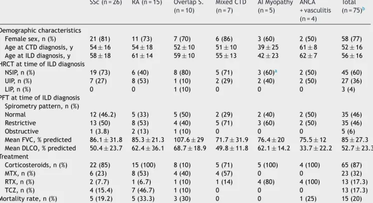
Table1 Descriptionofmaincharacteristicsanalyzedbydiseasesubgroupsandtheglobalcohort. SSc(n=26) RA(n=15) OverlapS. (n=10) MixedCTD (n=7) AIMyopathy (n=5) ANCA +vasculitis (n=4) Total (n=75)b Demographiccharacteristics Femalesex,n(%) 21(81) 11(73) 7(70) 6(86) 3(60) 2(50) 58(77) AgeatCTDdiagnosis,y 54±16 54±18 52±10 51±10 39±25 61±8 52±16 AgeatILDdiagnosis,y 58±18 61±14 59±10 55±13 42±23 62±7 56±16 HRCTattimeofILDdiagnosis
NSIP,n(%) 19(73) 6(40) 8(80) 5(71) 3(60)a 2(50) 45(60)
UIP,n(%) 7(27) 8(53) 1(10) 2(29) 2(40) 2(50) 27(36)
LIP,n(%) 0 0 1(10) 0 0 0 3(4)
PFTattimeofILDdiagnosis Spirometrypattern,n(%)
Normal 12(46.2) 5(33) 5(50) 2(29) 2(40) 2(50) 35(46) Restrictive 13(50) 8(53) 4(40) 5(71) 3(60) 2(50) 35(46) Obstructive 1(3.8) 2(13) 1(10) 0 0 0 5(6) MeanFVC,%predicted 86.1±31.8 85.3±21.3 107.6±29 71.7±31.9 76.4±20 75.5±12 85±27.3 MeanDLCO,%predicted 50.4±23.7 62.4±36.1 68.7±18.9 49.8±11.8 62.1±14.2 33.7±22.2 52.7±23.3 Treatment Corticosteroids,n(%) 22(85) 15(100) 8(10) 5(71) 5(100) 4(100) 65(87) MTX,n(%) 6(23) 8(53) 4(40) 4(57) 0 0 23(32) RTX,n(%) 2(7.7) 1(6.7) 1(10) 1(14) 4(80) 4(100) 13(17.3) TCZ,n(%) 4(15.4) 7(46.7) 1(10) 0 0 0 13(17.3) Mortalityrate,n(%) 5(19.2) 5(33.3) 3(30) 0 0 1(25) 15(20)
SSc-Systemicsclerosis;RA-RheumatoidArthritis;OverlapS.-Overlapsyndrome;CTD-connectivetissuedisorder;AImyopathy
-Autoimmunemyopathy;ILD-Interstitiallungdisease;HRCT-High-resolutioncomputerizedtomography;NSIP-Nonspecificinterstitial
pneumonia;UIP-Usualinterstitialpneumoniae;LIP-Limphocyticinterstitialpneumonia;PFT-Pulmonaryfunctiontest;FVC-Forced
vitalcapacity;DLCO-Diffusingcapacityforcarbonmonoxide;MTX-Methotrexate;RTX-Rituximab;TCZ---Tocilizumab.
aOnepatientwithanti-synthetasesyndromepresentedwithoverlapNSIP/OP.
b TotalincludessubgroupsshownplusundifferentiatedCTD(n=4)andSjögrensyndrome(n=2).
biasedsince this unit is primarily focused onCTD witha background in internal medicine. More importantly, diag-noses were simultaneous in 35% of patients, emphasizing therelevanceoflunginvolvementintheseconditions.Itis importanttonotethatallpatientshadadefinitediagnosis ofaCTD,hencenopatientfellwithintherecentlysuggested definitionof‘‘interstitialpneumoniawithautoimmune fea-tures’’.20
MostpatientspresentedwithaNSIPpatternonHRCTbut morecommonfindingssuchaspleuralthickeningsand effu-sionscanpointtowardsapossibleunderlyingCTDinanILD patient. The majority of SSc patients had extensive lung diseaseonHRCT at thetime ofILD diagnosis, again rein-forcingtherelevanceofthiscondition,particularlyinlight ofthefactthatitcanbeclinicallysilentduringasignificant amountof time.3 Lungbiopsywasseldomusedandrarely
providedadditionalinformation.
Regarding PFT, an isolated diminished DLCO (35% of patients)maybetheonlyabnormality.ANCA-positive vas-culitis and mixed CTD were the subgroups withthe most impactfuldisease at diagnosis. In ANCA-positivevasculitis theseverityofthe lungdiseasewasmainlyonaccountof averylow DLCO,whichwasprobablycausedby vasculitic diseaseratherthanILDperse.
Regarding treatment, corticosteroids remain the first-line but the use of other immunosuppressants such as mycophenolatemofetilisincreasing.
Methotrexate was stopped in most patients upon ILD diagnosis, even if only 3 patients presented with fibrosis admittedbeingrelatedtoit.OvertheyearsMTX-associated lung disease has been a controversial topic, with earlier studies pointing towards a slightly increased risk of lung fibrosis.21 MTX was stopped for most patients to prevent
it from being a confounding factor in the follow-up and becausesafer andequally effectivedrugs were available. Inretrospect,andinlightofarecentmultivariateanalysis showing that there was no association between develop-ment of RA-ILD and MTX use and that the drug could in facthaveaprotectiverole,thedrugcouldhavebeensafely continued.22
BiologicdrugsarebeingincreasinglyusedinCTD-ILD.The most usedones weretocilizumab andrituximaband their usewasmainlydictatedbytheextra-pulmonary manifesta-tionsoftheunderlyingCTD----tocilizumabwaspredominantly used in RA and rituximab for vasculitis and autoimmune myopathies.Inthispopulation,biologicsweredeemedsafe tostartinpatientswithILDastherewerenoexacerbations uponthestartofbiologics,contrarytosomepriorreports.23
Theuseofbiologicswasassociatedwithworsesurvivalbut thisisprobablyrelatedtodiseaseseverityratherthan drug-relatedcomplications.
The antifibrotic nintedanib was usedin 2 SSc patients with extensive lung fibrosis, after positive safety results in idiopathicpulmonary fibrosis,24 andmight bea
promis-ing option for slowing ILD progression in these patients, as shown by the SENSCIS Trial.17 However, criteria for
nintedanib use in SSc-ILD is up for debate,25 and both
ourpatientsremained onbackground immunossuppressive treatment.Evidenceisemergingfortheuseofantifibrotics inotherCTDs.TherecentlypublishedINBUILDTrialshowed thatnintedanib leadstoalowerannual rateofdeclinein FVC in patients withprogressive fibrosing interstitiallung diseases,includingpatientswithautoimmunediseases,the majority of them with RA-ILD and also includingpatients withmixedCTD.26
With 20% mortality rate and a mean survival of 37.8±20.9 months, ILD constitutes an important burden onCTD patients and significantlyimpacts their prognosis. Inthiscohort,pHwasthesinglemostimportantmortality predictor(OR14.41,p=0.006).
Even though this article provides an overview of the generalpopulationencounteredatanautoimmunediseases unit,someparticularsubsetsofpatientsarenotasstrongly represented in the cohort (e.g. autoimmune myopathies) andthiscouldleadtoanunderrepresentationofmore spe-cificandlesscommonfindings,asisthecaseoforganizing pneumonia.Attheotherend,itcouldleadtoan overrepre-sentationofrarefindings,suchastheLIPpatterninapatient withpolymyositis.Heterogeneity regardingtheunderlying CTDs, disease duration, treatment and follow-up and the retrospective nature of the study may limit the interpre-tationof someofthe results.The datapresentedreflects theclinicalexpertiseofasingleinstitutionthathasitsmain focusonCTDandmaynotbefullyrepresentativeof prac-ticeselsewhere.
Conclusion
To thebestof ourknowledge thisis thelargest cohortof CTD-ILDpresentedbyaPortuguesecentre.
ILD is a potentialcomplication of virtually every CTD, constitutes an important burden on these patients and significantlyimpactstheirprognosis.Asystemicand multi-disciplinaryoverviewisessentialforadequatemanagement, inordertofillintheremaininggapsregardingearly diagno-sis,follow-upandtreatmentofthesepatients.
Author
contributions
RPO conceived theideafor themanuscript,collected the data,wrotethefirstdraftandco-wrotethepaper.RR col-lectedthe dataandco-wrotethe paper.LMcollected the data,wasresponsiblefor statisticalanalysis andco-wrote the paper. BG, SO and JDA conceived the idea for the manuscript. All authors reviewed and approved the final versionofthismanuscript.
Conflicts
of
interests
Theauthorshavenoconflictsofintereststodeclare.
Acknowledgements
TheauthorswouldliketothanktheDepartmentof Respira-toryMedicineofHospitalProf.DoutorFernandoFonsecafor thecollaborationthroughouttheyears.
References
1.Fischer A, du Bois R. Interstitial lung disease in
con-nective tissue disorders. Lancet. 2012;380:689---98,
http://dx.doi.org/10.1016/S0140-6736(12)61079-4.
2.Olson A, Brown K, Fischer A. Connective tissue
disease---associated lung disease. Immunol Allergy Clin
N Am. 2012;32:513---36, http://dx.doi.org/10.1016/
j.iac.2012.09.002.
3.Steen V, Medsger T. Changes in causes of death in
sys-temic sclerosis, 1972-2002. Ann Rheum Dis. 2007;66:940---4,
http://dx.doi.org/10.1136/ard.2006.066068.
4.Olson A, Swigris J, Sprunger D, Fischer A, Fernandez-Perez
E, Solomon J, et al. Rheumatoid Arthritis---Interstitial
Lung Disease---associated Mortality. Am J Respir Crit
Care Med. 2011;183:372---8, http://dx.doi.org/10.
1164/rccm.201004-0622OC.
5.Fischer A, Strek M, Cottin V, Dellaripa P, Bernstein E,
Brown K, et al. Proceedings of the American College
of Rheumatology/Association of Physicians of Great Britain
and Ireland Connective Tissue Disease---Associated
Intersti-tial Lung Disease Summit: A Multidisciplinary Approach to
Address Challenges and Opportunities. Arthritis Rheumatol.
2019;71:182---95,http://dx.doi.org/10.1002/art.4076.
6.MathaiS,DanoffS.Managementofinterstitiallungdisease
asso-ciatedwithconnective tissue disease. BMJ. 2016;352:h6819,
http://dx.doi.org/10.1136/bmj.h6819.
7.Walsh S, Sverzellati N, Devaraj A, Keir G, Wells A, Hansell
D. Connective tissue disease related fibrotic lung disease:
highresolutioncomputedtomographicandpulmonaryfunction
indices asprognosticdeterminants.Thorax. 2014;69:216---22,
http://dx.doi.org/10.1136/thoraxjnl-2013-203843.
8.WinstoneT,AssayagD,WilcoxP,DunneJ,HagueC,LeipsicJ,
etal.Predictorsofmortalityandprogressionin
scleroderma-associated interstitiallung disease. Chest. 2014;146:422---36,
http://dx.doi.org/10.1378/chest.13-2626.
9.Assayag D, Lubin M, Lee J, King T, Collard H,
Ryer-son C. Predictors of mortality in rheumatoid
arthritis-relatedinterstitiallungdisease.Respirology.2014;19:493---500,
http://dx.doi.org/10.1111/resp.12234.
10.Aubart F, Crestani B, Nicaise-Roland P, Tubach F, Bollet C,
Dawidowicz K, et al. High Levels of anti-cyclic citrullinated
peptide autoantibodies are associated with co-occurrence
of pulmonary diseaseswith rheumatoidarthritis. J
Rheuma-tol. 2011;38:979---82, http://dx.doi.org/10.3899/jrheum.
101261.
11.TakahashiK,TaniguchiH,AndoM,SakamotoK,KondohY,
Watan-abeN,etal.Meanpulmonaryarterialpressureasaprognostic
indicatorinconnectivetissuediseaseassociatedwith
intersti-tiallungdisease:aretrospectivecohortstudy.BMCPulmMed.
2016;16:55,http://dx.doi.org/10.1186/s12890-016-0207-3.
12.Mittoo S, Frankel S, LeSage D, Strand V, Shah A,
Christopher-Stine L, et al. Patient Perspectives in
OMER-ACT provide an anchor for future metric development
and improved approaches to healthcare delivery in
connective tissue disease related interstitial lung
dis-ease (CTD-ILD). Curr Respir Med Rev. 2015;11:175---83
https://doi.org/10.2174%2F1573398X11666150619182624
13.Wallace B, Vummidi D, Khanna D. Management of
lung disease. Curr Opin Rheumatol. 2016;28:236---45
https://doi.org/10.1097%2FBOR.0000000000000270
14.Khanna D,DentonC,Jahreis A, vanLaar J,Frech T,
Ander-sonM,etal.Safetyandefficacyofsubcutaneoustocilizumab
in adults with systemic sclerosis (faSScinate): a phase
2, randomised, controlled trial. Lancet. 2016;387:2630---40,
http://dx.doi.org/10.1016/S0140-6736(16)00232-4.
15.SharpC,McCabeM,DoddsN,EdeyA,MayersL,AdamaliH,etal.
Rituximabinautoimmuneconnectivetissuedisease---associated
interstitial lung disease. Rheumatology. 2016;55:1318---24,
http://dx.doi.org/10.1093/rheumatology/kew195.
16.Khanna D, Tashkin D, Denton C, Lubell M,
Vasquez-Mateo C, Wax S. Ongoing clinical trials and treatment
options for patients with systemic sclerosis---associated
interstitial lung disease. Rheumatology. 2018;58:567---79,
http://dx.doi.org/10.1093/rheumatology/key151.
17.Distler O, Highland KB, Gahlemann M, Azuma A, Fischer A,
MayesMD,etal.NintedanibforSystemicSclerosis-Associated
Interstitial Lung Disease. N Engl J Med. 2019;380:2518---28,
http://dx.doi.org/10.1056/NEJMoa1903076.
18.Richeldi L, Launders N, Martinez F, Walsh S, Myers J, Wang B,etal.Thecharacterisationofinterstitiallungdisease mul-tidisciplinary team meetings: a global study.ERJ Open Res. 2019;5:00209---2018.
19.Goh N, Desai S, Veeraraghavan S, Hansell D, Copley
S, Maher T, et al. Interstitial Lung Disease in Systemic
Sclerosis. Am J Respir Crit Care Med. 2008;177:1248---54,
http://dx.doi.org/10.1164/rccm.200706-877OC.
20.FischerA,AntoniouK,BrownK,CadranelJ,Corte T,duBois
R, et al. An official European Respiratory Society/American
Thoracic Society research statement: interstitial pneumonia
with autoimmune features. Eur Respir J. 2015;46:976---87,
http://dx.doi.org/10.1183/13993003.00150-2015.
21.Conway R, Low C, Coughlan R, O’Donnell M, Carey J.
Methotrexateandlungdiseaseinrheumatoidarthritis:a
meta-analysisofrandomizedcontrolledtrials.ArthritisRheumatol.
2014;66:803---12,http://dx.doi.org/10.1002/art.38322.
22.Kiely P, Busby A, Nikiphorou E, Sullivan K, Walsh D,
Creamer P. Is incident rheumatoid arthritis interstitial
lung disease associated with methotrexate treatment?
Results from a multivariate analysis in the ERAS and
ERAN inception cohorts. BMJ Open. 2019;9:e028466,
http://dx.doi.org/10.1136/bmjopen-2018-028466.
23.Chen J, Chi S, Li F, Yang J, Cho W, Liu X.
Biologics-inducedinterstitiallungdiseasesinrheumaticpatients:facts
and controversies. Expert Opin Biol Ther. 2017;17:265---83,
http://dx.doi.org/10.1080/14712598.2017.1287169.
24.Richeldi L, du Bois R, Raghu G, Azuma A, Brown K,
Costabel U,et al. Efficacy and safetyof nintedanib in
idio-pathicpulmonary fibrosis. NEngl J Med. 2014;370:2071---82,
http://dx.doi.org/10.1056/NEJMoa1402584.
25.Antoniou K, Trachalaki A, Tzouvelekis A, Poletti V,
Vasarmidi E, Sfikakis P, et al. A role of antifibrotics in
the treatment of Scleroderma-ILD? Pulmonology. 2020,
http://dx.doi.org/10.1016/j.pulmoe.2019.08.004.
26.Flaherty K, Wells A, Cottin V, Devaraj A, Walsh S,
Inoue Y, et al. Nintedanib in Progressive Fibrosing
Inter-stitial Lung Diseases. N Engl J Med. 2019;381:1718---27,