DOI: http://dx.doi.org/10.18363/rbo.v75.2018.e1193 Literature Review/Oral Pathology
Peri-Implant Diseases: an update for dentistry
Álvaro Cavalheiro Soares,1 Andreza Maria de Oliveira Filgueiras,1 Viviane Vargas de Azevedo,1 Marcus Vinicius Rêgo Benevides,1 Bruna Michalski dos
Santos,2 Luciana Freitas Bastos,2,3 Marcelo Daniel Brito Faria,1,2 Ruth Tramontani Ramos,1 Marilia Heffer Cantisano1
1Department of Diagnosis and Therapeutics, School of Dentistry, Rio de Janeiro State University, Rio de Janeiro, RJ, Brazil
2Center of Dental Radiology and Care to Patients with Special Needs, Piquet Carneiro Polyclinic, Rio de Janeiro State University, Rio de Janeiro, RJ, Brazil 3Department of Preventive and Community Dentistry, School of Dentistry, Rio de Janeiro State University, RJ, Brazil
• Conflicts of interest: none declared.
AbstrAct
Objective: this study aims to present and describe the main pathological, benign and malignant alterations of the peri-implant mucosa. Material and Methods: an integrative review of the literature was carried out using the following descriptors: peri-implant pathology; reactive lesion dental implant; carcinoma dental implant; peri-im-plant disease dental imperi-im-plant; reactive lesion by dental imperi-im-plant; oral squamous cell carcinoma around dental imperi-im-plants; peri-imperi-im-plant disease; pathology; for the elaboration of the search strategy in the databases PubMed, LILACS, MEDLINE and SciElo to obtain the scientific productions. Results: conditions such as peri-implant mucositis and peri-implantitis are poorly reported in the literature, which makes it difficult to estimate their real prevalence. Among the benign reactive lesions, the pyogenic granuloma and peripheral giant-cell granuloma stand out, described as the most frequently associated with the implants. Regarding neoplasia, cases of hemangioma and OSCC were published, suggesting a possible influence of the implant in the development of these conditions. Conclusion: the occurrence of several pathologies associated with dental implants was observed in the literature, which demonstrates the importance of a detailed clinical examination, associated, in certain situations, with histopatholog-ical examination for an accurate diagnosis and adequate management of the various peri-implant diseases.
Keywords: Dental implant; Peri-implant mucositis; Peri-implantitis; Reactive lesion by dental implant; Squamous cell carcinoma.
Introduction
A
ttempts to replace teeth lost by prosthetic elements date back to the Mayan period (100-1500 A.D.). The dental implant is a titanium alloplastic material that has the root function of one or more teeth, used as a replace-ment for elereplace-ments lost by trauma, periodontal diseases or caries.1 Currently, oral rehabilitation with osseointegrateddental implants has become one of the best options for the treatment of edentulous patients, which has led to its univer-salization and, sometimes, its indication without adequate planning (Figure 1), leading to an increase in the number
of complications associated with its use (Figure 2), such as inflammatory processes affecting soft tissues and/or bones, known as peri-implant diseases (PIDs). The PIDs are sub-divided into peri-implant mucositis and peri-implantitis. These processes are analogous to gingivitis and periodonti-tis in natural teeth, respectively.2 Clinically, such conditions
are presented as edema, erythema, hypertrophy and soft tissue ulcers, with characteristics that sometimes require a differential diagnosis with malignant lesions, such as oral squamous cell carcinoma (OSCC), for example (Figure 3).3
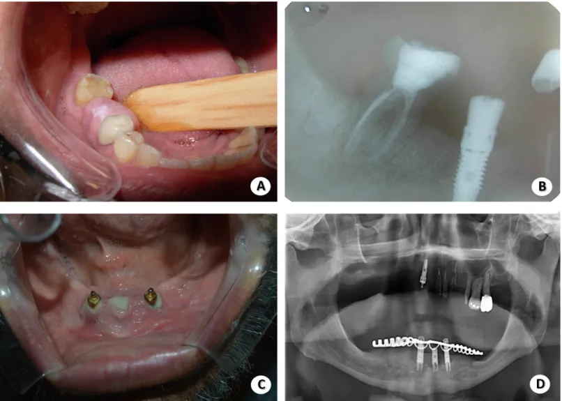
Figure 2. Examples of complications related to the installation of dental implants. “A” is the presence of a nodule located in the peri-implant re-gion of element 46, suggestive of reactional lesion; in “B” we have the periapical showing nodule association directly linked to the constituent of the dental implant; in “C” we have a case of peri-implant mucositis with purulent secretion drainage; and in “D”, panoramic radiograph of three implants associated with peri-implant mucositis
Figure 3. Ulcer with more than 30 days of evolution and no signs of healing, associated with the region of dental implants. Case to be investigated with biopsy examina-tion followed by anatomopathological analysis
Reactive lesions are those characterized by excessive pro-liferation of connective tissue in response to chronic irri-tation. Among these are included focal fibrous hyperplasia, fibroepithelial hyperplasia, peripheral ossifying fibroma, pe-ripheral giant-cell granuloma (PGCG), and pyogenic gran-uloma (PG). The last two are the most commonly diagnosed in the peri-implant mucosa.
In addition, some studies have reported the development of OSCC in the mucosa adjacent to the dental implant, which should be considered, therefore, as a diagnostic hypothesis in the case of peri-implant mucosal lesions, since it requires a precise and fast diagnosis for immediate treatment.4
Thus, in addition to the increase in the number of PID cases, such as mucositis and peri-implantitis, and the devel-opment of peri-implant mucosal lesions, this is associated with the fact that such conditions are little discussed in the training of dental surgeons and implant dentists. Therefore, this study aims to present and describe the main patholog-ical, benign and malignant alterations of the peri-implant mucosa aiming to update the professionals who perform the clinical follow-up of patients rehabilitated with dental im-plants, thus assisting the detection, diagnosis and adequate management of the most frequent changes.
Material and Methods
The study consists of an integrative review of the liter-ature, where the PubMed, LILACS, MEDLINE and SciElo databases were used to obtain the scientific productions. The bibliographic search was carried out in October 2016, establishing the time interval between 1989 and 2016 and using the following descriptors for the elaboration of the search strategy: peri-implant pathology; reactive lesion den-tal implant; carcinoma denden-tal implant; peri-implant disease dental implant; reactive lesion by dental implant; oral squa-mous cell carcinoma around dental implants; peri-implant disease; pathology.
Scientific articles in English, Spanish and Portuguese that were relevant, current and published in indexed journals, with editorial board and ISSN (International Standard Seri-al Number) were selected. Publications that did not have the complete text available for reading, if in different languages from those previously mentioned and that did not address the object of study, were excluded.
Results
Non-neoplastic Lesions Associated with Implants Peri-implant Mucositis
According to Berglungh et al.,6 studies on the prevalence
of peri-implant diseases are scarce, apart from the fact that cross-sectional studies in implant-rehabilitated patients and bleeding on probing data are hardly published. Considering this, to evaluate the prevalence of this condition is highly difficult.
Peri-implantitis
Peri-implantitis is an inflammatory and destructive process that affects peri-implant soft and hard tissues. The histopathological features found in peri-implant tissues in regions of implants removed due to the disease consisted of the presence of bacterial biofilm on the implant surface and a chronic inflammatory infiltrate in the connective tissue. Such histopathological features were described by Piatteli et
al.7 in a study of 230 implants recovered over a period of 8
years.
In their study, Roos-Jansåker et al.8 performed a clinical
and radiographic examination of 999 implants in 218 pa-tients between 9 and 14 years after the rehabilitation and observed a pocket depth greater than or equal to 4 mm, and bleeding on probing in 48% of the sites with dental implants.
Pyogenic Granuloma (PG)
The first reports of PG in association with dental im-plants were presented in the study by Olmedo et al. 4 where
2 clinical cases of reactive lesions of peri-implant mucosa were diagnosed, both initially considered as epulis. To the histopathological examination, the cases were diagnosed as PG and PGCG, with the presence of metallic particles in the tissues, suggesting that the etiology of the lesions may be associated with the corrosion process of the metallic struc-ture, as demonstrated by Rodrigues et al.9
Dojcinovic et al.10 reported the case of a 32-year-old male
patient who had an exophytic and ulcerated nodular lesion of 1.0 cm in the left posterior maxillary gingiva associated with a dental implant. A clinical diagnosis of inflammatory fibrous hyperplasia was made, and the lesion excised and re-ferred for histopathological analysis, and then diagnosed as PG. In this case, the etiology of the lesion was the inadequate choice of a healing abutment, which prevented the correct hygiene of the region, causing a chronic inflammatory pro-cess in the peri-implant tissue. Excision and replacement of the healing abutment were sufficient to resolve the case.
Peripheral Giant-Cell Granuloma (PGCG)
compli-to 2015. Hirshberg et al.12 reported PGCG in 12% of the 25
peri-implant soft tissue biopsies examined.
Bischof et al.13 described a case of PGCG in a 56-year-old
female patient with three implants in the posterior region of the mandible. The patient in question reported difficulty in performing hygiene in the region, causing dental plaque accumulation.
In the cases analyzed by Brown et al.,14 a recurrence rate
of 46.2% of PGCG associated with dental implants was ob-served, which is considerably higher than that of PGCG in general, where an average recurrence rate of 5 to 11% is re-ported.
Neoplastic Lesions Associated with Dental Implants Hemangioma
Kang et al.15 reported a case of hemangioma associated
with a dental implant (Figure 3). In their study, the co-de-velopment of PG and capillary hemangioma in the upper alveolar ridge associated with a dental implant in a patient using Warfarin was described. The lesion was excised and submitted to histopathological and immunohistochemical analysis, where PG and Hemangioma characteristics were observed. According to the authors, such different condi-tions coincidentally expanded in the same gingival mass.
Oral Squamous Cell Carcinoma (OSCC)
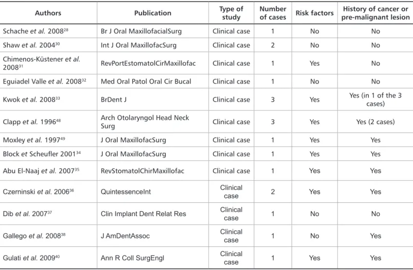
Jané-Salas et al.3 reviewed 13 articles published between
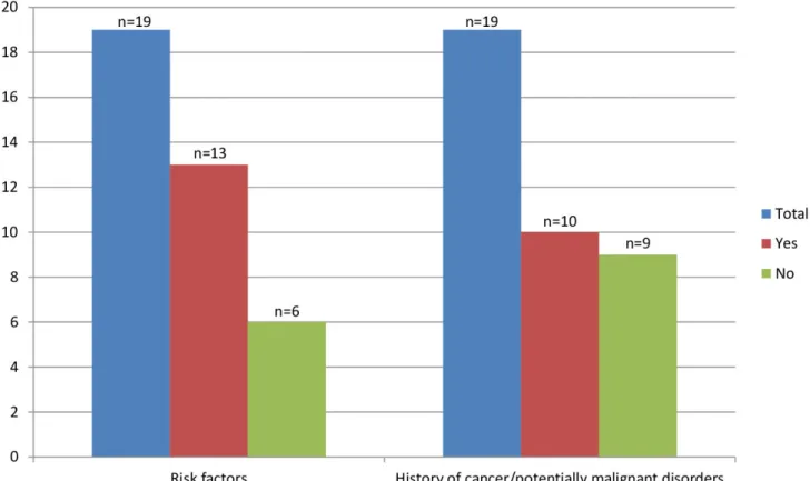
1996 and 2009 referring to 19 cases in which OSCC was de-veloped in patients with osseointegrated implants (Table 1). Figure 4 shows the number of patients who developed OSCC and shows in which cases there were risk factors or history of cancer and/or pre-malignant lesions.
Table 1. Cases of OSCC in patients rehabilitated with osseointegrated implants reported in the literature
Authors Publication Type of study Number of cases Risk factors pre-malignant lesionHistory of cancer or
Schache et al. 200828 Br J Oral MaxillofacialSurg Clinical case 1 No No
Shaw et al. 200430 Int J Oral MaxillofacSurg Clinical case 2 No No
Chimenos-Küstener et al.
200831 RevPortEstomatolCirMaxillofac Clinical case 1 Yes No
Eguiadel Valle et al. 200832 Med Oral Patol Oral Cir Bucal Clinical case 1 No No
Kwok et al. 200833 BrDent J Clinical case 3 Yes Yes (in 1 of the 3
cases)
Clapp et al. 199648 Arch Otolaryngol Head Neck
Surg Clinical case 3 Yes Yes (2 cases)
Moxley et al. 199749 J Oral MaxillofacSurg Clinical case 1 Yes Yes
Block et Scheufler 200134 J Oral MaxillofacSurg Clinical case 1 Yes Yes
Abu El-Naaj et al. 200735 RevStomatolChirMaxillofac Clinical case 1 Yes Yes
Czerninski et al. 200636 QuintessenceInt Clinical
case 2 Yes Yes
Dib et al. 200737 Clin Implant Dent Relat Res Clinical
case 1 No No
Gallego et al. 200838 J AmDentAssoc Clinical
case 1 No Yes
Gulati et al. 200940 Ann R Coll SurgEngl Clinical
case 1 Yes Yes
Raiser et al.16 reported cases of asymptomatic lesions
around long-standing implants that resembled peri-implan-titis and were subsequently diagnosed as malignant neo-plasms. Of 42 malignant tumors associated with implants, between 2000 and 2014, 85.7% were squamous cell carcino-mas (69% primary and 9.4% metastatic).Most patients had preexisting risk factors for oral cancer.
Discussion
According to Lang and Berglundh, PID is an inflammato-ry reaction of microbial etiology caused by the accumulation of bacterial plaque and affecting the peri-implant tissues, subdivided into: peri-implant mucositis and peri-implan-titis.17 As established during the I European Periodontics
Workshop, the term peri-implant mucositis refers to a re-versible inflammation that affects peri-implant soft tissues, considering peri-implantitis as an inflammatory process that strikes and destroys hard and soft tissues around the implant.18
Considering the increase of rehabilitated patients with implants, the development of lesions on peri-implant
muco-Figure 4. Number of patients rehabilitated with implants presenting risk factors for OSCC or history of cancer/potentially malignant disorders
tients with high hormonal levels (pregnant women, for ex-ample). Clinically, it is a tumor-like growth of the oral cavi-ty, most often located around the anterior teeth.19 The study
of Dojcinovic et al.10 demonstrated the case of a man who
had developed a PG due to the inadequate choice of a heal-ing abutment, thus allowheal-ing an accumulation of plaque and chronic inflammation of the peri-implant tissue. Olmedo et
al.4 histologically observed the presence of metal particles in
biopsied GPs, suggesting that the etiology of the lesions may be related to the corrosion process of the metallic structure, as shown also by Rodrigues et al.9 GPCG is a relatively
fre-quent benign reactive lesion of the oral cavity, originating from the periosteum or periodontal membrane, developed in response to local irritation or chronic trauma. Clinically, it presents itself as a red-purple nodule located in the region of the gingiva or edentulous alveolar ridge, occurring with greater prevalence in the fifth and sixth decades of life and presenting a slight predilection for females.20 Bischof et al.13
described a case of PGCG that evolved due to inadequate angulation of the implants, which hampered the hygiene of the region.This is confirmed by the Ozden et al.21 study, who
opment of PG and capillary hemangioma on the alveolar ridge of a patient using Warfarin rehabilitated with a den-tal implant. Therefore, patients with denden-tal implants un-der antithrombotic therapy should be closely monitored for a possible early detection of this condition.
OSCC is defined as a malignant neoplasm of epithelial origin that usually occurs in men over 60 years old.22 The
pathogenesis of OSCC is well established and is largely re-lated to lifestyle, especially excessive alcohol and tobacco consumption.1 Jané-Salas et al.3 study reviewed 19 cases of
OSCC associated with dental implants, where 9 of them had no history of neoplasia, neither in the oral cavity nor in another region of the body. Other reports regarding the occurrence of OSCC in patients rehabilitated with dental implants are set out in Table 1.
In addition to OSCC cases, there is a published osteo-sarcoma in the maxilla case associated with placement of the implant with the use of filling material in the maxil-lary sinus.23
The development of OSCC is more frequent in peri-im-plant mucosa of patients who have risk factors or prior history of cancer. The mechanism by which osseointegrat-ed dental implants may contribute to the development of OSCC is highly debatable, since the association and in-fluence of other risk factors for carcinogenesis, such as smoking and alcoholism, cannot be disregarded. Some studies propose that the development of OSCC may be re-lated to changes in the microbial profile of the oral cavity. Bacterias of the genera Porphyromonas and Fusobacteri-um, known as periodontopathogenic bacteria, are known to be found in larger amounts in SCC samples than in healthy adjacent tissues, and high concentrations of Strep-tococcus mitis, Capnocytophaga gingivalis and Prevotella melaninogenica have been reported in the saliva of pa-tients of OSCC when compared to healthy individuals.24
Since several of these species also colonize peri-implant tissues, further investigations are necessary in order for the hypothesis of the influence of bacterial colonization in the peri-implant region to be confirmed or not as the etiological factor for the development of OSCC. Based on this review, it is not possible to establish a direct causal relationship between implant rehabilitation and the devel-opment of malignant neoplasia, although, given the large number of cases reported in the literature, implant treat-ment may possibly be an additional risk factor for OSCC development, with an obvious necessity of studies with better methodological designs, such as case-control stud-ies or cohorts, so that this hypothesis can be confirmed or refuted.
Because it is an aggressive disease with a high rate of
relapse, patients with a history of OSCC should have any lesion in the peri-implant area removed and examined microscopically, to obtain an accurate diagnosis. The cas-es of Shaw et al. demonstrate that the development of a second primary tumor can be masked as benign peri-im-plant complications, being extremely necessary to have a high degree of vigilance with the periodic removal of fixed prostheses to perform peri-implant mucosa exam-ination.24
This study presented several factors that contribute to and/or are associated with the peri-implant mucosa pa-thologies, such as poor oral hygiene, history of cancer and the influence of tissue manipulation during surgical pro-cedure. Thus, prior to implant rehabilitation, an accurate history, as well as a thorough physical examination of the patient, should be carried out to detect possible risk fac-tors; and after rehabilitation, a rigorous follow-up proto-col should be instituted, where oral hygiene instruction, physical examination and, in specific cases, performance of biopsies play key roles in the prevention and/or diagno-sis of peri-implant mucosal diseases.
In terms of treatment, the therapy must be directed to the causal factor of the diagnosed condition. Mucositis treatment involves mechanical debridement to remove the plaque. Regarding peri-implantitis, mechanical debride-ment should be associated with surgical treatdebride-ment (open debridement) and treatment with local and/or systemic antibiotics. In general, in the case of reactional lesions (PG, PGCG), irritation factors such as prosthesis, dental calculus, fractured or poorly adapted restorations should be removed, followed by surgical excision of the lesion.6,18
Differences exist regarding the need for removal of the implant in these cases.10 As far as malignant neoplasia,
af-ter the histopathological diagnosis the patient should be referred for appropriate oncological treatment.
Conclusion
This study evidenced the relationship between dental implants and various PIDs. Among benign reactive le-sions, PG and PGCG are the most commonly associated with implants. The hypothesis that the treatment with dental implants constitute an additional risk factor for the development of OSCC, especially in patients with a his-tory of cancer or who have other associated risk factors, needs additional studies with better methodological de-signs to be confirmed or refuted. Clinicians should, still, suspect of any changes in the peri-implant mucosa in pa-tients with pre-existing risk factors for a correct diagnosis, which allows establishment of prognosis and definition of appropriate treatment plan for the patient with PID.
VC, Passador-Santos F. Peripheral giant cell granuloma associated with a dental implant: a case report and review of the literature. Case Rep Dent. 2015;2015:1-6. 15. Kang YH, Byun JH, Choi MJ, Lee JS, Jang JH, Kim YI, et al. Co-development of pyogenic granuloma and capillary hemangioma on the alveolar ridge associat-ed with a dental implant: a case report. J Massociat-ed Case Rep. 2014;8:192.
16. Raiser V, Abu-El Naaj I, Shlomi B, Fliss DM, Kaplan I. Primary oral malig-nancy imitating peri-implantitis. J Oral Maxillofac Surg. 2016;74(7):1383-90. 17. Lang N, Berglundh T. Working Group 4 of Seventh European Workshop on Periodontology. Periimplant diseases: where are we now? - Consensus of the Sev-enth European Workshop on Periodontology. J Clin Periodontol. 2011;38(Suppl 11):178-81.
18. Mombelli A., Lang N. The diagnosis and treatment of peri-implantitis.Peri-odontol. 2000;17:63-76.
19. Verma PK, Srivastava R, Baranwal H, Chaturvedi T, Gautam A, Singh A. “Pyogenic Granuloma - Hyperplastic Lesion of the Gingiva: Case Reports.” Open Dent J. 2012;6:153-6.
20. Galindo-Moreno P, Hernández-Cortés P, Rios R, Sánchez-Fernández E, Cámara M, O’Valle F. Immunophenotype of dental implant-associated peripher-al giant cell reparative granuloma in a representative case report. J Orperipher-al Implan-tol. 2016;42(1):55-60.
21. Ozden FO, Ozden B, Kurt M, Gündüz K, Günhan O. Peripheral giant cell granuloma associated with dental implants: a rare case report. Int J Oral Maxil-lofac Implants. 2009;24(6):1153-6.
22. Kwok J, Eyeson J, Thompson I, McGurk M. Dental implants and squa-mous cell carcinoma in the at risk patient--report of three cases. Br Dent J. 2008;205(10):543-5.
23. McGuff HS, Heim-Hall J, Holsinger FC, Jones AA, O’Dell DS, Hafemeister AC. Maxillary osteosarcoma associated with a dental implant: report of a case andreview of the literature regarding implant-related sarcomas. J Am Dent As-soc. 2008;139:1052-9.
24. Meyer MS, Joshipura K, Giovannucci E, Michaud DS. A review of the relationship between tooth loss, periodontal disease, and cancer. Cancer Causes Control (2008);19(9): 895-907.
25. Shaw R, Sutton D, Brown J, Cawood J. Further malignancy in field change ad-jacent to osseointegrated implants. Int J Oral Maxillofac Surg. 2004;33(4):353-5. References
1. Czerninski R, Kaplan I, Almoznino G, Maly A, Regev E. Oral squamous cell carcinoma around dental implants. Quintessence Int. 2006;37(9):707-11. 2. Henry PJ. Oral implant restoration for enhanced oral function. ClinExpPhar-macol Physiol. 2005;32(1-2):123-7.
3. Jané-Salas E, López-López J, Roselló-Llabrés X, Rodríguez-Argueta OF, Chi-menos-Küstner E. Relationship between oral cancer and implants: clinical cases and systematic literature review. Med Oral Patol Oral Cir Bucal. 2012;17(1):e23-8. 4. Olmedo DG, Paparella ML, Brandizzi D, Cabrini RL. Reactive lesions of peri-implant mucosa associated with titanium dental implants: a report of 2 cas-es. Int J Oral Maxillofac Surg. 2010;39(5):503-7.
5. Ferreira SD, Silva GL, Cortelli JR, Costa JE, Costa FO. Prevalence and risk variables for peri-implant disease in Brazilian subjects. J Clin Periodontol. 2006;33(12):929-35.
6. Berglundh T, Persson L., Klinge B. A systematic review on the incidence of biological and technical complications in implant dentistry reported in prospec-tive longitudinal studies of at least 5 years. Proceedings from the 4th European Workshop on Periodontology. J Clin Periodontol. 2002;29(suppl):197-212. 7. Piatteli A, Scarano A, Piatelli M. Histologic observations on 230 retrieved den-tal implants: 8 years’ experience (1989-1996). J Periodontol. 1998;69:178-84. 8. Roos-Jansåker AM, Lindahl C, Renvert H, Renvert S. Nine to fourteen year follow-up of implant treatment. Part II: Presence of peri-implant lesions. J Clin Periodontol. 2006b;33(4):290-5.
9. Rodrigues DC, Valderrama P, Wilson TG, Palmer K, Thomas A, Sridhar S, et al. Titanium corrosion mechanisms in the oral environment: A retrieval study. Materials. 2013;6(11):5258-74.
10. Dojcinovic I, Richter M, Lombardi T. Occurrence of a pyogenic granuloma in relation to a dental implant. J Oral Maxillofac Surg. 2010;68(8):1874-6.
11. Peñarrocha-Diago MA, Cervera-Ballester J, Maestre-Ferrín L, Peñarro-cha-Oltra D. Peripheral giant cell granuloma associated with dental implants: clinical case and literature review. J Oral Implantol. 2012;38(Spec No):527-32. 12. Hirshberg A, Kozlovsky A, Schwartz-Arad D, Mardinger O, Kaplan I. Pe-ripheral giant cell granuloma associated with dental implants. J Periodontol. 2003;74(9):1381-4.
13. Bischof M, Nedir R, Lombardi T. Peripheral giant cell granuloma associated with a dental implant. Int J Oral Maxillofac Implants. 2004;19(2):295-9. 14. Brown AL, Camargo de Moraes P, Sperandio M, Borges Soares A, Araújo
Submitted: 02/10/2018 / Accepted for publication: 05/20/2018 Corresponding Author
Álvaro Cavalheiro Soares E-mail: alvarosoares90@gmail.com
Mini Curriculum and Author’s Contribution
1. Álvaro Cavalheiro Soares - DDS. Contribution: performed the data collection, manuscript writing, manuscript review, work supervisor. 2. Andreza Maria de Oliveira Filgueiras - DDS. Contribution: performed the data collection, manuscript writing.
3. Viviane Vargas da Silveira – DDS. Contribution: performed the data collection and wrote the manuscript.
4. Marcus Vinícius Rêgo Benevides – DDS and MSc. Contribution: performed the data collection and wrote the manuscript. 5. Bruna Michalski dos Santos – DDS and MSc. Contribution: performed the data collection, manuscript writing, manuscript review. 6. Luciana Freitas Bastos – DDS and PhD. Contribution: performed the data collection, manuscript writing, manuscript review.
7. Marcelo Daniel Brito Faria – DDS and PhD. Contribution: performed the data collection, manuscript writing, manuscript review, work supervisor. 8. Ruth Tramontani Ramos – DDS and MSc. Contribution: performed the data collection, manuscript writing, manuscript review, work supervisor. 9. Marilia Heffer Cantisano – DDS and PhD. Contribution: performed the data collection, manuscript writing, manuscript review, work supervisor.