Toxic and Metabolic Myelopathies
Joana Ramalho, MD,
*,†Renato Hoffmann Nunes, MD,
*,‡Antonio José da Rocha, MD, PhD,
‡and Mauricio Castillo, MD
*Myelopathy describes any neurologic deficit related to the spinal cord. It is most commonly caused by its compression by neoplasms, degenerative disc disease, trauma, or infection. Less common causes of myelopathy include spinal cord tumors, infection, inflammatory, neurodegenerative, vascular, toxic, and metabolic disorders. Conditions affecting the spinal cord must be recognized as early as possible to prevent progression that may lead to permanent disability. Biopsy is rarely performed, thus the diagnosis and management rely on patient's history, physical examination, laboratory results, and imaging findings. Here we review the clinical presentations, pathophysiological mechanisms, and magnetic resonance imagingfindings of myelopathies related to metabolic or toxic etiologies.
Semin Ultrasound CT MRI 37:448-465C 2016 Elsevier Inc. All rights reserved.
Introduction
M
yelopathy is a term used to describe neurologic deficits related to the spinal cord. It is usually compressive in nature, typically associated with neoplasm, degenerative dis-ease, trauma, or infection, which may be managed surgically to relieve the cord compression in hopes of restoring normal function. Less common noncompressive myelopathies include inflammatory etiologies such as autoimmune, demyelinating, paraneoplastic, or infectious diseases and noninflammatory myelopathies, such as metabolic, toxic, or vascular disorders, which may not be amenable to surgical intervention and require different approaches to diagnosis and treatment.Toxic and metabolic myelopathies may be further subdi-vided into (1) metabolic myelopathies associated with an identified nutrient deficiency including cobalamin or B12 vitamin, folate, copper, or vitamin E deficiencies; (2) toxic myelopathies associated with drugs and chemical agents such as heroin abuse, organophosphate poisoning, chemotherapeu-tic agents, and radiation therapy; (3) metabolic myelopathies with geographical predilection such as lathyrism and Konzo seen in India, Bangladesh, and Ethiopia and, (4) other rare
noninfectious and noninflammatory myelopathies including ketoacidosis myelopathy, spinal cord decompression illness, and surfer's myelopathy (Table 1). Toxic and metabolic myelopathies share clinical, electrophysiological, neuropatho-logic, and imaging features. Variable degrees of peripheral nerve and optic nerve involvement are present and usually known as a myeloneuropathy and preferential involvement of the dorsal columns and corticospinal tracts is seen. Our purpose is to review the uncommon toxic and metabolic myelopathies giving special emphasis to their clinical presentation, pathophysiological mechanisms and magnetic resonance imaging (MRI)findings.
Myelopathies Associated With
Nutrient De
ficiencies
Cobalamin or B
12Vitamin De
ficiencies
Definition
Vitamin B12deficiency causes myelopathy, neuropathy, and neuropsychiatric disturbances. In the spinal cord, it manifests as a subacute combined degeneration, characterized by degen-eration of the lateral and posterior columns. It is considered the most common metabolic myelopathy.1-3
Pathophysiology
Vitamin B12 deficiency is due to inadequate intake and malabsorption. Inadequate intake is seen in strict vegetarians and is considered rare in the Western World. Pernicious anemia (the most common cause in the West), gastrectomy,
448 http://dx.doi.org/10.1053/j.sult.2016.05.010
0887-2171/& 2016 Elsevier Inc. All rights reserved.
*Radiology Department, University of North Carolina Hospital, Chapel Hill, NC.
†Neuroradiology Department, Centro Hospitalar de Lisboa Central, Lisboa, Portugal.
‡Neuroradiology Division, Santa Casa de Misericórdia de São Paulo, Sao Paulo, Brazil.
Address reprint requests to Joana Ramalho, MD, Lg Sao Joao Baptista, N5 Apt105, 1600-760 Lisboa, Portugal. E-mail: joana-ramalho@netcabo.pt
intestinal infections and ileal abnormalities (Helicobacter pylori, Diphyllobothrium latum, human immunodeficiency virus, intestinal tuberculosis or lymphoma, celiac disease, Whipple’s disease, inflammatory bowel disease, radiation enteritis, graft-versus-host disease, pancreatic disease, Zollinger-Ellison syndrome, and tropical sprue, bacterial over-growth) lead to malabsorption1,4. Other conditions include elderly (related to atrophic gastritis-related hypochlorhydria induced food-Vitamin B12malabsorption), hereditary enzy-matic defects and mutations in genes encoding endocytic receptors involved in ileal absorption and cellular Cbl uptake (Mutations in CUBN and AMN (selective Cbl malabsorption and proteinuria: Imerslund-Gräsbeck syndrome), mutations in gastric intrinsic factor (GIF) or TCN2 (transcobalamin) leading to absence of the protein or presence of abnormal protein, disorders of intracellular processing and utilization of vitamin B12, disorders involving the synthesis of cobalamin cofactors (cblA to cblG), and medications (Antacids, H2 blockers, metformin, sunitinib).1
Coenzyme vitamin B12 (cobalamin), which together with folate, is essential for the formation of methionine from homocysteine tetrahydrofolate. It is absorbed in the ileal microvilli after binding to the intrinsic factor. Tetrahydrofolate is necessary for normal DNA synthesis and methionine is converted to S-adenosylmethionine that is necessary for methylation of myelin sheath phospholipids. B12 deficiency results in decreased activity of cobalamin-dependent methyl-cobalamin esterase enzyme with resultant elevation of meth-ylmalonic acid and reduced myelin basic protein methylation with fragile myelin that is susceptible to demyelination and vacuolization.1,4
Clinical Presentations
Vitamin B12deficiency may lead to serious neurologic com-plications including neuropathy, myelopathy, neuropsychiat-ric disturbances, and optic neuropathy. These may be accompanied by hematologic manifestations of cobalamin deficiency such as anemia, macrocytosis, neutrophil hyper-segmentation, or megaloblastic marrow changes. Subacute
Table 1 Toxic and Metabolic Myelopathies
Key Findings*
Myelopathies associated with nutrient deficiency
Cobalamin or B12vitamin deficiency Subacute combined degeneration, peripheral neuropathy and neuropsychiatric
disturbances. Megaloblastic anemia may be present. Nitrous oxide toxicity (associated with
B12inactivation)
Previous exposure to N2O (“anesthesia paresthetica”).
Folate deficiency Similar to B12vitamin deficiency.
Copper deficiency Subacute combined degeneration and history of gastric surgery.
Vitamin E deficiency Progressive spinocerebellar syndrome with lesions in the posterior columns on MRI. AIDS-associated myelopathy
(mechanism not completely understood)
Patients who are HIV positive presenting with lesions in the posterior columns predominantly in the upper thoracic cord on MRI.
Toxic myelopathies associated with drugs and chemical agents
Heroin myelopathy Progressive involvement with selective involvement of the ventral pons and lateral and posterior columns of the spinal cord.
Hepatic myelopathy Pure motor spastic paraparesis with involvement of the lateral corticospinal tracts. Evidence of manganese deposition in the basal ganglia (high signal on T1-WI) is common.
Organophosphate poisoning Delayed myelopathy and neuropathy presenting with spinal cord atrophy that persists long after the cholinergic effects of organophosphate had subsided.
Chemotherapy and radiation therapy-related myelopathies
History of chemotherapy and radiation therapy. The affected spinal cord segment is in the same area as the radiation-induced bone marrow changes.
Toxic myelopathies with geographical predilection
Spinal sea stroke Spinal cord infarct in patients swimming in the sea of specific locations in the globe. Subacute myelo-optico neuropathy
(clioquinol toxicity)
Subacute myelo-optic neuropathy after clioquinol treatment. Lathyrism (seen in India, Bangladesh,
and Ethiopia) and Konzo (Africa)
Specific dietary habits.
Other noninfectious and noninflammatory myelopathies
Ketoacidosis myelopathy Rare complication of diabetic ketoacidosis.
Spinal cord decompression illness Patients diagnosed with decompression sickness presenting with patchy areas of increased T2 signal intensity in the spinal cord with no segmental distribution. Surfer's myelopathy Longitudinally extensive T2 hyperintense central cord lesion in the midthoracic region
in novice surfers.
combined degeneration presents as a myelopathy (clinical manifestations are summarized inTable 2).1
Pathology Findings
Neuropathologicfindings initially include swelling of myelin sheaths with little change in axons in the posterior columns of the upper cervical segments (funiculus gracilis and spinocer-ebellar tracts) and in the lateral columns, which typically commence in the upper thoracic cord but can extend to involve other levels. Later, myelin sheath and axonal degener-ation and loss are seen leading to Wallerian-type degenerdegener-ation of the long tracts which may extend symmetrically in the cranial-caudal direction. Other sites of involvement include peripheral nerves, cerebrum, and optic nerves.4
Imaging Findings
MRI shows increased signal intensity in the posterior and lateral white matter columns of spinal cord on T2-weighted images (T2-WI) (Fig. 1). The“inverted V” or “inverted rabbit ears” signs are often present and described as symmetrical T2 hyperintensities in the lateral aspects of the dorsal columns (Fig. 2).5Cervical and upper thoracic cord are most commonly and severely affected as a continuous long segment.2,4,6,7Mild cord expansion and contrast enhancement may be seen.8 Spinal cord lesions may resolve after appropriate therapy, however cord atrophy develops in some patients.9 Rarely, diffuse white matter abnormalities (supratentorial and infra-tentorial) suggestive of a leukoencephalopathy, particularly affecting the corticospinal tracts, may be seen.10,11
Diagnosis
Diagnosis is confirmed by low serum B12 or if the B12 is borderlined by elevated homocysteine and methylmalonic acid. Hematological changes such as megaloblastic anemia are not reliable markers of the disease.1
Treatment or Prognosis
Treatment goals are to reverse the signs and symptoms of deficiency, replete body stores, ascertain the cause of the
deficiency and monitor response to therapy. Severe impaired absorption requires parenteral therapy and most common recommended treatment is with B12intramuscular injections continued monthly for life. Spine and brain MRIfindings may resolve within a few months after starting therapy and the degree of recovery is inversely proportional to symptom duration and severity. Therefore, early diagnosis leads to full recovery.1
Nitrous Oxide Toxicity
DefinitionNitrous oxide (N2O or“laughing gas”) is a commonly used inhalational anesthetic that causes irreversible inactivation of vitamin B12 by oxidation and has been associated with a myeloneuropathy resembling subacute combined degenera-tion seen with B12deficiency.1,5
Pathophysiology
Methylcobalamine, the active form of intracellular vitamin B12, is required by the methionine synthetase enzyme to con-vert homocysteine into methionine and methyltetrahydrofo-late into tetrahydrofomethyltetrahydrofo-late. N2O inactivates vitamin B12 by irreversible oxidation of the cobalt center of methylcobalamine from the monovalent to bivalent and trivalent forms. The methionine synthesis pathway is thereby inhibited. In nor-mal subjects, blocking of methionine synthesis by N2O does not cause symptoms. However, subclinical vitamin B12 -deficient individuals are prone to neurologic degeneration because N2O causes exhaustion of the residual stocks of vitamin B121,12
Clinical Presentations
Myeloneuropathy results from chronic exposure or after a single exposure in individuals with B12deficiency. Signs and symptoms appear rapidly with N2O toxicity and may be seen despite a normal serum B12level. Less commonly, symptoms may be delayed up to 2 months after acute exposure. Neuro-logic manifestations may include a myelopathy (subacute
Table 2 Clinical Manifestations of Vitamin B12Deficiency*
Neurologic manifestations (may be the earliest and often the only manifestation of B12deficiency)
Myelopathy (subacute combined degeneration)
Numbness, weakness, and paresthesia of the extremities that primarily affect the lower extremities, often symmetric, and progresses in a distal-to-proximal manner. Later this myelopathy progresses to unsteady gait, poor coordination, sensory deficits, and bowel or bladder dysfunctions
Physical examination: signs ofdorsal column involvement include loss of position and vibration sense or ataxia. Lateral column involvement includes spasticity, hyperreflexia, and positive Babinski sign. Involvement of the spinothalamic tracts with sensory level may also be present
Peripheral neuropathy and optic neuropathy may be seen Psychiatric manifestations
Cognitive impairment
Personality and mood changes with depression and psychosis Hematologic manifestations
Megaloblastic anemia or megaloblastic marrow changes Pancytopenia (leukopenia and thrombocytopenia)
combined degeneration), neuropathy, myeloneuropathy, and mental status changes.1,12
Pathology Findings
Neuropathologically, thefindings are similar to Vitamin B12 related subacute spinal degeneration which are characterized by reversible swelling of myelin sheaths in the posterior and lateral columns. Later, myelin sheath and axonal degeneration are seen leading to irreversible Wallerian-type degeneration of the long tracts.12
Imaging Findings
MRI typically shows high T2 signal intensity in the posterior columns (sometimes in the lateral and anterior columns) of the cervical or thoracic spinal cord with often several vertebral bodies in length (Fig. 3). Modest expansion of the spinal cord
may be seen. Contrast enhancement is rare and generally mild when it present.9,12,13
Diagnosis
The diagnosis of myeloneuropathy because of N2O is a clinical one and should be considered in patients who develop neurologic symptoms following surgical and dental proce-dures (“anesthesia paresthetica”) or in recreational users.1,12 Treatment or Prognosis
Prevention includes prophylactic administration of vitamin B12 weeks before surgery in individuals with a borderline vitamin serum level who receives N2O anesthesia.1Treatment involves high doses of vitamin B12. Administration of methionine may also be required. Exogenous methionine provides a direct substrate for methionine synthetase whereas the body slowly replaces the inactive vitamin B12 and starts repletion of
A
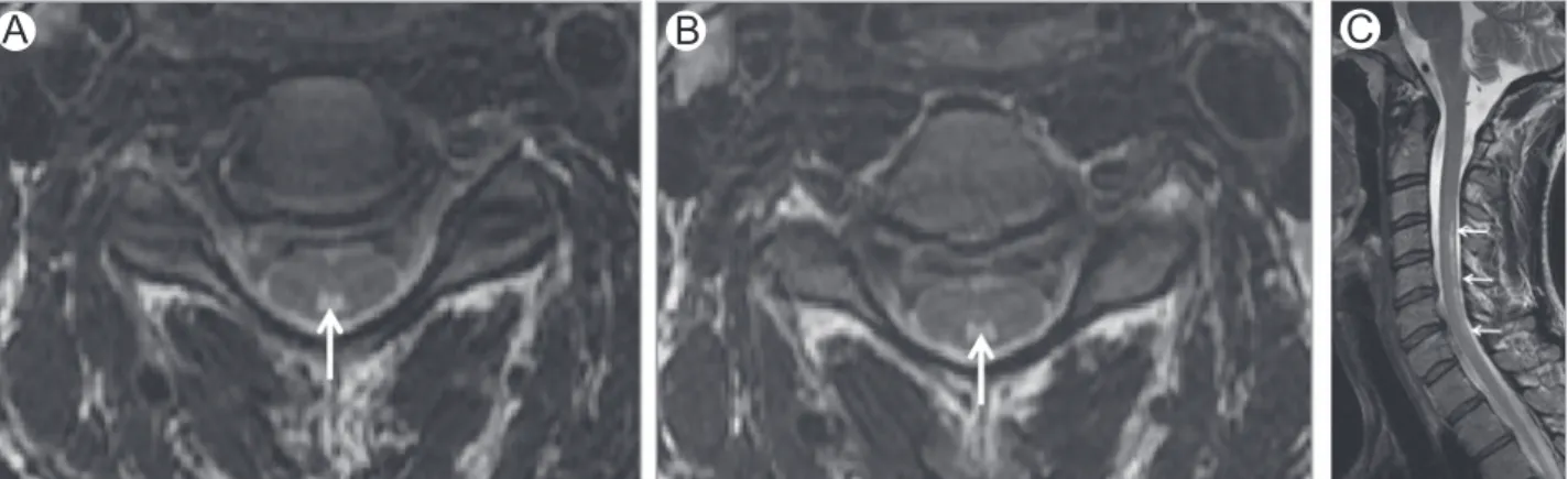
B
C
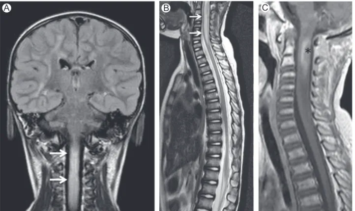
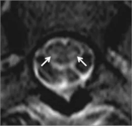
Figure 2 Vitamin B12deficiency. Axial (A and B) and sagittal (C) T2-WI in a patient with vitamin B12deficiency show
high-signal intensity involving the dorsal columns (arrows). Note typical“inverted V” or “inverted rabbit ears” on axial images.
A
B
Figure 1 Vitamin B12deficiency. Axial (A) T2-WI in a patient with vitamin B12deficiency shows high-signal intensity
endogenous methionine. Improvement of symptoms and MRI changes are seen after replacement therapy is started.12
Folate De
ficiency
Definition
Folate deficiency rarely exists in a pure state and is often associated with conditions that affect other nutrients. The most common manifestation of folate deficiency is a myelopathy or myeloneuropathy resembling subacute combined degenera-tion seen with B12deficiency.1
Pathophysiology
Folate is a water-soluble B vitamin present in fresh green vegetables, citrus fruits, legumes, and liver. As with B12, folate acts as an important cofactor in DNA synthesis and methyl-ation pathways. Its deficiency results from reduced intake of folate-rich products, impaired absorption, or increased folate metabolism. It is associated with alcoholism, small bowel disorders, and drugs such as methotrexate and trimethoprim. Folate absorption may be decreased with reduced gastric secretions in patients after gastric surgery, atrophic gastritis, acid-suppressive therapy, and acid neutralization by treatment of pancreatic insufficiency.1,14
Clinical Presentations
Folate deficiency causes similar manifestations to those of vitamin B12deficiency because of its importance in production of methionine and tetrahydrofolate. For unclear reasons, neurologic and hematologic manifestations due to folate deficiency are rare, mild, and confounding. Neurologic man-ifestations of folate deficiency include a myelopathy, peripheral neuropathy, optic neuropathy, and cognitive or behavioral manifestations.1,14
Pathology Findings
Pathologically, folate deficiency myelopathy is identical to vitamin B12 deficiency myelopathy and characterized by reversible swelling of myelin sheaths in the posterior and lateral columns.1,4,10,14
Imaging Findings
Subacute combined degeneration due to folate deficiency resembles other forms of subacute combined degeneration (Fig. 4).1,4,9,14
Diagnosis
Attribution of neurologic manifestations to pure folate de fi-ciency requires exclusion of other causes. Serum folate levels between 2.5 and 5μg/L may be indicative of a mildly compromised folate status. Erythrocyte folate is more reliable than plasma folate because its levels are less affected by short-term fluctuations in intake. Plasma homocysteine levels are typically elevated in patients with clinically significant folate deficiency.1,14
Treatment or Prognosis
Daily doses, as high as 20 mg, may be necessary in patients with malabsorption. Reduced folates are required only when folate metabolism is impaired by drugs such as methotrexate or inborn errors of metabolism. Plasma homocysteine is best for monitoring response to therapy as it decreases within a few days of instituting folate therapy but does not respond to inappropriate vitamin B12therapy.1,14
Copper De
ficiency
Definition
Acquired copper deficiency may cause myelopathy or
myeloneuropathy that resembles subacute combined
degeneration.1,15 Pathophysiology
Copper permits electron transfer in key enzymatic pathways. It is a component of metalloenzymes, including cytochrome-c oxidase, that have a critical role in the structure and function of the nervous system. Reduction in cytochrome-c oxidase activity may be the basis for neurologic dysfunction in copper deficiency.16Because of copper's ubiquitous distribution and low daily requirement, acquired dietary copper deficiency is rare. The coexistence of multiple causes of copper deficiency
A
B
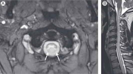
C
Figure 3 Nitrous oxide toxicity. Axial (A and B) and sagittal (C) T2-WI of the cervical spine in a patient with myelopathy after surgery with nitrous oxide anesthesia. Imagingfindings are typical of vitamin B12deficiency myelopathy with high
T2-WI signal involving the dorsal columns (arrows). This patient had undiagnosed B12 deficiency and nitrous oxide
increases the chances of development of a clinically deficiency state. The most common cause of acquired copper deficiency is a history of gastric surgery. Typically, neurologic manifesta-tions are delayed by years following gastric surgery. Excessive zinc ingestion is also a recognized cause of copper deficiency. Additionally, copper deficiency has been reported in entero-pathies associated with malabsorption such as cysticfibrosis, sprue, bacterial overgrowth, and inflammatory bowel disease.1
Clinical Presentations
Neurologic manifestations are similar to those seen with B12 deficiency with which they may coexist. The most common manifestation is that of a myelopathy presenting with a spastic gait and prominent sensory ataxia because of dorsal column dysfunction. Central nervous system (CNS) demyelination, peripheral neuropathy, and optic neuritis also occur.1,16
Pathology Findings
Copper deficiency–associated myelopathy has been well described in animal models. The typical distribution of spinal cord lesions is that of greater involvement of the cervical cord with less severe changes in the thoracic and lumbar segments. Wallerian degeneration and demyelination with microcavita-tion of the white matter of the spinal cord, particularly affecting the dorsal columns and brainstem, are typical.16
Imaging Findings
The most common abnormality on the spine MRI is increased T2-WI signal in the dorsal columns, mostly in the cervical cord; contrast enhancement is not seen (Fig. 5). Additionally, signal changes involving the lateral columns and extension of the pyramidal tract signal changes into the brainstem have been reported. These findings are similar to those seen in patients with vitamin B12deficiency.1,15-18
Diagnosis
Laboratory indicators of copper deficiency include a decrease in serum copper or ceruloplasmin and in 24-hour urinary copper excretion. Changes in serum copper usually parallel ceruloplasmin concentration. Serum zinc and 24-hour urinary zinc excretion levels should be obtained and an elevation in these should prompt an aggressive search for an exogenous source of zinc.1,16
Treatment or Prognosis
Oral copper supplement is the method of supplementation. Response of hematological parameters (including bone mar-row findings) is prompt and often complete. Recovery of neurologic signs and symptoms is variable. In patients in whom excess zinc ingestion is the cause, stopping zinc supplements may suffice and no additional copper supple-ments may be required.1,15,18
Vitamin E De
ficiency
Definition
Vitamin E deficiency manifests as a progressive spinocerebellar syndrome.
Pathophysiology
Vitamin E is as a fat-soluble antioxidant preventing peroxida-tion of membrane fatty acids. Most vitamin E in the human body is localized in adipose tissues. More than 2 years are required for adipose tissues to reach steady-state levels in response to changes in dietary intake. In adults, vitamin E deficiency is most often associated with fat malabsorption syndromes (celiac disease, cysticfibrosis, and cholestasis) and other intestinal disorders. In children, vitamin E deficiency may be seen because of genetic in alphatocopherol transfer protein, abetalipoproteinemia, or defects in chylomicron syn-thesis and secretion.1
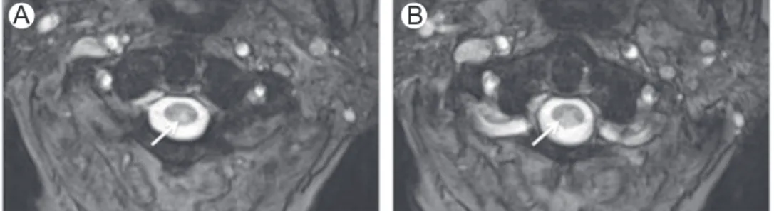
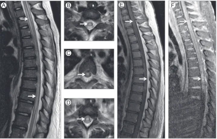
A
B
Figure 4 Folate deficiency. Axial (A) and sagittal (B) T2-WI in a patient with folate deficiency show high-signal intensity involving the dorsal columns (arrows).
Clinical Presentations
Neurologic manifestations of vitamin E deficiency include a progressive spinocerebellar syndrome, similar to those of Friedreich ataxia, and a peripheral neuropathy with gait difficulty, hyporeflexia or areflexia, pyramidal signs, impaired position and vibration perception, and dysarthria. Pigmented retinopathy, myopathy, movement disorders, and gaze palsies have also been reported.1,19
Pathology Findings
Lesions in the dorsal root ganglia, posterior medullary tracts, cerebellar cortex, spinocerebellar tracts, anterior horn cells, and peripheral nerves have been reported.20
Imaging Findings
MRI may show high-signal lesions on T2-WI in the posterior columns. Cerebellar atrophy may be present.21,22
Diagnosis
Serum vitamin E levels are dependent on concentrations of serum lipids. Hyperlipidemia or hypolipidemia can increase or decrease serum vitamin E without reflecting similar alterations in tissue levels. Effective serum vitamin E levels are calculated by dividing the serum vitamin E by the sum of serum cholesterol and triglycerides. In patients with neurologic manifestations serum vitamin E levels are frequently undetectable.1
Treatment or Prognosis
Replacement of vitamin E is treatment of choice. Improvement of neurologic symptoms or signs in severe cases may not occur.1
AIDS-Associated Myelopathy
DefinitionAIDS-associated myelopathy, also called vacuolar myelopathy, is a spinal cord disease in human immunodeficiency virus (HIV) positive patients. Clinical manifestations, imaging find-ings, and histopathology resemble that of subacute combined degeneration because of vitamin B12deficiency. It may occur at any stage during the course of HIV infection but is more common with advanced immunosuppression. Reliable data on the incidence and prevalence of myelopathy in HIV patients are not available.1,23
Pathophysiology
The cause of vacuolar myelopathy is unknown. Increased prevalence of B12 deficiency occurs in HIV patients with neurologic symptoms but its significance is unclear. Impair-ment in methylation pathways related or not to viral infection of the spinal cord have been proposed.1,23,24
Clinical Presentations
Manifestations of vacuolar myelopathy are varied and often include painless spastic paraparesis that is slowly progressive with a sensory ataxia. There may be an associated sphincter and erectile dysfunctions. Overlapping peripheral neuropathy may confound the diagnosis. Hyperreflexia at the knees with reduced ankle reflexes are common.23
Pathology Findings
Vacuolar myelopathy is characterized by intralamellar vacuo-lation in the spinal white matter throughout the lateral and posterior columns of the thoracic and cervical segments.23,25
Imaging Findings
MRI features of vacuolar myelopathy include high T2 intensity lesions in the posterior columns predominantly in the upper thoracic cord. Frequently, the spinal cord may be normal or show atrophy and nonspecific patchy T2-WI abnormalities.23,25
Diagnosis
Diagnosis is made by exclusion by ruling out vitamin B12 deficiency, human T-lymphotrophic virus-1, and neurosyphi-lis. Cerebrospinal fluid (CSF) may be normal or show mild pleocytosis with mild protein elevation. Similarfindings may be seen in asymptomatic HIV patients.
Treatment or Prognosis
There is no effective therapy for HIV-associated myelopathy. The role of antiretroviral therapy is not established, however following antiretroviral therapy MRI may show significant reduction of the T2 hyperintensity of the posterior columns.23,25
A
B
Figure 5 Copper deficiency. (A and B) Axial T2-WI at C1-2 level in a patient with Cu deficiency demonstrate high-signal intensity involving dorsal columns (arrows).
Toxic Myelopathies Associated
With Drugs and Chemical Agents
Heroin Myelopathy
Definition
Myelopathy induced by heroin is progressive with selective involvement of the ventral pons and lateral and posterior columns of the spinal cord and may be associated with inhalation (“chasing the dragon”) or intravenous use.1,26,27 Pathophysiology
Proposed mechanisms include direct toxicity, vasculitis, or hypersensitivity reaction. Isolated toxicity seems less probable as systemic toxins typically involve the nervous system more diffusely along with other organs. Generalized vasculitis would be unlikely to first affect the spinal cord.27 Thus, hyper-sensitivity is the predominant explanation as the myelopathy may occur after a period of abstinence followed by a single use.1,26-30 Hypotension may possibly result in border-zone infarctions in the cord.1However, hypotension is unlikely to be severe enough to damage the spinal cord and spare the more ischemia-sensitive neurons of the brain. Furthermore, although the level of injury varies among reported cases, no patternfitting the vascular “watershed zones” of the spinal cord has been documented.27
Clinical Presentations
Clinical picture consists of an initial flaccid paraparesis or paraplegia of sudden onset with sensory loss in the legs and lower thoracic dermatomes and urinary sphincter disturban-ces. Occasionally the sensory and motor levels extend to the cervical region,31resembling symptoms of transverse myelitis. Pathology Findings
Extensive necrosis of the cervical and thoracic cord has been described in some patients.31
Imaging Findings
MRI is frequently normal. Hyperintense lesions on T2-WI and fluid attenuation inversion recovery (FLAIR) images affecting the posterior and lateral columns, pontomedullary region, and ventral pons may be seen. Findings consistent with acute transverse myelitis have also been reported. Contrast enhance-ment is variable and usually patchy.27
Diagnosis
Most reports describe normal CSFfindings although pleocy-tosis and increased total protein may occur.28-30,32
Treatment or Prognosis
Treatment with either intravenous corticosteroids or immu-nopheresis may limit immune response preventing disability but their efficacy requires further validation.27 Prognosis is often poor, with residual spastic paraparesis and sensory deficits, and in some patients death.29Full recovery has been reported occasionally.31
Hepatic Myelopathy
DefinitionHepatic myelopathy or portosystemic myelopathy is a rare neurologic complication of chronic liver disease with portal hypertension usually associated with portosystemic shunting either surgically created or spontaneous. It is characterized by an insidious onset of pure motor spastic paraparesis without sensory or sphincter involvement.33
Pathophysiology
Pathophysiology is poorly understood and it is broadly accepted that nitrogenous products such as ammonia, fatty acids, indoles, and mercaptans bypassing the liver through a portocaval shunt play an important role.34These metabolites cause myelin damage resulting in demyelination in the brain and spinal cord. Selective predisposition of the motor system has been demonstrated by involvement of the lateral cortico-spinal tracts in autopsy studies. Other causative factors include nutrient deficiencies and deranged liver metabolism.33 Addi-tionally, the myelopathy may also be related to elevated manganese levels with MRI evidence of manganese deposition in the basal ganglia without extrapyramidal manifestations or clinical evidence of decompensated cirrhosis.1,26,35
Clinical Presentations
The typical clinical presentation is a progressive pure motor spastic paraparesis with minimal or no sensory deficits and no bowel or bladder involvement. Most patients report prior episodes of hepatic encephalopathy and often the development of myelopathy follow the creation of surgical shunts. However, preceding portosystemic encephalopathy is not invariably present.1Early and accurate diagnosis is important because patients with early stages of the disease can recover following liver transplantation.33,36
Pathology Findings
Neuropathologic studies show demyelination in the lateral corticospinal tracts with varying degrees of axonal loss.33,37 Imaging Findings
MRI usually shows symmetric high T2 signal intensity in the lateral corticospinal tracts and less commonly in the ventral pyramidal tracts, posterior columns, and spinocerebellar tracts.36Increased pallidal T1 signal in presence of elevated serum manganese may also be present.
Diagnosis
Hepatic myelopathy is diagnosis of exclusion. Motor evoked potential studies may be suitable for the early diagnosis even in patients in the preclinical stages of the disease.33,38
Treatment or Prognosis
Hepatic myelopathy is a rare and debilitating disorder with no effective treatment. However, its early identification and exclusion of other treatable causes is important. Some patients improve following liver transplantation26but the therapeutic potential of liver transplantation for preventing progression and allowing recovery needs to be further validated.33
Organophosphate Poisoning
DefinitionThe most common complication of organophosphate poison-ing is an acute intoxication with a cholinergic crisis because of the inhibition of acetylcholinesterase. Some patients subse-quently develop organophosphate-induced delayed myelop-athy and neuropmyelop-athy, also known as organophosphate-induced delayed neurotoxicity (OPIDN).
Pathophysiology
Organophosphates are common components of pesticides available in many homes. A major chemical that causes symptoms is triorthocresyl phosphate. Signs and symptoms of acute organophosphate toxicity are because of acetylcholi-nesterase inhibition and resulting muscarinic and nicotinic dysfunctions. In the neuromuscular junction, acetylcholine is released when a nerve impulse reaches terminal axonal end and it diffuses across the synaptic cleft and binds to cholinergic nicotinic receptors on the muscle fibers, causing them to contract. The enzyme, cholinesterase splits acetylcholine into acetic acid and choline, stopping its action. End products of the metabolism of acetylcholine are taken up by nervefibers and resynthesized into acetylcholine. In organophosphate poison-ing, cholinesterases are phosphorylated by the phosphate end of organophosphates resulting in excessive accumulation of acetyl chlorine with resultant effects on muscarinic, nicotinic receptors in the CNS.39After resolution of a cholinergic crisis, an intermediate syndrome can develop in some patients. It is likely a neuromuscular junction defect syndrome.40OPIDN is a late complication of some organophosphorus compounds and is possibly because of phosphorylation and subsequent aging of neurotoxic esterase in the nervous system.1,41
Clinical Presentations
After organophosphate poisoning, 3 well-defined clinical phases may be seen as follows: an initial acute cholinergic crisis, an intermediate syndrome, and OPIDPN. Pinpoint pupils, excessive salivation, generalized weakness, and respi-ratory collapse characterize the acute cholinergic crisis. The intermediate syndrome is characterized by weakness of neck flexors, proximal limbs, and respiratory muscles. OPIDN occurs 1-3 weeks after acute exposure and following an uncertain duration after chronic exposure. OPIDN may occur in absence of the cholinergic or intermediate phase. Symptoms include distal paresthesias, progressive leg weakness and wasting, and cramping muscle pain. There may be evidence of upper limb involvement and pyramidal tract dysfunction. Sensory loss when present is mild.1
Pathology Findings
Studies in chicks with organophosphate-induced delayed neuropathy show severe damage in the ventral and lateral tracts of the thoracic spinal cord. The same neuropathologic changes may be associated with prominent and diffuse spinal cord atrophy especially in the thoracic region.42
Imaging Findings
MRI at the late phase of exposure may reveal evidence of spinal cord atrophy that persists long after the cholinergic effects had subsided.
Diagnosis
There is usually a delay in onset of clinical symptoms after exposure to organophosphates. Measurement of cholinesterase activity in red blood cells helps confirm the diagnosis because red blood cells cholinesterase activity is less rapidly depressed than serum cholinesterase activity.1,26,43 In the late phase somatosensory evoked potentials are often abnormal. Treatment or Prognosis
Acute symptoms are best treated with pralidoxime and atropine. Pralidoxime is a reactivator of inhibited acetylcholi-nesterase and is its specific antidote.1
Chemotherapy-Related Myelopathies
DefinitionAcute myelopathy is a rare but devastating complication of chemotherapy treatment. It has been reported following administration of different chemotherapeutic agents including methotrexate, cytarabine, cisplatin, cladarabine, doxorubicin, vincristine, and cytosine arabinoside.26
Pathophysiology
Intrathecal methotrexate and cytarabine are the most fre-quently implicated agents.44Acute or chronic leukoencephal-opathy is the most common type of methotrexate-induced neurotoxicity and subacute myelopathy is rare.45The patho-genesis of methotrexate-induced myelopathy is not clear but it is probably multifactorial; Following 2 major mechanisms have been proposed: indirect inhibition of methionine synthesis and direct toxicity. Methotrexate inhibits the enzyme dihydrofolate reductase and prevents folic acid from converting to tetrahy-drofolic acid causing folate deficiency. This deficiency indi-rectly inhibits the synthesis of methionine, as folate is an important cofactor in DNA synthesis and methylation path-ways. As methionine is necessary for formation and main-tenance of myelin sheaths, its deficiency may lead to vacuolar degeneration of white matter. This mechanism is similar to that of subacute combined degeneration of the spinal cord because of vitamin B12deficiency. A constitutional predisposition or abnormal sensitivity to methotrexate such as genetic poly-morphisms or mutations related to methionine metabolism may also play a role in the pathogenesis as the reported frequency of severe methotrexate myelopathy is dispropor-tionately low considering the large number of patients receiv-ing the drug intrathecally.46Another proposed mechanism is direct injury of endothelial cells in venules and capillaries of the spinal cord with blood-brain barrier disruption, increased vascular permeability, and insinuation of plasma proteins into the vascular walls46 which lead to their degeneration and necrosis and vacuolar degeneration of white matter. Vascular injury also allows the drug to diffuse into the CNS causing direct toxic effects. Direct toxic effect of intrathecal
administration of methotrexate on the pia mater and white matter is probably caused by pia-glial barrier disruption and myelin sheaths and oligodendrocytes injuries.46
Known risk factors for development of methotrexate myelopathy include the following: cumulative intrathecal doses and continuous exposure to the drug more than a prolonged period of time, systemic administration, concurrent radiotherapy, and CNS involvement by the primary disease.47 Clinical Presentations
There is a variable clinical course in regard to time and rate of onset after therapy, severity of disease, and degree of recov-ery.48 Overall, 2 clinical patterns of paraparesis have been described.49A transient,flaccid, and areflexic paraparesis with pain and anesthesia may occur soon after intrathecal injection. Rarely, the paralysis may ascend with respiratory compromise and death. A less common form is a progressive spastic-ataxic paraparesis with sphincter dysfunction seen after weeks of a series of intrathecal treatments.1 The latency to onset of symptoms is variable and can be long.
Pathology Findings
Reports on the neuropathologic findings of methotrexate-induced myelopathy are rare andfindings vary from vacuolar degeneration of the lateral and posterior columns in association with severe loss of axons and myelin sheaths to complete transverse necrosis of the spinal cord or a combination of both.46The gray matter is relatively spared.46
Imaging Findings
MRIfindings are nonspecific and the initial imaging examina-tion is often normal. Symmetric increased T2 signal in dorsal and lateral columns of the cord without abnormal contrast enhancement similar to that of subacute combine degeneration has been described (Fig. 6). Cord swelling and contrast enhancement have also been reported. Long-term outcome is generally that of cord atrophy with normal T2 signal intensity demonstrated as early as 6 months after clinical onset of disease.48,50
Diagnosis
Chemotherapy-induced myelopathy is a diagnosis of exclu-sion. The main differential diagnosis includes malignancy, viruses, radiation, and paraneoplastic myelopathy.48
Treatment or Prognosis
Chemotherapy-induced myelopathy carries a poor prognosis. Preservative-containing chemotherapeutic agents or diluents should not be used intrathecally49and multiple and frequent intrathecal therapy should be avoided.1Meticulous neurologic monitoring of methotrexate concentration in CSF is important to prevent the occurrence of this complication.46Immediate discontinuation of intrathecal methotrexate treatment and supplementation of derivates of folic acid and
methio-nine or homocysteine metabolic pathway, such as
S-adenosylmethionine and cyanocobalamine, are the best treatment options.51However, further research is required to establish an effective and definite treatment.45
Radiation-Induced Myelopathies
DefinitionRadiation myelopathy is defined as injury caused by ionizing radiation.52There are 2 main reasons for spinal cord exposure to radiation—an intentional delivery to treat primary or secondary lesions or its inevitable inclusion in a radiationfield which intends to treat an adjacent neoplasm. In either case, factors such as total radiation dose, radiation dose per fraction, time between applications, spinal cord level and associated chemotherapy affect the incidence and severity of myelop-athy.52 Radiation myelopathy is divided into the following 4 categories: acute transient radiation myelopathy, acute para-plegia or quadripara-plegia, lower motor neuron findings, and chronic progressive radiation myelopathy also known as delayed radiation myelopathy (DRM). DRM is rare but is the most feared complication and typically occurs after a certain latent period that ranges from months to few years following irradiation.26,52
Pathophysiology
The pathogenesis of DRM is likely to be multifactorial with no single target cell and no single pathway solely responsible. Damage to vascular endothelial cells,53 direct injury of oligodendroglia and myelin sheaths,54and disturbance of the intra-spinal venous drainage because of lesions in the vein walls55have been considered most important.46
It is known that the spinal cord has an enhanced radio-sensitivity relative to other organs and that there is no uniformly accepted“spinal cord tolerance” and that no precise threshold dose is considered safe.56A total dose of 45 Gy with fractions of 1.8-2 Gy is generally accepted as dose limit using conventional radiotherapy,57although 40 Gy poses however a small risk of myelopathy. Doses of 45 Gy and 52 Gy to the cervical cord result myelopathy rates of 0.1% and 1% respectively with a lower incidence in the thoracic cord.48,58Reports of myelop-athy after stereotactic radiosurgery to spinal lesions are rare.59
Concomitant chemotherapy is a recognized risk factor for development of radiation myelopathy. In the past years, targeted antibody and antireceptor therapies have emerged, but the extent to which these agents contributed to the development of radiation myelopathy remains unknown.57 Previous exposure to radiation, particularly less than 2 years, may also carry an increased risk as spinal cord damage is most likely to be become overt during this period.56The volume and length of spinal cord as well as the spinal cord segment irradiated may also influence the incidence of radiation myelopathy.56
Other factors in the patients' medical history and physical status make the spinal cord sensitive to radiation injury. Hypertension, diabetes, autoimmune disease, congenital and acquired spinal cord abnormalities, pre-existing CNS injury, elevated hemoglobin, hypotension, alcohol abuse, and infec-tious disease also enhance the cord's sensitivity to radiation damage.60
Clinical Presentations
Overall, 4 clinical syndromes of radiation myelopathy have been described are as follows: (1) an acute transient radiation
myelopathy (most common) characterized by Lhermitte's sign with no other abnormalities on neurologic examination, (2) an acutely developing paraplegia or quadriplegia presumably secondary to ischemia and hemorrhage of the spinal cord caused by radiation-induced blood vessel damage, (3) a lower motor neuron disease in the upper or lower extremities, presumably caused by selective anterior horn-cell damage, and (4) DRM.52,56,61 Although the second and the third syndromes are exceedingly rare, the last one is the one most concerning.
Acute transient radiation myelopathy is a self-limiting early delayed reaction usually treated with corticosteroids. It occurs after a latent period of 2-4 months and is characterized by brief electric-like sensations in the spine and extremities triggered by neck flexion or extension (Lhermitte's sign). Clinical improvement is generally seen within 2-9 months followed by complete recovery. There is no correlation between acute transient radiation
myelopathy and DRM. Conversely DRM is a devastating condition, which may be permanent. It generally occurs after a latent period of time of 9-18 months but has been reported up to 3-4 years after completion of radiation treatment. Various combinations of motor and sensory deficits occur and depend on the anatomic level of cord injury. Development of bowel and bladder dysfunctions may occur. DRM may be fatal if it involves the upper cervical level.
Pathology Findings
Myelinated fibers and blood vessels are the most affected structures. In chronic progressive radiation myelopathy white matter necrosis and demyelination with vascular lesions including stenosis, thrombosis, hemorrhage,fibrinoid vascular necrosis, and venous wall thickening and hyalinization are seen.62 There is also gray matter involvement, including
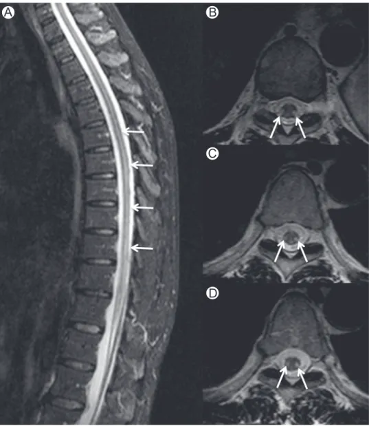
A
B
C
D
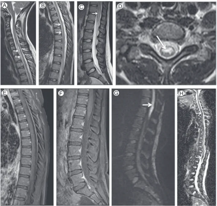
Figure 6 Methotrexate myelopathy. Midsagittal (A) and axial (B-D) STIR images in a patient who underwent intrathecal methotrexate therapy for acute lymphoblastic leukemia. Images show high-signal intensity in dorsal and lateral columns (arrows) mimicking subacute combined degeneration. STIR, short-tau inversion recovery. (Courtesy: Lazaro Amaral, Sao Paulo, Brazil.)
astrocytosis and hyalinosis of the vessel walls, but neurons are usually spared.60
Imaging Findings
In acute transient radiation myelopathy MRI is usually normal. In DRM, MRI shows cord swelling initially and a long intramedullary lesion with increased T2 signal and contrast enhancement (Fig. 7). The MRIfindings correlate with neuro-logic symptoms. In the chronic stage MRI demonstrates cord atrophy. Radiation-induced bone marrow changes indicate the extension of the irradiatedfield and may help in reaching the correct diagnosis.
Positron emission tomography-computed tomography studies might have a role in differentiating radiation myelop-athy from primary spinal cord lesions and spinal metastases. However, very few positron emission tomography-computed tomography studies have been conducted in the spinal cord and their results need validation.52
Diagnosis
Diagnostic criteria for DRM include—(1) the affected spinal cord segment must be in the irradiated zone, (2) symptomatol-ogy must correspond to the radiated spinal cord segment, (3) and the latency period should be more than 6 months. The differential diagnosis represents a challenging problem. Metastatic tumor is the most common cause of cord lesions in oncologic patients presenting with myelopathy.52 Primary
intramedullary neoplastic lesions and other neurologic path-ologies presenting with myelopathy must also be ruled out including paraneoplastic syndrome, zoster myelopathy, and myelopathy secondary to chemotherapy.52Nerve conduction studies show slowing or complete block in spinal conduction. CSF examination is usually normal but mild elevation of protein levels and lymphocytosis may be seen.52,63,64
Treatment or Prognosis
No treatment has conclusively been shown to be of value in DRM. Several therapies such as corticosteroids, anticoagula-tion, pentoxifylline, vitamin E, and hyperbaric oxygen have been tried with limited benefits. Some patients derive a short-term benefit from steroids which may be related to decreasing edema and inflammation.52
Toxic Myelopathies Associated
With Geographical Predilection
Spinal Sea Stroke
Definition
Spinal sea stroke is characterized by spinal and brainstem infarctions that occur in patients swimming in the North Carolina and Virginia coasts probably caused by contact with toxic marine animals.
A
B
C
Figure 7 Radiation therapy-related myelopathy. Brain MRI coronal FLAIR (A), spine MRI midsagittal T2 (B) and midsagittal postcontrast T1-WI (C) of a patient who underwent radiation therapy. Radiation-induced myelopathy is seen as an intramedullary area of high-signal intensity on FLAIR (arrows on A) and T2-WI (arrows on B) with mild enhancement after gadolinium administration (* on C). FLAIR,fluid attenuation inversion recovery. (Courtesy: Dr Quaghebeur Oxford, UK.)
Pathophysiology
Brainstem and spinal cord strokes in young adults are extremely rare. Jellyfish toxins have been proposed as a cause but the mechanism of neural tissue damage is unknown. It is also unclear why in all of the reported cases the abnormalities involved only the spinal cord and brainstem but spared the brain65Some jellyfish toxins affect the calcium channels of neurons, a process that could lead to cytotoxic edema, failure of cellular energetics, and eventually to irreversible damage such as seen in ischemia. Some authors have postulated that damage to the spinal cord is not direct but due to systemic hypotension with subsequent infarctions.65
Clinical Presentations
Patients typically present with an acute severe dysfunction of sensory and motor systems. Neurologic signs and symptoms depend on the level of spinal cord involvement and include severe back pain, loss of sphincter control, paraplegia or paraparesis, or quadriplegic or tetraplegia in patients with higher cord lesions.65
Pathology Findings
None of the published cases of spinal sea stroke report pathologyfindings.
Imaging Findings
The hallmark of spinal cord infarction is high T2 signal intensity in the cord (Fig. 8) especially involving the anterior two-third of the cord that correspond to the territory supplied by the anterior spinal artery. Diffusion weighted imaging shows restricted diffusion of the lesion.
Diagnosis
The diagnosis is generally made clinically. MRI should be obtained in all patients with suspected spinal cord infarction to confirm the diagnosis and to exclude other possible treatable causes of myelopathy.
Treatment or Prognosis
Spinal cord infarctions have a poor prognosis with little chance of recovery and significant sequelae.
Subacute Myelo-Optico Neuropathy
(Clioquinol Toxicity)
Clioquinol (iodochlorhydroxyquin) is an antifungal-antiprotozoal drug used to treat intestinal parasitic diseases. Clioquinol's use has been restricted or discontinued in some countries because of events in Japan (from 1955-1970) where more than 10,000 people developed a subacute myelo-optic neuropathy (SMON). SMON is characterized by subacute onset of lower limb paresthesias and spastic paraparesis with optic atrophy. Examination findings include hyperreflexia and extensor plantar responses. The physiopathology mecha-nism is unknown but clioquinol is a copper or zinc chelator
and this has led to the speculation that clioquinol-induced
neurotoxicity may be a consequence of copper de
fi-ciency.1,26,66-68
Lathyrism (India, Bangladesh, and Ethiopia)
and Konzo (Africa)
Lathyrism is a self-limiting neurotoxic disorder endemic in Bangladesh, India, and Ethiopia, caused by beta-N-oxalyl-amino-L-alanine, an excitotoxic amino acid present in Lathyrus sativus.1 Ingestion of this neurotoxin results in increased intracellular levels of reactive oxygen species and subsequent impairment of the mitochondrial oxidative phosphorylation chain. It presents as a subacute or insidious onset spastic paraparesis. Degeneration is most prominent in motor cortex and pyramidal tracts that are responsible for lower extremity function.1
Konzo is a tropical myelopathy seen in Africa resulting from weeks of high dietary cyanide exposure due to con-sumption of insufficiently processed cassava (Manihot esculenta Crantz) that contains cyanide. It is characterized by the abrupt onset of spastic non-progressive paraparesis.69Years of cassava consumption have also been implicated in a syndrome of slowly progressive ataxia, peripheral neuropathy, and optic atrophy seen in parts of Africa.1,70The diagnosis is based on a history of exposure and measurement of serum thiocyanate levels.
Lathyrism and Konzo are irreversible and non-progressive conditions. MRI studies are negative. Somatosensory evoked potentials may show abnormalities which indicate poor conduction through the spinal cord.26,71There are no specific treatments for these entities.
Other Noninfectious and
Nonin
flammatory Myelopathies
Ketoacidosis Myelopathy
DefinitionMyelopathy is a rare complication of diabetic ketoacidosis characterized by spinal cord edema and infarction usually associated with cerebralfindings.
Pathophysiology
There are no existing explanations for the spinal cord edema associated with diabetic ketoacidosis probably due its rarity. Current explanations are limited to diabetic children with cerebral edema but the pathophysiological mechanisms for cerebral edema and infarctions are not clear. Systemic hypo-tension and hypoxia, rapid changes in serum osmolality, disseminated intravascular coagulation, and thrombosis sec-ondary to dehydration; hemoconcentration and hyperviscosity have all been proposed as possible causes.72-75
Clinical Presentations
Clinical presentation depends on the affected spinal cord level. Pathology Findings
No neuropathologic findings have been described in
reported cases. Imaging Findings
Cord edema is seen as high T2 signal in the spinal cord usually associated with cord expansion (Fig. 9). Spinal cord infarction is seen as areas of bright T2 signal that show restricted diffusion. Patchy enhancement after gadolinium administra-tion has been reported. Atrophy may be seen in late stages.
Diagnosis
Ketoacidosis myelopathy is a diagnostic of exclusion. Treatment or Prognosis
There is no effective treatment. The prognosis is poor and varies according with the severity of the disease. Cardiac failure and pulmonary edema are rare but possible complications in critically ill patients with diabetic ketoacidosis.75
Spinal Cord Decompression Illness
DefinitionDecompression sickness is a clinical syndrome caused by alterations in environmental pressure resulting in liberation of
A
B
E
F
G
H
C
D
Figure 8 Spinal sea stroke. Sagittal T2-WI at the level of the cervical (A), thoracic (B), and lumbar (C) spine and axial T2-WI (D) show high T2-WI signal intensity in the anterior two-third of the cord. Sagittal postcontrast fat-suppressed T1-WI (E and F) show no contrast enhancement in areas of T2 signal abnormality. DWI (G) and ADC map (H) show restricted diffusion suggesting infarct. ADC, apparent diffusion coefficient.
inert gas bubbles in tissues or blood. Sport divers generally use compressed air as their breathing mixture and the inert gas that is liberated during decompression is nitrogen. There are following 2 types of decompression illness: type I is charac-terized by joint pain, skin marbling, small patchy hemorrhage, and lymphatic obstruction and type II is characterized by CNS involvement. CNS involvement may be further subdivided in cerebral and spinal cord subtypes. Spinal cord involvement accounts for 77% of CNS diving injuries. The population at risk includes, in addition to sport divers, naval and commercial divers, aviators, astronauts, and compressed air workers.
Pathophysiology
The pathophysiological mechanism of spinal cord decom-pression is not totally understood. Hypotheses include embo-lization of arterial bubbles via a right-to-left shunt, development of bubbles within the spinal cord, or emboliza-tion of the epidural vertebral venous system causing conges-tion and venous cord infarcconges-tions. The last hypothesis is the best documented one as the alteration of spinal cord venous draining resulting from the obstruction of the epidural venous system has been shown experimentally.76Secondary immuno-inflammatory processes may contribute to worsening of symptoms. Nitrogen bubbles lodge in spinal veins at the thoracic level; there the spine mobility is restricted by the rib cage.
Clinical Presentations
The great majority of spinal cord decompression sickness results in subjective sensory abnormalities distributed accord-ing to the dermatomes. A less frequent but more characteristic presentation corresponds to numbness in the limbs with a progressive ascending level of both sensory and motor deficits often accompanied by disturbances of bladder function.77,78
Pathologyfindings
Spinal cord lesions are scattered throughout the white matter with no segmental distribution. The dorsolateral white matter is more vulnerable because of its high fat content in which nitrogen is readily stored.
Imaging Findings
MRI is reliable in the early detection of spinal cord lesions that occur in decompression accidents leading to prompt baric treatment and in the follow-up during and after hyper-baric recompression. MRI may demonstrate patchy areas of increased T2 signal intensity in the white matter tracts (Fig. 10).
Diagnosis
The diagnosis relies on clinical history, physical examination, and MRIfindings.
A
B
E
F
C
D
Figure 9 Ketoacidosis myelopathy. Sagittal (A) and axial (B-D) T2-WI of thoracic spine show high T2-WI signal intensity involving the central aspect of the cord (arrows). Sagittal T1-WI (E) and postcontrast fat sat T1-WI (F) show areas of contrast enhancement.
Treatment or Prognosis
Hyperbaric oxygen therapy is the treatment of choice as it reduces the bubble volume and increases concentration of dissolved oxygen in ischemic tissues. Incomplete recovery is seen in 20%-30% of patients.79,80 Age, depth, bladder dysfunction, persistence or worsening of signs before recom-pression, and a Boussuges score more than 7 are independent predictive variables for poor clinical recovery.78
Surfer
's Myelopathy
Definition
Surfer's myelopathy is a rare non-traumatic injury that occurs in novice surfers who lay prone on a surfboard with the lumbar spine hyperextended for prolonged periods of time. It is probably underdiagnosed and underreported as it is a rare condition only recently recognized.81
Pathophysiology
The pathophysiology of surfer's myelopathy is unknown. Arterial insufficiency from hyperextension of the lumbar spine, venous hypertension associated with obstruction of the inferior vena cava by the liver whereas lying in a prone position, fibro-cartilaginous embolism caused by retrograde embolism of the nucleus pulpous into the radicular spinal cord arteries, or avulsion of perforating arteries have all been proposed.81,82 Clinical Presentations
Surfer's myelopathy is usually preceded by back discomfort followed by acute myelopathy with variable motor-sensory involvement and urinary incontinence.
Pathology Findings
No definite histopathologic confirmation has been published, but surfer's myelopathy probably is the result of a vascular phenomenon involving dynamic compression, vasospasm, or thrombotic infarction of the artery of Adamkiewicz that occurs
as the result of hyperextension of the spinal cord whereas individuals are lying prone on a surfboard.81,82
Imaging Findings
Surfer's myelopathy is usually associated with a longitudinally extensive T2 hyperintense central cord lesion in the midthora-cic region with variable cranial and caudal extension. Patchy increased T1 signal presumably related to petechial hemor-rhage have been described.83Enhancement of the ventral roots of the cauda equina have also reported.81,82,84,85
Diagnosis
The characteristic clinical history associated with the longitu-dinally extensive T2 hyperintense spinal cord lesion on MRI suggests the diagnosis.
Treatment or Prognosis
No treatment is available. Recovery is variable ranging from complete recovery of function to persistent paraplegia.
Conclusion
Toxic and metabolic myelopathies and myeloneuropathies are relatively rare clinical conditions that share clinical, neuro-pathologic, and imaging features. Acquaintance with these entities may lead to accurate diagnosis with successful identi-fication of the underlying cause and appropriate treatment which may revert or prevent progression of symptoms associated with these incapacitating disorders.
References
1. Kumar N: Metabolic and toxic myelopathies. Semin Neurol 32(2): 123-136, 2012
2. Timms SR, Curé JK, Kurent JE: Subacute combined degeneration of the spinal cord: MRfindings. AJNR Am J Neuroradiol 14(5):1224-1227, 1993
3. Goodman BP: Metabolic and toxic causes of myelopathy. Continuum (Minneap Minn) 21:84-99, 2015. [1 spinal cord disorders]
4. Katsaros VK, Glocker FX, Hemmer B, et al: MRI of spinal cord and brain lesions in subacute combined degeneration. Neuroradiology 40(11): 716-719, 1998
5. Sotirchos ES, Saidha S, Becker D: Neurological picture. Nitrous oxide-induced myelopathy with inverted V-sign on spinal MRI. J Neurol Neurosurg Psychiatr 83(9):915-916, 2012
6. Ravina B, Loevner LA, Bank W: MRfindings in subacute combined degeneration of the spinal cord: A case of reversible cervical myelopathy. AJR Am J Roentgenol 174(3):863-865, 2000
7. Jain KK, Malhotra HS, Garg RK, et al: Prevalence of MR imaging abnormalities in vitamin B12 deficiency patients presenting with clinical features of subacute combined degeneration of the spinal cord. J Neurol Sci 342(1-2):162-166, 2014
8. Küker W, Thron A: Subacute combined degeneration of the spinal cord: Demonstration of contrast enhancement. Neuroradiology 41(5):387, 1999 9. Lim CCT: Neuroimaging in postinfectious demyelination and nutritional disorders of the central nervous system. Neuroimaging Clin N Am 21(4): 843-858, 2011. [viii]
10. Su S, Libman RB, Diamond A, et al: Infratentorial and supratentorial leukoencephalopathy associated with vitamin B12deficiency. J Stroke Cerebrovasc Dis 9(3):136-138, 2000
11. Pacheco FT, Scoppetta TLPD, Sznirer M, et al: Acute demyelination with leukoencephalopathy and cerebellar involvement in vitamin B12 de fi-ciency. Arq Neuropsiquiatr 73(3):257, 2015
Figure 10 Spinal cord decompression sickness. Axial T2-WI at level of thoracic spine shows high-signal intensity in the dorsolateral white matter tracts. The lesions are usually scattered throughout the white matter with no segmental distribution. (Courtesy: A. Vidal, Santiago, Chile.)
12. Renard D, Dutray A, Remy A, et al: Subacute combined degeneration of the spinal cord caused by nitrous oxide anaesthesia. Neurol Sci 30(1): 75-76, 2009
13. Ernst LD, Brock K, Barraza LH, et al: Longitudinally extensive nitrous oxide myelopathy with novel radiographic features. J Am Med Assoc Neurol 72(11):1370-1371, 2015
14. Botez MI, Reynolds EH, eds. Folic acid in neurology, psychiatry and internal medicine. New York: Raven Press, 1-5, 1979
15. Kumar N, Ahlskog JE, Klein CJ, et al: Imaging features of copper deficiency myelopathy: A study of 25 cases. Neuroradiology 48(2): 78-83, 2006
16. Kumar N: Copper deficiency myelopathy (human swayback). Mayo Clin Proc 81(10):1371-1384, 2015
17. Page PS, Nazar RG, Park MC, et al: Copper deficiency myelopathy in the setting of advanced degenerative cervical spondylosis. Br J Neurosurg:1-3, 2015
18. Silva-Júnior FPD, Machado AAC, Lucato LT, et al: Resolution of MRI findings of copper deficiency myeloneuropathy in a patient with Wilson's disease. Arq Neuropsiquiatr 72(3):255-256, 2014
19. Nagappa M, Bindu PS, Adwani S, et al: Clinical, hematological, and imaging observations in a 25-year-old woman with abetalipoproteinemia. Ann Indian Acad Neurol 17(1):113-116, 2014
20. Nelson JS: Neuropathological studies of chronic vitamin E deficiency in mammals including humans. Ciba Found. Symp 101:92-105, 1983 21. Vorgerd M, Tegenthoff M, Kühne D, et al: Spinal MRI in progressive
myeloneuropathy associated with vitamin E deficiency. Neuroradiology 38(suppl 1):S111-S113, 1996
22. Müller KI, Bekkelund SI: Epilepsy in a patient with ataxia caused by vitamin E deficiency. Br Med J Case Rep 2011: 2011
23. Bilgrami M, O’Keefe P: Neurologic diseases in HIV-infected patients. Handb Clin Neurol 121:1321-1344, 2014
24. Tan SV, Guiloff RJ: Hypothesis on the pathogenesis of vacuolar myelopathy, dementia, and peripheral neuropathy in AIDS. J Neurol Neurosurg Psychiatr 65(1):23-28, 1998
25. Bizaare M, Dawood H, Moodley A: Vacuolar myelopathy: A case report of functional, clinical, and radiological improvement after highly active antiretroviral therapy. Int J Infect Dis 12(4):442-444, 2008
26. Schwendimann RN: Metabolic, nutritional, and toxic myelopathies. Neurol Clin 31(1):207-218, 2013
27. Haghi-Ashtiani B, Sina F, Ben Isa F, et al: Transverse myelitis and heroin abuse: A case report. Med J Islam Repub Iran 23(4):238-240, 2010 28. Richter RW, Rosenberg RN: Transverse myelitis associated with heroin
addiction. J Am Med Assoc 206(6):1255-1257, 1968
29. Ell JJ, Uttley D, Silver JR: Acute myelopathy in association with heroin addiction. J Neurol Neurosurg Psychiatr 44(5):448-450, 1981 30. Grassa C, Montanari E, Scaglioni A, et al: Acute heroin myelopathy—Case
report. Ital J Neurol Sci 5(1):63-66, 1984
31. Goodhart LC, Loizou LA, Anderson M: Heroin myelopathy. J Neurol Neurosurg Psychiatr 45(6):562-563, 1982
32. Lee MC, Randa DC, Gold LH: Transverse myelopathy following the use of heroin. Minn Med 59(2):82-83, 1976
33. Premkumar M, Bagchi A, Kapoor N, et al: Hepatic myelopathy in a patient with decompensated alcoholic cirrhosis and portal colopathy. Case Reports Hepatol 2012(4):1-4, 2012
34. Malhotra HS, Paliwal VK, Singh MK: Hepatic myelopathy: An unusual complication of advanced hepatic disease. Ann Neurosci 14(1), 2007 35. Gospe SM, Caruso RD, Clegg MS, et al: Paraparesis,
hypermanganesae-mia, and polycythaemia: A novel presentation of cirrhosis. Arch Dis Child 83(5):439-442, 2000
36. Weissenborn K, Tietge UJF, Bokemeyer M, et al: Liver transplantation improves hepatic myelopathy: Evidence by three cases. Gastroenterology 124(2):346-351, 2003
37. Zieve L, Mendelson DF, Goepfert M: Shunt encephalomyelopathy. II. Occurrence of permanent myelopathy. Ann Intern Med 53:53-63, 1960 38. Nardone R, Buratti T, Oliviero A, et al: Corticospinal involvement in patients with a portosystemic shunt due to liver cirrhosis: A MEP study. J Neurol 253(1):81-85, 2006
39. Azazh A: Severe organophosphate poisoning with delayed cholinergic crisis, intermediate syndrome and organophosphate induced delayed
polyneuropathy on succession. Ethiop J Health Sci 21(3):203-208, 2011
40. Senanayake N, Karalliedde L: Neurotoxic effects of organophosphorus insecticides. An intermediate syndrome. N Engl J Med 316(13):761-763, 1987
41. Lotti M, Becker CE, Aminoff MJ: Organophosphate polyneuropathy: Pathogenesis and prevention. Neurology 34(5):658-662, 1984 42. Funk KA, Henderson JD, Liu CH, et al: Neuropathology of
organophosphate-induced delayed neuropathy (OPIDN) in young chicks. Arch Toxicol 68(5):308-316, 1994
43. Chuang C-C, Lin T-S, Tsai M-C: Delayed neuropathy and myelopathy after organophosphate intoxication. N Engl J Med 347(14):1119-1121, 2002
44. Watterson J, Toogood I, Nieder M, et al: Excessive spinal cord toxicity from intensive central nervous system-directed therapies. Cancer 74(11): 3034-3041, 1994
45. Yi Y, Kang HJ, Shin HY, et al: Progressive myelopathy mimicking subacute combined degeneration after intrathecal chemotherapy. J Child Neurol 30(2):246-249, 2015
46. Shintaku M, Toyooka N, Koyama T, et al: Methotrexate myelopathy with extensive transverse necrosis: Report of an autopsy case. Neuropathology 34(6):547-553, 2014
47. Gosavi T, Diong CP, Lim S-H: Methotrexate-induced myelopathy mimicking subacute combined degeneration of the spinal cord. J Clin Neurosci 20(7):1025-1026, 2013
48. Counsel P, Khangure M: Myelopathy due to intrathecal chemotherapy: Magnetic resonance imagingfindings. Clin Radiol 62(2):172-176, 2007 49. Hahn AF, Feasby TE, Gilbert JJ: Paraparesis following intrathecal
chemo-therapy. Neurology 33(8):1032-1038, 1983
50. Lu C-H, Yao M, Liu H-M, et al: MRfindings of intrathecal chemotherapy-related myelopathy in two cases: Mimicker of subacute combined degeneration. J Neuroimaging 17(2):184-187, 2007
51. Schlegel U: Central nervous system toxicity of chemotherapy. Eur Assoc NeuroOncol Mag 1(1):25-29, 2001
52. Kadir T, Sarica FB, Ozgur K, et al: Delayed radiation myelopathy: Differential diagnosis with positron emission tomography/computed tomography examination. Asian J Neurosurg 7(4):206-209, 2012 53. Jellinger K, Sturm KW: Delayed radiation myelopathy in man. Report of
twelve necropsy cases. J Neurol Sci 14(4):389-408, 1971
54. Burns RJ, Jones AN, Robertson JS: Pathology of radiation myelopathy. J Neurol Neurosurg Psychiatr 35(6):888-898, 1972
55. Okeda R: Two autopsy cases of radiation myelopathy. Consideration about the pathogenesis. Shinkei Kenkyu No Shinpo 15(3):619-639, 1971 56. Gocheva L: Radiation tolerance of the spinal cord: Doctrine, dogmas, data.
Arch Oncol:1-4, 2000
57. Gibbs IC, Patil C, Gerszten PC, et al: Delayed radiation-induced myelopathy after spinal radiosurgery. Neurosurgery 64(suppl):A67-A72, 2009
58. Werner-Wasik M, Yu X, Marks LB, et al: Normal-tissue toxicities of thoracic radiation therapy: Esophagus, lung, and spinal cord as organs at risk. Hematol Oncol Clin North Am. 18(1):131-160, 2004. [x-xi] 59. Pompili A, Crispo F, Raus L, et al: Symptomatic spinal cord necrosis after
irradiation for vertebral metastatic breast cancer. J Clin Oncol 29(3):e53-6, 2011
60. Koehler PJ, Verbiest H, Jager J, et al: Delayed radiation myelopathy: Serial MR-imaging and pathology. Clin Neurol Neurosurg 98(2):197-201, 1996
61. Bowen J, Gregory R, Squier M, et al: The Post-Irradiation Lower Motor Neuron Syndrome Neuronopathy or Radiculopathy? Oxford University Press, 1996,http://dx.doi.org/10.1093/brain/119.5.1429
62. Soussain C, Ricard D, Fike JR, et al: CNS complications of radiotherapy and chemotherapy. Lancet 374(9701):1639-1651, 2009
63. Yasui T, Yagura H, Komiyama M, et al: Significance of gadolinium-enhanced magnetic resonance imaging in differentiating spinal cord radiation myelopathy from tumor: Case report. J Neurosurg 77(4): 628-631, 1992
64. Chao MW, Wirth A, Ryan G, et al: Radiation myelopathy following transplantation and radiotherapy for non-Hodgkin's lymphoma. Int J Radiat Oncol Biol Phys 41(5):1057-1061, 1998
65. Chiang F, Castillo M: Seastrokes: A new threat for North Carolina swimmers? Neuroradiol J 27:499-502, 2014
66. Schaumburg H, Herskovitz S: Copper deficiency myeloneuropathy: A clue to clioquinol-induced subacute myelo-optic neuropathy? Neurology 71(9):622-623, 2008
67. Mao X, Schimmer AD: The toxicology of Clioquinol. Toxicol Lett 182(1): 1-6, 2008
68. Konagaya M, Matsumoto A, Takase S, et al: Clinical analysis of long-standing subacute myelo-optico-neuropathy: Sequelae of clioquinol at 32 years after its ban. J Neurol Sci 218(1-2):85-90, 2004
69. Howlett WP, Brubaker GR, Mlingi N, et al: Konzo, an epidemic upper motor neuron disease studied in Tanzania. Brain. 113(Pt 1):223-235, 1990 70. Osuntokun BO: An ataxic neuropathy in Nigeria. A clinical, biochemical
and electrophysiological study. Brain 91(2):215-248, 1968
71. Nzwalo H, Cliff J: Konzo: From poverty, cassava, and cyanogen intake to toxico-nutritional neurological disease. PLoS Negl Trop Dis 5(6):e1051, 2011
72. Dixon AN, Jude EB, Banerjee AK, et al: Simultaneous pulmonary and cerebral oedema, and multiple CNS infarctions as complications of diabetic ketoacidosis: A case report. Diabet Med 23(5):571-573, 2006 73. Timperley WR, Preston FE, Ward JD: Cerebral intravascular coagulation
in diabetic ketoacidosis. Lancet 1(7864):952-956, 1974
74. Roe TF, Crawford TO, Huff KR, et al: Brain infarction in children with diabetic ketoacidosis. J Diabetes Complications 10(2):100-108, 1996 75. Christodoulidou M, Selmi F: Severe diabetic ketoacidosis leading to
cardiac failure, pulmonary oedema and spinal cord oedema resulting in tetraplegia. Br Med J Case Rep 2012, 2012
76. Hallenbeck JM: Cinephotomicrography of dog spinal vessels during cord-damaging decompression sickness. Neurology 26(2):190-199, 1976
77. Gempp E, Blatteau J-E: Risk factors and treatment outcome in scuba divers with spinal cord decompression sickness. J Crit Care 25(2): 236-242, 2010
78. Blatteau J-E, Gempp E, Simon O, et al: Prognostic factors of spinal cord decompression sickness in recreational diving: Retrospective and multicentric analysis of 279 cases. Neurocrit Care 15(1): 120-127, 2010
79. Divers Alert Network: The DAN annual review of recreational SCUBA diving injuries and fatalities based on 1999 data. Report on Decom-pression Illness, Diving Fatalities and Project Dive Exploration. Durham, Divers Alert Network, 2001.
80. Divers Alert Network. The DAN annual review of recreational SCUBA diving injuries and fatalities based on 2003 data. Report on Decom-pression Illness, Diving Fatalities and Project Dive Exploration. Durham, Divers Alert Network, 2005.
81. Nakamoto BK, Siu AM, Hashiba KA, et al: Surfer's Myelopathy: A Radiologic Study of 23 Cases. American Journal of Neuroradiology. 34(12):2393-2398, 2013
82. Chang C, Donovan DJ, Liem LK, et al: Surfers' myelopathy. A case series of 19 novice surfers with nontraumatic myelopathy. Neurology 79(22): 2171-2176, 2012
83. Avilés-Hernández I, García-Zozaya I, DeVillasante JM: Nontraumatic myelopathy associated with surfing. J Spinal Cord Med 30(3):288-293, 2007
84. Thompson TP, Pearce J, Chang G, et al: Surfer's myelopathy. Spine (Phila Pa 1976) 29(16):E353-E356, 2004
85. Dhaliwal PPS, Cenic A, Eesa M, et al: An unusual case of myelopathy: Surfer's myelopathy. Can J Neurol Sci 38(2):354-356, 2011