Clinical, imagiological and etiological
spectrum of posterior reversible
encephalopathy syndrome
Espectro clínico, imagiológico e etiológico da síndrome de encefalopatia posterior reversível
P Ricardo Pereira1, João Pinho2, Margarida Rodrigues2, João Rocha2, Filipa Sousa2, José Amorim3, Manuel Ribeiro3, Jaime Rocha3, Carla Ferreira2
ABSTRACT
Objective:Analyze the cases of posterior reversible encephalopathy syndrome (PRES) admitted in a Neurology Department during an
8-year period.Method:Retrospective observational study in a central hospital in the north of Portugal.Results:14 patients were identified, mean age 52.3 years. Precipitating factors included: eclampsia, isolated arterial hypertension, spinal trauma and autonomic dysreflexia, Guillain-Barré syndrome, sepsis, sarcoidosis and pulmonary cryptococcosis and drugs. Most patients presented posterior-predominant vasogenic edema lesions, however 64.2% presented frontal lesions and in 42.8% cerebellum was involved. Four patients also had acute ischemic lesions and 1 had hemorrhagic lesions. During follow-up 10 patients recovered fully, 2 recovered partially, 1 suffered a recurrence and 2 died in hospital.Conclusion:PRES has many etiological factors. The terms posterior and reversible should be revised because PRES frequently involves other brain regions and it is not always reversible. PRES patients may develop life-threatening complications and mortality is not negligible.
Keywords:posterior reversible encephalopathy syndrome, vasogenic edema, arterial hypertension.
RESUMO
Objetivo:Análise dos casos de síndrome de encefalopatia posterior reversível (PRES) internados em um Serviço de Neurologia durante oito anos.Método:Estudo restrospectivo observacional num hospital central do norte de Portugal.Resultados:Identificaram-se 14 casos, idade média de 52,3 anos. Os factores precipitantes foram: eclâmpsia, hipertensão arterial isolada, traumatismos vertebro-medulares com disfunção autonómica, síndrome de Guillain-Barré, sépsis, sarcoidose e criptococose pulmonar e fármacos. A maioria dos doentes apresentou lesões edematosas de predomínio posterior, contudo 64,2% apresentaram lesões frontais e 42,8% apresentaram também lesões cerebelosas. Quatro doentes tinham lesões isquémicas agudas e um apresentou lesões hemorrágicas. Durante o seguimento, 10 doentes recuperaram totalmente, 2 recuperaram com sequelas, 1 teve recidiva e 2 faleceram durante o internamento.Conclusão:A PRES apresenta muitos factores precipitantes. As designações posterior e reversível deverão ser reequacionadas dado que a PRES afecta outras zonas do cérebro e nem sempre é reversível, apresentado complicações e mortalidade não negligenciáveis.
Palavras-chave:síndrome de encefalopatia posterior reversível, edema vasogénico, hipertensão arterial.
Posterior reversible encephalopathy syndrome (PRES) was initially described in patients presenting with sudden onset headache, mental status changes, visual disturbances and seizures associated with a predominantly posterior leu-koencephalopathy1. Reversibility and posterior region
pre-dominance of lesions were main features in its original description. The first reported cases were mainly related to arterial hypertension (hypertensive encephalopathy),
hypertension in pregnancy (namely eclampsia) and immunosuppressive therapy1,2. PRES is an increasingly
recognized syndrome and since its original description other series have been published3, presenting cases with atypical
features and revealing new causes and associated conditions (inflammatory or auto-immune diseases, systemic inflam-matory response syndrome, electrolyte imbalances, spinal injuries, Guillain-Barré syndrome (GBS), vasoactive drugs,
1Departamento de Medicina Interna, Unidade Local de Saúde de Matosinhos, Hospital Pedro Hispano, Matosinhos, Portugal;
2Departamento de Neurologia, Hospital de Braga, Braga, Portugal;
3Departamento de Neuroradiologia, Hospital de Braga, Braga, Portugal.
Correspondence:P Ricardo Pereira; Hospital Pedro Hispano; Rua Dr. Eduardo Torres, 4464-513; Senhora da Hora, Matosinhos, Portugal; E-mail: [email protected]
Conflict of interest:There is no conflict of interest to declare.
Received 16 April 2014; Received in final form 05 September 2014; Accepted 25 September 2014.
chemotherapy agents and monoclonal antibodies)3,4. PRES
pathophysiology remains unclear, but cerebral autoregulation impairment as well as endothelial dysfunction are proposed to be the most important underlying mechanisms5.
Neuroimaging has a major role in the diagnosis of this entity6,7.
MRI is the best exam to diagnose PRES although CT is also useful. Lesions are usually located in the parieto-occipital region sparing calcarine and paramedian regions of the occi-pital lobe. Nevertheless, there may be lesions in the frontal lobe, cerebellum and diencephalon. Based on their location, four topographic patterns were described: mainly parietal-occipital pattern, superior frontal sulcus pattern, holo-hemispheric watershed pattern, and partial or asymmetric expression of the primary patterns8. PRES usually has a benign
prognosis and is reversible after correction or removal of the precipitating factor and blood pressure control1,2.
The aim of this paper is to review our series of patients with PRES, comparing clinical, imagiological and etiological aspects with previous literature.
METHOD
Retrospective and descriptive study developed in a cent-ral hospital in the north of Portugal. Patient selection and data collection was performed using the electronic patient’s database. Patients admitted with the diagnosis of PRES between January 2005 and September 2013 were included in this study. Two cases included were previously reported9.
The criteria of inclusion were: (1) acute or subacute neuro-logic syndrome characterized by seizures, encephalopathy, headache, visual disturbance or focal deficit; (2) neuroima-ging findings consistent with the diagnosis of PRES, namely otherwise unexplained focal or diffuse vasogenic edema lesions. Patients with edematous lesions secondary to ischemic, hemorrhagic, infectious, inflammatory or space-occupying lesions were excluded. Hypertension was defined as a systolic blood pressure of 140 mmHg or greater and/or a diastolic blood pressure of 90 mmHg or greater.
Clinical and radiological files were systematically ana-lyzed and the following variables were recorded: gender, age, signs and symptoms, blood pressure at admission, lesions topography in brain imaging, presence of ischemic and hemorrhagic lesions, etiological factors, association with previous cardiovascular risk factors, complications, recur-rences and mortality. This study was conducted according to local Ethics Committee requirements.
RESULTS
Fourteen patients were included (57% males), mean age was 52.3 years (20-89). Half of the patients presented with
seizures (7/14), three patients with headaches, three with visual disturbances (visual hallucinations, campimetric defi-cits and cortical blindness with Anton syndrome) and one patient presented with focal motor deficit. Table 1 displays clinical manifestations in the course of the disease of all patients. Most patients (13/14) had high blood pressure on admission: mean systolic pressure (sBP) was 172.6 mmHg (111-206), mean diastolic pressure (dBP) was 92.2 mmHg (64-121) and mean mean blood pressure (mBP) was 109.1 mmHg (80-149). Four patients had previous history of hypertension. The only patient presenting with normal blood pressure had a spinal trauma with autonomic dysfunc-tion. Regarding other cardiovascular risk factors, 5 patients had dyslipidemia, 4 patients had diabetes and 2 patients were smokers. Six patients presented more than one risk factor simultaneously.
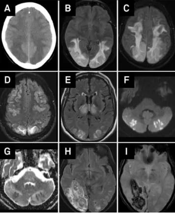
Thirteen patients performed MRI. In one patient, diagnosis was supported exclusively by brain CT. Two patients had no abnormalities in CT, but subsequent MRI revealed findings suggestive of PRES. Table 2 shows brain lesions characteristics found in these patients. The majority of patients presented lesions in other locations besides the posterior parieto-occipital region and in 3/14 patients this region was not pre-dominantly affected. Almost one third of patients presented ischemic or hemorrhagic lesions. Figure shows the main radiological findings. Seven patients performed video electro-encephalogram and among those 6 had slowing of back-ground activity, localized or generalized slowing of activity, suggesting encephalopathic cortical dysfunction.
Table 1.Clinical manifestations during the course of disease.
N
Encephalopathy 11
Seizures 10
Visual disturbances 9
Headache 5
Focal neurologic deficit.24h 4
Focal neurologic deficit,24h 2
N: Number of cases.
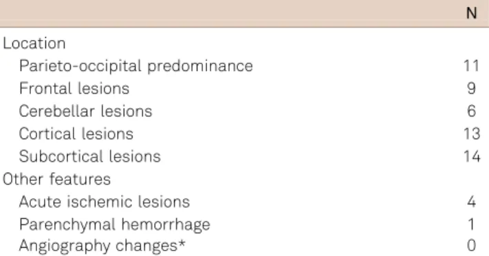
Table 2.Radiological characteristics of brain lesions.
N
Location
Parieto-occipital predominance 11
Frontal lesions 9
Cerebellar lesions 6
Cortical lesions 13
Subcortical lesions 14
Other features
Acute ischemic lesions 4
Parenchymal hemorrhage 1
Angiography changes* 0
Among etiological factors, eclampsia was the most fre-quent (4 patients), followed by isolated high blood pressure (3 patients) and spinal trauma with autonomic dysfunction (2 patients). Other factors identified were sepsis, sarcoidosis and concomitant pulmonary cryptococcosis, GBS, octreotide perfusion and chemotherapy (Table 3).
Duration of follow-up was variable (3 weeks-8 years). Most patients presented a partial or complete recovery
(Table 4). Brain MRI was repeated in 8 patients and in seven patients lesions disappeared. Two patients developed neuro-logical sequelae (epilepsy, severe focal motor deficit). Two patients died after onset. One patient developed rapidly pro-gressive bilateral edema and acute ischemic lesions, with mass effect, intracranial hypertension refractory to medical therapy, and died 19 days after admission. The other patient had been submitted to abdominal surgery for gastric cancer, presented with PRES after octeotride perfusion and died of sepsis related to esophago-jejunal anastomosis leak 3 weeks later. One case of recurrence was observed two months after the first event. This patient had a persistent autonomic dys-function related to spinal trauma.
DISCUSSION
This study describes the clinical and radiological features of 14 patients with PRES, affecting individuals of all ages. In this series there was a male predominance, although in other series there is a slight female predominance, even when eclampsia associated cases are not taken into account10,11.
In most patients of our series, the identified causes were those classically described, namely, eclampsia and high blood pressure. Nevertheless, the cases associated with autonomic dysfunction related to spinal trauma, sepsis, sar-coidosis and pulmonary cryptococcosis, GBS and drugs demonstrate the diversity of precipitating factors that cul-minate in a common clinical-radiological syndrome3,12,13.
High blood pressure was present in the majority of patients, supporting the hypothesis of cerebral autoregulation impair-ment as the main pathogenic mechanism in PRES1,10. When
cerebral autoregulation capacity is overcome by systemic blood pressure, cerebral hyperperfusion emerges, damaging the blood brain barrier and originating liquid leakage to the extracellular space and vasogenic edema, mainly in arterial border territories1,10,14,15,16. The fact that only 4
patients had previous high blood pressure supports the idea that sudden elevation of blood pressure prevents vascular adaptation to take place. The mean mBP in our patients ser-ies (109 mmHg) was inferior to the considered upper limit value of cerebral autoregulation, situated around 150 mmHg17. However, Liman et al.11 demonstrated that
mBP was not correlated with edema severity, while sBP had a positive correlation with edema severity supporting
Figure.PRES radiological characteristics. (A) CT, bilateral posterior parietal and frontal cortico and subcortical hypo-dense lesions; (B) MRI, FLAIR, bilateral temporo-occipital hyperintense lesions; (C-D) MRI, FLAIR, bilateral frontal cortical and subcortical lesions; (E) MRI, FLAIR, typical hyperintense bilateral occipital associated with bilateral thalamic lesions; (F-G) MRI, DWI, multiple bilateral cerebellar lesions presenting restriction to diffusion (recent ischemic lesions); and (H-I) RMI, FLAIR and T2 echo-gradient, revealing typical predominately posterior PRES lesions associated with a right temporo-occipital hemorrhage.
Table 3.Identified precipitating factors.
N
Eclampsia 4
High blood pressure 3
Autonomic dysfunction related to spinal trauma 2
Sepsis 1
Pulmonary sarcoidosis and cryptococcosis 1
Guillain-Barré syndrome 1
Octreotide perfusion 1
Chemotherapy (Cisplatin, Gemcitabine, Bevacizumab)* 1 *Hypomagnesemia was also present (cisplatin side effect but also potentially pathogenic). N: Number of cases.
Table 4.Clinical outcome.
N
Full recovery 10
Partial recovery 2
Recurrence 1
Death 2
the hypothesis of the acute lesion of the blood-brain barrier. In this series there was only one patient with normal blood pressure, contrasting with other reports where 20%-30% of patients with PRES presented without high blood pressure9.
The 2 cases related to autonomic dysfunction associated with spinal trauma support the role of the sympathetic nerv-ous system dysfunction in PRES at least in some patients9
. In addition to blood pressure, other mechanisms that cause endothelial dysfunction, blood brain barrier impairment and cerebral edema could also be involved, and the cases asso-ciated with sepsis, sarcoidosis and drugs may be illustrative of this hypothesis. In this series one patient treated with bevacizumab developed PRES. In fact, there is a growing number of PRES cases occurring in patients treated with anti-vascular endothelial growth factor agent (anti-VEGF) as bevacizumab. This drug interferes with vascular permeability and with endothelium intracellular signaling pathways sup-porting the endothelial dysfunction role in this disease18.
Concerning topography of lesions, most patients pre-sented predominantly posterior parietal-occipital lesions which may be explained by the lower density of sympathetic fibers in the vertebrobasilar territory, making it more suscept-ible to systemic blood pressure oscillations1,19. Nevertheless,
64.2% presented frontal lobe lesions and 42.8% also presented cerebellar lesions, in accordance to other series3,10,11,13.
Thus, posterior in PRES designation may be inadequate. Furthermore, this syndrome was initially described as a white matter disease1, but in 92.8% of our patients, cortical
involve-ment was demonstrated.
It has been suggested that in PRES, arteriolar vasocon-striction occurs in response to cerebral hyperperfusion, thus leading to hypoperfusion, ischemia and subsequent edema10,15,16. Although angiographic studies demonstrate
focal or diffuse vasoconstriction, ischemic complications are not frequent20. Acute ischemic complications occurred
in 4/14 (28.6%) of our patients, in accordance to what is described in the literature11,16. There was only one case of
cerebral hemorrhage (7.1%) in this series. However in the literature, hemorrhagic complications occur in 15 to 32% of cases, mostly in the form of petechial bleeding11,16.
Ten of 14 patients experienced a complete clinical recovery, supporting the reversibility of this syndrome. Follow-up MRI showed partial or complete improvement of brain lesions in 7/8 patients. Liman et al.11 reported a
partial or complete recovery of lesions in 82% of patients during follow-up. Recurrent PRES episodes are rare11,13,
and occurred in 1/14 patient in this series. The adequate control of blood pressure and the elimination of all other precipitating factors should be achieved during acute man-agement and follow-up21. Among patients that recovered
partially, one developed motor sequelae and the other developed epilepsy. Furthermore, 2 patients (14.3%) died, and death was attributed directly to PRES in one of them. Rarely this entity may complicate with progressive cerebral edema, intracranial hypertension and death1,10,11. Morbidity
and mortality in this series (28.6%) prove that PRES is not always reversible and benign.
This study had several limitations associated with its retrospective design and related to the population size. Imagiological evaluation was not uniform in all patients and angiographic study was absent in 9/14 patients. Follow-up was not homogeneous, mainly in what concerns to radiological reevaluation. In depth analysis of clinical and analytical variables with prognostic significance was not performed due to the lack of statistical power.
In conclusion, PRES should be rapidly recognized in emergency settings. It is a clinical-radiological entity whose pathophysiology is not fully understood and with a diversity of precipitating factors. The denomination posterior and reversible has its origin in the first description by Hinchey1
but the growing number of reported cases, including the pre-sent series, show the frequent involvement of other brain regions and less favorable outcomes with significant morbid-ity and mortalmorbid-ity. While pathophysiological mechanisms are not fully understood the current designation remains useful, although it does not encompass its clinical and radiological diversity. PRES, reversible vasoconstriction syndrome and cerebral hyperperfusion syndrome may partially share com-mon pathological mechanisms and they may all belong to a larger spectrum of acquired vasculopathies.
References
1. Hinchey J, Chaves C, Appignani B, et al. A reversible posterior leukoencephalopathy syndrome. N Engl J Med 1996;334:494.
2. Schwartz R B, Mulkern R V, Gudbjartsson H, Jolesz F. Diffusion-weighted MR imaging in hypertensive encephalopathy: clues to pathogenesis. Am J Neuroradiol 1998;19:859-862.
3. Hinchey J. Reversible posterior leukoencephalopathy syndrome: what have we learned in the last 10 years? Arch Neurol 2008;65:175-176.
4. Lee VH, Wijdicks EFM, Manno EM, Rabinstein AA. Clinical spectrum of reversible posterior leukoencephalopathy syndrome. Arch Neurol 2008;5:205-210.
5. Bartynski WS. Posterior reversible encephalopathy syndrome, part 2: controversies surrounding pathophysiology of vasogenic edema. AJNR Am J Neuroradiol 2008;29:1043-1049.
6. Bartynski WS. Posterior reversible encephalopathy syndrome, part 1: fundamental imaging and clinical features. AJNR Am J Neuroradiol 2008;29:1036-1042.
7. Lamy C, Oppenheim C, Méder JF, Mas JL Neuroimaging in posterior reversible encephalopathy syndrome. J Neuroimaging 2004;14:89.
9. Matias AC, Rocha J, Cerqueira ME, Pereira JM. Autonomic dysreflexia and posterior reversible encephalopathy syndrome. Am J Phys Med Rehabil 2013;92:453-458.
10. Stott VL, Hurrell MA, Anderson TJ. Reversible posterior leukoencepha-lopathy syndrome: a misnomer reviewed. Intern Med J 2005;35:83.
11. Liman TG, Bohner G, Heuschmann PU, Endres M, Siebert E. The clinical and radiological spectrum of posterior reversible encephalo-pathy syndrome: the retrospective Berlin PRES study. J Neurol 2012;259:155-164.
12. Stevens CJ, Heran MKS. The many faces of posterior reversible encephalopathy syndrome. Br J Radiol 2012;85:1566-1575.
13. Fugate JE, Claassen DO, Cloft HJ, Kallmes DF, Kozak OS, Rabinstein AA. Posterior reversible encephalopathy syndrome: associated clinical and radiological findings. Mayo Clin Proc 2010;85:427-432.
14. Vaughan CJ, Delanty N. Hypertensive emergencies. Lancet 2000;356:411-417.
15. Ito Y, Niwa H, Iida T, et al. Post-transfusion reversible posterior leukoencephalopathy syndrome with cerebral vasoconstriction. Neurology 1997;49:1174-1175.
16. Tajima Y, Isonishi K, Kashiwaba T, Tashiro K. Two similar cases of encephalopathy, possibly a reversible posterior leukoencephalopathy syndrome: serial findings of magnetic resonance imaging, SPECT and angiography. Intern Med 1999;38:54-58.
17. Ziai WC, Mirski MA. Blood pressure management in the neurocritical care patient. In: Suarez JI (Ed). Critical care neurology and neurosurgery. New Jersey: Humana Press Inc; 2004:247-266.
18. Tlemsani C, Mir O, Boudou-Rouquette P, et al. Posterior reversible encephalopathy syndrome induced by anti-VEGF agents. Targ Oncol 2011;6:253-258.
19. Sheth RD, Riggs JE, Bodenstenier JB, Gutierrez AR, Ketonen LM, Ortiz OA. Parietal occipital edema in hypertensive encephalopathy: a pathogenic mechanism. Eur Neurol 1996;36:25-28.
20. Bartynski WS, Boardman JF. Catheter angiography, MR angiography, and MR perfusion in posterior reversible encephalopathy syndrome. AJNR Am J Neuroradiol 2008;29:447-455.