CASE REPORT
Gliosarcoma with neuroaxis metastases
Rui Ramos,
1Nuno Morais,
2Ana Isabel Silva,
3Rui Almeida
11Department of Neurosurgery, Hospital de Braga, Braga, Portugal
2Department of Neurosurgery, Hospital CUF, Porto, Portugal 3Department of Pathology, Hospital de Braga, Braga, Portugal
Correspondence to
Dr Rui Ramos, [email protected]
Accepted 15 November 2015
To cite:Ramos R, Morais N, Silva AI,et al.
BMJ Case RepPublished online: [please includeDay Month Year] doi:10.1136/ bcr-2015-212970
SUMMARY
Gliosarcomas are rare tumours of the central nervous system, with a well-known capacity for metastasis. When they metastasise, the dissemination occurs more frequently via the haematogenous route to extraneural sites. Metastasis-spread through the cerebrospinalfluid is extremely rare. We present the case of a 58-year-old man who underwent a gross total resection of a lesion in the left temporal lobe. The histologicalfindings revealed a gliosarcoma and the patient received radiotherapy followed by chemotherapy. Seven months after surgery, while the patient remained neurologically intact, brain and spinal cord MRI revealed tumour recurrence and neuroaxis metastases through the traffic routes of the cerebrospinalfluid. The patient died 8 months after the diagnosis. A PubMed search regarding metastatic gliosarcoma up to June 2015 was also carried out. To the best of our knowledge, this is thefirst case report of gliosarcoma metastases to the brain and spinal cord leptomeninges.
BACKGROUND
Gliosarcomas werefirst described in 1895 as
glio-blastomas with a sarcomatous component.1 The
current definition is based on the 2007 WHO clas-sification, which considers them well-defined brain lesions with a clearly identifiable biphasic pattern
of glial and mesenchymal components.2
Gliosarcomas comprise 0.48% of all intracranial
tumours and 2–8% of glioblastomas.3They
prefer-entially affect individuals between the sixth and seventh decades of life, with a male:female ratio of 1.4–1.8:1.4 5 The most frequent locations, in des-cending order, are the temporal, frontal, parietal and occipital lobes.6
Clinical features depend on the location of the tumour and are similar to those of glioblastomas. The most common symptoms are headache, vomit-ing, seizures, hemiparesis, cognitive decline and other symptoms associated with intracranial hyper-tension.4 The imaging features are variable. They may present with central necrotic areas and hetero-geneous contrast uptake similar to a glioblastoma, or with homogeneous contrast enhancement and well-defined margins similar to a meningioma.4 5 Histologically, two distinct cell populations can be identified, one composed of neoplastic astrocytes meeting the criteria for glioblastoma and the other consisting of a spindle cell sarcomatous compo-nent.7The glial component exhibits strong staining
for glial fibrillar acidic protein (GFAP), unlike the GFAP-negative sarcoma-like component.
The metastatic capacity of gliosarcomas is well known with an incidence that can reach 11%, which is much higher than that for glioblastomas (0.2–1.2%).8 As far back as 1958, some authors
have reported cases of metastasis with mixed
elements, namely glial and sarcomatous.5
Subsequently, Smith et al observed two cases of
metastasis that were composed only of sarcomatous cells, raising the possibility that the metastatic potential of gliosarcoma is linked to its sarcomatous component.5 The greater propensity of gliosarcoma for metastasis could also be related to its
frequent temporal location, near the dura and venous sinuses.1 The major sites of metastasis are lungs, liver and lymph nodes.8
Other reported sites are the spleen, adrenal glands, kidneys, oral mucosa, skin, bone marrow, skull, ribs and spine.5 Metastatic
disease is more common in young male individuals who have undergone adjuvant radiotherapy.9
Beaumont et alreported a case involving a gliosarcoma with
multiple extracranial metastases and intravascular tumour emboli revealed in the postmortem examination. This is consist-ent with a greater propensity for haematogenous dissemination.9
The increased capacity for haematogenous metastasis is also related to the fact that sarcomatous tumours have a higher ten-dency to spread using this pathway.1However, there are other routes of spread, such as metastasis through the cerebrospinal
fluid, where the tumour cells reach the subarachnoid space or ventricular cavities through the leptomeninges or transependy-maly.10In these cases, ventricular, cranial nerve, spinal cord and leptomeningeal invasion can occur.1
Regarding the therapeutic approach to gliosarcoma, there are no specific protocols. Thefirst review published in the literature considered a number of clinical and biological similarities with glioblastomas, and since then they have been treated using the same protocols; these involve maximum surgical removal fol-lowed by radiotherapy and chemotherapy.11 12 In the presence
of metastasis, the ideal treatment remains unknown but the common chemotherapy regimens for soft tissue sarcomas seem to offer no benefit.8Even with treatment, the survival times of patients with gliosarcoma are short and range from 6 to 14.8 months.1
Figure 2 Postoperative brain MRI. Gadolinium-enhanced T1-weighted axial image demonstrating radical tumour excision.
Figure 3 Histological tissue sections. (A) H&E showing a biphasic tumour with glial (left) and fusiform (right) cells (×40 original magnification). (B) Glialfibrillar acidic protein with strong immunoreactivity in the glial
component (left) and virtually no staining in the mesenchymal tissue (right; ×40 original magnification).
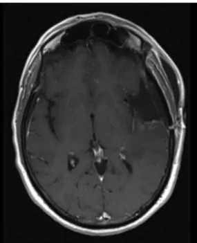
Figure 4 MRIs of the control brain at 7 months after craniotomy.
Figure 5 Vertebrospinal MRI at 7 months after craniotomy. Gadolinium-enhanced T1-weighted sagittal images revealing: (A) retroclival enhancement, diffuse meningeal spread, and nodular lesions in the C5 and (B) along the cauda equina nerve roots, indicative of‘drop’
metastases.
Table 1 Reports on metastatic gliosarcomas published until June 2015
Author Year Sex Age
(years) Localisation Type Resection
Adjuvant
treatment Metastasis location Metastasis histology
Survival (months)
Ehrenreich13 1958 M 44 Parietal P Unknown RT Lung Mixed 8
Feigin14 1958 M 6 Temporal P Unknown Unknown Lung Mixed 15
Garret15 1958 F 55 Temporal P Radical RT Lymph nodes Mixed 9
Smith16 1969 M 49 Frontal P Partial RT+CT Liver Sarcomatous 8
Smith16 1969 M 44 Temporal P Partial RT Liver Mixed 8
Smith16 1969 M 58 Temporal P Biopsy RT Liver, lung Sarcomatous 8
Smith16 1969 M 63 Temporal P Partial Unknown Lung, liver, adrenal gland Mixed 8
Smith16 1969 M 64 Temporal P Partial Unknown Liver, lung Mixed 11
Smith16 1969 M 6 Temporal P Biopsy RT Lung Mixed 13
Smith16 1969 F 63 Frontal P Partial Unknown Vertebrae, lung Mixed 11
Slowik17 1980 F 46 Parietal S Unknown RT Lung, liver, kidney, lymph nodes Mixed 19
Ojeda18 1984 F 83 Frontal P Not
operated
None Lung Sarcomatous 3
Weaver19 1984 M 63 Parietal S Biopsy RT Lung, omentum Sarcomatous 7
Cerame20 1985 F 11 Temporal P Unknown RT+CT Lung Mixed 1
Yokoyama21 1985 F 22 Occipital P Radical RT+CT Lung, pleura, lymph nodes, bone marrow, liver
Mixed 4
Matsuyama22 1989 M 68 Temporal P Radical RT+CT Liver, spleen, spinal cord, scalp Mixed 5
Gjerdrum23 1999 M 61 Temporo-parietal P Unknown RT Oral mucosa, palpebra, lung Sarcomatous 6
Witwer10 2000 M 48 Temporal P Radical RT+CT Spinal cord Unknown 3
Wharton24 2001 M 53 Temporal P Unknown RT Liver, ileum, vertebrae, skull, ribs Gliosarcoma with primitive neuroepithelial differentiation
5
Beaumont9 2007 M 47 Temporal S Radical RT+CT+G Thyroid, chest wall, pleura, lung, pericardium, myocardium, diaphragm, pancreas, liver, scalp, spleen, kidney, stomach, lip mucosa
Sarcomatous 20
Fischer25 2007 M 50 Multifocal P Biopsy RT+CT Spinal cord Mixed 5
Demirci1 2008 F 68 Frontal P Radical RT Spinal cord Sarcomatous 10
Maeda26 2010 F 51 Temporal P Radical RT+CT Lung Unknown 5
Mesfin27 2010 F 51 Temporal P Radical RT+CT+G Lung Mixed 17
Rapp28 2011 M 67 Temporooccipital P Unknown RT+CT Lung, skeletal system Mixed 12
Chen29 2012 F 31 Temporal P Unknown RT+CT Liver, lymph nodes, spinal cord, lung, scalp, neck soft tissue, ileum, humeri, collarbone
Mixed 92
Dawar8 2013 F 57 Temporal S Radical RT+CT Lung, pleura, lymph nodes Mixed 64
Mansouri30 2013 M 62 Frontal P Radical RT+CT Brain leptomeninges and dura Mixed Unknown
Oberndorfer31 2013 M 37 Temporal S Radical RT+CT Diaphragm Sarcomatous 11
Asencio-Cortés32 2014 F 48 Frontotemporal P Radical RT+CT Spinal cord Unknown 15
Schindler33 2014 F 64 Frontal S Radical RT+CT Spinal cord Sarcomatous 23
CASE PRESENTATION
A 58-year-old man with no relevant clinical history presented with bilateral tinnitus, which had developed over a 2-week period. Neurological examination on admission revealed no abnormalities.
INVESTIGATIONS
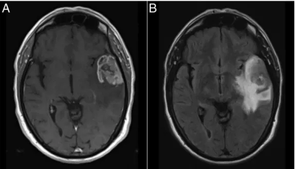
The patient’s symptoms encouraged investigation using brain CT, and a lesion in the left temporal lobe was detected. In MRI,
the lesion measured 32×30 mm, and had ill-defined contrast
enhancement, central necrotic areas and marked vasogenic oedema; it caused a small uncal herniation (figure 1).
TREATMENT
The patient underwent pterional craniotomy with radical tumour excision (figure 2). The surgery was performed without complications and the patient remained neurologically intact. Histological examination revealed a gliosarcoma, according to the WHO criteria (figure 3). Adjuvant treatments were carried out. Radiotherapy was given in 30 fractions,five times a week, to a total dose of 60 Gy, and a chemotherapy regimen consisting of temozolomide (Stupp protocol) was instituted.
OUTCOME AND FOLLOW-UP
At 7 months after surgery, the patient’s neurological status remained unchanged; MRI examination was performed on the control brain. The study revealed tumour recurrence and menin-geal, parenchymal and perineural spread (figure 4). As a result of these findings, an MRI scan of the spinal cord and a thora-coabdominopelvic CT scan were carried out. The MRI revealed meningeal spread and‘drop’metastases in the C5 and the cauda equina nerve roots (figure 5), consistent with dissemination through the traffic routes of the cerebrospinalfluid. The thora-coabdominopelvic CT scan revealed no suspect tumoural lesions. It was decided to interrupt chemotherapy because of the leptomeningeal dissemination and a gradual deterioration in the neurological status of the patient. The patient died at 8 months after surgery.
DISCUSSION
A PubMed search of studies published up until June 2015, using the term‘gliosarcoma’, revealed 31 cases of metastatic gliosar-coma (table 1). Three additional case reports have been pub-lished but we were unable to access the full-text articles. Of the cases, the vast majority reported on haematogenous spread and subsequent visceral metastasis. Nevertheless, in eight patients, there may have been spread through the traffic routes of the
cerebrospinal fluid to the spinal cord or leptomeninges. The present case might be the ninth to report cerebrospinalfluid dis-semination, the second with metastasis to the leptomeninges, and the first with simultaneous spread to the leptomeninges of the brain and spinal cord.
Acknowledgements The authors would like to thank Dr Carlos Alegria, Dr Afonso Almeida Pinto, Dr Miguel Afonso and Dr Ricardo Moreira, for their time spent, and help with, improving this article.
Competing interests None declared.
Patient consent Obtained.
Provenance and peer reviewNot commissioned; externally peer reviewed.
REFERENCES
1 Demirci S, Akalin T, Islekel S,et al. Multiple spinal metastases of cranial gliosarcoma: a case report and review of the literature.J Neurooncol
2008;88:199–204.
2 Han SJ, Yang I, Ahn BJ,et al. Clinical characteristics and outcomes for a modern series of primary gliosarcoma patients.Cancer2010;116:1358–66.
3 Guney Y, Hiçsönmez A, Yilmaz S,et al. Gliosarcoma: a study of four cases.
Rare Tumors2010;2:e37.
4 Biswas A, Kumar N, Kumar P,et al. Primary gliosarcoma-clinical experience from a regional cancer centre in north India.Br J Neurosurg2011;25:723–9.
5 Han SJ, Yang I, Tihan T,et al. Primary gliosarcoma: key clinical and pathologic distinctions from glioblastoma with implications as a unique oncologic entity.
J Neurooncol2010;96:313–20.
6 Moiyadi A, Sridhar E, Jalali R. Intraventricular gliosarcoma: unusual location of an uncommon tumor.J Neurooncol2010;96:291–4.
7 Pakos EE, Goussia AC, Zina VP,et al. Multi-focal gliosarcoma: a case report and review of the literature.J Neurooncol2005;74:301–4.
8 Dawar R, Fabiano AJ, Qiu J,et al. Secondary gliosarcoma with extra-cranial metastases: a report and review of the literature.Clin Neurol Neurosurg
2013;115:375–80.
9 Beaumont TL, Kupsky WJ, Barger GR,et al. Gliosarcoma with multiple extracranial metastases: case report and review of the literature.J Neurooncol2007;83:39–46. 10 Witwer BP, Salamat MS, Resnick DK. Gliosarcoma metastatic to the cervical spinal
cord: case report and review of the literature.Surg Neurol2000;54:373–9. 11 Damodaran O, van Heerden J, Nowak AK,et al. Clinical management and survival
outcomes of gliosarcomas in the era of multimodality therapy.J Clin Neurosci
2014;21:478–81.
12 Han SJ, Yang I, Otero JJ,et al. Secondary gliosarcoma after diagnosis of glioblastoma: clinical experience with 30 consecutive patients.J Neurosurg
2010;112:990–6.
13 Ehrenreich T, Devlin JF. A complex of glioblastoma and spindle-cell sarcoma with pulmonary metastasis.AMA Arch Pathol1958;66:536–49.
14 Feigin I, Allen LB, Lipkin L,et al. The endothelial hyperplasia of the cerebral blood vessels with brain tumors, and its sarcomatous transformation.Cancer
1958;11:264–77.
15 Garret R. Glioblastoma andfibrosarcoma of the brain with extracranial metastases.
Cancer1958;11:888–94.
16 Smith DR, Hardman JM, Earle KM. Contiguous glioblastoma multiforme and fibrosarcoma with extracranial metastasis.Cancer1969;24:270–6.
17 Slowik F, Balogh I. Extracranial spreading of glioblastoma multiforme.Zentralbl Neurochir1980;41:57–68.
18 Ojeda VJ, Sterrett GF. Cerebral gliosarcoma, pulmonary adenoid-cystic carcinoma, and pulmonary metastatic gliosarcoma: report of an untreated case.Pathology
1984;16:217–21.
19 Weaver D, Vandenberg S, Park TS,et al. Selective peripancreatic sarcoma metastases from primary gliosarcoma. Case report.J Neurosurg1984;61:599–601. 20 Cerame MA, Guthikonda M, Kohli CM. Extraneural metastases in gliosarcoma:
a case report and review of the literature.Neurosurgery1985;17:413–18. 21 Yokoyama H, Ono H, Mori K,et al. Extracranial metastasis of glioblastoma with
sarcomatous component.Surg Neurol1985;24:641–5.
22 Matsuyama J, Mori T, Hori S,et al. [Gliosarcoma with multiple extracranial metastases. Case report].Neurol Med Chir (Tokyo)1989;29:938–43.
23 Gjerdrum LM, Bojsen-Møller M. 61 year old male with brain tumor and oral, lung, and palpebral masses.Brain Pathol1999;9:421–2.
24 Wharton SB, Whittle IR, Collie DA,et al. Gliosarcoma with areas of primitive neuroepithelial differentiation and extracranial metastasis.Clin Neuropathol
2001;20:212–18.
25 Fischer S, Lee W, Aulisi E,et al. Gliosarcoma with intramedullary spinal metastases: a case report and review of the literature.J Clin Oncol2007;25:447–9. 26 Maeda D, Miyazawa T, Toyooka T,et al. Temporal gliosarcoma with extraneural
metastasis: case report.Neurol Med Chir (Tokyo)2010;50:343–5. Learning points
▸ Although the route of gliosarcoma metastasis is
preferentially haematogenous, its capacity to metastasise through the cerebrospinalfluid routes should not be underestimated.
▸ Owing to the metastatic capacity of gliosarcomas, a
whole-body CT and neuroaxis MRI should be performed after diagnosis.
▸ Although they appear normal on neurological examination,
patients can present with extensive metastatic spread through the cerebrospinalfluid routes.
▸ Because of the rarity of these tumours, further studies will
27 Mesfin FB, Deshaies EM, Patel R,et al. Metastatic gliosarcoma with a unique presentation and progression: case report and review of the literature.Clin Neuropathol2010;29:147–50.
28 Rapp M, Felsberg J, Sorg RV,et al. Case report: extracranial metastasis from gliosarcoma-the influence of immune system.Br J Neurosurg2011;25:286–8. 29 Chen L, Xiao H, Xu L,et al. A case study of a patient with gliosarcoma with an
extended survival and spinal cord metastases.Cell Biochem Biophys2012;62:391–5. 30 Mansouri B, Barboriak DP, Kilani RK. Gliosarcoma metastatic to the leptomeninges
and dura.J Neuroimaging2013;23:245–7.
31 Oberndorfer S, Wöhrer A, Hainfellner JA,et al. Secondary gliosarcoma with massive invasion of meninges, skull base, and soft tissue, and systemic metastasis.Clin Neuropathol2013;32:522–4.
32 Asencio-Cortés C, de Quintana-Schmidt C, Clavel-Laria P,et al. Metástasis medulares de gliosarcoma: presentación de un caso y revisión de la literatura.
Neurocirugia2014;25:132–5.
33 Schindler G, Capper D, Korshunov A,et al. Spinal metastasis of gliosarcoma: array-based comparative genomic hybridization for confirmation of metastatic spread.J Clin Neurosci2014;21:1945–50.
Copyright 2015 BMJ Publishing Group. All rights reserved. For permission to reuse any of this content visit http://group.bmj.com/group/rights-licensing/permissions.
BMJ Case Report Fellows may re-use this article for personal use and teaching without any further permission.
Become a Fellow of BMJ Case Reports today and you can: ▸ Submit as many cases as you like
▸ Enjoy fast sympathetic peer review and rapid publication of accepted articles ▸ Access all the published articles
▸ Re-use any of the published material for personal use and teaching without further permission
For information on Institutional Fellowships contact [email protected]