Carolina Leça de Azevedo Pereira Tavares
Licenciada em Bioquímica
Interplay of Endoplasmic Reticulum Stress
and Autophagy in Parkinson’s Disease
Dissertação para obtenção do Grau de Mestre em
Genética Molecular e Biomedicina
Orientador: Maria João Gama, PhD,
Faculdade de Farmácia da Universidade de Lisboa
Co-orientador: Andreia Neves Carvalho, PhD
Faculdade de Farmácia da Universidade de Lisboa
Setembro 2017
i | P a g e
Interplay of Endoplasmic Reticulum Stress and Autophagy in Parkinson’s disease
Copyright © Carolina Leça de Azevedo Pereira Tavares, Faculdade de Ciências e Tecnologia,
Universidade Nova de Lisboa
A Faculdade de Ciências e Tecnologia e a Universidade Nova de Lisboa têm o direito,
perpétuo e sem limites geográficos, de arquivar e publicar esta dissertação através de
exemplares impressos reproduzidos em papel ou de forma digital, ou por qualquer outro meio
conhecido ou que venha a ser inventado, e de a divulgar através de repositórios científicos e
de admitir a sua cópia ou distribuição com objetivos educacionais ou de investigação, não
comerciais, desde que seja dado crédito ao autor e editor.
iii | P a g e
Part of the results discussed in this thesis were presented in the following meetings:
Tavares C.L.A., Rodrigues E., Castro-Caldas M., Neves Carvalho A., Gama M.J. Interplay of Endoplasmic Reticulum Stress and Autophagy in Parkinson’s disease. Role of Glutathione S-Transferase pi. Jornadas intercalares das Dissertações Anuais dos Mestrados, Universidade Nova de Lisboa, 2nd February 2017, Caparica [Oral Communication]
Tavares C.L.A., Rodrigues E., Castro-Caldas M., Neves Carvalho A., Gama M.J. Expression Levels of ER Stress and Autophagy Markers in Mice Brain and N2a Cells Under MPTP/MPP+ - Induced Oxidative
Stress. 9th iMed.UL Postgraduate Students Meeting, 13th July 2017, Lisbon, Portugal [Abstract and
Poster]
The results regarding mRNA quantitative analysis of UPR factors were obtained in the setting of an active collaboration with W. Scheper, J.J. Hoozemans and J. van Horssen, VU University Medical Center, Amsterdam, The Netherlands.
This work was supported by National Funds (Fundação para a Ciência e Tecnologia – FCT, Portugal) with the project PTDC/NEU-OSD/0502/2012 and SFRH/BPD/98023/2013 (to ANC).
v | P a g e
AGRADECIMENTOS
As minhas primeiras palavras de agradecimento não poderiam deixar de ser para a Professora Doutora Maria João. Não por se tratar do politicamente correto e do que seria esperado, mas sim por ter sido a melhor orientadora que eu poderia ter desejado para a minha tese. Obrigada por todas as palavras de incentivo, compreensão e coragem. Obrigada por perceber todos os imprevistos pessoais e laboratoriais que foram surgindo pelo caminho. Obrigada pela boa disposição e dedicação constantes e por nunca me ter feito duvidar de ter escolhido o caminho certo. De um modo geral, obrigada por ser um exemplo para mim e um marco muito importante na minha vida científica.
Andreia, se existisse uma palavra superior ao “obrigada” era para ti que a guardava. Ao longo desta jornada foste a minha parceira de alegrias e frustrações (temos de admitir que mais de frustrações do que alegrias) e desde o início que soube que podia sempre contar contigo. Obrigada por me tentares ensinar que “há sempre o dia de amanhã” apesar de muitas vezes eu achar que aquele dia é o fim do mundo. Obrigada também por me teres feito perceber que antes de ser mãe tenho de comprar um bloco de notas gigante para apontar todos os detalhes e mais alguns. Mais do que tudo, obrigada por nunca me fazeres sentir a tua aluna, mas sim fazeres de mim uma companheira. Admiro imenso tudo o que fazes e tornaste-te para mim um exemplo a seguir.
Agradeço também à Professora Doutora Cecília Rodrigues a oportunidade de fazer parte do seu grupo “Cellular Function and Therapeutic Targeting”, enriquecendo os meus conhecimentos e preparando-me para o mundo da investigação.
Á Maria, um grande obrigada por estares sempre disponível a ajudar e esclarecer toda e qualquer dúvida e por seres o maior exemplo de organização que conheço. O laboratório sem ti não é o mesmo. Á professora Elsa tenho de agradecer a sua boa disposição e diversão e por me ter mostrado o seu livro de “experiências falhadas”. Sei que provavelmente este parágrafo entra nos agradecimentos de todas as pessoas que passam pelo CellFun, mas de facto é muito importante para quem está do lado de cá. À professora Margarida quero agradecer todo o interesse mostrado ao longo da elaboração da minha tese e por toda a disponibilidade e espirito de entreajuda.
Tenho de agradecer aos restantes membros do CellFun, Vanda, Maria, Diane, Dionísio, Pedro, André, Alexandra, Marta, Sara, Simão, Hugo e Sofia por todos os almoços de descontração que tornavam o dia um bocadinho melhor. Obrigada por me ajudarem sempre que eu precisei e me mostrarem que um laboratório é muito mais do que um local de trabalho.
Marta e Mariana, este ano não teria sido o mesmo sem vocês. Companheiras de queixas, cafés e sorrisos foram um dos meus grandes escapes dentro do CPM. Estou muito orgulhosa da tese que conseguiram desenvolver. Mariana, para ti um grande obrigada por seres a melhor companheira que podia ter tido e, como não poderia faltar, obrigada por todas as vezes que puseste as minhas membranas em primário.
Durante este ano, o apoio que tive fora do laboratório tornou-se tão crucial para o desenvolvimento desta tese como todo o apoio que me proporcionaram dentro das paredes do CPM. Um enorme obrigada aos meus “Grandes Babes Lindões” por tornarem as minhas idas ao Porto tão especiais e por sempre me terem feito sentir que a distância se trata apenas de um número. Ao João,
vi | P a g e um obrigada por nunca dizeres “não” e por estares sempre disponível para mim. Mostraste-me que os amigos não precisam de concordar a 100% para que a amizade seja especial. Maria, obrigada por seres sempre a minha melhor amiga, estando eu no Porto, em Lisboa ou na China. És dos meus maiores apoios na vida e quero que a nossa amizade continue sempre assim. Gosto muito de ti.
Cláudia e Rita, foram a melhor coisa que a capital me proporcionou. Nunca pensei que fosse possível sentir uma amizade tão forte por pessoas que conheço há tão pouco tempo. Obrigada por todas as idas ao cinema, ao ginásio e ao laréu. São ambas um enorme exemplo para mim e não podia estar mais orgulhosa. Gosto muito de vocês! Tiago, obrigada por todas as boleias e boa disposição. Obrigada por me fazeres sempre rir. Joana, Filipe e João obrigada pela amizade e pelos almoços na “casa do povo”, sempre tão especiais.
Filipa, Teresa e Patrícia, as melhores colegas de casa que poderia ter tido, saiu-me a sorte grande. Obrigada por todas as vezes que compreenderam a minha ausência de tempo e me alimentaram. Obrigada por todas as conversas até de madrugada e por me fazerem sempre sentir melhor. Nunca pensei sentir-me em casa tão fora de casa.
Os meus agradecimentos mais especiais estão guardados para a minha família. Mãe e Vitor, obrigada por apostarem na minha educação e por nunca me recusarem nada. Obrigada por perceberem todos os finais de semana em que não pude voltar a casa e também todos aqueles em que precisava do vosso conforto. Obrigada por me apoiarem não só ao longo deste ano, mas também ao longo de toda a minha vida. Helena, obrigada por me mostrares que a família não é só sangue. Obrigada por teres sido uma segunda mãe para mim hoje e sempre, principalmente no pior ano das nossas vidas. Maria, obrigada por me mostrares o que é amor de irmã. Tenho muito orgulho em ti e sei que te estão reservadas grandes coisas no futuro. Adoro-te, e podes contar comigo sempre.
Silvestre, obrigada por me tentares sempre fazer ver que a vida é muito mais para além desta tese. Obrigada por seres o meu saco de boxe nos dias menos bons e por (quase) nunca te queixares. Obrigada pela amizade e companheirismo, mas sobretudo obrigada pelo amor. És o que de mais especial tenho na minha vida.
Pai, esta tese é para ti. Dava tudo para que pudesses lê-la com todo o interesse que demonstravas em tudo na minha vida. És a minha estrelinha e espero que fiques orgulhoso de mim, onde quer que estejas. Amo-te.
Com carinho, Carolina
vii | P a g e
ABSTRACT
Parkinson’s disease (PD) is characterized by the selective loss of dopaminergic neurons of the
substantia nigra pars compacta, and by the accumulation of misfolded proteins. Evidence from studies
in human PD brain indicates that endoplasmic reticulum (ER) stress and consequent unfolded protein response (UPR) are common features of the disease, placing ER dysfunction as an early component of PD pathogenesis.
It is well established that autophagy is up-regulated in response to ER stress, probably as a compensatory mechanism. However, in the face of impaired UPR and autophagy, there is inefficient clearance, leading to protein accumulation that will result in development and progression of neurodegeneration.
The main objective of this study is to evaluate the expression levels of ER stress and autophagy markers in C57BL/6 wild-type mice and N2a cells, a mouse neuroblastoma cell line, upon treatment with the neurotoxin 1-methyl-4-phenyl-1,2,3,6-tetrahydropyridine (MPTP) and 1-methyl-4-phenylpyridinium (MPP+), respectively. In parallel, mice and cells were also treated with Tauroursodeoxycholic acid
(TUDCA), a chemical chaperone that modulates ER adaptive capacity, in order to assess if the neuroprotetive effect of this molecule results from modulation of autophagy. Finally, we seek to understand if TUDCA modulates the expression levels of glutathione-S-transferase pi (GSTp), an antioxidant enzyme.
Expression of ER stress and autophagy markers was analysed using Western blot analysis and qPCR. Our results show that MPTP/MPP+ administration increases the expression of ER stress
responsive genes and that this effect is attenuated with administration of TUDCA prior or after MPTP/MPP+ administration, namely by stimulation of autophagy. We also demonstrated by Western
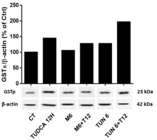
blot and immunocytochemistry that TUDCA increases the levels of GSTp.
Together, our results suggest that TUDCA modulates ER stress by stimulation of UPR pathways as an early response and by stimulation of autophagy when ER stress is persistent, therefore TUDCA remains a promising therapeutic agent to be implemented in PD treatment.
ix | P a g e
RESUMO
A doença de Parkinson (DP) é caracterizada pela perda seletiva de neurónios dopaminérgicos da substantia nigra pars compacta e pela acumulação de proteínas misfolded. Estudos feitos em cérebro de pacientes com DP mostram fortes evidências que o stress do reticulo endoplasmático (RE) e consequente unfolded protein response (UPR) são características típicas da doença, acentuando o papel da disfunção do RE como um componente inicial na patogénese da DP.
Sabe-se que a autofagia é induzida em resposta ao stress do reticulo, provavelmente como um mecanismo compensatório. No entanto, na presença de UPR e autofagia comprometidos não existe remoção eficiente de proteínas misfolded, levando à sua acumulação e consequente desenvolvimento e progressão de degenerescência neuronal.
Assim, o principal objetivo desta tese é a avaliação dos níveis de expressão de marcadores do stress do RE e de autofagia em ratinhos C57BL/6 e em células N2a, uma linha celular de neuroblastoma de ratinho, após tratamento com a neurotoxina 1-metil-4-fenil-1,2,3,6-tetrahidropiridina (MPTP) e com 1-metil-4-fenilpiridina (MPP+), respetivamente. Em paralelo, os ratinhos e as células foram tratados com
o ácido tauroursodeoxicólico (TUDCA), um chaperone químico que influencia a capacidade adaptativa do RE, de maneira a perceber se o efeito neuroprotetor desta molécula resulta da modulação da autofagia. Por fim, pretendemos também perceber se o TUDCA altera os níveis de expressão de glutationo-S-transferase pi (GSTp), um enzima antioxidante, nas células N2a.
Os níveis de expressão de marcadores do stress do RE e de autofagia foram analisados por Western blot e análise por reação em cadeia da polimerase por método quantitativo (qRT-PCR). Os resultados obtidos mostram que a administração de MPTP/MPP+ aumenta a expressão de genes
suscetíveis ao stress do RE e que este efeito é atenuado pela administração de TUDCA antes ou após MPTP/MPP+, provavelmente por estimulação da autofagia. Demonstramos também por Western blot e
imunocitoquímica que os níveis de expressão de GSTp aumentam na presença de TUDCA.
Estes resultados sugerem que o TUDCA influencia o stress do RE por estimulação da UPR como primeira resposta e por estimulação da autofagia em casos de stress do RE persistente. Sendo assim, o TUDCA é um agente terapêutico promissor no tratamento da DP.
xi | P a g e
TABLE OF CONTENTS
ABBREVIATIONS ... xvii
I . INTRODUCTION ... 1
1. Parkinson’s Disease ... 1
1.1. Pathophysiology, clinical presentation, diagnosis and treatment ... 3
1.2. Role of oxidative stress in Parkinson’s disease ... 5
1.3. Autophagy and Parkinson’s disease ... 6
2. The Endoplasmic Reticulum and Oxidative Stress ... 7
2.1. The Unfolded Protein Response ... 8
2.1.1. PERK/EIF2AK3 signalling ... 10
2.1.2. IRE1α/ERN1 signalling ... 10
2.1.3. ATF6 signalling ... 11
2.2. ER stress and UPR in Parkinson’s Disease ... 12
3. The autophagy machinery ... 13
3.1. mTOR signalling pathways ... 13
3.2. Autophagy and ER stress ... 15
4. Glutathione-S-transferases ... 17 4.1. Glutathione S-transferase Pi ... 17 5. Tauroursodeoxycholic acid ... 19 6. Experimental models of PD ... 20 6.1. Neurotoxin-based models of PD ... 20 6.1.1. MPTP mechanism of action ... 20 6.2. PD Animal Models ... 22
6.2.1. The MPTP rodent model ... 22
6.3. Cellular models of PD ... 23
7. Main goals ... 24
II . MATERIALS AND METHODS ... 25
1. Materials ... 25
1.1. Supplements and chemicals ... 25
xii | P a g e
2. Methods ... 27
2.1. Animal treatments ... 27
2.2. Culture conditions and N2a treatment ... 29
2.3. Western Blot Analysis ... 30
2.4. Immunocytochemistry ... 31
2.5. Total RNA isolation and qRT-PCR analysis ... 31
2.6. Statistical analysis ... 32
III. RESULTS ... 33
1. Evaluation of autophagy markers in N2a cells and C57BL/6 mouse model ... 33
1.1. TUDCA alters autophagy markers expression through time as well as mTORC1 complex proteins, in N2a cells ... 33
1.2. TUDCA alters autophagy markers expression through time as well as mTORC1 complex proteins, in C57BL/6 mice brain ... 36
2. Evaluation of ER stress and UPR markers in N2a cells and C575BL/6 mouse model ... 39
2.1. Expression levels of mediators and downstream effectors of the UPR pathways are altered in N2a cells ... 39
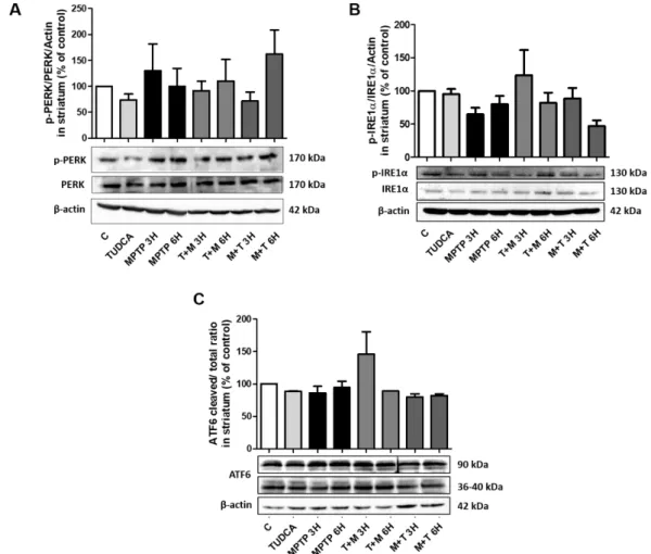
2.2. TUDCA regulates mRNA levels as well as proteins expression of ER stress related factors in C57BL/6 mice brain ... 41
3. GSTp may play an important role in TUDCA potential neuronal defense ... 46
3.1. TUDCA modulates GSTp expression levels in N2a cells ... 46
3.2. CHOP mRNA levels are altered in GSTp KO C57BL/6 mice midbrain ... 48
IV. DISCUSSION ... 49
xiii | P a g e
INDEX OF FIGURES
I. Introduction
Figure I.1 – Schematic representation of the UPR.. ... 9
Figure I 2 - Simplified schematic representation of mTORC1 autophagy signalling pathway ... 14
Figure I.3 – Schemactic representation of MPTP metabolism and intracelular pathways. ... 21
II. Materials and Methods
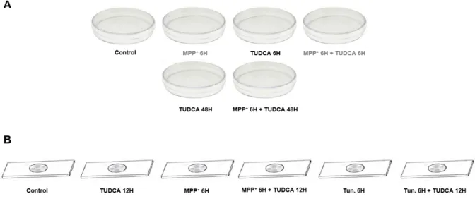
Figure II.1 - Schematic representation of C57BL/6 mice treatment course ... 28Figure II 2 – Different treatment conditions of N2a ... 29
III. Results
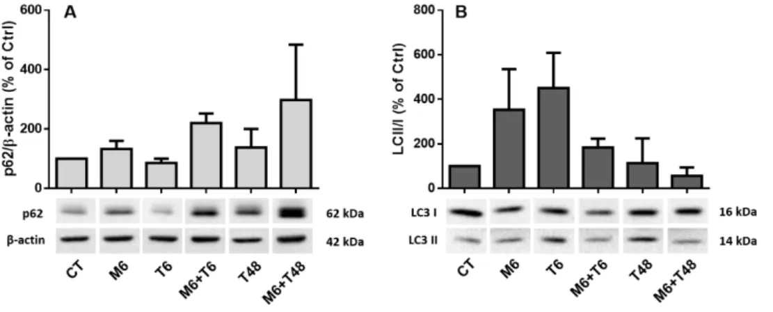
Figure III 1 – TUDCA modulates levels of LC3II and p62 in N2a cells. ... 33Figure III.2 – mTORC1 proteins phosphorylation levels in response to MPP+ and TUDC administration in N2a cells.. ... 35
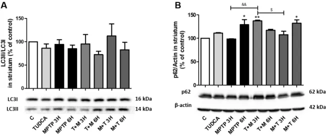
Figure III.3 – Expression levels of LC3II and p62 in the presence of MPTP and TUDCA in C57BL/6 mice striatum. ... 36
Figure III 4 - mTORC1 proteins phosphorylation levels in response to MPTP and TUDCA administration in C57BL/6 mice striatum ... 38
Figure III.5 – UPR related proteins expression levels in N2a cells after treatment with MPP+ or TUDCA ... 40
Figure III.6 - TUDCA modulates mRNA levels of ER stress related factors in C57BL/6 mice striatum. ... 42
Figure III.7 - Protein expression levels of the three main initiators of UPR response. ... 43
Figure III.8 – TUDCA and MPTP modulates the levels of UPR downstream effectors... 45
Figure III 9 – GSTp levels are altered in the presence of TUDCA in N2a cells ... 46
Figure III.10 – GSTp fluorescence in N2a cells treated with MPP+, TUDCA and Tunicamycin. ... 47
xv | P a g e
INDEX OF TABLES
I. Introduction
Table I.1 – PARK designated PD related loci... 2
Table I.2 – Clinical features of Parkinson Disease. Motor and non-motor symptoms ... 4
II. Materials and Methods
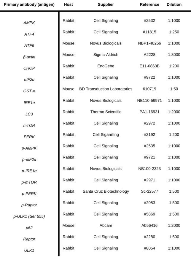
Table II.1 – List of the primary antibodies used in western blot and immunofluorescence assays. ... 26 Table II.2 – Sequences of primers used for qRT.PCR analysis ... 31 Table II.3 – Cycle treatments used for DNase sample treatment and cDNA synthesis ... 32
xvii | P a g e
ABBREVIATIONS
6-OHDA 6-hydroxydopamine
ADP Adenosine diphosphate
AMPK Adenosine monophosphate activated protein kinase
ASK1 Apoptosis signal-regulating kinase 1
ATF4 Activating transcription factor-4
ATF6 Activating transcription factor-6
Atg Autophagy-related proteins
ATP Adenosine triphosphate
BBB Blood brain barrier
Bcl-2 B-cell lymphoma 2
BSA Albumin bovine serum
Ca2+ Calcium
cDNA Complementary DNA
CHOP C/EBP-homologous protein
DA Dopamine
DAT Dopamine transporter
DJ-1 Parkinson protein 7
DNA Deoxyribonucleic acid
eIF2α Eukaryotic initiation factor 2
ER Endoplasmic reticulum
ERAD Endoplasmic reticulum associated degradation
ETC Electron transport chain
FADH2 Flavin adenine dinucleotide
FBS Fetal bovine serum
GBA Glucocerebrosidase
GRP78/BiP Immunoglobulin-heavy-chain-binding protein
GSH Glutathione
GST Glutathione S-transferase
GSTP Glutathione S-transferase Pi
xviii | P a g e
IP3R Inositol triphosphate receptor
IRE1α Inositol-requiring protein 1α
JNK c-JUN NH2-terminal kinases
KEAP1 Kelch ECH associating protein
KO Knockout
LBs Lewy Bodies
LC3 Microtubule-associated light chain
LRRK2 Leucine-rich repeat kinase 2
MAO-B Monoamine oxidase type B
MPDP+ 1-methyl-4-phenyl-2,3-dihydropyridinium
MPP+ 1-methyl-4-phenylpyridinium
MPTP 1-methyl-4-phenyl-1,2,3,6-tetrahydropyridine
mRNA Messenger ribonucleic acid
mTOR Mammalian target of rapamycin
mTORC1 Mammalian target of rapamycin complex one
NADH Nicotinamide adenine dinucleotide
NMS Non-motor symptoms
Nrf2 Nuclear factor erythroid 2-related factor 2
PBS Phosphate buffered saline
PD Parkinson’s Disease
PERK Protein kinase RNA-like ER kinase
PFA Paraformaldehyde
PI3K Phosphoinositide 3-kinase
PINK1 Phosphatase and tensin (PTEN) induced putative kinase 1
PRKN Parkin
PVDF Polyvinyl difluoride
qRT-PCR Quantitative reverse transcription polymerase chain reaction
Raptor Regulatory-associated protein of mTOR
RNA Ribonucleic acid
ROS Reactive oxygen species
S1P Site 1 protease
xix | P a g e
SDS Sodium dodecyl sulphate
SNCA α-synuclein gene
SNpc Substantia nigra pars compacta
ST Striatum
TBS-T Tris-buffered saline-Tween 20
TRAF2 Tumor necrosis factor-α receptor-associated factor 2
TUDCA Tauroursodeoxycholic acid
Tun Tunicamycin
UDCA Ursodeoxicholic acid
ULK1 UNC-51-like kinase
UPR Unfolded protein response
UPS Ubiquitin-proteasome system
VMAT2 Vesicular monoamine transporter-2
VPS35 Vacuolar sorting protein 5
1 | Page
I . INTRODUCTION
1.
Parkinson’s Disease
Parkinson's disease (PD) is estimated to affect more than 10 million individuals worldwide representing the most common neurodegenerative movement disorder (Abdullah et al., 2015). PD is an idiopathic neurodegenerative movement disorder mainly characterized by the selective loss of dopaminergic (DA) neurons in the substantia nigra pars compacta (SNpc), that project their terminals into the striatum (ST), leading to the subsequent depletion of dopamine and loss of signalling in the nigrostriatal pathway (Martin et al., 2011; Moore et al., 2005). Loss of DA neurons leads to the major clinical symptoms of PD, but there is widespread neuropathology and the SNpc only becomes involved towards the middle stages of the disease (Braak et al., 2003). Lewy bodies (LBs) – abnormal aggregates of α-synuclein protein- and dystrophic neurites (Lewy neurites) in the surviving neurons are classical pathologic hallmarks of PD (Kalia & Lang, 2015).
The etiology of PD is multifactorial, which probably results from an interplay of mostly unknown factors: several genes modifying effects by susceptibility alleles, environmental exposures and gene-environment interactions (e.g., influence of gene-environmental agents on gene expression), and their direct impact on the developing and aging of the brain (Lill, 2016). Therefore PD is considered a sporadic disorder with occult causes in 95% of the cases (designated as sporadic PD) with only the remaining 5% of the cases being due to genetic factors (referred as familial PD) (Ranjita Betarbet et al., 2000; Piccini et al., 1999).
In the case of Parkinson’s, a clear Mendelian inheritance is rarely seen, and the familial PD comprises the autosomal dominant form and autosomal recessive form (Bonifati, 2014). Dominant forms manifest in the presence of mutations on genes SNCA, LRRK2, VPS35 and GBA. Mutations in the
alpha-synuclein gene (SNCA) are rare and include point mutations and whole-locus multiplications
(duplications or triplications) and are believed to cause PD through a toxic gain of function and Lewy Bodies may represent the attempt to purge the cell of toxic damaged alpha-synuclein (Bertoncini et al., 2005; Chen & Feany, 2005). The most common known cause of autosomal dominant PD results from mutations in leucine-rich repeat kinase 2 (LRRK2) a large protein with many domains capable of protein– protein interactions, and thus it is plausible that changes in these domains would influence the LRRK2’s relationship with other proteins, although its pathogenic mechanism is still uncertain (Lill, 2016). Mutations in the vacuolar protein sorting 35 (VPS35) were identified as a cause of dominant autosomal disease in 2011 but the brain pathology in carriers of this mutation remains unknown (Vilarino-Guell et al., 2011). The gene encodes a subunit of the retromer complex linked to the trafficking and recycling of proteins and synaptic vesicles (Bonifati, 2014). Loss of function mutations in glucocerebrosidase (GBA) cause impairment in glycolipid metabolism and are a frequent and strong risk factor for PD (Sidransky et al., 2009).
Autosomal recessive forms can result in an early-onset parkinsonism or juvenile atypical parkinsonism. Mutations in parkin (PRKN) are the most common known cause of these PD forms and
2 | Page explain up to half of familial PD. Parkin protein functions as an ubiquitin ligase in the process of ubiquitination (Bonifati, 2014). Phosphatase and tensin (PTEN) induced putative kinase 1 (PINK 1) codes for a protein with a kinase domain located in the mitochondria. PINK1 and parkin proteins interact with each other, with PINK1 acting upstream of parkin. Together they are important to tag damaged mitochondria for degradation by autophagy (Narendra et al., 2010). Parkinson protein 7 (DJ-1) gene codes for a ubiquitous protein and functions as a cellular sensor of oxidative stress (Junn et al., 2005).
PRKN, PINK1 and DJ-1 are all implicated in mitochondrial health, however the pathology of DJ-1 PD
patients remains unknown. Main mutations involved in PD are described in Table I.1.
Despite the intensive search in the field of PD it remains unknown if the disease results from either environmental factors, genetic causes or a combination of both. Loss of dopaminergic neurons is shown to be associated with mitochondrial dysfunction with related oxidative stress and deficient adenosine triphosphate (ATP) production, neuroinflammation and impaired proteolytic degradation. These seem to be observed in both familial and sporadic forms of PD (Dauer & Przedborski, 2003).
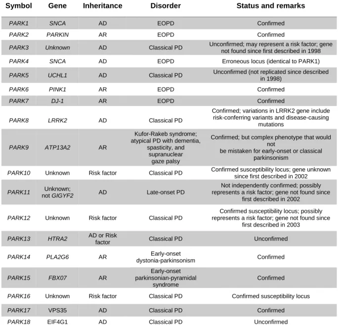
Table I.1 – PARK designated PD related loci. Adapted from (Klein & Westenberger, 2012)
Symbol Gene Inheritance Disorder Status and remarks
PARK1 SNCA AD EOPD Confirmed
PARK2 PARKIN AR EOPD Confirmed
PARK3 Unknown AD Classical PD Unconfirmed; may represent a risk factor; gene
not found since first described in 1998
PARK4 SNCA AD EOPD Erroneous locus (identical to PARK1)
PARK5 UCHL1 AD Classical PD Unconfirmed (not replicated since described
in 1998)
PARK6 PINK1 AR EOPD Confirmed
PARK7 DJ-1 AR EOPD Confirmed
PARK8 LRRK2 AD Classical PD
Confirmed; variations in LRRK2 gene include risk-conferring variants and disease-causing
mutations
PARK9 ATP13A2 AR
Kufor-Rakeb syndrome; atypical PD with dementia,
spasticity, and supranuclear
gaze palsy
Confirmed; but complex phenotype that would not
be mistaken for early-onset or classical parkinsonism
PARK10 Unknown Risk factor Classical PD Confirmed susceptibility locus; gene unknown since first described in 2002
PARK11 Unknown;
not GIGYF2 AD Late-onset PD
Not independently confirmed; possibly represents a risk factor; gene not found since
first described in 2002
PARK12 Unknown Risk factor Classical PD
Confirmed susceptibility locus; possibly represents a risk factor; gene not found since
first described in 2003
PARK13 HTRA2 AD or Risk
factor Classical PD Unconfirmed
PARK14 PLA2G6 AR Early-onset
dystonia-parkinsonism Confirmed PARK15 FBX07 AR Early-onset parkinsonian-pyramidal syndrome Confirmed
PARK16 Unknown Risk factor Classical PD Confirmed susceptibility locus
PARK17 VPS35 AD Classical PD Confirmed
3 | Page
1.1.
Pathophysiology, clinical presentation, diagnosis and treatment
PD is a progressive disease which incidence increases markedly with age, with a mean age at onset of 60 years old. PD affects more men than women, with both incidence and prevalence 1.5 to 2.0 higher in men (A. Lee & Gilbert, 2016). Symptoms tend to worsen overtime with most PD patients suffering considerable motor disability after 5-10 years of disease, even when treated with symptomatic medications (Dauer & Przedborski, 2003). However, in about 10% of the times the onset occurs earlier, between 20 and 50 years of age, being classified as young onset (Dexter & Jenner, 2013).
Loss of dopaminergic neurons, also enriched in neuromelanin, in the SNpc, is the main neuropathological feature of PD. Cell bodies of dopaminergic neurons are located in the SNpc, sending their projections to the caudate and putamen nucleus, in the striatum, creating the nigrostriatal pathway, key to motor function and voluntary movement control. The subsequent loss of striatal dopamine and neuromelanin content is accepted as being responsible for the classical motor features of PD and complex depigmentation (Dexter & Jenner, 2013). Remaining neurons develop intraneuronal inclusions, known as LBs and Lewy neurites, composed mostly of aggregated pre-synaptic α-synuclein together with phosphorylated neurofilaments and ubiquitin, that impair optimal neuronal functioning (Braak et al., 2003). The neuropathology observed in PD extends well beyond dopaminergic neurons as neurodegeneration and LBs formation are found in noradrenergic, serotonergic and cholinergic systems, as well as in the cerebral cortex, olfactory bulb and autonomic nervous system. Degeneration of hippocampal structures and cholinergic cortical inputs contribute to the high rate of dementia that accompanies PD, particularly in older patients (Dauer & Przedborski, 2003).
At the onset of symptoms, putamenal dopamine is depleted in about 80%, and approximately 60% of SNpc dopaminergic neurons have already been lost (Dauer & Przedborski, 2003). Symptoms of PD can be divided into two main groups: motor symptoms and non-motor symptoms. The earliest symptoms might be unnoticed or misinterpreted for a long time. Usually impaired motor function is used to make a clinical diagnosis of PD, as it is secondary to progressive loss of dopaminergic neurons. The main motor symptoms are bradykinesia (slowness of initiating movement), rigidity (resistance of muscles to passive movement around a joint), resting tremor (present in about 70% of PD patients) and postural instability with an asymmetric onset spreading to become bilateral with time (Dexter & Jenner, 2013; Hess & Okun, 2016). Non-motor symptoms (NMSs) are broadly classified as autonomic nervous system dysfunction, cognitive changes, pain, cognitive abnormalities and sleep disturbances. NMSs represent some of the challenges to life quality in PD since they usually do not respond to dopamine therapy as well as motor symptoms (Beitz, 2014; H. M. Lee & Koh, 2015). Table I.2 summarizes the main motor and non-motor symptoms of PD. Whereas the causes of motor dysfunction in PD are reasonably well understood, the cause of NMS in PD remains poorly researched and they may largely relate to pathology outside of the basal ganglia (Dexter & Jenner, 2013).
4 | Page Table I.2 – Clinical features of Parkinson Disease. Motor and non-motor symptoms. Adapted from (Beitz,
2014; Garcia Ruiz et al., 2011; Hess & Okun, 2016; H. M. Lee & Koh, 2015)
Motor symptoms
The onset is insidious where individuals may attribute the symptoms to aging processes. PD symptoms are progressive but rates of motor progression are highly
variable • Resting tremor (initially unilateral)
• Bradykinesia (slowness, clumsiness, and difficulty in making voluntary and automatic semi voluntary movements)
• Rigidity
• Shuffling gait (sensation of heavy legs, slowness of walking, shorter and insecure steps)
• Loss of postural reflexes
• Hypomimia (masked facial expression) • Dystonia
• Stooped posture
Non-motor symptoms
Affect all patients with PD, the frequency of which increases with disease severity. They are often poorly
diagnosed and treated. Later in the disease, the disability from nonmotor symptoms often overshadows that from motor symptoms • Autonomic dysfunction (sexual dysfunction,
swallowing disorder, incontinence,
constipation, excessive salivation, orthostatic hypotension)
• Sensory disorders (pain syndromes, abnormal sensations, olfactory dysfunction) • Integumentary (malignant melanoma, drug
rashes, skin denervation, flush) • Visual (diplopia, blurred vision) • Neurobehavioral (anxiety, depression,
psychosis, cognitive dysfunction, dementia, apathy)
• Sleep problems (insomnia, REM sleep disorder, restless leg syndrome)
Diagnosis of PD is based upon the presence or manifestation of resting tremor, rigidity, postural instability and bradykinesia. Differential diagnosis is challenging given the fact that the classic PD symptoms can be present in other neurodegenerative disorders (Beitz, 2014). Despite diagnosis of PD being made on clinical grounds the gold standard for diagnosis of Parkinson’s disease is the neuropathological assessment. However, there are no generally accepted standard pathological diagnostic criteria for Parkinson’s disease (Klein & Westenberger, 2012). There is no definitive test able to confirm the diagnosis during life, except for gene testing in a reduced number of cases. Therefore, a definite diagnosis of PD requires a post-mortem confirmation (Massano & Bhatia, 2012).
PD is still an incurable progressive disease. Drugs like levodopa, dopamine agonists, monoamine oxidase type B (MAO-B) inhibitors, and, less commonly, amantadine enhance the intracerebral dopamine concentrations or stimulate the dopamine receptors and are still the standard treatment for motor symptoms (Kalia & Lang, 2015). Other targets include mitochondrial dysfunction and oxidative stress, calcium channel activity, among others (AlDakheel et al., 2014). When symptoms
5 | Page cannot be managed using medication, surgical interventions, namely live targeted gene therapy or deep brain stimulation of bilateral subthalamic nuclei, may be the answer (Kordower & Bjorklund, 2013; Li et al., 2017). Nowadays, a major goal of PD research is the development of disease-modifying drugs that slow or stop the inevitable neurodegeneration phenomenon.
1.2.
Role of oxidative stress in Parkinson’s disease
Oxidative stress can be defined as a condition in which the cell is not able to keep the levels of reactive oxygen species (ROS) below a toxic threshold, caused either by failure of cell-buffering mechanisms or over-production of ROS (Gaki & Papavassiliou, 2014). The production of ROS requires the activation of molecular oxygen, being continuously produced in vivo as a result of oxygen metabolism. To achieve cellular energy, within the mitochondria occurs the transport of electrons resulting from oxidation of reduced nicotinamide adenine dinucleotide (NADH) or flavin adenine dinucleotide (FADH2) along the electron transport chain (ETC) until there is a reduction of oxygen to
water at complex IV (Winklhofer & Haass, 2010). The transport of electrons generates a proton flow, creating an electrochemical gradient resulting in ATP production from adenosine diphosphate (ADP), through the ATP synthase located in complex V of ETC (Keane et al., 2011). This process, known as oxidative phosphorylation, provokes leakage of electrons that can react with molecular oxygen originating ROS such as superoxide anion, hydrogen peroxide and nitric oxide that can produce oxidative damage by reacting with deoxyribonucleic acid (DNA), lipids and proteins (Kirkinezos & Moraes, 2001).
The brain consumes about 20% of the oxygen supply of the body, and a significant portion of that oxygen is converted to ROS (W. M. Johnson et al., 2012). The presence of oxidative damage to lipids, proteins and DNA in the SNpc of sporadic PD patients’ brains have implicated oxidative damage in the pathogenesis of the disease, once it contributes to neuronal degeneration of sensitive neurons (Jenner, 2003). Also, the oxidation of dopamine by monoamine oxidase is a reaction known to cause production of superoxide and hydrogen peroxide (Miller et al., 2009). Deficits in the subunits and activity of mitochondrial complex I of the ETC are a prominent phenomenon in blood platelets and SNpc of PD patients, as well as reduced complex I activity in cytoplasmic hybrid cell lines containing mitochondrial DNA from PD patients (Beal, 2005; Swerdlow et al., 1996). These findings suggest an important and determinant role of oxidative stress in the pathogenesis of PD.
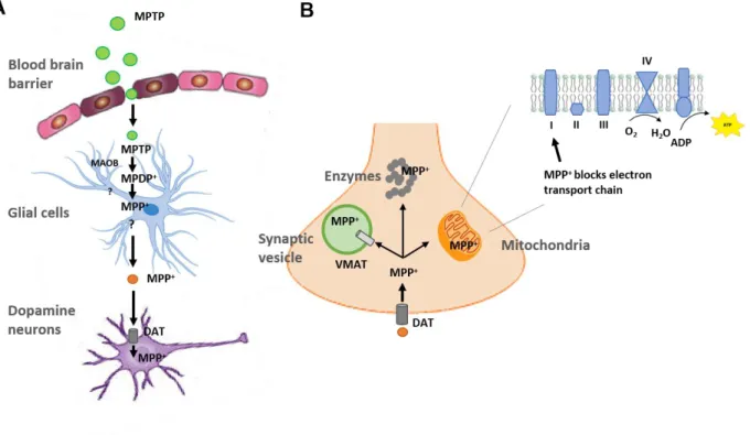
The source of the oxidative stress remains contested, with both neuronal and glial sources being implicated, but there seems to exist little doubt that the most likely contributor is increased radical formation originating from mitochondria and endoplasmic reticulum (Dexter & Jenner, 2013). Considerable studies show that ROS may accumulate due to dopamine metabolism, low glutathione (GSH) and high levels of iron and calcium in the SNpc. The brain also contains high levels of polyunsaturated fatty acids, which under oxidative stress conditions result in lipid peroxidation and generation of toxic products (Vera Dias, 2014). Also, studies suggest that pesticides, such as rotenone, and other environmental toxins that inhibit complex-I are involved in the pathogenesis of sporadic PD. One of these toxins is 1-methyl-4-phenyl-1,2,3,6-tetrahydropyridine (MPTP), that replicates most
6 | Page features of PD in humans and animals, through inhibition of complex-I, in a mechanism explained in section 6.1.1 (Moore et al., 2005).
1.3.
Autophagy and Parkinson’s disease
Clearance of misfolded proteins is done through the ubiquitin-proteasome and autophagy-lysosome pathways in healthy cells, however, in PD patients the process is compromised leading to the accumulation of misfolded and damaged proteins. Phenomena’s that induce protein alterations induce autophagy to minimize errors. Stimuli such as ROS, nutritional deprivation and chemicals are among these (Abdullah et al., 2015).
Previous studies showed that overexpression of wildtype α-synuclein inhibits autophagosome synthesis in mammalian cells, compromising autophagy as well as enhances the onset of tremors and weight loss, whereas deletion of α-synuclein increases autophagy, thereby significantly delaying the onset of disease phenotypes (Sarkar, 2013). Also, the downregulation of proteins involved in autophagy contributes to the accumulation of SNCA protein, and the pathogenic mutations on this gene can block the uptake of damaged proteins by lysosomes causing inhibition of autophagy (Alvarez-Erviti et al., 2013; Cuervo et al., 2004). It appears that as neurons age there is a decrease in autophagy and in the ability of clearing misfolded proteins, such as SNCA, therefore leading to an increase in the susceptibility to Parkinson’s (Abdullah et al., 2015). Last, studies showed an autophagosome accumulation in the brain of PD patients and in MPTP-induced PD animal models (Su et al., 2015).
Studies on rapamycin and the mammalian target of rapamycin (mTOR) pathway help to highlight the link between PD and autophagy. The mTOR pathway is a major negative regulatory axis of autophagy so rapamycin, an mTOR inhibitor, leads to autophagy induction and protein clearing (Abdullah et al., 2015). However, studies also show that L-DOPA – precursor of dopamine - administration to PD mice results in dopamine D1 receptor-mediated activation of the mTOR pathway (Santini et al., 2009). Regardless of this, it is clear that compromised autophagy contributes to PD susceptibility as the brain ages.
7 | Page
2. The Endoplasmic Reticulum and Oxidative Stress
Although the molecular mechanisms of DA neuronal demise are still unclear, solid evidence has been implicating oxidative stress, mitochondrial dysfunction and failure of protein degradation pathways with consequent accumulation of protein aggregates as critical players in the pathogenesis of PD (Henchcliffe & Beal, 2008; Jenner, 2003; McCoy & Cookson, 2012). All of the mentioned processes are associated with ROS and reactive nitrogen species production, that are important for execution of physiological functions of the cell but become detrimental to cell membranes and can cause cell death when produced in excess (Loh et al., 2006). In order to cope with the many oxidative reactions, cells produce antioxidants such as glutathione as well as antioxidants enzymes like superoxide dismutase and catalase to maintain the balance between ROS formation and antioxidant capacity. However, when the production of ROS overwhelms the antioxidant defense, damage can be done in cellular proteins, lipids and DNA and apoptotic pathways can be activated (Carvalho et al., 2017; Miller et al., 2009).
The brain consumes about 20% of the oxygen supply of the body and brain cells are quite susceptible to oxidative damage due to high levels of polyunsaturated fatty acids in their membranes and relatively low activity of endogenous antioxidant enzymes (Mariani et al., 2005). Further it is also known that oxidation of dopamine via monoamine oxidase, causes production of superoxide and hydrogen peroxide and consequently DA neurons are in a continuous state of oxidative stress (Miller et al., 2009). Post-mortem studies in patients suffering from PD also showed increased levels of 4-hydroxyl-2-nonenal, a by-product of lipid peroxidation, carbonyl modifications of soluble proteins and DNA and ribonucleic acid (RNA) oxidation products (Vera Dias, 2014). Finally, the link between oxidative stress and PD is further supported by the reduction in mitochondrial complex-I activity (Mythri et al., 2011). The increase in oxidative stress together with the decline in endogenous antioxidants are important underlying risk factors for older people to develop neurodegenerative diseases like PD (Cardoso et al., 2005).
Mitochondrial oxidative phosphorylation is the primary source of high energy compounds in the cell and the dysfunction of mitochondrial metabolism leads to reduced ATP production, impaired calcium buffering and generation of ROS (Surmeier et al., 2011). The endoplasmic reticulum (ER) is composed of a single continuous phospholipid membrane that is comprised of the outer nuclear envelope, flattened peripheral sheets with ribosomes (rough ER), and a complex network of smooth tubules (smooth ER) that extend throughout the cell (Deegan et al., 2013). ER enables protein and lipid synthesis, ion homeostasis, quality control of newly synthesized proteins and organelle communication (Borgese et al., 2006). It is involved in several metabolic processes like gluconeogenesis and is the major intracellular calcium reservoir in the cell (Chaudhari et al., 2014). Rough ER is mainly responsible for protein synthesis and secretions as smooth endoplasmic reticulum, lacking ribosomes, is important for fatty acid and phospholipid synthesis, carbohydrate metabolism, lipid bilayer assembly and regulation of calcium homeostasis (Chaudhari et al., 2014).
Being responsible for quality control of new proteins, the ER needs to be able to degrade misfolded proteins, therefore it contains an array of chaperone systems as glycosidases, calcium (Ca2+
)-dependent chaperones, and members of the protein disulfide isomerase family that are responsible for the correct folding of proteins under normal physiological conditions (Deegan et al., 2013). These
8 | Page processes contribute to the levels of ROS in the cell, with its production increasing with the increase of ER stress by protein overload, impaired redox homeostasis and calcium release, which in turn can augment the production and accumulation of mitochondrial ROS, influencing their functions (Malhotra & Kaufman, 2011).
The correct folding of proteins is under the vigilance of ER resident molecular chaperones and folding enzymes that secure appropriate conformation and maturation of proteins. The Endoplasmic Reticulum Quality Control System is composed by glucose-regulated proteins and ER lectin-like chaperone system among others and has a vital role in converting a protein from nascent to native state (Araki & Nagata, 2011; Kampinga & Craig, 2010; Rutkevich & Williams, 2011). However, these mechanisms fail in the presence of accumulation of unfolded or misfolded proteins, disturbances in cellular redox regulation and endogenous ROS production, hypoxia, hyperglycaemia and hyperlipidaemia, alterations in calcium regulation and viral infections, that alter the ER homeostasis, causing ER stress (Chaudhari et al., 2014). In response to such diverse signals, ER elicits a protective or adaptive response called the unfolded-protein response (UPR) with an aim to restore ER homeostasis. However, if the stress signal is severe and/or prolonged, cell death pathways can be triggered (Benbrook & Long, 2012).
2.1.
The Unfolded Protein Response
One consequence of accumulating misfolded proteins is the generation of ER stress, which triggers a coordinated and rapid response known as the UPR (Hetz & Saxena, 2017). The UPR is a protective or adaptive response provided by the ER to cope with protein-folding alterations with the final aim to restore homeostasis (Hetz, 2012). UPR induction involves a shift towards a more reductive environment in the ER transducing information about the protein-folding status in the ER lumen to the nucleus and cytosol in order to buffer fluctuations in unfolded protein load through several pro-survival mechanisms, including the expansion of the ER membrane, the selective synthesis of key components of the protein folding and quality control machinery and the attenuation of the influx of proteins into the ER (Hetz, 2012; Pani et al., 2009).
The UPR is responsible for controlling the stability of RNAs and the rate of proteins synthesis, activating the transcription of genes involved in the secretory pathway. Usually UPR target genes encode for proteins associated with protein folding, ER-associated degradation (ERAD), vesicular trafficking, autophagy, ER redox control, amino acid metabolism and lipid synthesis (Hetz & Saxena, 2017; M. Wang & Kaufman, 2016). Two main components are involved in the UPR: stress sensors at the ER membrane and downstream transcription factors that reprogramme gene expression toward stress mitigation or the induction of proapoptotic programmes (Chow et al., 2015) . The activation of different genes varies with the physiological perturbation that causes ER stress and tissue context and this variation might result from the formation of different heterodimers between transcription factors, post-translational modifications and epigenetic changes (Hetz et al., 2015).
Three main type-I transmembrane proteins initiate the UPR: protein kinase RNA-like ER kinase (PERK/EIF2AK3), inositol-requiring protein 1α (IRE1α/ERN1) and activating transcription factor-6
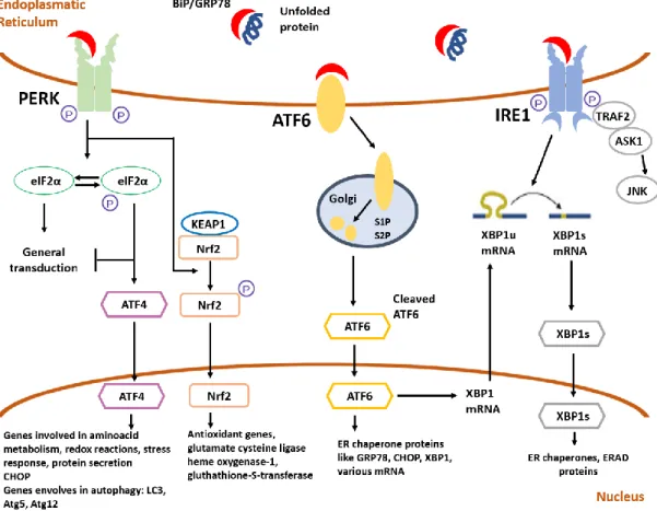
9 | Page (ATF6, α and β) (Hetz & Saxena, 2017) (Figure I.1). Each of these transmembrane proteins has an ER-luminal domain that senses unfolded proteins, a transmembrane domain by which it is targeted to the ER membrane, and a cytosolic domain that transmits signals to the transcriptional or translational apparatus (K. Zhang & Kaufman, 2008). Under normal physiological conditions all three effectors are negatively regulated by the ER chaperone GRP78/BiP (immunoglobulin-heavy-chain-binding protein), which supresses their activity by binding to their luminal ends (Bertolotti et al., 2000). In the presence of misfolded proteins, complex GRP78/BiP releases PERK, IRE1α and ATF6, these become activated and GRP78 binds to misfolded proteins (Hoozemans et al., 2007). These three sensors transmit signals from the ER to the cytoplasm or nucleus and activate the following three pathways: suppression of protein translation to stop the production of more unfolded proteins, induction of genes encoding ER molecular chaperones to ease protein folding, and activation of ERAD to decrease the accumulation of unfolded proteins in the ER (R. J. Lee et al., 2004).
Figure I.1 – Schematic representation of the UPR. Accumulation of unfolded proteins in the ER lumen results in
the binding of GRP78/BiP to those proteins, with consequent release of UPR stress sensors PERK, ATF6 and IRE1α. PERK, after self-phosphorylation, inactivates eIF2α through phosphorylation, inhibiting general proteins synthesis permiting non-canonical translation of ATF4 mRNA. Activated PERK also acts in Nrf2, breaking the complex with KEAP1. Activated ATF6 is processed in the Golgi by site 1 (S1P) and site 2 proteases (S2P), resulting in the transcription of XBP1 mRNA. IRE1α has a domain responsible for processing unspliced XBP1 mRNA, translating it into an active transcription factor. Activation of IRE1α, similar to PERK, results from dimerization and autophosphorylation. Last, IRE1α recruits TRAF2 and ASK1 leading to the activation of JNK. ASK1 – Apoptosis signal-regulating kinase 1; ATF4 – Activating transcription factor-4; ATF6 – Activation transcription factor-6; eIF2α
10 | Page
- Eukaryotic initiation factor 2; BiP/GRP78 - Immunoglobulin-heavy-chain-binding protein; IRE1α - Inositol-requiring protein 1α; JNK - c-JUN NH2-terminal kinases; KEAP1 - Kelch ECH associating protein 1; Nrf2- Nuclear factor erythroid 2-related factor 2; PERK - Protein kinase RNA-like ER kinase; S1P – Site 1 protease; S2P – Site 2 protease; TRAF2 - tumor necrosis factor-α receptor-associated factor 2; XBP1 - X-box-binding protein 1. Adapted
from (Deegan et al., 2013).
2.1.1. PERK/EIF2AK3 signalling
PERK is the major protein responsible for lessening of mRNA translation under ER stress, preventing the influx of newly synthesized proteins into the stressed ER compartment. It is a type I ER transmembrane protein with serine/threonine kinase activity, with its N-terminus in the ER lumen, involved in the regulation of its dimerization and interaction with BiP, while the C-terminus is cytosolic and harbours its autophosphorylation sites and the kinase domain (Deegan et al., 2013; Song et al., 2017). When sensing ER stress, the cytosolic kinase domain of PERK oligomerizes and is activated by
trans-autophosphorylation. The activated cytosolic kinase domain of PERK then recruits and
phosphorylates the α subunit of eukaryotic initiation factor 2 (eIF2α), inactivating it (P. Wang et al., 2016). The inactivation of eIF2α results in a decrease of protein load and a translation attenuation of mRNAs with 5’ cap which usually encode for proteins involved in cell growth and proliferation and also results in non-canonical translation of ATF4 mRNA via an open reading frame in its 5’-untranslated region that is bypassed only when eIF2α is inactivated (Deegan et al., 2013). ATF4 mRNA encodes for a cAMP response element binding transcription factor which activates genes important for amino acid metabolism, redox balance, protein folding, autophagy and apoptosis (Deegan et al., 2013). The role of ATF4 in apoptosis is associated with its cooperation with C/EBP-homologous protein (CHOP/GADD153), that is reported to downregulate B cell lymphoma 2 (Bcl-2) and up-regulate transcription of proteins that lead to mitochondrial outer-membrane permeabilization and initiation of the intrinsic apoptotic cascade (H. Kim et al., 2009; McCullough et al., 2001).
PERK also phosphorylates nuclear factor erythroid 2-related factor 2 (Nrf2), causing its nuclear translocation after release from complex with Kelch ECH associating protein 1 (KEAP1). Once activated, Nrf2 binds the antioxidant response element (ARE), located in the promoter or enhancer regions of antioxidant and cytoprotective genes (M. Zhang et al., 2013). Nrf2 is involved in free radical scavenging, detoxification of xenobiotics and maintenance of redox potential (Chan & Kwong, 2000). Known targets of Nrf2 include glutathione S-transferase (GST) isoforms that will be described in section I-4.
2.1.2. IRE1
α/ERN1 signalling
The IRE1 branch is the most conserved and sole branch of the UPR in lower eukaryotes (Mori, 2009). IRE1 is also a type I ER transmembrane protein containing a serine/threonine kinase domain and a site-specific endoribonuclease (RNase) that, in response to ER stress, is autophosphorylated. IRE1 is divided in two isomers in humans: IRE1α that is ubiquitously express, and IRE1β, which expression is restricted to epithelial cells of the intestine and the lungs (Deegan et al., 2013). After
11 | Page initiation of the signalling cascade it has been observed a self-assembly of the cytosolic region followed by IRE1α oligomerization, leading to its RNase activation. It was also described autophosphorylation at serine 724 and ADP binding (Hocking et al., 2010; Korennykh et al., 2009).
The cytoplasmic part of IRE1α forms a complex with tumor necrosis factor-α (TNF-α) receptor-associated factor 2 (TRAF2) and apoptosis signal-regulating kinase-1 (ASK1), resulting in an activation of c-JUN NH2-terminal kinases (JNK), promoting autophagy or apoptosis (Ron & Hubbard, 2008). The
signal of this branch of the UPR is transmitted through excision of a 26-base nucleotide intron from X-box-binding protein 1 (XBP1) mRNA, which is then ligated by an uncharacterized RNA ligase and translated to produce XBP1s, that has been reported to be important for the activation of unfolded protein response element, controlling the expression of the ERAD, thus helping to degrade unfolded proteins in the ER (Korennykh et al., 2009; Yamamoto et al., 2004). The ERAD pathway is constituted by different components including chaperones, protein transporters, and ubiquitin-related enzymes that sense, deliver, and retro translocate misfolded proteins to the cytoplasm for proteasome mediated degradation (Vembar & Brodsky, 2008).
Overall, IRE1α has been suggested to have a role in development, metabolism, immunity, inflammation, and neurodegeneration (Kaufman & Cao, 2010). In addition, IRE1 can selectively degrade mRNAs encoding for proteins that are predicted to be difficult to fold and micro-RNAs (Hetz, 2012).
2.1.3. ATF6 signalling
ATF6 is a type II transmembrane receptor and a member of the leucine zipper protein family that is synthesized as an ER membrane-tethered precursor, with its C terminal domain located in the ER lumen and its N-terminal DNA binding domain facing the cytosol (Haze et al., 1999). There are two isoforms of ATF6 – α and β – that are ubiquitously expressed in the ER. Upon ER stress, BiP dissociates from ATF6, unravelling its two Golgi localization signals, allowing ATF6 to interact with the protein trafficking complex COPIII, which causes translocation of ATF6 to the Golgi for processing, to become active (Shen et al., 2002). The 90-kDA protein is cleaved by site 1 protease (S1P) and site 2 protease (S2P), originating a 50-kDa cleaved N-terminal cytosolic domain that translocates to the nucleus where it acts as a transcriptional factor, activating target genes such as GRP78/BiP, CHOP and XBP1 (Adachi et al., 2008). Then ATF6 indirectly regulates autophagy or apoptosis via XBP1 and CHOP. It has also been reported that ATF6 regulates an array of miRNAs to alleviate ER stress (Belmont et al., 2012). Moreover, it is thought that the ATF6 and IRE1α pathways merge through the regulation of XBP1 activity: ATF6 increases the amount of XBP1 mRNA whereas IRE1α removes the 26-nucleotide intron, increasing XBP1 activation potential (K. Lee et al., 2002).
12 | Page
2.2.
ER stress and UPR in Parkinson’s Disease
A lot of neurodegenerative disorders share a common neuropathology associated with the accumulation of abnormal protein aggregates or inclusions in the brain containing specific misfolded proteins. These diseases include PD, amyotrophic lateral sclerosis, Alzheimer’s disease, Huntington’s disease, and many others (Selkoe, 2003; Taylor et al., 2002). The presence of aggregates of misfolded proteins indicates that protein quality control mechanisms have been compromised or are incompetent to restore protein homeostasis and provides potential targets for therapeutic intervention (Hoozemans & Scheper, 2012).
Accumulating evidence shows an important role of UPR and ER stress in PD. The involvement of the UPR in PD has been shown in cellular and in vivo models using 6-hydroxydopamine (6-OHDA), MPTP and rotenone (Hoozemans et al., 2007). Generation of ROS by these toxins lead to a rapid accumulation of oxidized proteins that can activate the UPR (Blesa & Przedborski, 2014). Presence of ER stress in human tissue derived from PD patients has been reported, showing the UPR as an early event of the disease (Hoozemans & Scheper, 2012). Previous studies show the activation of PERK-eIF2α pathway of the UPR in dopaminergic neurons in the substantia nigra of PD patients, and the neurons presenting activated PERK were positive for αSyn inclusions (Hoozemans et al., 2007). Studies also showed that XBP1 deletion in DA neurons produce different outcomes, related with the age of the animals and reflecting compensatory changes in UPR-related factors. During development, the lack of XBP1 induced mild ER stress with protection of DA neurons against neurotoxicity of 6-OHDA. Down-regulation of XBP1 in adult mice nigral dopaminergic neurons triggered chronic ER stress, with CHOP induction, which caused spontaneous degeneration (Valdes et al., 2014).
Recent evidence from both toxicological and genetic models of PD indicates that activation of the UPR has a beneficial effect on the survival of dopaminergic neurons. The loss of DA neurons and the accumulation of ubiquitin-positive inclusions induced by MPTP are enhanced in ATF6α-deficient animals, showing the importance of this stress sensor as it controls the levels of BiP, ERAD components and promotes astrogial activation under resting conditions in DA neurons (Egawa et al., 2011; Hashida et al., 2012). Similarly, deletion of the gene encoding for CHOP protects DA neurons against exposure to 6-OHDA and MPTP in different experimental settings (Silva et al., 2005).
Given the dual role of the UPR in maintaining cell viability and the engagement of cell death, it is predicted that low levels of ER stress during the early stages of PD may actually protect dopaminergic neurons against proteostasis defects. Results showed that low concentrations of tunicamycin (Tun), an ER stress inducing agent, increased neuronal surviving in genetical and pharmacological models of the disease, since Tun triggered a preconditioning effect in which sub lethal levels of ER stress selectively engaged adaptive UPR signalling events involving the expression of XBP1s in the brain but not the proapoptotic factor CHOP (Mercado et al., 2016).
Despite these results, characterization of ER markers is still very poor and the precise mechanisms of interplay between oxidative stress and ER stress in dopaminergic neurons have been sparsely described. The mechanisms leading to ER stress in PD and the impact of the UPR on the degeneration cascade in the disease are just starting to be uncovered (Mercado et al., 2013).
13 | Page
3. The autophagy machinery
Autophagy is now considered a major survival mechanism of the cell, being involved in the removal of misfolded proteins and turnover of organelles for cellular homeostasis during starvation by recycling of cytosolic components. The autophagic flux comprises the fusion of autophagosomes with late endosomes to form amphisomes which subsequently fuse with lysosomes forming autolysosomes for degrading its load (Ravikumar et al., 2010). Basal levels of autophagy are very low but, in the presence of stress or extracellular cues, an efficient mechanism of autophagy is crucial to regulate the turnover of long-lived proteins and getting rid of damaged structures (He & Klionsky, 2009). Regulation of autophagy can be done by the mTOR pathway, the classical regulation which negatively controls this process, or by mTOR independent pathways, like the inositol signalling pathway (Sarkar, 2013). Here, we will focus of the mTOR dependent signalling pathways and diverse upstream and downstream signals impinging on it.
3.1.
mTOR signalling pathways
The mammalian target of rapamycin is a serine threonine kinase that belongs to the phosphoinositide 3-kinase (PI3K)-related kinase family that is involved in cell metabolism, growth, proliferation and survival and is conserved throughout evolution, nucleating at least two distinct multi protein complexes, mTOR complex 1 (mTORC1) and mTOR complex 2 (Laplante & Sabatini, 2009). Studies have shown that mTORC1 inhibition increases autophagy, whereas stimulation of mTORC1 reduces this process (Codogno & Meijer, 2005). mTORC1 is a complex formed by five proteins, being the catalytic subunit regulatory-associated protein of mTOR (Raptor) one of them. Previous studies shown that Raptor binds directly to mTOR, functioning as a scaffold protein that regulates complex assembly in addition to substrate recognition, determining downstream signalling of mTORC1. Therefore, in order to inhibit autophagy, mTOR needs to be linked to Raptor (L. Wang et al., 2009). Importantly, mTORC1 can be phosphorylated in S2488 by one of the downstream targets of mTOR, p70S6 kinase, an important reaction for the correct signalling of mTORC1 (Chiang & Abraham, 2005).
In conditions of low ATP/ADP ratio, AMP-activated protein kinase (AMPK), a master-sensor of intracellular energy status, is activated through phosphorylation in residue T172 and directly phosphorylates Raptor at residues S792 and S722, inhibiting mTOR (L. Wang et al., 2009). Also, the activated AMPK inhibits mTORC1 through phosphorylation and activation of tuberous sclerosis complex 2 (TSC2), activating its GAP activity towards Rheb, to inhibit mTORC1 signalling. Thus, there are two separate pathways that transmit AMPK signalling to mTORC1 (Jung et al., 2010).
The initiation, elongation, maturation and fusion states of autophagy’s pathway are orchestrated by several autophagy-related (Atg) proteins. Atg13 is involved in the biogenesis of autophagosomes, namely through binding to UNC-51-like kinase (ULK1) and FIP200, leading to the formation of a ULK1– Atg13–FIP200 stable complex that signals to the autophagic machinery downstream of mTORC1 (Sarkar, 2013). Under physiological conditions, mTOR inhibits Atg13 and ULK1 through phosphorylation. Under stress conditions, mTOR dissociates from mTORC1, resulting in the inhibition of mTORC1-mediated phosphorylation of Atg13 and ULK1. This leads to dephosphorylation dependent
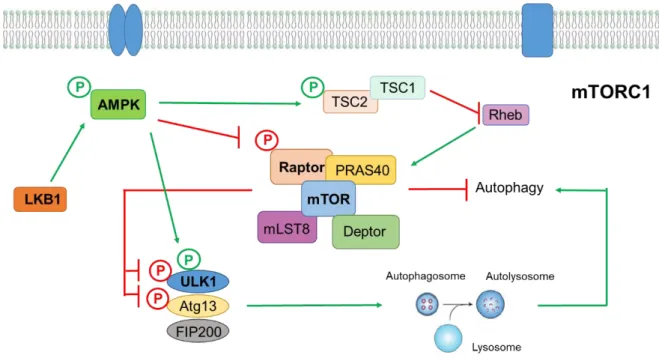
14 | Page activation of ULK1 and ULK1-mediated phosphorylation of Atg13, FIP200 and ULK1 itself, which regulates autophagosomes synthesis and triggers autophagy (Laplante & Sabatini, 2009). Besides phosphorylating Raptor and inhibiting mTORC1 activation, activated AMPK is also able to decrease phosphorylation levels of ULK1 in residue S757 (via inactivation of mTOR), therefore promoting the phosphorylation of ULK1 in other sites like S555 and S777, although there is not much consensus about the residues of ULK1 phosphorylated by AMPK (J. Kim et al., 2011). A simplified scheme of mTORC1 signalling pathway is represented in figure I.2.
Two ubiquitin-like conjugation systems function in the initiation of autophagy. The first one involves several Atg as the second one involves the conjugation of microtubule associated protein 1 LC3 (light chain 3) to phosphatidylethanolamine. LC3 undergoes post-translational modifications originating the cytosolic LC3-I form, that conjugated with phosphatidylethanolamine generate the autophagosome associated LC3-II form. Therefore, the levels of LC3-II correlate with the steady-state number of autophagosomes (Sarkar, 2013). The polyubiquitin-binding protein p62/SQSTM1 acts as a scaffold to facilitate aggregation of ubiquitylated mutant proteins and is also a specific autophagy substrate that targets the mutant proteins for autophagic degradation, anchoring substrates of autophagy to LC3I (Sarkar, 2013).
Figure I.2 - Simplified schematic representation of mTORC1 autophagy signalling pathway. Diverse signals
like amino acids, growth factors, energy status and stressors activate mTORC1, which negatively regulates autophagy. mTORC1 complex is formed by five proteins that interact with each other – Raptor, mTOR, PRAS40, mLST8 and Deptor. To inhibit autophagy, mTOR needs to be linked to Raptor, that regulates complex assembly. Even more, mTOR inhibits ULK1 and Atg13 through phosphorylation, preventing formation of autophagosomes. In conditions of low energy levels, LKB1 activates AMPK by phosphorylation and AMPK can inhibit mTORC1 in two ways – it phosphorylates and activates TSC2, that through its GTPase-activating protein activity, inactivates the small GTPase Rheb, that is necessary to activate mTORC1 complex; and inactivates Raptor through phosphorylation. Furthermore, AMPK can phosphorylate ULK1, activating it, therefore promoting autophagy. AMPK - AMP-activated protein kinase; Atg13 – Autophagy related gene 13; FIP200 – focal adhesion kinase