Edited by: Alessandro Tozzi, University of Perugia, Italy Reviewed by: Gunnar Keppler Gouras, Lund University, Sweden Davide Tampellini, Institut National de la Santé et de la Recherche Médicale (INSERM), France *Correspondence: Cláudia Guimas Almeida claudia.almeida@nms.unl.pt Specialty section: This article was submitted to Cellular Neuropathology, a section of the journal Frontiers in Cellular Neuroscience
Received: 02 February 2020 Accepted: 12 March 2020 Published: 17 April 2020 Citation: Perdigão C, Barata MA, Araújo MN, Mirfakhar FS, Castanheira J and Guimas Almeida C (2020) Intracellular Trafficking Mechanisms of Synaptic Dysfunction in Alzheimer’s Disease. Front. Cell. Neurosci. 14:72. doi: 10.3389/fncel.2020.00072
Intracellular Trafficking Mechanisms
of Synaptic Dysfunction in
Alzheimer’s Disease
Catarina Perdigão
,
Mariana A. Barata
,
Margarida N. Araújo
,
Farzaneh S. Mirfakhar
,
Jorge Castanheira
and
Cláudia Guimas Almeida*
Laboratory Neuronal Trafficking in Aging, CEDOC Chronic Diseases Research Center, NOVA Medical School, Universidade NOVA de Lisboa, Lisbon, Portugal
Alzheimer’s disease (AD) is the most common neurodegenerative disease characterized
by progressive memory loss. Although AD neuropathological hallmarks are extracellular
amyloid plaques and intracellular tau tangles, the best correlate of disease progression
is synapse loss. What causes synapse loss has been the focus of several researchers
in the AD field. Synapses become dysfunctional before plaques and tangles form.
Studies based on early-onset familial AD (eFAD) models have supported that synaptic
transmission is depressed by
β-amyloid (Aβ) triggered mechanisms. Since eFAD is
rare, affecting only 1% of patients, research has shifted to the study of the most
common late-onset AD (LOAD). Intracellular trafficking has emerged as one of the
pathways of LOAD genes. Few studies have assessed the impact of trafficking LOAD
genes on synapse dysfunction. Since endocytic traffic is essential for synaptic function,
we reviewed A
β-dependent and independent mechanisms of the earliest synaptic
dysfunction in AD. We have focused on the role of intraneuronal and secreted A
β
oligomers, highlighting the dysfunction of endocytic trafficking as an Aβ-dependent
mechanism of synapse dysfunction in AD. Here, we reviewed the LOAD trafficking genes
APOE4, ABCA7, BIN1, CD2AP, PICALM, EPH1A, and SORL1, for which there is a
synaptic link. We conclude that in eFAD and LOAD, the earliest synaptic dysfunctions
are characterized by disruptions of the presynaptic vesicle exo- and endocytosis and
of postsynaptic glutamate receptor endocytosis. While in eFAD synapse dysfunction
seems to be triggered by A
β, in LOAD, there might be a direct synaptic disruption by
LOAD trafficking genes. To identify promising therapeutic targets and biomarkers of the
earliest synaptic dysfunction in AD, it will be necessary to join efforts in further dissecting
the mechanisms used by A
β and by LOAD genes to disrupt synapses.
Keywords: late-onset Alzheimer’s disease, synapses, endocytosis,β-amyloid, APOE4, PICALM, BIN1, CD2AP
INTRODUCTION
Alzheimer’s disease (AD), the most common neurodegenerative disease, affecting 1 in 10 people
over 65 years old, remains without adequate treatment. By the age of 85, one in three people
develops the disease (
Alzheimer’s Association, 2019
). Functionally, a progressive loss of memory
and cognitive impairment leads to a loss of autonomy and independance. Neuropathologically,
AD is characterized by two hallmark proteinaceous aggregates,
extracellular amyloid plaques and intracellular neurofibrillary
tangles. The AD brain volume is dramatically reduced,
mainly due to extensive cortical neuronal death. These are
hallmark phenotypes of a disease in which pathological
mechanisms can start 30 years before diagnosis. Importantly,
the synapse loss is the best pathological correlate of cognitive
impairment in AD, which can exceed and precede the existing
neuronal cell death (
Tampellini and Gouras, 2010
). Among
the earliest disease phenotypes are intracellular
β-amyloid (Aβ)
accumulation and synapse dysfunction. Synapse dysfunction
can remain silent, at least initially, due to the reserve brain
capacity to compensate for the affected neurons. Cognitive
impairment likely manifests when the pathology spreads
throughout the brain. Thus, it has become apparent that the
next step for AD research is to focus on identifying novel
therapeutic strategies targeted at the earliest mechanisms of
synaptic dysfunction.
AD has two forms (
Katzman, 1976
), the familial
early-onset (eFAD) and sporadic late-early-onset (LOAD), depending
on if it occurs before or after the age of 65. eFAD is the
result of dominantly inherited mutations in the amyloid
precursor
protein
(APP)
and
presenilin
(PSEN1
and
PSEN2;
Selkoe and Hardy, 2016
). These mutations lead
to increased production of Aβ, the main component of
amyloid plaques, or a higher ratio of longer Aβ peptides
(Aβ42/43) to Aβ40. Aβ42, while accounting for only 10%
of the total Aβ produced, is more hydrophobic and has a
higher capacity of aggregating into oligomers. Currently,
evidence strongly supports that soluble Aβ oligomers are
more toxic than insoluble fibrils (plaques). The production of
Aβ42 occurs intracellularly, where it accumulates before being
secreted and deposited into amyloid plaques extracellularly
(
Gouras et al., 2005
). Aβ oligomerization, particularly
of Aβ42 peptides, begins intracellularly (
Tampellini and
Gouras, 2010
). Intraneuronal Aβ correlates with cognitive
dysfunction better than amyloid plaques (
Billings et al., 2005;
Takahashi et al., 2017
). Aβ42 oligomerization precedes tau
aggregation (
Billings et al., 2005; Bilousova et al., 2016
).
Thus, we did not include tau-dependent synaptic dysfunction
in this review; nevertheless, since tau aggregation is a
determinant for the propagation and progression of the
pathology, we recommend a recent review on the topic
(
Tracy and Gan, 2018
).
In contrast with eFAD, LOAD is likely multifactorial,
caused by a combination of aging, lifestyle, and genetic
factors. Given the prediction for heritability of AD to be
between 58 and 79%, geneticists have been looking for gene
variants in LOAD patients (
Gatz et al., 2006
). Apolipoprotein
E-ε4 was the first genetic risk factor identified and is the
most significant one (
Strittmatter et al., 1993
). However,
it does not account for all genetic susceptibility (
Stocker
et al., 2018
). Thus, genome-wide association studies (GWAS)
conducted since 2007 have identified several additional genetic
risk factors associated with LOAD (
Grupe et al., 2007; Harold
et al., 2009; Hollingworth et al., 2011; Naj et al., 2014
).
A recent meta-analysis of GWAS expanded the initial ‘‘Top
10’’ genes (
Lambert et al., 2013
1) to 37 LOAD putative
risk factors (
Jansen et al., 2019; Kunkle et al., 2019
). Now
it is crucial to determine how these genes contribute to
AD functionally. We, along with other resesarchers, have
performed gene ontology (GO) analysis and LOAD risk factors
group in three main pathways: endocytic trafficking, immune
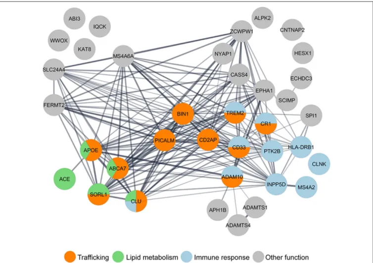
response, and lipid metabolism (Figure 1). Interestingly, some
genes, such as ABCA7 and TREM2, are grouped in more
than one pathway. Indeed, it is essential to keep in mind
that these biological pathways intersect; for example, lipid
metabolism influences trafficking at many levels (
Huijbregts
et al., 2000
) and trafficking in microglia influences the
immune response (
McQuade and Blurton-Jones, 2019
). Most
attention has been given to how the loss of function of
these genes impacts Aβ, generation (reviewed in
Guimas
Almeida et al., 2018
), aggregation (
Zhang et al., 2018
), and
clearance (
Van Acker et al., 2019
). Recently reviewed data
indicates that LOAD microglia risk genes may impact synapses
(
Rajendran and Paolicelli, 2018
).
Here, we will review the importance of neuronal endocytic
trafficking for synapses (
Barry and Ziff, 2002
), how Aβ can
have a physiological synaptic role, and how intraneuronal
and secreted Aβ oligomers affect synapses, highlighting
Aβ-dependent consequences to synaptic endocytic trafficking.
Importantly, we will highlight recent data on how LOAD risk
genes related to trafficking may impact synapse dysfunction,
in particular, the ones related to endocytic protein trafficking
(BIN1, SORL1, PICALM, CD2AP), to lipid trafficking (APOE
and ABCA7), and synapses (EPH1A).
SYNAPTIC ENDOCYTIC TRAFFICKING
Presynaptic Trafficking
Cycles of synaptic vesicles (SV) exocytosis and endocytosis at
the presynaptic active zone membrane mediate neurotransmitter
release. This presynaptic trafficking begins with the arrival of
an action potential that opens Ca
2+channels, which triggers
exocytosis of readily releasable SV pool (RRP), which comprises
the SVs that are docked and primed for exocytosis. There is also
the SV reserve pool, which is mobilized only during periods of
intense neuronal activity. After exocytosis, there are different
routes of endocytosis to recycle SVs. SV components and newly
added membrane need to be recycled to ensure repeated rounds
of release.
The SV fusion machinery is composed of the SNARE
(soluble N-ethylmaleimide-sensitive factor attachment protein
receptor) complex proteins synaptobrevin/VAMP2, syntaxin-1,
and SNAP-25 and SM proteins (‘‘Sec1/Munc-18-like proteins’’),
which promote the assembly of the fusogenic SNARE complex.
To trigger exocytosis, Ca
2+binds to synaptotagmin-1, the
SV calcium sensor protein that, by interacting with the
SNARE complex, modulates its assembly or its coupling to
the plasma membrane, enabling fusion (
Tucker et al., 2004;
Wang et al., 2011; Dittman and Ryan, 2019
). Upon fusion, NSF
promotes the disassembly and recycling of the SNARE complex
FIGURE 1 | Gene ontology (GO) analysis of LOAD risk factors reveals the enrichment of genes in the trafficking pathway. Nearly half of the 37 LOAD putative risk factors identified by genome-wide association studies (GWAS) meta-analysis (Lambert et al., 2013; Jansen et al., 2019; Kunkle et al., 2019), have been functionally grouped by GO analysis in three main pathways: trafficking (orange), immune response (blue), and lipid metabolism (green). GO analysis was performed using the Cytoscape StringApp (Doncheva et al., 2019).
(
Südhof, 2013
). SV recycling follows exocytosis to regulate
the presynaptic plasma membrane area and locally regenerate
SVs (
Kononenko and Haucke, 2015; Azarnia Tehran et al.,
2018; Chanaday et al., 2019
). There are probably several routes
of SV recycling. Here, we focus on endocytic recycling that
starts with endocytosis of SV proteins. When SVs fuse, they
intermix with the plasma membrane (
Fernández-Alfonso et al.,
2006
); therefore, SV recycling requires sorting and reformation.
Considering the high number of SVs, their identity and
uniformity, protein sorting, and SV reformation are significant
tasks for the presynaptic endocytic machinery. Presynaptic
endocytosis can be mediated by clathrin, by bulk endocytosis, or
by ultrafast endocytosis. Upon the formation of the presynaptic
early endosome, SVs need to be accurately reformed, which
requires endocytic adaptors such as the assembly protein
complex 2 (AP-2) to sort cargo into clathrin-coated pits. AP-3
is an alternative adaptor that can sort SV cargo at endosomes
into reforming SVs but independently of clathrin (
Milosevic,
2018
). Other clathrin adaptors are neuronal-specific such as
AP180 and LOAD risk factor CALM (for LOAD risk factors,
see below). These proteins are essential for VAMP2 sorting
by still unclear mechanisms. Other endocytic adaptors are
required to ensure sorting fidelity, such as stonin 2 and synaptic
vesicle 2 (SV2A;
Kononenko and Haucke, 2015; Gordon and
Cousin, 2016
). After the assembly of endocytic vesicles, the BAR
domain proteins, endophilin, amphiphysin, and SNX9/18 form
the vesicle neck and recruit dynamin, which is responsible for
membrane scission. Then, endophilin recruits synaptojanin-1 to
shed the coat off of the released endocytic vesicles (
Kononenko
and Haucke, 2015
).
Postsynaptic Trafficking
In the postsynaptic site, activation of
neurotransmitter-gated ion channels occurs by the presynaptic release of
neurotransmitters. In an excitatory synapse, the principal
neurotransmitter released is glutamate, which binds NMDA
(N-methyl-D-aspartic acid) receptors, kainate receptors, and
AMPA (α-amino-3-hydroxy-5-methyl-4-isoxazole propionic
acid) receptors (
Waites et al., 2005
). Brief periods of high
neuronal excitability activate NMDARs and induce Ca
2+influx, leading to a long-lasting increase in synaptic efficacy.
Repetitive low-frequency stimulation produces long-term
depression (LTD), with a decrease in synaptic strength (
Kessels
and Malinow, 2009
). Plasticity at synapses, i.e., changes in the
onset or magnitude of long-term potentiation (LTP) and LTD,
can be regulated postsynaptically by changing the number,
types, or properties of neurotransmitter receptors (reviewed in
Henley and Wilkinson, 2016
). Regulated trafficking of AMPARs
underlies activity-induced changes in synaptic transmission,
and therefore their abundance at synapses can significantly
strengthen or weaken synaptic transmission (
Malinow and
Malenka, 2002; Bredt and Nicoll, 2003; Newpher and Ehlers,
2008
). AMPAR subunits can have long- (GluA1, GluA2L, and
GluA4) or short-tailed carboxyl-terminals (GluA2, GluA3, and
GluA4S), which impact their trafficking (
Shepherd and Huganir,
2007; Hanley, 2008
). The recycling or functional insertion of
AMPARs into the postsynaptic membrane is dependent on
AMPAR-binding proteins, phosphorylation, and ubiquitination
events (
Derkach et al., 1999, 2007; Malinow and Malenka, 2002;
Schwarz et al., 2010
). The addition of long-tailed AMPARs to the
postsynaptic membrane is correlated with synaptic strengthening
and, therefore, with LTP (
Hayashi et al., 2000; Kakegawa et al.,
2004
), while LTD is associated with the removal of synaptic
long- or short-tailed AMPARs (
Malinow and Malenka, 2002;
Sheng and Hyoung Lee, 2003
).
A
β Physiological Role at Synapses
Although in AD, high Aβ concentration has a toxic effect,
endogenous Aβ concentration has a physiological synaptic
role. Aβ physiological function was first proposed in 2003 by
the Malinow lab and described that blocking Aβ endogenous
production by gamma-secretase inhibition potentiated synaptic
transmission (
Kamenetz et al., 2003
). In contrast, in 2008, the
Arancio lab showed that exogenous picomolar concentrations
of Aβ, monomers and oligomers, increase LTP, while high
nanomolar amounts of Aβ reduce LTP (
Puzzo et al., 2008
). In
agreement, the Slutsky lab showed that increasing endogenous
Aβ, by inhibition of neprilysin-mediated degradation, potentiates
synaptic transmission (
Abramov et al., 2009
). Importantly, SV
recycling was first identified to be endogenously regulated by
Aβ. The Slutsky lab elegantly demonstrated that increasing Aβ
by neprilysin inhibition increases SV recycling, while decreasing
physiological
Aβ levels by anti-Aβ antibody-promoted
degradation, in contrast, decreases SV recycling (
Abramov
et al., 2009
). More recent data indicate that exogenous picomolar
preparations of oligomeric Aβ42 can augment neurotransmitter
release and the length of the postsynaptic density, resulting
in a late-LTP (
Gulisano et al., 2019
). The mechanisms of how
endogenous Aβ regulates synaptic vesicle recycling and PSD
recruitment for modulating synaptic transmission and plasticity
are unclear. Some evidence indicates that Aβ can bind to
alpha7-nicotinic acetylcholine receptors (
Puzzo et al., 2008; Gulisano
et al., 2019
) to induce presynaptic calcium entry. Overall, the
evidence points to an Aβ physiological role, but it is still not
clear which form of Aβ is relevant. Does Aβ40 being more
abundant play a different physiological role from Aβ42? Other
products of APP processing, as well as APP full length, also have
synaptic physiological functions that have been recently reviewed
(
Ludewig and Korte, 2016
).
A
β-DIRECT IMPACT ON SYNAPTIC
TRAFFICKING
There are several lines of evidence demonstrating that Aβ
is synaptotoxic. In most experimental conditions, exogenous
preparations of synthetic or brain-derived oligomeric Aβ were
used (reviewed by
Mucke and Selkoe, 2012
), while other
experimental conditions mimicked chronic and time-dependent
Aβ accumulation with overexpression of mutant APP, with only
a few assessing the contribution of intracellular Aβ. Here we
will cover intracellular Aβ-dependent mechanisms focusing on
trafficking dysfunction.
Aβ progressively accumulates within neurons (
Gouras et al.,
2000; Mochizuki et al., 2000
) in the limiting membrane of
late endosomes or MVBs pre- and especially postsynaptically
(
Takahashi et al., 2002; Willén et al., 2017; Yu et al., 2018
). This
intracellular Aβ can oligomerize and disrupt synapses (
Takahashi
et al., 2004; Pickett et al., 2016
). Increased Aβ production,
as a result of overexpression of exogenous APP or
β-CTF in
hippocampal slices, is sufficient to depress synaptic transmission,
since inhibiting beta- or gamma-secretase prevented synaptic
depression (
Kamenetz et al., 2003
).
More mechanistically, increased Aβ42 production, as a
result of overexpression of APP with eFAD Swedish mutation
(APPsw) in primary neurons, is enough to trigger postsynaptic
dysfunction with loss of PSD-95, AMPA and NMDA glutamate
receptors (
Almeida et al., 2005; Snyder et al., 2005
). Loss
of PSD-95 and spines also occurs
in vivo in AD mice and
human AD brain (
Gylys et al., 2004; Pham et al., 2010; Proctor
et al., 2010; Sultana et al., 2010; Baglietto-Vargas et al., 2018
).
Since PSD-95 drives AMPA receptors’ incorporation in the
postsynaptic density, its loss may underlie the synaptic removal
of these receptors (
Ehrlich and Malinow, 2004
). The loss of
AMPA receptors likely causes the reduced AMPA
receptor-mediated currents observed even when APP is overexpressed
(
Ting et al., 2007
). Aβ42 requirement for loss of AMPA
transmission was confirmed when a mutation that inhibits
BACE1 cleavage (APPM596V) blocked synaptic depression
(
Ting et al., 2007
). Indeed, Aβ-dependent synaptic endocytosis
of AMPA receptors is sufficient to account for spine loss and
reduced NMDA synaptic response (
Hsieh et al., 2006
). One
interesting study found that intracellular Aβ oligomerization,
induced by overexpression of APP with the Osaka mutation,
reduced spines
via dysfunction of BDNF, mitochondria, and
endosomes transport (
Umeda et al., 2015
). Intracellular Aβ
also interferes with the BDNF TrkB receptors’ endosomal
sorting for lysosomal degradation, which could disturb synapses
(
Almeida et al., 2006
).
The presynaptic compartment may get affected after the
postsynaptic compartment since the loss of synaptophysin, a
major component of SVs, only occurred after AMPA receptor
synaptic loss (
Almeida et al., 2005
). Indeed, the presynaptic
decrease of synaptophysin and synapsin mark AD synaptic
loss. Interestingly, the presynaptic compartments of APPsw
neurons are enlarged but undergo SV recycling (
Almeida
et al., 2005
). Also, upon sustained neuronal activation of
APPwt neurons, SV recycling is reduced (
Ting et al., 2007
).
The defects in SV endocytosis could be partially due to
dynamin-1 depletion induced by APPsw overexpression in
eFAD mice (
Kelly et al., 2005; Parodi et al., 2010
). The
contribution of intracellular Aβ to SV cycle dysfunction remains
mostly unstudied.
Secreted Aβ can affect synapses extracellularly and by
contributing to intracellular Aβ via endocytosis of extracellular
Aβ (
Lai and McLaurin, 2010
). Endocytosed Aβ oligomers
could translocate to synapses where interaction with the SV
marker synaptophysin can be detected (
Russell et al., 2012
).
Extracellular Aβ can also form a complex with secreted
ApoE. This complex can bind to low-density lipoprotein
receptor (LDLR) and LRP1, internalize, and accumulate into
endosomes within synapses (
Bilousova et al., 2019
). It is not
clear how endosomal Aβ can interact with cytosolic proteins.
An answer may be provided by a study that showed that
endocytosed Aβ42 could accumulate in endosomes, increasing
their membrane permeability and facilitating Aβ cytosolic
accumulation and neuronal toxicity (
Yang et al., 1998
).
Moreover, a recent study demonstrated that Aβ oligomers
in vitro could inhibit the SNARE fusion complex assembly
by direct binding to syntaxin-1a (
Yang et al., 2015
). Besides,
in vitro, Aβ seems to be able to block synaptophysin complex
formation with VAMP2 with relevance for SV exocytosis (
Russell
et al., 2012
). Thus, when Aβ oligomers rupture endosomes
and reach the cytosol, Aβ could directly inhibit the SV
cycle. This mechanism is similar to the one used by
alpha-synuclein in Parkinson’s disease, where cytosolic alpha-alpha-synuclein
oligomerization inhibits SNARE-mediated vesicle fusion in
dopaminergic neurons (
Garcia-Reitböck et al., 2010; DeWitt and
Rhoades, 2013
).
Secreted Aβ, or exogenous synthetic Aβ, can also bind
extracellularly to excitatory synapses, namely postsynaptically
(
Wang et al., 2017; Willén et al., 2017
), increasing calcium influx,
which triggers AMPA and NMDA receptor phosphorylation
leading to their removal from synapses by endocytosis (
Snyder
et al., 2005; Liu et al., 2010; Miñano-Molina et al., 2011; Sinnen
et al., 2016; Baglietto-Vargas et al., 2018
). The mechanisms
of disruption of glutamate receptor trafficking induced by
extracellular Aβ have been extensively reviewed (
Guntupalli
et al., 2016
). Other synaptic receptors may also be affected
by exposure to Aβ oligomers, such as EphB2, the degradation
of which increases upon Aβ binding (
Cissé et al., 2011
).
EphB2 regulates NMDAR expression and currents, for long-term
plasticity in the dentate gyrus, consequently regulating memory
(
Cissé et al., 2011
). Extracellular Aβ may also contribute to
spine loss
via F-actin disassembly (
Kommaddi et al., 2018
). The
mechanisms of SV cycle disruption by extracellular Aβ that seem
to involve calcium influx have been recently reviewed (
Marsh and
Alifragis, 2018
).
Overall, extracellular Aβ has the same targets of intracellular
Aβ. While the acute treatment with Aβ oligomers promotes
synaptic receptor dysfunction, chronic treatment results in
abnormal spine morphology, with the induction of long thin
spines that, ultimately, cause a significant decrease in spine
density (
Lacor et al., 2007
). Alternatively, evidence indicates that
extracellular Aβ depends on intracellular Aβ for synaptotoxicity
as follows: Aβ binds to APP with high affinity (
Lorenzo et al.,
2000; Lu et al., 2003; Lacor et al., 2004; Fogel et al., 2014; Wang
et al., 2017
); extracellular Aβ can promote its processing and
intracellular Aβ accumulation (
Tampellini et al., 2009
); APP
enriched at synapses can be the synaptic receptor for Aβ (
Laßek
et al., 2013; Del Prete et al., 2014; Fanutza et al., 2015; Montagna
et al., 2017
); APP KO neurons are resistant to exogenous Aβ
toxicity (
Wang et al., 2017
); and, extracellular synthetic Aβ
no longer reduces PSD-95 when Aβ production is inhibited
(
Tampellini et al., 2009
).
In vivo in normal rodent hippocampus, acute exposure to
Aβ dimers extracted from the AD brain reduced dendritic spine
density and potently inhibited LTP and enhanced LTD (
Shankar
et al., 2008
). Interestingly while LTD required mGluR5, spine loss
required NMDA receptors (
Shankar et al., 2008
). In a 3xTg-AD
mouse model, the chronic accumulation of Aβ correlated
with the impaired synaptic insertion of GluA1-containing
AMPARs during chemical LTP stimulation (
Baglietto-Vargas
et al., 2018
). However, in some AD models (APP/PS1), a
postsynaptic reduction of AMPA receptors or spine loss was not
a significant phenotype; instead, the presynaptic SV release was
affected (
He et al., 2019
). A reduction of the phosphoinositol
PtdIns(4, 5)P2 (PIP2) triggered by endogenous or exogenous Aβ
oligomers could account for the presynaptic deficits (
Berman
et al., 2008; He et al., 2019
). PIP2 is a plasma membrane
phosphoinositide that binds to synaptotagmin-1 on the SV to
allow for its docking and fusion (
Lee et al., 2010
). Selective
inhibition of Aβ-induced PIP2 hydrolysis via presynaptic
mGluR5 could rescue the presynaptic release of glutamate and
restore synaptic transmission in APP/PS1 mice (
He et al., 2019;
Kunkle et al., 2019
).
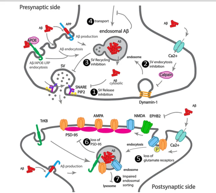
We summarized how Aβ accumulates at the synapse, as well
as the earliest Aβ-dependent mechanisms of synapse dysfunction
(Figure 2).
LOAD GENETIC RISK-DRIVEN
TRAFFICKING DEFECTS IMPACT
SYNAPSES INDEPENDENTLY OF A
β
Among the 11 LOAD risk genes involved in intracellular
trafficking, we will review the evidence that supports a direct
synaptic (dys)function for APOE4, ABCA7, BIN1, CD2AP,
PICALM, SORL1, and EPH1A.
APOE4
Apolipoprotein E gene (APOE) has a risk LOAD variant
(APOE4). Two single nucleotide polymorphisms (SNPs) generate
three allelic variants of the human APOE gene,
ε2, ε3, and ε4 that
affect the structure of the ApoE protein, as well as, the binding to
lipids, receptors, and Aβ (
Liu et al., 2013; Yamazaki et al., 2019
).
APOE
ε3 is the most common allele, with a prevalence of 77.9%,
the
ε2 allele is the less prevalent (8.4%), and the ε4 allele has a
FIGURE 2 | Scheme illustrating how Aβ impacts synaptic trafficking. Scheme of a dysfunctional early-onset familial AD (eFAD) synapse. At the presynaptic terminal: 1. Synaptic vesicle release inhibition, extracellular Aβ endocytosed, alone or via ApoE-LRP endocytosis can accumulate at endosomes. Intracellular Aβ, produced from APP endocytosis, accumulates at endosomes. Endosomal Aβ can disrupt the endosomal membrane and reach the cytosol, where it can potentially bind to SNARE subunit syntaxin-1a, or indirectly inhibit synaptophysin or PIP2, thus inhibiting synaptic vesicles (SV) fusion and neurotransmitter release. 2. SV endocytosis inhibition, extracellular Aβ increases calcium influx, triggers calcium-dependent calpain to degrade dynamin1, reducing endocytosis. 3. SV recycling inhibition by intracellular Aβ. 4. Transport to synapses inhibition by intracellular Aβ oligomerization. At the postsynaptic terminal: 5. Increased endocytosis of AMPA and NMDA receptors, by intracellular and extracellular Aβ. 6. Loss of PSD-95 by intracellular Aβ. 7. Impaired endosomal sorting of TrkB receptor, by endosomal Aβ.
frequency of 13.7% that increases in LOAD patients (
Farrer et al.,
1997; Michaelson, 2014
).
In the brain, ApoE is the primary lipoprotein of high-density
lipoprotein (HDL), mainly secreted by astrocytes and also
produced by neurons (
Xu et al., 1999; Hauser et al., 2011
).
Secreted ApoE scavenges lipids from the local environment for
cellular delivery. Upon receptor-mediated endocytosis and lipid
unloading, ApoE is released from its receptor at the endosomal
pH and sorted for recycling or degradation (
Hauser et al., 2011
).
ApoE knockdown altered the cholesterol distribution within
synaptic membranes (
Igbavboa et al., 1997
). ApoE mediates
cholesterol transport into neurons, increasing synapse formation
(
Mauch et al., 2001; Liu et al., 2013
). Cholesterol binding
to synaptophysin may enable the correct sorting of SV
constituents at the plasma membrane necessary for SV recycling
(
Thiele et al., 2000
).
ApoE4 differs from ApoE3 by one aminoacid, which is
sufficient to cause ApoE4 misfolding and abnormal trafficking.
Reduced ApoE4 recycling and endosomal accumulation
may lead to increased cholesterol intracellular accumulation
(
Heeren et al., 2004
), as well as increased ApoE4 lysosomal
degradation, accounting for the reduction of ApoE in the
brain of APOE4 carriers (
Riddell et al., 2008
). ApoE4 is
hypolipidated compared with ApoE3 and ApoE2, the most
lipidated (
Heinsinger et al., 2016
). The APOE state of lipidation
may be more critical than ApoE levels. There is some evidence
that ApoE4 transports cholesterol into and out of neurons
less efficiently (
Michikawa et al., 2000; Rapp et al., 2006
).
Controversy exists since ApoE2 may delay AD development by
reducing brain cholesterol (
Oikawa et al., 2014
).
APOE4 knock-in (KI) increased excitatory postsynaptic
currents in human-induced neurons, suggesting enhanced SV
release or synapse density (
Lin et al., 2018
). Importantly,
APOE4 KI and APOE KO mice show reduced neuronal
complexity and impaired synaptic plasticity (
Trommer et al.,
2004
). Importantly, in APOE4 KI mice, LTP is diminished
and LTP maintenance unimpaired (
Trommer et al., 2004;
Korwek et al., 2009
).
Morphologically, APOE KO and APOE4 KI present less spine
density and shorter spines (
Wang et al., 2005; Dumanis et al.,
2009; Rodriguez et al., 2013
). Reduced spines could be due to
both a delay in spine formation and increased spine elimination,
as observed in APOE4 KI neurons
in vitro (
Nwabuisi-Heath
et al., 2014
). APOE4 KI show increased calcineurin activity
(
Neustadtl et al., 2017
), a phosphatase that can dephosphorylate
AMPA receptors, promoting their synaptic removal and
increasing LTD (
Zhou et al., 2004
). Presynaptically, APOE4 KI
show reduced production of the neurotransmitter glutamate
(
Dumanis et al., 2013
) and glutamate transporter vGlut1
(
Liraz et al., 2013
).
ApoE receptors, members of the low-density lipoprotein
(LDL) receptor family, include ApoE receptor 2 (ApoEr2),
very low-density lipoprotein receptor (VLDLR), LDLR, and
LDLR-related protein 1 (LRP1). ApoE receptors allow for
its endocytosis and are also present at synapses, where they
have specific functions (
May et al., 2004; Bal et al., 2013;
Bilousova et al., 2019
). Presynaptically, ApoE receptors regulate
SV release (
Bal et al., 2013
). Presynaptic activation of Apoer2 and
VLDLR by Reelin, another ligand, leads to a rise in Ca
2+and
SV fusion (
Bal et al., 2013
). The SNARE complex involved,
containing VAMP7 and SNAP-25, was specifically up-regulated
(
Bal et al., 2013
). Interestingly, SNAP-25 levels were higher in
APOE
ε4 carriers CSF compared to non-carriers with
mild-cognitive impairment (MCI;
Wang et al., 2018
). Postsynaptically,
LRP1 interaction with PSD-95 is disrupted upon NMDA
activation to modulate NMDA receptors signaling (
Bacskai
et al., 2000; May et al., 2004
). LRP1 regulates NMDA receptor
function (
May et al., 2004
) and endocytosis, promoting, during
development, the switch from NR2B to NR2A subunits at
synapses (
Maier et al., 2013
). ApoER2 can get trapped in
postsynaptic recycling endosomes upon ApoE4 binding, thus
preventing reelin dependent activation of the NMDA receptor,
decreasing calcium influx and LTP (
Chen et al., 2010; Yong et al.,
2014
). Long-term spatial memory could require LDLR binding to
ApoE4 (
Johnson et al., 2014
).
Interestingly, clusterin (or apolipoprotein J), another putative
LOAD genetic risk factor, accumulates in synapses of human
post-mortem brains of APOE4 AD carriers (
Jackson et al., 2019
).
Research is still needed to address if the detrimental synaptic
effects of ApoE4 are independent of ApoE4 impact on Aβ brain
accumulation. Importantly, genetically correcting APOE4 to
APOE3 in LOAD IPSCs reduced synaptic release and synaptic
density without however rescuing secreted Aβ (
Lin et al.,
2018
). Complementarity may also exist since APOE KO or
APOE4 KI can potentiate LTP inhibition induced by Aβ
oligomers (
Trommer et al., 2005
). Also, APOE4 KI in an
early onset model (5xTg mice) increases amyloid pathology
and enhances age-dependent decline in cognitive function by
down-regulating an NMDA receptor pathway (
Liu et al., 2015
).
ABCA7
ATP-binding cassette transporter A7 (ABCA7) belongs to
the ABC transporter family that transports lipids, including
cholesterol and lipophilic proteins, across membranes. While
some ABCA7 variants increase the risk of developing AD
(
Carrasquillo et al., 2015; Cuyvers et al., 2015; Steinberg et al.,
2015; Allen et al., 2017
), others are protective (
Vasquez et al.,
2013
). In LOAD, it is not clear whether ABCA7 expression
is altered early in the disease; surprisingly, in advanced AD,
ABCA7’s higher expression was associated with advanced
cognitive decline (
Karch et al., 2012
). ABCA7 knockout mice
have altered brain lipid profiles, and when aged, show impaired
spatial memory (
Sakae et al., 2016
) with more pronounced
effects in female mice (
Logge et al., 2012
). ABCA7 knockout
may accelerate amyloid pathology in some eFAD mouse
models (APP/PS1;
Sakae et al., 2016
), but not in others
(J20;
Kim et al., 2013
). Whether or not ABCA7 knockout
impacts cognitive function needs to be studied. Suggestive of an
effect on synaptic function, a recent study found that healthy
ABCA7 carriers show impaired cortical connectivity (
Sinha
et al., 2019
). Interestingly, there might be genetic interactions
between APOE4 with ABCA7 that impact memory (
Chang et al.,
2019
). ABCA7 expression in the brain is highest in neurons
(
Zhang et al., 2014
).
BIN1
Bridging integrator-1 gene (BIN1) encodes for Bin1, a
member of the BAR (Bin–Amphiphysin–Rvsp) superfamily
(
Sakamuro et al., 1996
), homologous to the previously reported
amphiphysin-1 (
Lichte et al., 1992; Leprince et al., 1997
). Bin1,
with highest expression in brain and skeletal muscle (
Sakamuro
et al., 1996; Butler et al., 1997
), undergoes alternative splicing
(
Tsutsui et al., 1997
), originating at least 10 isoforms. The main
isoforms are a longer neuronal-specific isoform with an extra
clathrin binding domain (CLAP), two ubiquitous, and a muscle
isoform (
Ramjaun et al., 1997
). All contain an N-BAR domain,
through which Bin1 confers and senses membrane curvature,
and a C-terminal SH3 domain that mediates Bin1 interaction
with proteins involved in endocytosis, such as amphiphysin-1,
dynamin (
Butler et al., 1997; Leprince et al., 1997; Ramjaun et al.,
1997; Wigge et al., 1997
) and endophilin (
Micheva et al., 1997
).
The brain expresses mainly the Bin1 neuronal-specific isoform
and at least one ubiquitous isoform (
Prokic et al., 2014
). Initially,
Bin1 was detected in brain synaptosomes and was shown to
localize to axon initial segments and nodes of Ranvier (
Butler
et al., 1997; Wigge et al., 1997; Wigge and McMahon, 1998
).
Bin1 can dimerize, through its BAR domain, with itself or with
amphiphysin-1 (
Wigge et al., 1997
). Overexpressed Bin1 inhibits
clathrin-mediated endocytosis (
Wigge et al., 1997
). In contrast,
BIN1 knockdown reduced the recycling but not endocytosis of
the transferrin receptor in fibroblasts and HeLa cells (
Muller
et al., 2003; Pant et al., 2009
). In neurons, Bin1 polarizes to axons,
where its knockdown reduced BACE1 recycling and degradation
but not its endocytosis (
Miyagawa et al., 2016; Ubelmann
et al., 2017
). Mechanistically, Bin1 contributes to the scission
of recycling carriers from early endosomes (
Ubelmann et al.,
2017
). Neuronal BIN1 knockdown enlarges early endosomes by
cargo accumulation or over-activation of Rab5, a small GTPase
and regulator of early endosome formation, by interaction
with RIN3, a Rab5 GEF and a putative risk factor for LOAD
(
Calafate et al., 2016
).
The expression of Bin1 in the AD brain remains controversial.
In AD human brains,
BIN1 transcription increases (
Chapuis
et al., 2013
). In contrast, BIN1 protein decreases in sporadic AD
human brains (
Glennon et al., 2013
). In eFAD models, there is
BIN1 accumulation adjacent to amyloid plaques (
De Rossi et al.,
2019
). Subsequently, a separate analysis of two Bin1 isoforms,
neuronal and ubiquitous, indicated reduced neuronal
BIN1 but
increased ubiquitous
BIN1 in AD human brains (
Holler et al.,
2014; De Rossi et al., 2016
).
To investigate the impact of Bin1 overexpression, the Lambert
lab generated a human BIN1 transgenic mouse (hBin1-Tg;
Daudin et al., 2018
). The hBin1-Tg mice showed an early
phenotype of neurodegeneration starting at 3 months, with
structural impairment of fiber pathways and structural and
functional changes in entorhinal cortex-dentate gyrus (EC-DG)
pathway, the earliest brain region impacted in LOAD (
Khan
et al., 2014; Small, 2014
). BIN1 overexpression seems to affect
hippocampal physiology, even in the absence of amyloid plaques
(
Daudin et al., 2018
). These data show that the BIN1 risk factor
can be sufficient to generate phenotypic changes before the
accumulation of Aβ plaques or tau aggregates.
The di Camilli lab identified a synaptic role for Bin1 (
Di Paolo
et al., 2002
). They found Bin1 depleted in the amphiphysin-1
knockout mice. Amphiphysin-1 knockout and
Bin1 knockdown
mice synaptosomes exhibited SV recycling defects, such as slower
recycling and a smaller pool of SVs and irreversible seizures
(
Di Paolo et al., 2002
). Unfortunately, BIN1 knockout mice
die after birth due to muscle defects (
Muller et al., 2003
). In
neurons, Bin1 enriched in axons (
Ubelmann et al., 2017
) has a
presynaptic localization (
Di Paolo et al., 2002
) and
in vitro binds
to SV glycoprotein 2A (SV2A;
Yao et al., 2010
), suggesting a
presynaptic role for Bin1.
Additionally,
a
proteomic
analysis
of
postsynaptic
compartments identified Bin1, suggesting a postsynaptic
role for Bin1 (
Bayés et al., 2011
). Subsequently, both an increase
and a decrease in Bin1 levels led to a small impact on spine
morphology (
Schürmann et al., 2019
). BIN1 knockdown resulted
in a reduction of the mean amplitude of AMPAR currents and
reduced surface expression of GluA1 in spines and the dendritic
shaft (
Schürmann et al., 2019
). Given that Bin1 is a regulator of
endocytic recycling (
Pant et al., 2009; Ubelmann et al., 2017
), it
could be necessary for GluA1 synaptic insertion,
via recycling
carriers from postsynaptic endosomes (
Oku and Huganir, 2013
).
CD2AP
CD2AP, CD2-associated protein, belongs to the CIN85/CD2AP
protein family, contains three SH3 domains in the N-terminal,
a proline-rich domain, and a coil-coiled domain in the
C-terminal. CD2AP enriched in podocytes functions in kidney
glomeruli (
Shih et al., 1999; Dikic, 2002
). In podocytes foot
processes, in T-cell contacts, at the lamellipodia of migrating
cells, CD2AP is at the interface between endocytic trafficking and
actin cytoskeleton (
Dustin et al., 1998; Li et al., 2000; Welsch
et al., 2005; Zhao et al., 2013
).
It is still unknown whether CD2AP has a synaptic function.
Interestingly, in the kidney, CD2AP interacts with endophilin
and, as CIN85, with dynamin and synaptojanin, proteins that
have synaptic functions in neurons (
Soda et al., 2012
). In
neurons, a pool of CD2AP localizes to dendritic but not axonal
early endosomes (
Ubelmann et al., 2017
). Functionally, there
is only one recent study of Cindr, CD2AP/CIN85 homolog in
Drosophila melanogaster (
Ojelade et al., 2019
). Cindr localizes to
presynaptic boutons. Importantly, Cindr depletion leads to the
impairment of synaptic maturation and SV recycling and release.
Mechanistically, Cindr forms a complex with 14–3–3, a regulator
of UPS-mediated degradation, to control the levels of calcium
channels and synapsin (
Ojelade et al., 2019
). This study suggests
that CD2AP, like Cindr, could regulate neurotransmission and
synaptic plasticity.
In the future, it would be imperative to understand if in AD
brain CD2AP expression is altered and if its loss of function
has an impact on synapses, whether if it is through presynaptic
channels degradation or postsynaptic endosomal missorting.
PICALM
PICALM encodes for CALM (clathrin assembly lymphoid
myeloid leukemia), a ubiquitous expressed endocytic clathrin
adaptor, the paralog of the neuronal-specific AP180 (
Dreyling
et al., 1996; Yao et al., 2005
). Both bind to membrane lipids, such
as PI(4, 5)P2, through their
N-terminal domain and interact
with clathrin through their C-terminal (
Yao et al., 2005
). In
neurons, AP180 is mostly presynaptic, while CALM is both
pre- and postsynaptic (
Yao et al., 2005
). AP180 and CALM
are involved in neuronal growth, but are not functionally
redundant;
AP180
controls
axonal
development,
while
CALM controls dendritic morphology (
Bushlin et al., 2008
).
AP180 participates specifically in clathrin-mediated SV recycling
(
Vanlandingham et al., 2014
).
At the plasma membrane, CALM sorts cargo into nascent
clathrin-coated vesicles and recruits R-SNARES (Vamp2, 3, 4,
7, and 8) to endocytic vesicles allowing for fusion into early
endosomes (
Meyerholz et al., 2005; Sahlender et al., 2013
).
Presynaptically,
CALM
facilitates
VAMP2
synaptic
endocytosis (
Harel et al., 2008; Koo et al., 2011; Gimber
et al., 2015
). Interestingly, VAMP2 is essential for the fusion of
SVs and neurotransmitter release (
Fernández-Alfonso and Ryan,
2006
). CALM can also recluster associated and integral synaptic
proteins, such as synaptophysin and synaptotagmin, after SV
exocytosis, for sorting into endocytic vesicles for SV recycling
(
Gimber et al., 2015; Xu et al., 2015
).
Postsynaptically, CALM can modulate the abundance of
GluA2 at the postsynaptic membrane by influencing its
endocytosis (
Harel et al., 2011
), indicating a possible role of
CALM in AMPA receptor dysfunction in AD.
EPH1A
Eph receptors are a large family of receptor tyrosine kinases
(RTK). Based on their structural similarity and the interaction
with their ligands, the Eph family members are divided into two
types, Eph type-A (EphA) and Eph type-B receptors (EphB), with
nine and five members respectively. Their natural ligands are
membrane-anchored molecules bound to the cell surface called
ephrins. EphrinAs, with higher affinity to EphA, are ligands
bound to the cell surface through a glycosylphosphatidylinositol
(GPI)-linked moiety, and ephrinBs preferentially interact with
transmembrane EphB (
Pasquale, 2004; Héroult et al., 2006
).
Ephrins also present reverse signaling properties, and
Eph-ephrin binding can function in a bidirectional way between
two opposing cells. Therefore, Ephs and ephrins localize both
pre- or postsynaptically; their signaling can be initiated at the
presynapse and move forward or reverse
via the postsynaptic Eph
receptor, and signaling can be initiated at the postsynapse and
translated through a presynaptic Eph receptor (
Klein, 2009
).
Ephs and ephrins function in the formation and regulation
of excitatory synapses is well established (
Hruska and Dalva,
2012; Sloniowski and Ethell, 2012
). EphA4 regulates dendritic
spine morphogenesis in the hippocampus, triggering dendritic
spine retraction (
Bourgin et al., 2007
) in a process involving
actin (
Zhou et al., 2012
), Cdk5 (
Fu et al., 2007
) and integrins
(
Bourgin et al., 2007
). Also, postsynaptic EphA4, but not axonal,
is required for synaptic plasticity (
Filosa et al., 2009
). EphB is
necessary for spine formation
via N-WASP and Cdc42 (
Irie and
Yamaguchi, 2002
), in an RhoA-GEF-dependent manner (
Penzes
et al., 2003; Margolis et al., 2010
). Also, EphB is necessary for
spine morphogenesis and synapse formation in the hippocampus
(
Henkemeyer et al., 2003
), as well as for the recruitment,
localization, and function of NMDA receptors (
Dalva et al., 2000;
Takasu et al., 2002; Nolt et al., 2011
). EphBs are also crucial for
synaptic plasticity (
Grunwald et al., 2004
).
In AD-related models, Aβ oligomers can bind to EphB2,
depleting them, and consequently impairing synaptic plasticity
and cognitive functions (
Cissé et al., 2011
).
Although the EPHA1 gene has been known as a putative
risk factor since 2011 (
Hollingworth et al., 2011; Naj et al.,
2011
), it is still unclear how EPHA1 could contribute to LOAD
synapse dysfunction.
SORL1
SORL1 encodes for the sortilin-related receptor with A-type
repeats (Sorla), also known as LR11, a member of the vacuolar
protein sorting ten domain (VPS10) receptor family and
LDLR family (
Jacobsen et al., 1996; Yamazaki et al., 1996
).
SORL1 variants were first associated with LOAD in 2007
(
Rogaeva et al., 2007
), but a decrease in Sorla expression in the
brain of AD patients had been already observed in 2004 by the
Lah lab (
Scherzer et al., 2004
) and in 2005 by the Willnow lab
(
Andersen et al., 2005
). Even in mild cognitive impairment, there
is less Sorla expression (
Sager et al., 2007
). Sorla expression is
widespread in the brain (
Motoi et al., 1999
) and localizes to early
endosomes and Golgi compartments (
Andersen et al., 2005; Offe
et al., 2006
) as well as pre- and postsynaptic compartments (
Rohe
et al., 2013; Hartl et al., 2016
).
SORL1’s role in synaptic function is unclear. In neurons,
Sorla may function as a neuroprotective factor supported by
findings that Sorla is a sorting factor for the tropomyosin-related
kinase receptor B (TrkB) to facilitate trafficking between synaptic
plasma membrane, postsynaptic densities, and cell soma (
Rohe
et al., 2013
). TrkB is a receptor of brain-derived neurotrophic
factor (BDNF), a growth factor involved in neuronal survival
(
Chao, 2003
).
Presynaptically, Sorla can interact with phosphorylated
synapsin,
via 14–3–3 adaptor proteins to sort it for degradation
by the protease calpain (
Hartl et al., 2016
). Also, supporting a
role for Sorla in regulating synapsin turnover, a proteomic study
of cortices and hippocampus of SORL1-deficient mice revealed
an increase in synapsin 1 and 2 (
Hartl et al., 2016
).
Postsynaptically,
Sorla
interacts
with
the
synaptic
EphA4 receptor tyrosine kinase attenuating its activation by
ephrinA1 (
Huang et al., 2017
). Interestingly, EPhA4 activation
by ephrinA1 induces spine retraction (
Sloniowski and Ethell,
2012
). Sorla overexpression
in vivo decreases EPhA4 activation
and reduces the deficits caused by Aβ in LTP and memory (
Fu
et al., 2014; Huang et al., 2017
). This study indicates that in
LOAD, decreased Sorla may account for increased activation of
spine retraction, maybe even in the absence of Aβ.
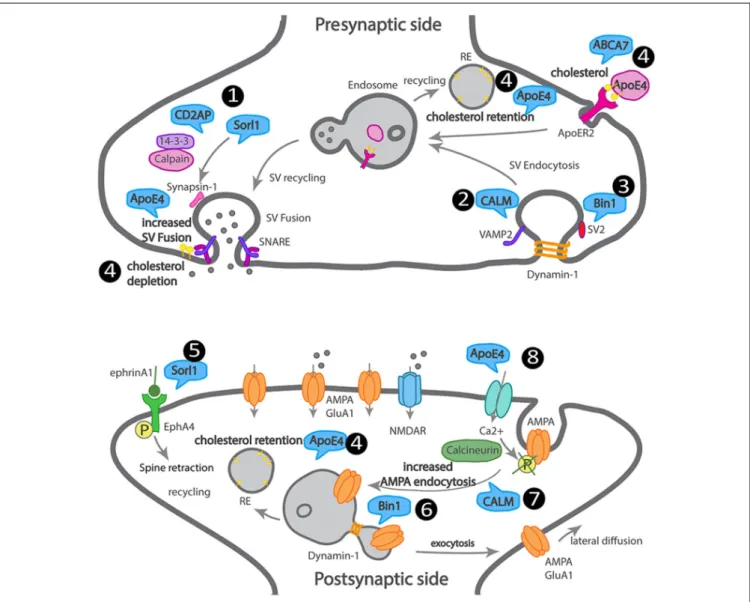
We attempted to summarize how LOAD trafficking genes
may affect synapses, although direct supporting data is missing
in most cases (Figure 3).
CONCLUSION
For many years, LOAD etiology has been a debatable topic
among researchers. The amyloid hypothesis has dominated
the field as the leading cause of AD. Experimental models of
eFAD support this hypothesis, as well as the delay of disease
onset in carriers of the AD protective mutation in APP that
reduces Aβ production (
Jonsson et al., 2012
). It remains unclear
if Aβ accumulation causes the earliest synaptic dysfunction
in LOAD.
Moreover, most clinical trials targeting Aβ aggregation
cleared amyloid plaques, but the cognitive improvement was
limited at best. Today, clinical trials include patients early in the
disease, but it is still unclear if once disease manifests, it will be
possible to revert synapse damage. In the future, based on the
genetic risk, it will be possible to predict the age of onset, and thus
preventive therapeutic strategies targeted at the still reversible
synapse dysfunction may be the solution. Now, we need to
determine the targets for future preventive therapies based on the
mechanisms that drive the initial synaptic dysfunction in AD.
FIGURE 3 | Scheme of a dysfunctional LOAD synapse illustrating how LOAD trafficking genes impact synaptic trafficking. At the presynaptic terminal, 1. Sorl1 and Cindr (CD2AP homolog) interact with synapsin, via 14–3–3, and their loss of function leads to aberrant synapsin accumulation. 2. CALM mediates
VAMP2 endocytosis and SNARE complex recycling. 3. Bin1 interacts with dynamin and SV2 and could play a role in SV endocytosis. 4. ApoE4 and ABCA7 alter the transport of cholesterol and other lipids into neurons, leading to cholesterol retention in recycling endosomes and depletion from the synaptic membranes. At the postsynaptic terminal, 5. Sorl1 attenuates EphA4 activation by ephrinA1, causing spine retraction. 6. Bin1 regulates GluA1 availability at synapses via recycling. 7. CALM regulates GluA2 availability at synapses via endocytosis. 8. ApoE4 regulates AMPA receptor availability at synapses via calcium influx, calcineurin activation, that dephosphorylates AMPA receptors triggering their endocytosis.
Here we reviewed the Aβ-dependent mechanisms of synapse
function and dysfunction as well as potentially Aβ-independent
mechanisms. We found evidence in the literature supporting that
Aβ, at the physiological concentration (picomolar), facilitates
synaptic transmission and plasticity while reducing it when at
pathological levels (nanomolar). Pathological Aβ has multiple
synaptic targets indicating a broad impact on synapse or a lack of
specificity. Alternatively, the multiplication of Aβ targets reflects
the need for an effort from the research community to improve
cellular and
in vivo models, to uniformize the experimental
conditions, and to integrate individual findings. Overall, we
find that presynaptically, Aβ targets the synaptic vesicle cycle
and neurotransmitter release. However, the mechanism thus
far identified depends on the disruption of the endosomal
membrane by Aβ. Once at the cytosol, at least in vitro,
Aβ can bind to the SV release machinery and disrupt SV
release. Postsynaptically, Aβ targets spine loss and glutamate
receptors endocytosis, depressing synaptic transmission; the
mechanism involved seems to depend on extracellular Aβ-driven
calcium influx and endosomal Aβ. However, the mechanisms
by which endosomal Aβ interferes with synapses remain
mostly unknown.
Moreover, the generalized use of exogenous Aβ treatments
or transgenic models overexpressing APP or presenilin with
eFAD mutations may disguise relevant Aβ-dependent synaptic
dysfunction mechanisms, such as the ones mediated by aging or
by LOAD risk factors. So, there is an increasing need for better
models to understand the physiological targets of Aβ in AD.
Regarding
Aβ-independent mechanisms of synapse
dysfunction, we focused on LOAD genetic risk factors linked to
endocytic trafficking, given its importance for synapses. There
are few studies on the impact of these genes’ loss of function
and even less on the effects of patient’s variants. Nevertheless,
it is interesting to find that most of the LOAD genes, similarly
to Aβ, control the synaptic vesicle cycle and the trafficking of
glutamate receptors.
Knowing the synaptic dysfunction mechanisms used by
each LOAD gene will likely reveal novel targets that may
be independent of Aβ. It would be essential to correlate the
mechanism of each LOAD gene with the cognitive decline and
the carrier genotype.
Therefore, new preclinical animal and cellular models bearing
LOAD genetic risk are being established and will hopefully soon
contribute to identifying early LOAD therapeutic targets and
potential biomarkers. Thus, with these new medical strategies,
we might approach LOAD during its prodromal phase, and we
might minimize the disease burden, expanding life span with a
better quality of life.
AUTHOR CONTRIBUTIONS
CP: review of presynaptic mechanisms and LOAD genetic risk
factors presynaptic (dys)functions. MB: Go analysis of LOAD
genes and preparation of Figure 1. MB and JC: introduction.
MB, JC, and MA: review of Abeta synaptotoxicity. MA: review
of postsynaptic mechanisms and LOAD genetic risk factors
postsynaptic (dys)functions. FM: illustration of Figures 2, 3. CP
and FM: writing modifications and feedback. CG: supervision,
writing, interpretation, and editing of the manuscript.
FUNDING
The
Almeida
lab
has
been
supported
by
FCT-JPCOFUND/0004/2015;
Alzheimer’s
Association
Research
Grant (AARG-19-618007); Maratona da Saúde; H2020 Spreading
Excellence and Widening Participation,
H2020-WIDESPREAD-01-2016-2017-TeamingPhase2-GA739572;
iNOVA4Health
(UID/Multi/04462/2019), a program financially supported by
Fundação para a Ciˆencia e Tecnologia (FCT)/Ministério da
Educação e Ciˆencia, through national funds and co-funded by
FEDER under the PT2020 Partnership Agreement. CG’s salary
is supported by FCT-CEECIND/00410/2017. FM has been the
recipient of an FCT doctoral fellowship (PD/BD/128344/2017).
CP has been the recipient of an FCT doctoral fellowship
(SFRH/BD/128374/2017).
ACKNOWLEDGMENTS
We acknowledge Tatiana Burrinha for critically reading
the manuscript.
REFERENCES
Abramov, E., Dolev, I., Fogel, H., Ciccotosto, G. D., Ruff, E., and Slutsky, I. (2009). Amyloid-β as a positive endogenous regulator of release probability at hippocampal synapses. Nat. Neurosci. 12, 1567–1576. doi: 10.1038/ nn.2433
Allen, M., Lincoln, S. J., Corda, M., Watzlawik, J. O., Carrasquillo, M. M., Reddy, J. S., et al. (2017). ABCA7 loss-of-function variants, expression, and neurologic disease risk.Neurol. Genet. 3:e126. doi: 10.1212/nxg.00000000000 00126
Almeida, C. G., Takahashi, R. H., and Gouras, G. K. (2006). β-amyloid accumulation impairs multivesicular body sorting by inhibiting the ubiquitin-proteasome system.J. Neurosci. 26, 4277–4288. doi: 10.1523/jneurosci.5078-05.2006
Almeida, C. G., Tampellini, D., Takahashi, R. H., Greengard, P., Lin, M. T., Snyder, E. M., et al. (2005). β-amyloid accumulation in APP mutant neurons reduces PSD-95 and GluR1 in synapses.Neurobiol. Dis. 20, 187–198. doi: 10.1016/j.nbd.2005.02.008
Alzheimer’s Association. (2019). 2019 Alzheimer’s disease facts and figures. Alzheimers Dement. 15, 321–387. doi: 10.1016/j.jalz.2019.01.010
Andersen, O. M., Reiche, J., Schmidt, V., Gotthardt, M., Spoelgen, R., Behlke, J., et al. (2005). Neuronal sorting protein-related receptor sorLA/LR11 regulates processing of the amyloid precursor protein.Proc. Natl. Acad. Sci. U S A 102, 13461–13466. doi: 10.1073/pnas.0503689102
Azarnia Tehran, D., Kuijpers, M., and Haucke, V. (2018). Presynaptic endocytic factors in autophagy and neurodegeneration. Curr. Opin. Neurobiol. 48, 153–159. doi: 10.1016/j.conb.2017.12.018
Bacskai, B. J., Xia, M. Q., Strickland, D. K., Rebeck, G. W., and Hyman, B. T. (2000). The endocytic receptor protein LRP also mediates neuronal calcium signalingvia N-methyl-D-aspartate receptors. Proc. Natl. Acad. Sci. U S A 97, 11551–11556. doi: 10.1073/pnas.200238297
Baglietto-Vargas, D., Prieto, G. A., Limon, A., Forner, S., Rodriguez-Ortiz, C. J., Ikemura, K., et al. (2018). Impaired AMPA signaling and cytoskeletal
alterations induce early synaptic dysfunction in a mouse model of Alzheimer’s disease.Aging Cell 17:e12791. doi: 10.1111/acel.12791
Bal, M., Leitz, J., Reese, A. L., Ramirez, D. M. O., Durakoglugil, M., Herz, J., et al. (2013). Reelin mobilizes a VAMP7-dependent synaptic vesicle pool and selectively augments spontaneous neurotransmission.Neuron 80, 934–946. doi: 10.1016/j.neuron.2013.08.024
Barry, M. F., and Ziff, E. B. (2002). Receptor trafficking and the plasticity of excitatory synapses.Curr. Opin. Neurobiol. 12, 279–286. doi: 10.1016/s0959-4388(02)00329-x
Bayés, A., van de Lagemaat, L. N., Collins, M. O., Croning, M. D. R., Whittle, I. R., Choudhary, J. S., et al. (2011). Characterization of the proteome, diseases and evolution of the human postsynaptic density.Nat. Neurosci. 14, 19–21. doi: 10.1038/nn.2719
Berman, D. E., Dall’Armi, C., Voronov, S. V., McIntire, L. B. J., Zhang, H., Moore, A. Z., et al. (2008). Oligomeric amyloid-β peptide disrupts phosphatidylinositol-4,5-bisphosphate metabolism. Nat. Neurosci. 11, 547–554. doi: 10.1038/nn.2100
Billings, L. M., Oddo, S., Green, K. N., McGaugh, J. L., and LaFerla, F. M. (2005). Intraneuronal Aβ causes the onset of early Alzheimer’s disease-related cognitive deficits in transgenic mice.Neuron 45, 675–688. doi: 10.1016/j. neuron.2005.01.040
Bilousova, T., Melnik, M., Miyoshi, E., Gonzalez, B. L., Poon, W. W., Vinters, H. V., et al. (2019). Apolipoprotein E/amyloid-β complex accumulates in alzheimer disease cortical synapsesvia apolipoprotein e receptors and is enhanced by APOE4.Am. J. Pathol. 189, 1621–1636. doi: 10.1016/j.ajpath.2019. 04.010
Bilousova, T., Miller, C. A., Poon, W. W., Vinters, H. V., Corrada, M., Kawas, C., et al. (2016). Synaptic amyloid-β oligomers precede p-Tau and differentiate high pathology control cases.Am. J. Pathol. 186, 185–198. doi: 10.1016/j.ajpath. 2015.09.018
Bourgin, C., Murai, K. K., Richter, M., and Pasquale, E. B. (2007). The EphA4 receptor regulates dendritic spine remodeling by affectingβ1-integrin signaling pathways.J. Cell Biol. 178, 1295–1307. doi: 10.1083/jcb.200610139
Bredt, D. S., and Nicoll, R. A. (2003). AMPA receptor trafficking at excitatory synapses. Neuron 40, 361–379. doi: 10.1016/s0896-6273(03) 00640-8
Bushlin, I., Petralia, R. S., Wu, F., Harel, A., Mughal, M. R., Mattson, M. P., et al. (2008). Clathrin assembly protein AP180 and CALM differentially control axogenesis and dendrite outgrowth in embryonic hippocampal neurons. J. Neurosci. 28, 10257–10271. doi: 10.1523/jneurosci.2471-08.2008
Butler, M. H., David, C., Ochoa, G. C., Freyberg, Z., Daniell, L., Grabs, D., et al. (1997). Amphiphysin II (SH3P9; BIN1), a member of the amphiphysin/Rvs family, is concentrated in the cortical cytomatrix of axon initial segments and nodes of ranvier in brain and around T tubules in skeletal muscle.J. Cell Biol. 137, 1355–1367. doi: 10.1083/jcb.137.6.1355
Calafate, S., Flavin, W., Verstreken, P., and Moechars, D. (2016). Loss of bin1 promotes the propagation of tau pathology. Cell Rep. 17, 931–940. doi: 10.1016/j.celrep.2016.09.063
Carrasquillo, M. M., Crook, J. E., Pedraza, O., Thomas, C. S., Pankratz, V. S., Allen, M., et al. (2015). Late-onset alzheimer’s risk variants in memory decline, incident mild cognitive impairment and Alzheimer’s disease.Neurobiol. Aging 36, 60–67. doi: 10.1016/j.neurobiolaging.2014.07.042
Chanaday, N. L., Cousin, M. A., Milosevic, I., Watanabe, S., and Morgan, J. R. (2019). The synaptic vesicle cycle revisited: new insights into the modes and mechanisms.J. Neurosci. 39, 8209–8216. doi: 10.1523/jneurosci.1158-19.2019 Chapuis, J., Hansmannel, F., Gistelinck, M., Mounier, A., Van Cauwenberghe, C.,
Kolen, K. V., et al. (2013). Increased expression of BIN1 mediates Alzheimer genetic risk by modulating tau pathology.Mol. Psychiatry 18, 1225–1234. doi: 10.1038/mp.2013.1
Chang, Y.-T., Hsu, S.-W., Huang, S.-H., Huang, C.-W., Chang, W.-N., Lien, C.-Y., et al. (2019). ABCA7 polymorphisms correlate with memory impairment and default mode network in patients with APOEε4-associated Alzheimer’s disease. Alzheimers Res. Ther. 11:103. doi: 10.1186/s13195-019-0563-3
Chao, M. V. (2003). Neurotrophins and their receptors: a convergence point for many signalling pathways. Nat. Rev. Neurosci. 4, 299–309. doi: 10.1038/nrn1078
Chen, Y., Durakoglugil, M. S., Xian, X., and Herz, J. (2010). ApoE4 reduces glutamate receptor function and synaptic plasticity by selectively impairing ApoE receptor recycling.Proc. Natl. Acad. Sci. U S A 107, 12011–12016. doi: 10.1073/pnas.0914984107
Cissé, M., Halabisky, B., Harris, J., Devidze, N., Dubal, D. B., Sun, B., et al. (2011). Reversing EphB2 depletion rescues cognitive functions in Alzheimer model. Nature 469, 47–52. doi: 10.1038/nature09635
Cuyvers, E., De Roeck, A., Van den Bossche, T., Van Cauwenberghe, C., Bettens, K., Vermeulen, S., et al. (2015). Mutations in ABCA7 in a Belgian cohort of Alzheimer’s disease patients: a targeted resequencing study.Lancet Neurol. 14, 814–822. doi: 10.1016/S1474-4422(15)00133-7
Dalva, M. B., Takasu, M. A., Lin, M. Z., Shamah, S. M., Hu, L., Gale, N. W., et al. (2000). EphB receptors interact with NMDA receptors and regulate excitatory synapse formation.Cell 103, 945–956. doi: 10.1016/s0092-8674(00) 00197-5
Daudin, R., Marechal, D., Wang, Q., Abe, Y., Bourg, N., Sartori, M., et al. (2018). BIN1 genetic risk factor for Alzheimer is sufficient to induce early structural tract alterations in entorhinal cortex-dentate gyrus pathway and related hippocampal multi-scale impairments.BioRxiv [Preprint]. doi: 10.1101/ 437228
Del Prete, D., Lombino, F., Liu, X., and D’Adamio, L. (2014). APP is cleaved by Bace1 in pre-synaptic vesicles and establishes a pre-synaptic interactome,via its intracellular domain, with molecular complexes that regulate pre-synaptic vesicles functions. PLoS One 9:e108576. doi: 10.1371/journal.pone. 0108576
Derkach, V. A., Oh, M. C., Guire, E. S., and Soderling, T. R. (2007). Regulatory mechanisms of AMPA receptors in synaptic plasticity.Nat. Rev. Neurosci. 8, 101–113. doi: 10.1038/nrn2055
Derkach, V., Barria, A., and Soderling, T. R. (1999). Ca2+ /calmodulin-kinase II enhances channel conductance of α-amino-3-hydroxy-5-methyl-4-isoxazolepropionate type glutamate receptors.Proc. Natl. Acad. Sci. U S A 96, 3269–3274. doi: 10.1073/pnas.96.6.3269
De Rossi, P., Buggia-Prévot, V., Clayton, B. L., Vasquez, J. B., van Sanford, C., Andrew, R. J., et al. (2016). Predominant expression of Alzheimer’s
disease-associated BIN1 in mature oligodendrocytes and localization to white matter tracts.Mol. Neurodegener. 11:59. doi: 10.1186/s13024-016-0124-1 De Rossi, P., Andrew, R. J., Musial, T. F., Buggia-Prevot, V., Xu, G.,
Ponnusamy, M., et al. (2019). Aberrant accrual of BIN1 near Alzheimer’s disease amyloid deposits in transgenic models.Brain Pathol. 29, 485–501. doi: 10.1111/bpa.12687
DeWitt, D. C., and Rhoades, E. (2013).α-synuclein can inhibit SNARE-mediated vesicle fusion through direct interactions with lipid bilayers.Biochemistry 52, 2385–2387. doi: 10.1021/bi4002369
Di Paolo, G., Sankaranarayanan, S., Wenk, M. R., Daniell, L., Perucco, E., Caldarone, B. J., et al. (2002). Decreased synaptic vesicle recycling efficiency and cognitive deficits in amphiphysin 1 knockout mice.Neuron 33, 789–804. doi: 10.1016/s0896-6273(02)00601-3
Dikic, I. (2002). CIN85/CMS family of adaptor molecules.FEBS Lett. 529, 110–115. doi: 10.1016/s0014-5793(02)03188-5
Dittman, J. S., and Ryan, T. A. (2019). The control of release probability at nerve terminals.Nat. Rev. Neurosci. 20, 177–186. doi: 10.1038/s41583-018-0111-3 Doncheva, N. T., Morris, J. H., Gorodkin, J., and Jensen, L. J. (2019). Cytoscape
stringapp: network analysis and visualization of proteomics data.J. Proteome Res. 18, 623–632. doi: 10.1021/acs.jproteome.8b00702
Dreyling, M. H., Martinez-Climent, J. A., Zheng, M., Mao, J., Rowley, J. D., and Bohlander, S. K. (1996). The t(10;11)(p13;q14) in the U937 cell line results in the fusion of the AF10 gene and CALM, encoding a new member of the AP-3 clathrin assembly protein family.Proc. Natl. Acad. Sci. U S A 93, 4804–4809. doi: 10.1073/pnas.93.10.4804
Dumanis, S. B., DiBattista, A. M., Miessau, M., Moussa, C. E. H., and Rebeck, G. W. (2013). APOE genotype affects the pre-synaptic compartment of glutamatergic nerve terminals.J. Neurochem. 124, 4–14. doi: 10.1111/j.1471-4159.2012.07908.x
Dumanis, S. B., Tesoriero, J. A., Babus, L. W., Nguyen, M. T., Trotter, J. H., Ladu, M. J., et al. (2009). ApoE4 decreases spine density and dendritic complexity in cortical neurons in vivo. J. Neurosci. 29, 15317–15322. doi: 10.1523/jneurosci.4026-09.2009
Dustin, M. L., Olszowy, M. W., Holdorf, A. D., Li, J., Bromley, S., Desai, N., et al. (1998). A novel adaptor protein orchestrates receptor patterning and cytoskeletal polarity in T-cell contacts.Cell 94, 667–677. doi: 10.1016/s0092-8674(00)81608-6
Ehrlich, I., and Malinow, R. (2004). Postsynaptic density 95 controls AMPA receptor incorporation during long-term potentiation and experience-driven synaptic plasticity.J. Neurosci. 24, 916–927. doi: 10.1523/jneurosci.4733-03. 2004
Fanutza, T., Del Prete, D., Ford, M. J., Castillo, P. E., and D’Adamio, L. (2015). APP and APLP2 interact with the synaptic release machinery and facilitate transmitter release at hippocampal synapses.Elife 4:e09743. doi: 10.7554/elife. 09743
Farrer, L. A., Cupples, L. A., Haines, J. L., Hyman, B., Kukull, W. A., Mayeux, R., et al. (1997). Effects of age, sex, and ethnicity on the association between apolipoprotein E genotype and Alzheimer disease. A meta-analysis. APOE and Alzheimer Disease Meta Analysis Consortium.JAMA 278, 1349–1356. doi: 10.1001/jama.278.16.1349
Fernández-Alfonso, T., Kwan, R., and Ryan, T. A. (2006). Synaptic vesicles interchange their membrane proteins with a large surface reservoir during recycling.Neuron 51, 179–186. doi: 10.1016/j.neuron.2006.06.008
Fernández-Alfonso, T., and Ryan, T. A. (2006). The efficiency of the synaptic vesicle cycle at central nervous system synapses.Trends Cell Biol. 16, 413–420. doi: 10.1016/j.tcb.2006.06.007
Filosa, A., Paixão, S., Honsek, S. D., Carmona, M. A., Becker, L., Feddersen, B., et al. (2009). Neuron-glia communicationvia EphA4/ephrin-A3 modulates LTP through glial glutamate transport. Nat. Neurosci. 12, 1285–1292. doi: 10.1038/nn.2394
Fogel, H., Frere, S., Segev, O., Bharill, S., Shapira, I., Gazit, N., et al. (2014). APP homodimers transduce an amyloid-β-mediated increase in release probability at excitatory synapses. Cell Rep. 7, 1560–1576. doi: 10.1016/j.celrep.2014. 04.024
Fu, W.-Y., Chen, Y., Sahin, M., Zhao, X.-S., Shi, L., Bikoff, J. B., et al. (2007). Cdk5 regulates EphA4-mediated dendritic spine retraction through an ephexin1-dependent mechanism. Nat. Neurosci. 10, 67–76. doi: 10.1038/nn1811