This journal is © The Royal Society of Chemistry 2015 Chem. Commun.,2015,51, 9141--9144 | 9141 Cite this:Chem. Commun.,2015,
51, 9141
Selective endocytic trafficking in live cells with
fluorescent naphthoxazoles and their boron
complexes
†
Gleiston G. Dias,aBernardo L. Rodrigues,aJarbas M. Resende,a
Ha´llen D. R. Calado,aCarlos A. de Simone,bValter H. C. Silva,cBrenno A. D. Neto,d
Marilia O. F. Goulart,eFabricia R. Ferreira,eAssuero S. Meira,f Claudia Pessoa,fg
Jose´ R. Correa*d
and Eufraˆnio N. da Silva Ju´nior*a
Fluorescent naphthoxazoles and their boron derivatives have been synthesized and applied as superior and selective probes for endo-cytic pathway tracking in live cancer cells. The best fluorophores were compared with the commercially available acridine orange (co-staining experiments), showing far better selectivity.
Endocytosis, as the key regulator process for molecular inter-nalization in eukaryotic cells, has a fundamental role in cell homeostasis.1 Signal transduction, neurotransmission, inter-cellular communication and immune response are known to be readily affected by endocytic pathway dysfunction.2The complexity of the cellular environment renders the understanding of endo-cytosis an outstanding challenge. Cellular homeostasis, for instance, depends on the proper communication of the cellular organelles and all other components associated with correct signals throughout the highly complex machinery. Deregulation in any of these signalizing pathways could be the answer which triggers a specific pathological process. All homeostasis unbalances may also be associated with endocytosis dysfunctions. Fluorescence techniques, in this sense, rose up, shedding light onto many doubts over cellular endocytic mechanisms and pathways.3 Despite the great advance of using fluorescent bioprobes,4–7many unanswered
questions and challenges still remain to be tackled. For some, the capacity of a bioprobe to transpose the cell membrane is one of the most desirable features.8Very recently, some have shown the great potential of fluorogenic probes for studying dynamic cellular uptake in live cells.9
With the knowledge that many quinonoid derivatives sustaining the basic carbon scaffold are capable of transposing the cell-membrane and can present luminescence, as we10–12and others13–15have already demonstrated, we envisaged the synthesis of fluorescent bioprobes from an affordable naturally occurring naphthoquinone. In this sense, we aimed to use the cell membrane permeability feature of such compounds to synthesize fluorescent derivatives for dynamics and uptake in live cells using the endocytic pathway. Herein, we disclose the rational design, synthesis, characterization, single crystal X-ray, photophysics, DFT calculations, bioimaging, cellular uptake and dynamics in living cells (cancer cells) of new fluor-escent derivatives which have allowed for selectively visualizing the whole endocytic pathway.
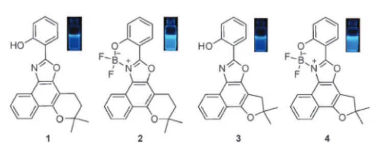
The structures of the synthesized fluorescent compounds are visualized in Fig. 1 and the synthetic details are found in the ESI†(Scheme S1).
All compounds afforded single crystals suitable for X-ray characterization (Fig. 2 and Tables S5 and S6, ESI†) with different packing depending on the size of the side ring.
Molecular structures of1and2are basically planar except for the side pyrane ring. Compound4is in turn perfectly planar with methyl groups and fluorine atoms out of the plane.
Fig. 1 Structures of the fluorescent compounds evaluated in this work. Inset: fluorescence images under UV light (365 nm) in CH2Cl2(50mM).
aInstitute of Exact Sciences, Department of Chemistry, Federal University of Minas
Gerais, Belo Horizonte, 31270-901, MG, Brazil. E-mail: [email protected] bInstitute of Physics, Department of Physics and Informatics, University of Sa˜o
Paulo, Sa˜o Carlos, 13560-160, SP, Brazil c
University Unit of Exact Sciences and Technology, State University from Goia´s, Ana´polis, 75024-010, GO, Brazil
d
Laboratory of Medicinal & Technological Chemistry, Institute of Chemistry, University of Brasilia, P.O. Box 4478, Brasilia, 70904970, DF, Brazil. E-mail: [email protected]
eInstitute of Chemistry and Biotechnology, Federal University of Alagoas, Maceio´,
57072-970, AL, Brazil
fDepartment of Physiology and Pharmacology, Federal University of Ceara´,
Fortaleza, 60430-270, CE, Brazil
gFiocruz-CE, Fortaleza, 60180-900, CE, Brazil
†Electronic supplementary information (ESI) available. CCDC 1030235–1030237 and 1033804. For ESI and crystallographic data in CIF or other electronic format see DOI: 10.1039/c5cc02383a
Received 23rd March 2015, Accepted 24th April 2015
DOI: 10.1039/c5cc02383a
www.rsc.org/chemcomm
ChemComm
COMMUNICATION
Published on 27 April 2015. Downloaded by Federal University of Ceará on 18/07/2016 12:59:47.
View Article Online
9142 | Chem. Commun.,2015,51, 9141--9144 This journal is © The Royal Society of Chemistry 2015
Distinct crystal packing for all compounds are also noted when comparing the four structures (Fig. 2).
The photophysical properties of these molecules were inves-tigated and pointed to the stability of these compounds in the excited state, as shown in Table S7 (ESI†).
In a general way, the boron complexes showed larger Stokes shift values, most likely due to the higher planarity and more efficient
p-conjugation. Bathochromic displacements are observed when
structures1and3were compared with their correspondent boron complexes, showing that the planarity introduced with the for-mation of the complex favors more electronic conjugation in the structure. Beyond the red shift,3 and4 displayed a shoulder at 356 and 360 nm, respectively, indicating a slight effect over its conformation as a consequence of the solvatochromic effect. The Stokes shifts for the BODIPY-like compound2are larger than those observed for the other structures (Table S7, ESI†).
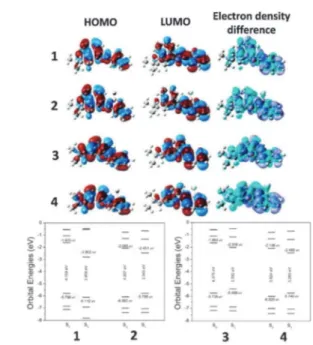
Considering that the solvent effect is important for investi-gating this sort of derivatives, especially in the presence of polar solvents, some theoretical calculations were performed aiming to shed some light on their behaviour in the ground and excited
state. In this sense, DMSO was used (implicit treatment) as the medium for the calculations at the PBE1PBE/6-311+G(2d,p)// CAM-B3LYP/6-31+g(d) level of theory. HOMO, LUMO (basically ofp-type) and electron density difference were also calculated
and are in accordance with the good stability observed in the excited state for compounds1–4(Fig. 3).
Electrochemical (cyclic voltammetry, CV) analyses were also performed (Fig. S45 and S46, ESI†). All the analysed compounds were reduced in two reduction waves (Fig. S45 and Table S9, ESI†). The first electron uptake was associated with a relatively stable radical anion whereas the second one is, presumably, a dihydro-derivative, in accordance with similar compounds.16 Differences in current were noticed comparing the boron-free and boron-containing derivatives. As shown in Fig. S45 and S46 (ESI†), comparison of the synthesized compounds allowed us to suggest that complexation with boron slightly facilitates the reduction process (Table S9, ESI†), withEpcvalues less negative than those for the boron-free compounds.
Finally, live cell-imaging experiments (PC3 cell lineages – human prostate cancer cells) were conducted to evaluate the potential of the fluorescent compounds as bioprobes. Dye 1 showed no fluorescent signal when incubated with live cells (Fig. S41, ESI†) whereas compounds2–4displayed a bright blue emission signal. These three compounds proved to be capable of staining the canonical endocytic pathway, meaning they are capable of selectively staining early endosome, late endosome and lysosome (Fig. 4–6) during the cellular uptake process. Fig. 2 Molecular structure of1–4(top) and packing (bottom) depicted
from single crystal X-ray analysis.
Fig. 3 Optimized geometries, HOMO and LUMO plots, and electron density difference map. Diagram energy levels calculated with DMSO as the solvent. Calculations at the PBE1PBE/6-311+G(2d,p)//CAM-B3LYP/6-31+g(d) level of theory. Structures calculated with implicit DMSO as the solvent showed similar orbitals localizations.
Fig. 4 Live PC3 cancer cells stained with2(blue emission). (A) Positive control with cells stained with acridine orange (green emission). (B)2as the bioprobe and (C) overlay of (A) and (B).2was found accumulated inside of spherical membranous organelles associated with endosomes (white arrow heads). White arrows show the nucleus stained with acridine orange (non-specific staining). N = cell nucleus. Reference scale bar 20mm.
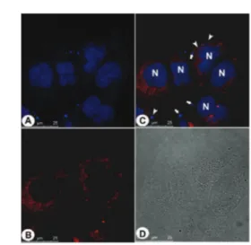
Fig. 5 Live PC3 cancer cells stained with3(blue emission). (A) Positive control with cells stained with acridine orange (green emission). (B)3as the bioprobe and (C) overlay of (A) and (B).3was found accumulated inside of spherical membranous organelle correlated with early endosomes (white arrow heads), late endosomes (yellow arrow heads) and lysosomes (red arrows). White arrows show the nucleus stained with acridine orange (non-specific staining). N = cell nucleus. Reference scale bar 20mm.
Communication ChemComm
Published on 27 April 2015. Downloaded by Federal University of Ceará on 18/07/2016 12:59:47.
This journal is © The Royal Society of Chemistry 2015 Chem. Commun.,2015,51, 9141--9144 | 9143 Panels (A) and (C) in Fig. 4 and 5 show all membranous
compartments strongly stained; there are no fluorescent sig-nals inside of the nucleus. Acridine orange (AO) is widely used in order to stain endocytic pathways, however, AO also stains nucleic acids as can be observed in Fig. 4–6. Under the same experimental conditions,2–4 did not stain nucleic acids and the cell nuclei are visualized as black voids. Compounds 2–4 could be found at different localizations inside the cytoplasm, and therefore are found associated with different membranous compartments. Vesicles found at the cell periphery close to the plasmatic membrane is correlated with early endosomes (Fig. 4 and 5). Vesicles found between the plasmatic membrane and the nucleus are correlated with late endosomes (Fig. 5), whereas vesicles found near to the nucleus are correlated with lysosomes (Fig. 6). The overlay images between the tested compounds and acridine orange (panel (C) in the cited figures) provided the evidence that these compounds are staining the membranous compartments belonging to the endocytic pathway, but in this case selectively. AO is widely used to stain acidic organelles in studies regarding the endocytic and autophagic pathways applied to eukaryotic cells, however, as demonstrated, with high affinity to nucleic acid components, which produce a non-specific pattern in the cells nuclei.2–4were able to stain specifically the membra-nous compartments belonging to the endocytic pathway without nucleus cross staining.
The possibility of caveolae-mediated endocytosis was also evaluated to confirm the selective endocytic trafficking. Cells through caveolae-mediated endocytosis may also internalize the tested compounds irrespective of any affinity with the caveolar structure. Caveolar vesicles formation is a stable structure due to the lipids composition in these regions.17,18 This feature keeps the vesicles open by a time period greater than that observed in membrane regions associated with clathrin-mediated vesicles formation.17,19Most likely the tested compounds accumulate in the open vesicles during their formation. As a consequence they are found trapped inside the vesicles during their bud off from the cells membrane therefore targeting the endoplasmic reticulum and matching with our findings related to vesicles staining near to the nuclei. To be sure about this hypothesis, an assay using the commer-cially available anti-caveolin-1 antibody (red emitter), which is
the universal marker specific for caveolar vesicle formation, has been performed (Fig. 7–9).
Fig. 7 shows few vesicles stained near to cytoplasmic membrane correlated with caveolar vesicle bud off and near to the nucleus of the cell. This feature indicates the vesicles target endoplasmic reticulum which is the main docking point Fig. 6 Live PC3 cancer cells stained with4(blue emission). (A) Positive
control with cells stained with acridine orange (green emission). (B)4as the bioprobe and (C) overlay of (A) and (B). 4was localized inside of spherical membranous organelles belonging to the endocytic pathway. Red arrows show the staining membranous compartment correlated with lysosomes. White arrows show the nucleus stained with acridine orange (non-specific staining). N = cell nucleus. Reference scale bar 20mm.
Fig. 7 Live PC3 cancer cells stained with 2 and immunodetection of caveolin-1 (universal caveolar marker). (A) Shows the cell nucleus stained with commercially available DAPI (blue), and the cytoplasmic endocytic vesicles stained by2(also in blue). (B) Shows the fluorescence distribution related to caveolin-1 immunodetection (red). (C) Shows the overlay of the two fluorescent signals (blue and red). Arrows show the vesicles belonging to canonical endocytic pathways (absence of caveolin-1 detection) and arrowheads show the vesicles belonging to caveolae-mediated endo-cytosis (vesicle with double staining – blue and red). (D) Shows the cell normal morphological aspects. Reference scale bar 25mm.
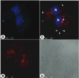
Fig. 8 Live PC3 cancer cells stained with 3 and immunodetection of caveolin-1. (A) Shows the cell nucleus stained with DAPI (blue), and the cytoplasmic endocytic vesicles stained by3(also in blue). (B) Shows the fluorescence distribution related to caveolin-1 immunodetection (red). (C) Shows the overlay of the two fluorescent signals (blue and red). Arrows show the vesicles belonging to canonical endocytic pathways (absence of caveolin-1 detection) and arrowheads show the vesicles belonging to caveolae-mediated endocytosis (vesicle with double staining – blue and red). (D) Shows the cell normal morphological aspects. Reference scale bar 25mm.
ChemComm Communication
Published on 27 April 2015. Downloaded by Federal University of Ceará on 18/07/2016 12:59:47.
9144 | Chem. Commun.,2015,51, 9141--9144 This journal is © The Royal Society of Chemistry 2015
for caveolar vesicles. In other words, the caveolar entering is one possibility as well as the canonical endocytic pathway. The same behaviour could be noted for all tested compounds indi-cating that the new bioprobes are capable of probing selectively the endocytic trafficking in live cells irrespective of the preferred internalization path. Similar results from that in Fig. 7 have been observed in Fig. 8 and 9.
In summary, four new fluorescent derivatives have been synthesized, characterized and successfully used as live cell-imaging probes, capable of selectively staining the endocytic pathway. The compounds also showed no cytotoxicity when evaluated by MTT assay. Compounds2–4were able to permeate intact live cells, thus being trapped inside of acidic compart-ments, turning them into suitable molecules for investigating the lysosome biogenesis, host pathogens interaction and auto-phagic processes, as will be disclosed in due course. The selective endocytic trafficking in live cells and its dynamics have been probed using the synthesized compounds, which in turn, were capable of selectively reveal the internalization endocytic process in live cell through both the caveolar vesicles or the
canonical pathway. The new designed bioprobes showed by far better results than the commercially available AO.
The authors would like to thank CAPES, CNPq, FAPDF, DPP-UnB, FINATEC and FAPEMIG.
Notes and references
1 M. N. Levine, T. T. Hoang and R. T. Raines,Chem. Biol., 2013,20, 614–618.
2 S. D. Conner and S. L. Schmid,Nature, 2003,422, 37–44.
3 A. A. Marti, S. Jockusch, N. Stevens, J. Ju and N. J. Turro,Acc. Chem. Res., 2007,40, 402–409.
4 L. D. Lavis and R. T. Raines,ACS Chem. Biol., 2014,9, 855–866.
5 L. D. Lavis and R. T. Raines,ACS Chem. Biol., 2008,3, 142–155. 6 N. Johnsson and K. Johnsson,ACS Chem. Biol., 2007,2, 31–38. 7 (a) B. A. D. Neto, J. R. Correa and R. G. Silva,RSC Adv., 2013, 3,
5291–5301; (b) B. A. D. Neto, J. R. Correˆa, P. H. P. R. Carvalho, D. C. B. D. Santos, B. C. Guido, C. C. Gatto, H. C. B. de Oliveira, M. Fasciotti, M. N. Eberlin and E. N. da Silva Ju´nior,J. Braz. Chem. Soc., 2012, 23, 770–781; (c) P. H. P. R. Carvalho, J. R. Correa,
B. C. Guido, C. C. Gatto, H. C. B. De Oliveira, T. A. Soares and B. A. D. Neto,Chem. – Eur. J., 2014,20, 15360–15374; (d) C. D’Angelis
do E. S. Barbosa, J. R. Correˆa, G. A. Medeiros, G. Barreto, K. G. Magalha˜es, A. L. de Oliveira, J. Spencer, M. O. Rodrigues and B. A. D. Neto,Chem. – Eur. J., 2015,21, 5055–5060.
8 A. Nadler and C. Schultz,Angew. Chem., Int. Ed., 2013,52, 2408–2410. 9 A. A. R. Mota, P. H. P. R. Carvalho, B. C. Guido, H. C. B. de Oliveira, T. A. Soares, J. R. Correa and B. A. D. Neto,Chem. Sci., 2014,5, 3995–4003.
10 E. H. G. da Cruz, P. H. P. R. Carvalho, J. R. Correa, D. A. C. Silva, E. B. T. Diogo, J. D. de Souza Filho, B. C. Cavalcanti, C. Pessoa, H. C. B. de Oliveira, B. C. Guido, D. A. da Silva Filho, B. A. D. Neto and E. N. da Silva Ju´nior,New J. Chem., 2014,38, 2569–2580.
11 S. N. Sunassee, C. G. L. Veale, N. Shunmoogam-Gounden, O. Osoniyi, D. T. Hendricks, M. R. Caira, J.-A. de la Mare, A. L. Edkins, A. V. Pinto, E. N. da Silva Ju´nior and M. T. Davies-Coleman,Eur. J. Med. Chem., 2013,62, 98–110.
12 E. N. da Silva Ju´nior, C. F. de Deus, B. C. Cavalcanti, C. Pessoa, L. V. Costa-Lotufo, R. C. Montenegro, M. O. de Moraes, M. C. F. R. Pinto, C. A. de Simone, V. F. Ferreira, M. O. F. Goulart, C. K. Z. Andrade and A. V. Pinto,J. Med. Chem., 2010,53, 504–508. 13 (a) R. Araya-Maturana, W. Cardona, B. K. Cassels, T. Delgado-Castro, J. Ferreira, D. Miranda, M. Pavani, H. Pessoa-Mahana, J. Soto-Delgado and B. Weiss-Lopez, Bioorg. Med. Chem., 2006, 14,
4664–4669; (b) C. E. M. Carvalho, A. S. Silva, I. M. Brinn, A. V. Pinto, M. C. F. R. Pinto, S. Lin, T. A. Moore, D. Gust and M. Maeder,Phys. Chem. Chem. Phys., 2002,4, 3383–3389; (c) C. E. M.
Carvalho, I. M. Brinna, A. V. Pinto and M. C. F. R. Pinto,J. Photochem. Photobiol., A, 1999,123, 61–65.
14 V. P. Torchilin,Adv. Drug Delivery Rev., 2005,57, 95–109.
15 E. M. Perchellet, B. J. Sperfslage, G. Qabaja, G. A. Jones and J. P. Perchellet,Anti-Cancer Drugs, 2001,12, 401–417.
16 L. Henning and H. Ole,Organic Electrochemistry, Marcel Dekker, Inc., New York, 4th edn, 2001.
17 L. Pelkmans and A. Helenius,Traffic, 2002,3, 311–320.
18 A. Zloza, D. W. Kim, J. Broucek, J. M. Schenkel and H. L. Kaufman,
J. Interferon Cytokine Res., 2014,34, 915–919. 19 G. Sowa,Front. Physiol., 2012,2, 120.
Fig. 9 Live PC3 cancer cells stained with4 and immunodetection of caveolin-1. (A) Shows the cell nucleus stained with DAPI (blue), and the cytoplasmic endocytic vesicles stained by4(also in blue). (B) Shows the fluorescence distribution related to caveolin-1 immunodetection (red). (C) Shows the overlay of the two fluorescent signals (blue and red). Arrows show the vesicles belonging to canonical endocytic pathways (absence of caveolin-1 detection) and arrowheads show the vesicles belonging to caveolae-mediated endocytosis (vesicle with double staining – blue and red). (D) Shows the cell normal morphological aspects. Reference scale bar 25mm.
Communication ChemComm
Published on 27 April 2015. Downloaded by Federal University of Ceará on 18/07/2016 12:59:47.