Prognostic Indicators in Foals with Neonatal
Encephalopathy
Dissertação de Mestrado Integrado em Medicina Veterinária
Ana Alexandra Vieira Vilela
Orientador: Professor Doutor Mário Pedro Gonçalves Cotovio
Prognostic Indicators in Foals with Neonatal
Encephalopathy
Dissertação de Mestrado Integrado em Medicina Veterinária
Ana Alexandra Vieira Vilela
Orientador: Professor Doutor Mário Pedro Gonçalves Cotovio
Composição do Júri:
Professora Doutora Maria da Conceição Medeiros Castro Fontes Professor Doutor Filipe da Costa Silva
Professor Doutor Mário Pedro Gonçalves Cotovio
NOME: Ana Alexandra Vieira Vilela
C.C: 14516655
TELEMÓVEL: (+351) 914887267
CORREIO ELETRÓNICO: [email protected]
DESIGNAÇÃO DO MESTRADO: Mestrado Integrado em Medicina Veterinária
TÍTULO DA DISSERTAÇÃO DE MESTRADO EM MEDICINA VETERINÁRIA:
Prognostic Indicators in Foals with Neonatal Encephalopathy
ORIENTADOR: Mário Pedro Gonçalves Cotovio
ANO DE CONCLUSÃO: 2019
Declaro que esta Dissertação de Mestrado é resultado da minha pesquisa e trabalho pessoal e das orientações dos meus supervisores. O conteúdo é original e as fontes consultadas estão devidamente mencionadas no texto e na bibliografia final. Declaro ainda que este trabalho não foi apresentado em nenhuma outra instituição para obtenção de qualquer grau académico.
Vila Real, 2019
“The love for all living creatures is the most noble attribute of man.”
Ao Professor Doutor Mário Cotovio, por todo o apoio, empenho, ajuda e orientação que demonstrou, sem os quais a realização desta dissertação não seria possível.
Ao Professor Doutor Jorge Colaço, por ser extremamente prestável em disponibilizar o seu tempo e ajuda na componente estatística deste estudo.
À Professora Doutora Maria João Pires e ao seu irmão João Pires, pela valiosa contribuição no estudo estatístico desta tese.
A toda a equipa do Hospital Equino da Universidade do Tennessee - Faculdade de Medicina Veterinária, pela simpatia, disponibilidade, por todos os conhecimentos que me transmitiram e por me ajudarem a crescer quer profissionalmente, quer como pessoa. Foram dois meses que vivi num país diferente, num continente diferente mas que, graças a vocês, me consegui sentir em casa.
A toda a equipa do Hospital La Equina, em Málaga, pela amizade, por me terem ensinado tanto e por me terem proporcionado momentos que nunca me vou esquecer.
Aos meus pais, por me ajudarem a alcançar todos os meus sonhos. Não há palavras que descrevam a enorme gratidão e orgulho que sinto por ser vossa filha. Vocês são o meu porto de abrigo e sempre me ensinaram que nada é impossível se lutarmos pelos nossos sonhos, fundamento esse que levo comigo para a vida. Muito obrigada por tudo.
Ao Luís, por haver tanto para agradecer que nem sei por onde começar. Obrigada por seres o meu maior apoio e por me ajudares sempre em tudo o que preciso. A minha vida não seria a mesma sem ti.
Aos meus avós, tios e primos por acreditarem sempre em mim e por, apesar da distância, estarem sempre disponíveis para me ajudar e apoiar.
À D. Felícia, por me ter visto crescer e por ter estado sempre presente na minha vida. Aos meus amigos de Vila Real, por serem a família que eu escolhi. Vocês são os melhores amigos que eu podia ter pedido e só tenho de agradecer por terem tornado os 6 anos de faculdade nos melhores anos da minha vida. Levo-vos comigo para a vida toda! Aos meus animais de estimação, por me lembrarem todos os dias do quão maravilhoso é este sonho de puder ser Médica Veterinária e por todo o tempo em que estive em casa, à frente do computador a realizar este trabalho, nunca me terem deixado sentir sozinha.
Neonatal Encephalopathy is the most common neurological disorder in neonatal foals. It is usually associated with peripartum events that can lead to acute or chronic hypoxia, but neither the etiology nor the pathophysiology are still fully understood. Clinical signs often observed include recumbency, abnormal nursing behavior, loss of awareness of the environment, and seizures. However, a multi-system evolvement can also be found. There is no definitive ante-mortem diagnostic test, so diagnosis is based on history, clinical presentation, elimination of other differentials, and supplementary diagnostic tests. Treatment is mainly supportive, relying on resolution of secondary complications. Prognosis is usually good (60%-80%) in uncomplicated cases.
The purpose of this dissertation is to stablish prognostic indicators in a population of foals with Neonatal Encephalopathy. Medical records from 61 foals with the diagnosis of Neonatal Encephalopathy between 1982 and 2018 were collected from the Equine Hospital of the University of Tennessee, College of Veterinary Medicine, in the United States of America and from the Hospital La Equina, in Málaga, Spain. Variables included in the study were surviving rate, animal identification, predisposing factors of the mare and foal, physical exam and laboratory findings on admission, concurrent diseases, and treatment. In order to determine factors associated with the outcome, these variables were statistically compared between survivors and non-survivors.
The overall surviving rate was 57.4%. Neither gender nor breed were associated with the surviving rate. There were no predisposing factors associated with survival as well. This study demonstrated that normal body temperature, normal glycemia levels, normal creatinine concentration, or absence of recumbency were associated with a good surviving rate, while hypothermia, abnormal glycemia levels, creatinine levels >4 mg/dL, pneumonia, anemia, or sepsis were associated with mortality. Foals with hypothermia, hypoglycemia or hyperglycemia had greater odds of non-survival. The use of antibiotics or non-steroidal anti-inflammatory drugs were positively associated with survival.
In conclusion, body temperature, glycemia, creatinine concentration, absence of recumbency, presence of pneumonia, anemia, and sepsis are the main prognostic indicators in the population in study. Given its influence in survival, using antibiotics or non-steroidal anti-inflammatory drugs are recommended in foals with NE.
A Encefalopatia Neonatal é a doença neurológica mais comum em poldros neonatos. Está, normalmente, associada com problemas no pré-, durante e no pós-parto que podem conduzir a episódios agudos ou crónicos de hipóxia. No entanto, atualmente, nem a etiologia nem a fisiopatologia estão completamente compreendidas. Os sinais clínicos observados mais frequentemente incluem decúbito, perda do reflexo de sucção, perda de afinidade pela mãe, alteração do estado de alerta e convulsões. No entanto, também pode ocorrer um envolvimento multissistémico. Uma vez que não existe um teste ante-mortem definitivo, o diagnóstico é feito com base na história clínica, sinais clínicos, eliminação de diagnósticos diferenciais e exames diagnósticos complementares. O tratamento é de suporte, baseando-se na resolução de complicações secundárias. O prognóstico é normalmente bom (60%-80%) em casos não complicados.
O objetivo desta dissertação é definir fatores de prognóstico numa população de poldros com Encefalopatia Neonatal. Foram recolhidos os registos médicos de 61 poldros com o diagnóstico de Encefalopatia Neonatal do Hospital Equino da Universidade do Tennessee, Faculdade de Medicina Veterinária, Estados Unidos da América e do Hospital La Equina, em Málaga, Espanha, datados desde 1982 até 2018. As variáveis incluídas neste estudo foram a taxa de sobrevivência, identificação do animal, fatores predisponentes da égua e do poldro, exame físico e exames laboratoriais, doenças concorrentes e tratamento. De forma a determinar os fatores associados com o prognóstico, estas variáveis foram estatisticamente comparadas entre sobreviventes e não-sobreviventes.
A taxa de sobrevivência desta população foi de 57.4%. Nem o género, nem a raça tiveram influência na taxa de sobrevivência. Os fatores predisponentes relativos à égua ou poldro também não mostraram nenhuma associação com a sobrevivência. Este estudo demonstrou que temperatura corporal normal, níveis de glicémia normais, concentrações de creatinina normais e ausência de decúbito tiveram uma influência positiva na sobrevivência. Por outro lado, a presença de hipotermia, níveis de glicémia anormais, valores de creatinina superiores a 4 mg/dL, pneumonia, anemia ou septicémia estiveram associados a mortalidade. De facto, poldros com hipotermia, hipoglicémia ou hiperglicémia tiveram mais chances de morrer. O uso de antibióticos ou de anti-inflamatórios não esteroides tiveram uma associação positiva com a taxa de sobrevivência.
de decúbito, presença de pneumonia, anemia ou septicémia são os indicadores de prognóstico desta população. Dada a sua influência na sobrevivência, o uso de antibióticos e anti-inflamatórios não esteroides estão recomendados em poldros com Encefalopatia Neonatal.
CONTENTS
AGRADECIMENTOS ... ix
ABSTRACT ... xi
RESUMO ... xiii
CHAPTER I – BIBLIOGRAPHIC REVIEW ... 1
1. INTRODUCTION ... 1 2. PREDISPOSING FACTORS ... 3 3. PATOPHYSIOLOGY ... 4 4. CLINICAL SIGNS ... 8 5. DIFFERENTIAL DIAGNOSES ... 10 6. DIAGNOSIS ... 11 7. TREATMENT ... 17 8. PROGNOSIS ... 25
CHAPTER II – RETROSPECTIVE STUDY ... 29
1. OBJECTIVES ... 29
2. MATERIALS AND METHODS ... 29
3. RESULTS... 32
4. DISCUSSION ... 46
5. CONCLUSIONS ... 55
BIBLIOGRAPHIC REFERENCES ... 57
Figure 1 - Schematic representation of the pathogenesis of NE, as a result of a
hypoxic-ischemic event (HIE) ... 5
Figure 2 and 3– Foal with NE, showing signs of loss of awareness of the environment and
abnormal nursing behavior by nursing the fence (left) ... 9
Figure 4 - Foal with somnolence and difficulty to arouse, often seen in foals with NE ... 9 Figure 5 - Tongue protrusion in foal with NE ... 9 Figure 6 - Head protection and environment adaptation to a recumbent, convulsive foal with
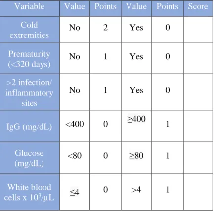
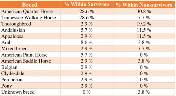
Table 1 - APGAR Scoring System ... 13 Table 2 and 3 - Survival Scoring System (Left) and Probability of Survival (Right) ... 28 Table 4 - Percentage of the breeds included in this study among survivors and non-survivors.
... 33
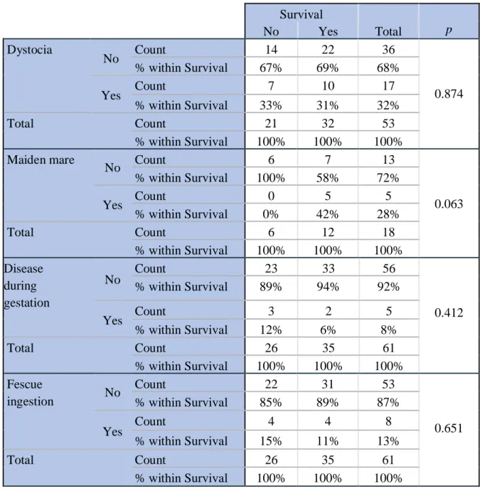
Table 5 - Comparison between several predisposing factors of the mare and survival, using the
Pearson Chi-Squared Test. ... 34
Table 6 - Univariate logistic regression models to study the influence of a delivery deemed a
dystocia in the mortality of the population in study. ... 34
Table 7 - Comparison between predisposing factors of the foal and survival, using the Pearson
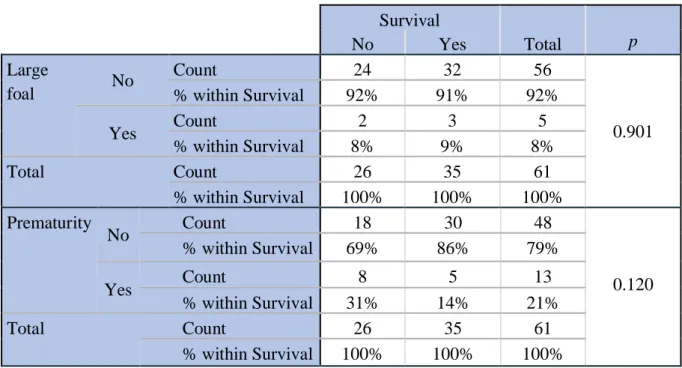
Chi-Squared Test. ... 35
Table 8 - Univariate logistic regression model to study the influence of prematurity in the
mortality of the population in study. ... 35
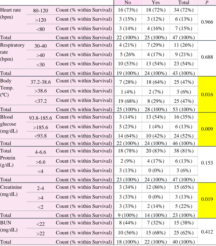
Table 9 - Comparison between intervals of the numerical variables on admission and survival,
using the Pearson Chi-Squared Test... 36
Table 10 - Comparison of means and standard deviation of the numerical variables, between
survivors and non-survivors, through the ANOVA test. ... 37
Table 11 - Univariate logistic regression models to study the numerical variables obtained
during the physical examination ... 39
Table 12 - Comparison between the presence of several clinical signs and survival, using the
Pearson Chi-Squared Test. ... 41
Table 13 - Univariate logistic regression models to study the influence of some clinical signs
the Pearson Chi-Squared Test. ... 43
Table 15 - Univariate logistic regression models to study the influence of some concurrent
diseases in the mortality of the population in study. ... 44
Table 16 - Comparison between therapeutic interventions and survival, using the Pearson
Graphic 1 - Number of animals with each clinical sign found in the population in study ... 40
Graphic 2 - Percentage of concurrent diseases in the population in study ... 42
Graphic 3 - Percentage of the therapeutic interventions performed in the population in study
Abbreviations
ATP Adenosine Triphosphate CI Confidence Interval CNS Central Nervous System
CT Computed tomography
DMSO Dimethyl sulfoxide EEG Electroencephalography
FPTI Failure of passive transfer immunity GI Gastrointestinal
HIE Hypoxic-Ischemic Encephalopathy HPA Hypothalamic-Pituitary-Adrenal Axis
IgG Immunoglobulins G
IV Intravenous
Mg Magnesium
MRI Magnetic Resonance Imaging NE Neonatal Encephalopathy PAS Perinatal Asphyxia Syndrome
pNF-H Plasma Concentration of the Phosphorylated Axonal forms of Neurofilament H PRP Platelet Rich Plasma
REE Resting Energy Requirements
OR Odds Ratio
TH Thyroid Hormones
T3 Triiodothyronine
T4 Total Thyroxine
UTCVM University of Tennessee, College of Veterinary Medicine UCHL 1 Ubiquitin C-Terminal Hydrolase 1
CHAPTER I – BIBLIOGRAPHIC REVIEW
1. INTRODUCTION
Normal neonatal foals, as precocious species (Gold, 2017) are born with an immediate post-birth capacity for survival in the wild (Mellor, 2015). The foal is delivered in 20-40 minutes of active labor and it is essential to achieve a successful transition from the intrauterine state to an extrauterine state of consciousness, which includes the ability to stand up, ambulate, and suck from the mare (Aleman, 2017). It is expected that foals will stand and nurse within 2-3h after birth and stay close to the mare for protection and to bond (Grogan, 2005). It has been reported that skin-to-skin contact, similar as in humans, benefits newborns in short and long-term outcomes (McCallie, 2017), with a reported more accelerated neurophysiological development (Scher, 2009). These critical milestones must be achieved within the first few minutes and hours after birth, otherwise, significant problems associated with inadequate colostrum and milk intake may occur, leading to energy depletion, weakness, difficulty to rise, hypothermia, failure of passive immunoglobulin transfer, infection and death (UC Davis International Animal Welfare Training Institute, 2018).
Neonatal encephalopathy (NE) is a very broad syndrome that includes any neonate with neurologic signs, regardless of etiology. Such signs include difficulty with the initiation and maintenance of respiration, depression of tone and reflexes, subnormal levels of consciousness and frequently seizures (McKenzie III, 2018; Bernard, 2018a). It is a noninfectious syndrome and is primarily characterized by central nervous system (CNS) dysfunction (MacKay, 2005; Tennent-Brown, 2015). Besides that, it is now known that it can affect many organ systems beyond the neurologic one, such as the cardiopulmonary, endocrine, gastrointestinal and renal systems and can cause behavioral dysfunctions (Gold, 2016).
It is recognized as the most common neurological disorder in neonatal foals (Lyle-Dugas, 2017) with an estimated incidence of 1-2% of all births (Bernard, 1995; Bernard, 2018a). It is usually associated with peri-partum events that may result in acute or chronic hypoxia, such as dystocia, cesarean section, premature placental separation, or placentitis (Tennent-Brown, 2015). Other terms commonly used are hypoxic-ischemic encephalopathy (HIE), perinatal asphyxia syndrome (PAS), neonatal maladjustment
syndrome, dummy foal syndrome, wanderer, convulsive and barker foal (Vaala, 2003; Mackay, 2005; Wong, 2011; McKenzie III, 2018).
The confusion associated with the nomenclature is based on the fact that: • Clinical signs are unspecific for NE;
• Diagnosis is made based on elimination of other potential etiologies;
• It is difficult to determine the exact cause of neurologic dysfunction in foals; • Elevations in neurosteroids concentrations may be associated with NE,
particularly in those without a hypoxic episode.
Therefore, to overcome that, there has been an effort to restrict the term HIE to the subset of cases of NE in which a hypoxic-ischemic episode occurred (McKenzie III, 2018). Although this disorder has often been compared with PAS in human infants, it seems likely that oxygen deprivation may not be the only pathophysiologic mechanism by which NE occurs. An inflammatory insult or failure to correctly transition from intrauterine to extrauterine life can also cause NE.
Since the etiology and pathophysiology are still not fully understood, the vast part of information available is extrapolated from human and other animal models (Gold, 2017). However, some situations differ significantly from their human counterparts, like the fact that many foals recover quickly and demonstrate no neurologic sequelae (McKenzie III, 2018), while about 60% of human infants with PAS die, and 25% of the survivors have some neurological disability, like cerebral palsy (Ballet, 2010). Additionally, long-term outcome for foals is significantly better than for infants (Gold, 2017).
The clinical signs associated with NE can range from mild depression with loss of the suck reflex (Wilkins, 2015a) to severe neurologic signs including central blindness, seizures, coma and death (McKenzie III, 2018). In fact, equine neonatal sepsis and perinatal asphyxia syndrome are the most important causes of morbidity and mortality in newborn foals (Galvin, 2010).
2. PREDISPOSING FACTORS
In order to understand the causal pathways and develop preventive strategies, it is essential to identify the risk factors for NE (Kurinczuk, 2010).
Conditions associated with neonatal encephalopathy include maternal diseases, such as endotoxemia, respiratory disease, hemorrhage/anemia, fescue toxicity, placentitis, chronic or acute/premature uteroplacental separation, and surgery (McSloy, 2008; Gold, 2017), meaning that the health of the mare during gestation is an important key point in the survival of foals with this condition (Gold, 2016). Furthermore, advanced mare’s age and multiple parity can also contribute to placental insufficiency and, subsequently, to foal’s disease and gross abnormalities in the placenta (Wilsher, 2003).
Problems during parturition, such as induced parturition, dystocia, caesarean delivery and post-term pregnancy can also lead to hypoxia and ultimately to NE (Wong, 2011; Hahn, 2008; Gold, 2017; McKenzie III, 2018).
Conditions associated with the fetus/foal include meconium aspiration, twinning, fetal infection (Wong, 2011; McKenzie III, 2018), congenital abnormalities, umbilical cord compression, sepsis, prematurity, and dysmaturity (Gold, 2017).
Nevertheless, it is still not fully understood how these factors contribute to non-survival in foals with NE, which complicates the determination of prognosis and decision making (Gold, 2016).
3. PATOPHYSIOLOGY
The underlying pathophysiology of NE is complex (McKenzie III, 2018) and likely multifactorial (Wilkins, 2015a). It is still not completely understood in the foal or in human infants (Gold, 2017), being most information regarding the pathophysiology extrapolated from human studies to veterinary medicine (Wong, 2011).
A primary hypoxic or anoxic insult, as a result of a reduction in the uterine and umbilical circulation, leads to the activation of the sympathetic adrenergic nervous system (Wilkins, 2004a). This causes a critical redistribution of cardiac output, resulting in preferential blood flow to the heart, brain and adrenal glands and decreased perfusion to other organs like the lungs, gastrointestinal tract, liver, spleen, kidneys, muscles and skin (Hahn, 2008; Harteman, 2013; Magdesian, 2014); hence the likelihood of multisystem evolvement (Hahn, 2008; Wilkins, 2015a). If the initial insult continues to a point where the fetus cannot maintain this centralization of circulation, the cardiac output falls, and cerebral circulation decreases (Wilkins, 2004a). An alteration of the blood flow can cause a decrease in cerebral oxygenation (Gold, 2017) which initiates a cascade of deleterious events (McKenzie III, 2018) leading to neuronal cell death and injury (Wilkins, 2015a), either during the ante-, peri- or post-partum period (Gold, 2017).
Furthermore, studies in human infants have suggested that inflammatory mediators may contribute to the cascade of events that leads to neuronal brain injury (Shalak, 2002), meaning that mares with placentitis or that experienced some illness during pregnancy, will induce an inflammatory response from the fetus, that may be an important contributor to the pathogenic cascade (Hagberg, 2015; Gold, 2017). Thus, NE might occur after in utero exposure to infection or inflammation, leading to the cascade of inflammatory cytokine and causing CNS injury through direct cytotoxicity, changes in vascular tone and tissue perfusion and increased blood-barrier permeability and release of excitotoxic neurotransmitters (Tennent-Brown, 2015). Besides that, microglia can also be activated by hypoxic-ischemic events and consequently produce proinflammatory cytokines, leading to an increase in cerebral blood flow, with alteration of neuronal and microglial function, causing further brain injury and cytotoxic edema (Wong, 2011; Hagberg, 2015).
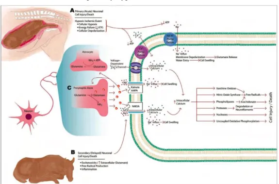
1) Primary neuronal cell death: the hypoxic/anoxic event results in a significant decrease in oxidative phosphorylation in the brain, with concomitant shift toward anaerobic metabolism. This leads to the depletion of adenosine triphosphate (ATP) reserves, accumulation of lactate, and a failure of cellular homeostasis. (Wassink, 2014; McKenzie III, 2018). The Na/K transcellular pumps cannot maintain the ionic gradient, the membrane potential is lost (Wilkins, 2004a) and an intracellular accumulation of sodium, calcium and water occurs (Hahn, 2008; Wassink, 2014; McKenzie III, 2018). This increased intracellular concentration of calcium in the neurons activates calcium-dependent lipases, proteases, and endonucleases (Figure 1). Also, protein synthesis is ceased (Wilkins, 2004a). Following the anoxic cellular depolarization there is the release of the potent excitatory neurotransmitter glutamate from presynaptic vesicles. Glutamate allows calcium to enter the cells by acting on the N-metyl-D-aspartate receptor and opening its channels, leading to neuronal injury by potentiating calcium influx into the neurons. Ultimately, there is a perpetuation of excitotoxicity, causing neuronal cell death and brain injury (Ferriero, 2004; Hahn, 2008; Johnston, 2001; McKenzie III, 2018). When the insult is over, protein synthesis returns to normal in less vulnerable areas of the brain but remains inhibited in specific areas. It seems likely that the loss of protein synthesis can be an early indicator of cell death associated with the primary hypoxic/anoxic insult (Wilkins, 2004a);
Figure 1 - Schematic representation of the pathogenesis of NE, as a result of a hypoxic-ischemic event (HIE). Retrieved from Wong, D., 2011.
2) Delayed neuronal cell death: When the initial acute phase injury is stabilized, it is followed by the second wave of delayed neuronal cell death (McKenzie III, 2018), which is associated with reperfusion injury and can occur during the hypoxic-ischemic episode or hours to days after (Gold, 2017). It is caused by the increased production of oxygen radicals, nitric oxide and inflammatory cell infiltration (McKenzie III, 2018). Also, there’s an imbalance between excitatory and inhibitory neurotransmitters (Wilkins, 2004a). This ultimately leads to cell death and activation of apoptotic cascades, resulting in further tissue damage (Johnston, 2001; McKenzie III, 2018). Besides that, an inflammatory response causes an increase in the blood flow and vascular permeability, which can be associated with the development of edema. It is documented that this excitatory cascade extends over several days from the time of insult and is modifiable (Wilkins, 2015a). In fact, after restoration of oxygenation and circulation, the destructive processes continue creating damage in the brain, over hours, days and possibly weeks (Whitelaw, 2000; Ferriero, 2004). This period gives a therapeutic window during which the full extent of the brain’s damage may be reduced (Whitelaw, 2000). Moreover, it is believed that neurochemical changes (excessive neurotransmitter release), are pivotal in the pathophysiology of secondary neuronal death (Wilkins, 2004a).
Also in human literature, we can find an association between the nature of the event and the severity and distribution of the neurologic damage. For example, a chronic hypoxic-ischemic insult was related with periventricular leukomalacia, whereas an acute hypoxic episode affected mainly the basal ganglia and thalamus. This can be useful in defining neuroprotective strategies and therapies for neonates with NE (Wilkins, 2004a). In situations of decreased oxygen availability, the fetus decreases its rate of growth, being this the reason why some of these foals are born with physical disproportions, such as large head, little muscle mass, small frail body, and little to no fat (Hahn, 2008).
Some mild cases of foals with NE were born without any complications and had a fast recovery, with no behavioral or neurologic deficits (Gold, 2017). Some of these foals have persistence of neuroinhibitory steroids, such as progesterone, pregnenolone, androstenedione, dehydroepiandrosterone, and episterone, which could possibly indicate
suggestive that other factors may contribute to the pathogenesis of this disease (Gold, 2017). It is hypothesize that a post-natal persistence of fetal physiological conditions linked to aberrant activity of the fetal hypothalmic-pituitary-adrenal axis (HPA) and/or elevated pregnane concentrations may also play a role in the pathogenesis of NE (Diesch, 2013).
The HPA function is dynamic in the neonatal foal, being responsible for the maintenance of water homeostasis and cardiovascular, immunologic, and metabolic functions, helping the transition from intra to extrauterine life (Ousey, 2004; Hart, 2009; Barrett, 2010). Before 290 days of gestation, the HPA axis has a basal, unresponsive to stimuli activity, being activated after 300 days of gestation. After this time, HPA axis’s activity increases, leading to the release of cortisol in fetal circulation immediately before birth (Fowden, 2012). Cortisol has a key-role not only in the terminal maturation of the fetus but also for neonatal adaptation for the extrauterine life (Hillman, 2012). This is important in the perinatal period because it integrates a series of events, such as the onset of labor, the acceleration of fetal tissue maturation immediately before birth, the timely onset of colostrum/milk production by the mother, and the impetus to engage in mother-young bonding (Mellor, 1988). All of these processes are of major importance for foal survival and any alteration can seriously jeopardized it (Diesch, 2013).
While in utero, the fetus is under the neuroinhibitory effects of high concentrations of adenosine, progesterone, allopregnanolone, pregnanolone, prostaglandin D2 and a placental neuroinhibitory peptide. This combined with buoyancy, warmth and cushioned tactile stimulation produces somnolence, contributing to the sleep-like state of the fetus (Mellor, 1988; Mellor, 2006; Mellor, 2010; Diesch, 2013). After birth, the onset of consciousness is achieved by the reduction in cerebral cortical inhibition by factors that are unique to life in utero, as well as to an increase in cerebral cortical activation associated with birth (Diesch, 2013). Normal healthy newborn foals have high concentrations of pregnanes at birth that decrease promptly during the first 48 hours of life. A study performed by Aleman et al. (2013) demonstrated that pregnane concentrations in foals with NE remained increased over the normal 48 hour time period, while in healthy neonatal foals declined rapidly to, essentially, zero in the same time frame. Additionally, sick foals affected with other disorders than NE, also had significantly lower progesterone and pregnenolone concentrations at 48 hours compared with birth. This supports the theory that a lack of the normal transition from synthesis to
inhibition of specific neurosteroids for readiness for birth may have a role in the pathogenesis of NE (Aleman, 2013).
As seen in another study, an infusion of allopregnanolone in a healthy foal resulted in signs of sedation and decreased responsiveness to the environment. As the concentration was increased, these signs were more notorious, resulting in dramatic neurobehavioral effects, with the foal in recumbency, stupor, unresponsive to the mare, environment, sound and tactile stimulation. These signs persisted during the infusion and began to fade within 8 minutes after cessation. They kept fading until completely disappear at around 30 minutes after cessation of the infusion, with no long-term effects observed following the infusion (Madigan, 2012).
With this being said, it is thought that NE may comprise of more than one phenotype: foals that suffered hypoxia and ischemia and foals with persistence of fetal hypothalamic-pituitary-adrenocortical axis and increased pregnanes (pregnenolone, progesterone and metabolites) concentrations (Aleman, 2013).
4. CLINICAL SIGNS
Neonatal encephalopathy can produce a wide range of clinical signs (Magdesian, 2014). Most foals may appear healthy at birth but will typically develop central nervous system abnormalities within the first 72 hours of life (McKenzie III, 2018; Bernard, 2018a). In fact, two major categories of NE-affected foals can be distinguished:
• Category 1: includes foals that are normal at birth but develop signs within the first 48 hours of life (MacKay, 2005; Wong, 2011; McKenzie III, 2018);
• Category 2: includes foals that are abnormal at birth, usually associated with documented predisposing factors for NE (MacKay, 2005; McKenzie III, 2018).
Foals from Category 1 usually have an excellent prognosis while foals from Category 2 are associated with worst prognosis for survival (Gold, 2017). The clinical presentation is extremely variable and can include:
• Behavioral changes (Figure 2 and 3): loss of affinity for the mare, inability to find the udder (Wilkins, 2015a), loss of awareness of the environment, inappropriate nursing behavior, head-pressing, and abnormal vocalization (Hahn, 2008; Wong, 2011; McKenzie III, 2018);
• Altered mentation (Figure 4): varying from depression, stupor, somnolence, difficult to arouse or coma to hyperresponsiveness (Hahn, 2008; Wong, 2011; McKenzie III, 2018);
• Cranial nerve dysfunction (Figure 5): loss of suckle reflex, weak tongue tone, tongue protrusion, and dysphagia (Hahn, 2008; McKenzie III, 2018);
• Central nervous system (CNS) dysfunction: hypotonia, tremors, hypertonia, proprioceptive deficits, central blindness, irregular respiratory patterns (Wong, 2011; McKenzie III, 2018) (sometimes leading to
Figure 2 and 3– Foal with NE, showing signs of loss of awareness of the environment and abnormal nursing behavior by nursing the fence (left). Images kindly provided by the
University of Tennessee.
Figure 4 -Foal with somnolence and difficulty to arouse, often seen in foals with NE. Image
kindly provided by the Hospital La Equina.
Figure 5 -Tongue protrusion in foal with NE.
Image kindly provided by the University of Tennessee.
respiratory acidosis, hypoxemia and/or hypercapnia (Giguère, 2008; Tennent-Brown, 2015), opisthotonus, and seizures (McKenzie III, 2018). These can range from mild, abnormal movement of the face and jaw to generalized seizures with recumbency and paddling (MacKay, 2005; Tennent-Brown, 2015). NE is the most common cause of neonatal seizures. They usually manifest within 2 days of life and are associated with long-term seizure risk (Shetty, 2015).
• Other common clinical signs include loss of menace response, fixed dilated pupils, nystagmus, dysuria and wandering (Magdesian, 2014).
Clinical signs are usually related to the cerebrum, but some foals also show signs of brain stem or spinal cord dysfunction, (Tennent-Brown, 2015) often from Category 2 (MacKay, 2005).
The gastrointestinal tract and renal system are commonly affected but the cardiovascular, respiratory, and endocrine systems may also be involved (Wilkins, 2015a). In fact, foals with NE regularly have gastric reflux, feeding intolerance, bloat, meconium retention, colic, and persistent increases of creatinine concentration (Tennent-Brown, 2015; Bernard, 2018a). Oliguria with peripheral edema formation can also be found (Vaala, 2003). As shown in one review of PAS in human neonates, CNS was the most affected body system (82%), followed by the renal system (42%), cardiac (29%), gastrointestinal (29%) and respiratory (26%) systems (Wong, 2011). Limb deficits and generalized spasticity are rare (Vaala, 2003).
5. DIFFERENTIAL DIAGNOSES
The most common differential diagnoses for NE include sepsis-associated encephalopathy, prematurity/dysmaturity and hypoglycemia (Tennent-Brown, 2015). Less common differentials include bacterial meningitis, cerebral trauma, cerebral vascular accidents, congenital abnormalities such as hydrocephalus (Wilkins, 2015a), cerebellar abiotrophy and occipitoatlantoaxial malformation, epilepsy, narcolepsy and cataplexy, botulism, tetanus, metabolic encephalopathies and seizures disorders (McKenzie III, 2018). Hypothermia and hypovolemia should also be considered (Wilkins, 2006).
6. DIAGNOSIS
It is important to clarify that there is no definitive diagnostic test for neonatal encephalopathy (Volpe, 2012; Mackenzie, 2010), so diagnosis is made based on historical information, clinical signs, neurologic examination, elimination of other differentials, and supplementary diagnostic exams (MacKay, 2005; Wong, 2011; McKenzie III, 2018; Bernard, 2018a). These include electroencephalography, brain imaging (computed tomography and magnetic resonance imaging), complete blood count, serum biochemistry, arterial blood gas analysis, blood culture, urinalysis, and measurement of IgG (Wong, 2011).
The clinical signs associated with NE are not specific of this disorder and share similarities with other causes of NE, like sepsis, hypoglycemia and prematurity (Wong, 2011; Gold, 2017). Also, NE can act alone or be complicated by other problems (Gold, 2017), making the diagnosis of NE challenging and very limited (Cruz, 2017; McKenzie III, 2018).
If the birth is attended, gross evaluation of the placenta after delivery can give information regarding fetal well-being. If the membranes are markedly lighter (less than 11% of the foal’s birth mass), it means that their surface area was smaller than normal, therefore not being able to give the adequate nutritional support to the fetus (Pirrone, 2014).
1. History
Foals with history of known risk factors of NE, such as dystocia, delivery by caesarian section, premature delivery, abnormal placenta of the mare, premature placental separation or that required resuscitation after delivery, are expected to develop this condition (Hahn, 2008).
2. Neurologic Exam
A concise neurologic examination is indispensable during the initial evaluation of foals with neurologic signs and when monitoring their progression (MacKay, 2005; Tennent-Brown, 2015). Before initiating this exam, objective history-taking and a complete physical examination should be done (MacKay, 2005; Bernard, 2018a). Some
characteristics that can give the clinician early insights into the neurologic status of the newborn foal are:
• Normal foals should be sternal within the first 5 minutes of life and have a suckle reflex within 20 minutes (it is usually present at birth);
• They should stand within an hour, except if the floor is hard and/or slippery or if the foal is very large (in these cases, a little extra time may be needed); • They are expected to find the mare’s udder and nurse by 2h of age;
• It is expected newborn foals to be inquisitive about the environment surrounding them and to remain close to their dams. Their movements are also typically swift and may appear jerky;
• Normal foals usually defecate soon after standing or following their first meal but some do not defecate for 24 hours;
• When the foal is manipulated or stimulated, it is expected to observe an exaggerated response that must not be confused with the hyperresponsiveness associated with NE. When manipulating the head, they can also have an exaggerated physiologic, horizontal nystagmus, with the fast phase in the direction of head movement;
• Healthy foals may allow their tongue to hang out of the mouth but are able to completely withdraw it when stimulated;
• During the ophthalmologic exam, the clinician must take into consideration that pupils in foals 1 to 2 days old should be equal in size, large, and circular. Over the first week of life, they should decrease in size and becoming more ovoid. Newborn foals do not have menace response until about 2 weeks of life, since it is a learned behavior. Also, direct and indirect pupillary light reflexes should be present, with its rate of constriction being influenced by the degree of excitement;
• A crossed extensor reflex that can last for up 3 weeks and an abrupt patellar reflex may also be present in normal newborn foals (Tennent-Brown, 2015).
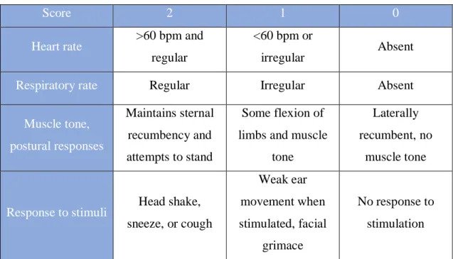
3. APGAR Score
The modified APGAR Scoring System (Table 1) is a visual classifying system used to evaluate neonatal vitality. It can range from zero to ten (Cruz, 2017) and has been developed as a guide of probable level of fetal compromise, identifying the need for medical intervention and defining when resuscitation should be initiated. This method does not alleviate the need of further monitoring (Wilkins, 2006). A low APGAR score and metabolic acidosis are associated with neurologic dysfunction (Volpe, 2012).
Table 1 - APGAR Scoring System. Adapted from Palmer, J. E., 2009.
Score 2 1 0
Heart rate >60 bpm and
regular
<60 bpm or
irregular Absent
Respiratory rate Regular Irregular Absent
Muscle tone, postural responses Maintains sternal recumbency and attempts to stand Some flexion of limbs and muscle
tone
Laterally recumbent, no
muscle tone
Response to stimuli Head shake,
sneeze, or cough Weak ear movement when stimulated, facial grimace No response to stimulation
*The scores of the parcels are summed to derive the APGAR score, which is interpreted the following way: Score 7-8: normal; Score 4-6: mild to moderate asphyxia. The foal should be stimulated and oxygen therapy should be implemented; Score 0-3: severe asphyxia. Aggressive cardiopulmonary resuscitation must be done.
4. Laboratory parameters
Careful analysis of the biochemistry panel in critically ill foals, particularly those with neurological signs, is imperative in the assessment of these patients (Johnson, 2012). We can identify some alterations that are usually present in foals with NE, such as low blood glucose before suckling (normal range values are 93.8-185.6 mg/dL (McAuliffe and Slovis (2008)), hyperlactatemia, hypoxemia and respiratory acidosis (PO2 <60 mmHg; PCO2 >65 mmHg), hypercapnia, metabolic acidosis (ph <7.3;
bicarbonate concentration <20 mEq/L), hypocalcemia and increased serum concentrations of creatinine (normal range values for creatinine are <2 mg/dL and for BUN are <22 mg/dL (Stoneham (2004)) and creatinine kinase (Vaala, 2003; Chaney, 2010). In fact, a study reported that among a 28 foals population with spurious hypercreatininemia included in the study, 20 (71%) had clinical signs of NE. This suggests that these foals did not suffered from renal damage, but rather adverse maternal and/or placental conditions (Chaney, 2010). However, all of these alterations can also be present in a large number of other neonatal diseases (Gold, 2017).
The pancreas and the liver can also be affected, so an insulin-responsive hyperglycemia and increased concentrations of bilirubin and hepatocellular enzyme sorbitol dehydrogenase can also be present (Vaala, 2003).
It has been reported that foals with NE have significantly higher Mg concentration at admission, comparing with healthy, septic, premature/dysmature foals or with other pathologies. This can be explained by the extracellular movement of Mg (an intracellular ion), as consequence of hypoxia, acidosis and probable cellular injury or competition of hydrogen ions with Mg for protein binding (Mariella, 2016). With this being said, plasma total Mg may be helpful in the diagnosis of NE, in addition to historical information and clinical signs (Mariella, 2016).
Thyroid hormones (THs) concentrations are higher in healthy neonatal foals than in adults (Breuhaus, 2014). Total thyroxine (T4) and triiodothyronine (T3) concentrations in normal foals are 14 times higher and free T4 and T3 concentrations are 5 times higher than those in adults (Irvine, 1975). They slowly decrease over the first weeks of life until the adult’s concentrations be achieved (Breuhaus, 2014). Pirrone et al. (2013) studied plasma THs concentrations (T3 and T4) in critically ill foals suffering from NE during their first 7 days of hospitalization and compared those values with those in healthy foals. This study suggests that NE may cause lower THs concentrations than the healthy ones (Pirrone, 2013). However, lower THs concentrations have also been reported for other systemic diseases, such as sepsis and prematurity. In fact, a prognostic value of these hormones in these illnesses has been reported, since there are lower THs concentrations in non-surviving septic and premature foals than in surviving septic and premature foals (Himler, 2012).
5. Imaging modalities
In human medicine, other exams are also made, including electroencephalography (EEG), computed tomography (CT) and magnetic resonance imaging (MRI) (McKenzie III, 2018). MRI is one of the best diagnostic and prognostic modalities used in human infants with neonatal encephalopathy, however, there is a lack of published reports of MRI findings in foals with this condition. In 2017, D. M. Wong published the first report describing the MRI findings associated with a suspected diagnosis of neonatal encephalopathy in a foal and compared these images with those described in human literature. This study showed imaging changes consistent with the counterpart disease in human neonates, such as hyperintensity of the basal nuclei, superficial layers of the ventral cerebral hemispheres, ventral thalamic nuclei and rostral aspect of the ventral midbrain (Wong, 2017). Nevertheless, MRI machines are usually limited to referral centers (McKenzie III, 2018).
The EEG has been used as a diagnostic tool in human medicine to assess cerebral cortical function (Williams, 2008). It might provide information regarding the pathophysiology and the prognosis of this condition. Besides that, it has the advantage that it can be performed continuously to detect seizure activity (Tennent-Brown, 2015). However, an EEG is difficult to perform in an unsedated and unrestrained foal and the sedation and movements can derail the results. (McKenzie III, 2018).
This led to the search of non-invasive alternatives like ultrasonography of the optic nerve sheath diameter and ultrasonographic assessment of the atlanto-occipital space. A recent study showed that the ultrasonographic assessment of the atlanto-occipital space was easier to perform and appeared to offer more potential for obtaining standardized image planes, comparing to the ultrasonography of the optic nerve sheath diameter. It compared the ultrasound measurements in healthy foals with the ones obtained in foals with NE and the results were: the dimension of the spinal cord was smaller in foals with neonatal encephalopathy than healthy foals, with no significant change in the overall size of the spinal canal; the dorsoventral diameter of the ventral spinal artery in longitudinal images was also smaller in foals with NE than in the healthy ones; and spinal canal to spinal cord ratios for both cross sectional area and width were significantly larger in foals with NE than in healthy foals; which shows that the differences between the two groups were independent of the size of the foal. It is also possible that the differences in spinal cord dimensions could be related to increased
intracranial pressure, usually found in some critically ill foals. This approach has been described in the past to be useful to aid the diagnosis of subarachnoid hemorrhage or meningitis (Mackenzie, 2010).
6. Biomarkers
Because of the limitation associated with clinical imaging, the use of body fluid analysis in foals with NE to monitor brain injury and evaluate neuroprotective effects may allow early diagnosis and interventions (Lv, 2015). Two potential biomarkers of NE have been recently documented in a study where the plasma concentration of the phosphorylated axonal forms of neurofilament H (pNF-H) and ubiquitin C-terminal hydrolase 1 (UCHL1) were measured. It was found that the diagnostic performance of UCHL1 was significantly higher than that of pNF-H, with sensitivity of 70% and specificity of 94% for diagnosis of NE. This demonstrates that UCHL1 may have potential as a biomarker of neuronal injury in neonatal foals with signs consistent with NE. This study also referred that pNF-H was heavily concentrated in the white matter and in deep regions of the brain, like the medulla and midbrain, while UCHL1 was more concentrated in the gray matter. This suggests that a blood UCHL1/pNF-H ratio can be useful in localizing the region of the brain damage in foals with NE and other kinds of brain injury (UCHL1 should be increased in any neurologic disease with marked neuron cell death, being unspecific for NE). Despite that, there are some limitations in this study, like the fact that only 33 foals with clinical diagnosis of NE and 17 healthy foals were included and that the measurement of neurobiomarkers is still not available for clinical practice. Nevertheless, this can be a future aid in the ante-mortem diagnosis of NE (Ringger, 2011).
7. Other alternatives
Cerebrospinal fluid collection and analysis is not commonly done but it can help rule out meningitis (Tennent-Brown, 2015). It can be either normal or xanthochromic, with an increase in protein and red blood cells concentration (MacKay, 2005).
Measurement of intracranial pressure has been a diagnostic tool used in infants with NE. There were attempts to adapt this procedure to foals, however, since it is an
invasive technique (requires placement of a subdural catheter) and is associated with certain risks, it led to the search of non-invasive alternatives (Mackenzie, 2010).
Furthermore, measuring the plasma concentrations of neuroactive progestogens derivatives may also be helpful in the diagnosis of NE, but these are also increased in foals with other diseases (Aleman, 2013).
8. Necropsy findings
There are still no post-mortem findings specific of NE, however, neuronal necrosis and/or degeneration within the central nervous system are consistent with ischemia (Lyle-Dugas, 2017). Some lesions usually associated with this condition are epidural subarachnoid, parenchymal and nerve root hemorrhage of the brain and spinal cord, as well as central nervous system edema and necrosis, and hepatic and renal lesions (Gold, 2017). However, in many cases, an obvious cause of hypoxia is not apparent and there is no evidence of hypoxic-ischemic or hemorrhagic brain injury or cerebral edema in foals clinically diagnosed with NE (Tennent-Brown, 2015).
7. TREATMENT
It is difficult to define one single treatment strategy since a wide range of disease processes are associated with NE in foals (McKenzie III, 2018). The treatment of these foals can be not only difficult and very costly (Gold, 2016), but also stressful for the owner (Wilkins, 2015b).
It usually relies on supportive care and treatment of other complications that may arise (Gold, 2017; Bernard, 2018a), with prevention of CNS injury and normalization of nervous system function as the main treatment goals (McKenzie III, 2018). Some standard therapies include oxygen supplementation, maintenance of blood pressure and glucose, control of seizures, general cerebral support (neuroprotectants), correction of metabolic abnormalities, maintenance of tissue perfusion, maintenance of renal function, treatment of gastrointestinal dysfunction, prevention and early treatment of secondary infections (Wilkins, 2004a; Gold, 2017). Excellent nursing care and monitorization are essential to ensure good outcomes (Tennent-Brown, 2015).
1. Supportive care
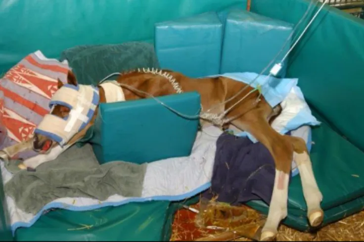
A very important thing to be kept in mind is that outside stimuli need to be minimized, especially if the foals are convulsing or with hyperexcitability (Galvin, 2004). Wrapping the limbs may be
necessary to protect the foal from self-trauma during seizures (Vaala, 2003). Recumbent foals require a dry and warm place, often maintained in sternal recumbency and turned in order to avoid pressure sores (Galvin, 2004) (Figure 6). To prevent secondary corneal ulceration, artificial tears can be applied to the eyes of the foal (Vaala, 2003).
2. Stabilization of the patient systemically
This is the first thing to do, in order to restore CNS perfusion and ensure an adequate delivery of oxygen and glucose (Douglas-Escobar, 2015; McKenzie III, 2018). Identifying the cause(s) of hypotension and correct it with judicious intravenous fluid support is one of the main goals of treatment. If the foal is unable to maintain tissue perfusion with IV fluids alone, administration of inotropes and vasopressors may also be necessary to support the cardiovascular function and to normalize perfusion pressures. (Wilkins, 2004a; Wong, 2011; Tennent-Brown, 2015). Attention to avoid overhydration and hypertension should be kept, because these may cause cerebral edema and potentiate CNS further injury (Wong, 2011; McKenzie III, 2018). These foals need maintenance-type fluids with lower sodium and chloride concentrations and higher potassium concentrations (Tennent-Brown, 2015). Maintenance requirements should be 5 to 6 ml/kg bwt/h of a balanced polyionic fluid. If there is hypokalemia, potassium supplementation is necessary (Galvin, 2004).
Figure 6 - Head protection and environment adaptation to a recumbent, convulsive foal with NE. Image kindly
Blood glucose concentration also need to be normalize without causing hyperglycemia, since it is detrimental in foals with NE (Corley, 2005) and critically ill foals may be intolerant to dextrose infusions (McKenzie III, 2018).
If the foal is anemic, blood transfusion may be needed (McKenzie III, 2018). Also, if there is failure of passive transfer of immunoglobulins (IgG levels <800 mg/dL),an intravenous hyperimmune plasma may be required (Vaala, 2003; Galvin, 2004). Bottle, bucket or nasogastric tube feeding of colostrum or artificial IgG supplement can also be done (Vaala, 2003; Grogan, 2005). Foals in need of colostrum can be fed 250 mL/h for the first 6h-10h. To ensure its quality, the specific density has to be greater than 1.060 (Grogan, 2005).
In mild to severe cases of NE, acidosis may be present with a pH less than 7.3 and bicarbonate concentration less than 20 mmol/L, which needs to be fixed (Galvin, 2004).
The most accurate way to determine the foal’s fluid status is thought to be with urine output paired with non-invasive blood pressure monitoring. If the urine output falls below 66% of all fluids administered, there should be an intervention in order to restore circulating volume and tissue perfusion. Also, the normal reference range of specific gravity of the urine is 1.001-1.009, which can be higher in cases of dehydration or hypovolemia (Galvin, 2004).
3. Broad-spectrum antimicrobial therapy
Because foals with NE can be predisposed to sepsis, prompt and aggressive broad-spectrum antimicrobial therapy is indicated. (Tennent-Brown, 2015; Bernard, 2018a). Attention must be paid to renal function because some antimicrobials have nephrotoxic effects (Tennent-Brown, 2015).
4. Respiratory support
Lower respiratory rates and/or periods of apnea will cause hypoxemia (PaO2 <60 mmHg) and hypercapnia (PaCO3 > 65 mmHg), that need to be corrected (Galvin, 2004). Intranasal oxygen therapy can either be used as a preventive measure or as a direct treatment (Tennent-Brown, 2015). It is often used an indwelling nasal cannula, at an initial rate of 9-10 L/min of oxygen (Corley, 2005).
In weak or recumbent foals, or with centrally mediated hypoventilation, periods of apnea and abnormal breathing patterns, with documented arterial hypoxemia, may need additional respiratory support. This can be done with doxapram (0.02-0.05 mg/Kg/h as a constant rate infusion) (Tennent-Brown, 2015). Although both doxapram and caffeine have been recommended and used as respiratory stimulants in foals with NE, there is not much data regarding its efficacy, being the dosage regimens extrapolated from human studies. Besides that, a study that compared the effects of caffeine and doxapram on respiratory and cardiovascular function in foals with induced respiratory acidosis showed that caffeine is unlikely to improve ventilation and decrease hypercapnia in neonatal foals, since its effects were undistinguishable from those of the placebo group (Giguère, 2007). It has also been demonstrated that doxapram is more effective than caffeine for the fast correction of hypercapnia in foals with NE (Giguère, 2007; Giguère, 2008).
Short-term positive pressure ventilation may also be required in foals with persistent or severe hypoxemia and hypercapnia (Wilkins, 2004a; Tennent-Brown, 2015). Other therapies that have shown some results are the use of melatonin and desferrioxamine, which are free radical scavengers, and the hyperbaric oxygen therapy, which can reduce apoptosis, enhance oxygen radical scavengers, increase brain oxygenation and promote neuronal stem cells in rats with NE (Gold, 2017).
5. Seizure control
In foals with rare or very subtle signs of seizures, specific treatment may not be necessary, however, when there is repeated, generalized seizure activity, it is undoubtedly indicated (McKenzie III, 2018). For seizure control:
Benzodiazepines (diazepam and midazolam): the first-choice therapy in acute seizures
because they have a quick onset of action with minimal depressive effects. IV bolus of Diazepam are ideal to control of emergency and single episode seizures (Morresey, 2009; McKenzie III, 2018). If it show no effect or if there is more than two seizures, then it should be replaced by a constant rate infusion of midazolam (McKenzie III, 2018). This latter has the advantage of allowing frequent assessment of neurologic function, since it has a short half-life and can be reversed if necessary (Tennent-Brown, 2015).
Phenobarbital: is often used in the management of acute seizure episodes that do not
respond to the previous drugs and in recurrent seizure activity (Morresey, 2009). It can cause significant CNS depression when first administered and its half-life can be up to 100 hours in the foal, so special attention must be paid when monitoring neurologic function after administration of this drug (Wilkins, 2004a; McKenzie III, 2018). The therapeutic range is 5-30 µg/mL and serum levels must be monitored to make sure the range is respected (McKenzie III, 2018). If phenobarbital shows no effect in seizure control, then phenytoin therapy should be used (Wilkins, 2004a).
Pentobarbital: only indicated in foals with status epilepticus that failed to be controlled
with other drugs. It is associated with high risk because it causes profound respiratory and cardiovascular depression, hypotension, and low cardiac output (Morresey, 2009; McKenzie III, 2018).
Phenytoin: not commonly used and has unpredictable kinetics, however, its use has been
reported as an anticonvulsant in foals (Morresey, 2009; McKenzie III, 2018).
Potassium bromide: less side effects than phenobarbital and is well tolerated by foals. It
should be used in long-term maintenance in foals with epilepsy. Although the therapeutic range of this drug has not been described for foals, in other species it is 70-240 mg/dL (McKenzie III, 2018).
Besides the use of the anticonvulsants, other measures need to be taken during a seizure, like protect the foal from injury and clean its airways, in order to prevent the onset of negative pressure pulmonary edema (Wilkins, 2004a). The combination of xylazine and ketamine should be avoided in foals with NE because of their association with increase of the intracranial pressure (Wilkins, 2004a).
6. Pharmacological approaches to neuroprotection
Dimethyl sulfoxide (DMSO): has been one of the most widely used drugs for decades.
It has anti-inflammatory, osmotic and diuretic effects and alleged free radical scavenging properties, blocks sodium channel activation, suppresses calcium influx and prevents glutamate excitotoxic cell death. It’s also easy to administrate and has a low cost. Despite
that, there is almost none scientific evidence regarding the use of DMSO in patients with NE and there’s a lack of clinical consensus on its use as well (McKenzie III, 2018). That is probably why the use of dimethyl sulfoxide in neonates has decreased a lot in the past years, being rarely used, as no difference in outcome has been noted (Wilkins, 2004a).
Mannitol: is used to treat cerebral interstitial edema, however, it is minimally effective
in treating cellular edema, which is present in the majority of the cases. Mannitol and dimethyl sulfoxide (DMSO) are only indicated when cellular necrosis and vasogenic edema are present, which is usually in the worst cases (Wilkins, 2004a). One study in human infants showed that the use of mannitol infusion decreased the intracranial pressure and improved cerebral perfusion pressure within 60 minutes (Whitelaw, 2000). However, evidence of efficacy is also lacking and is not frequently used (McKenzie III, 2018).
Magnesium sulfate: has a N-metyl-D-aspartate-receptor antagonist effects, can stabilize
cell membranes, inhibit free radical production, and reduce secondary CNS inflammation and injury (McKenzie III, 2018). Early infusion of magnesium sulfate has been showed to be effective in improving outcome in neonates with severe NE. Its combination with hypothermia could be very beneficial (Bhat, 2009).
Pentoxifyline: has anti-inflammatory and immune-modulating effects and can improve
local tissue perfusion. It is also thought to inhibit the Tumor Necrosis Factor alpha production in foals with NE (McKenzie III, 2018).
Antioxidants: may be a valuable approach to control CNS inflammation. Vitamins C, E
and B1 (thiamine) have all been used in the treatment of foals with NE (McKenzie III, 2018). It has been described the use of thiamine supplementation in IV fluids to support metabolic processes, specially mitochondrial metabolism and membrane Na+, K+ ATPases involved in maintaining cellular fluid balance, however, it’s still unproven in efficacy (Wilkins, 2004a). On the other hand, although allopurinol has shown some promising results in human neonates (McKenzie III, 2018) and good evidence from animal experiments (Whitelaw, 2000), it still remains preclinical in foals, as there are no reports of its use in this specie yet (McKenzie III, 2018).
7. Anti-inflammatories
Nonsteroidal anti-inflammatory drugs, such as flunixin meglumine, are frequently used (Hahn, 2008). Corticosteroids have no role in the treatment of NE (MacKay, 2005).
8. Nutritional support
Critically ill foals have minimal reserves in the form of glycogen and fat and a high metabolic rate relative to body mass. This makes the nutritional support of these animals extremely important, in order to meet their needs. Healthy foals should consume daily 23%-28% of their bodyweight as milk (120-150 kcal/kg bwt/day) during the first 2 to 3 weeks of life, in order to maintain a high metabolic rate and a rapid rate of growth (1-1.3 kg/day in a 50 kg foal). Jose-Cunilleras et al. (2012) stablished the nutritional requirements of critically ill foals, concluding that the resting energy requirements (REE) in these animals were 50 kcal/kg bwt/day, which is much lower than the REE for healthy foals. Also, as critically ill foals recover, their REE increases to values similar with the healthy ones (65-70 kcal/kg bwt/day). So, sick foals that need enteral nutrition, should receive at least 10% of bodyweight in mare’s milk over a 24-hour period, which has 500-570 kcal/L, in order to meet their REE (Jose-Cunilleras, 2012; Bernard, 2018a). This feedings should be made every 1-2 hours (Bernard, 2018a). If parenteral nutrition is required, 50 kcal/kg bwt/day is a reasonable goal of energy provision (Jose-Cunilleras, 2012).
9. Squeeze-induced somnolence technique
First described in 2012 by Toth et al. with promising results. This approach was based on the fact that normal foals may collapse and become flaccid in lateral recumbency during a particular type of restraint, with this phenomenon being called as the flopping reaction or reflex relaxation. All that is needed is a soft linen rope (6.1 m in length and 1.27 cm in diameter). First, a bow-line knot is used to secure the rope around the neck and under the shoulder, to prevent tightening of that segment (it could cause pressure in the trachea and jugular veins). Then, two half-hitch knots are used to loop the rope around the thorax and abdomen 5 to 25 cm from each other and perpendicular to the vertebral column. The half-hitch knots are positioned directly on the dorsal thoracolumbar area.
After that, one person should stand behind the foal and pull on the rope, resulting in a generalized squeezing of the foal, while another person holds the foal and assists as it lays down. Tension on the rope must be maintained during 20 minutes. After that, pressure is released and it has been reported that foals woke up without signs of NE (Aleman, 2017).
A survey showed that foals that received this procedure with or without medical therapy were 3.7 times more likely to have a faster recovery than the ones that did not receive it. Squeezed foals had faster and higher recovery rates at different time points and were 15.1 times more likely to recover in less than 1h than non-squeezed foals. In addition, foals receiving only the squeeze procedure had 17.5 times more chances to recover within the first 24h than foals treated only medically (Aleman, 2017). This technique showed no adverse effects, so it can be considered a safe procedure (Toth, 2012; Aleman, 2017).
10. Hypothermia
In human infants with PAS, the use of regional hypothermia is the actual major treatment modality. It appears to slow the metabolism down with an inhibition of inflammation and apoptosis (Douglas-Escobar, 2015; Sarkar, 2015; Gold, 2017) and decrease intracellular edema and neuronal death (Wilkins, 2004a). This implies cooling the patient to 33.5ºC - 35ºC either with whole body hypothermia or selective head-cooling approaches. This procedure has been confirmed to be beneficial in human neonates with NE, as it reduces mortality without increasing long-term neurologic damage in the survivors (Douglas-Escobar, 2015; Sarkar, 2015; McKenzie III, 2018). As any procedure, it is also associated with some side effects, such as sinus bradycardia, thrombocytopenia, overcooling, skin problems, altered drug metabolism, and an increased risk of seizure during the rewarming period (Sarkar, 2015; McKenzie III, 2018). However, as promising this new approach seem to be, there is still not much information regardless to the use of hypothermia in neonatal foals (Gold, 2017), so the logistics regarding the appropriate technique of cooling, patient selection, and duration of the cooling period needs to be defined before its clinical application in equine medicine (McKenzie III, 2018).
11. Other treatment modalities
There’s a wide range of new therapies being investigated in human infants with NE with promising results, including inhaled xenon (anti-excitotoxic), melatonin (antioxidant), erythropoietin (growth factor), and stem cells therapy (McKenzie III, 2018).
8. PROGNOSIS
It is highly desirable to provide the owner a prognosis for both survival and athletic outcome of the foal as soon as possible (Wilkins, 2015b). The main reasons leading to euthanasia in foals are poor prognosis for survival, poor athletic outcome, and financial constraint (Dembek, 2014).
Prognosis of NE depends on the severity of injury (Hagberg, 2015; Bernard, 2018a) but is usually good to excellent in uncomplicated cases (Lyle-Dugas, 2017). It is expected that 60% to 80% of foals with this condition will recover fully without neurological sequelae (Magdesian, 2014) and with productive athletics outcomes (Wilkins, 2004a). Long-term neurologic disabilities are rare and may include inability to suck from the dam, recurrent seizures, prolonged visual impairment, residual spasticity, and unusual docility as adults (Vaala, 1994; Bernard, 1995; Vaala, 2003). It was observed that if the foal survives the first 5 days with neurologic improvement, the prognosis is good with no long-term neurologic disabilities (Bernard, 1995). In humans, it was stablished that infants who survive the first 72h of age, typically improve over the next days/weeks (Wong, 2011).
A recent study showed that foals with failure of passive transfer immunity or with at least one complication/comorbidity during hospitalization were less likely to survive. Foals with seizure activity within the first 24h of hospitalization were less likely to survive as well (Gold, 2016).
According to another study, the reasons associated with death or euthanasia were the presence of pneumonia, sepsis or sepsis-associated complications, primary neurological disease or multi-organ failure secondary to ischemia (Lyle-Dugas, 2017). Besides that, foals with high total calcium or low alkaline phosphatase at admission, in recumbency, treated with vasopressors/inotropes or that had multiple comorbidities had significantly less chance to survive (Lyle-Dugas, 2017). On the other hand, pH greater