Accepted Manuscript
Antileishmanial activity of meroditerpenoids from the macroalgae Cystoseira baccata Carolina Bruno-de-Sousa, Katkam N. Gangadhar, Thiago R. Morais, Geanne A.A. Conserva, Catarina Vizetto-Duarte, Hugo Pereira, Márcia D. Laurenti, Lenea Campino, Debora Levy, Miriam Uemi, Luísa Barreira, Luísa Custódio, Luiz Felipe D. Passero, João Henrique G. Lago, João Varela
PII: S0014-4894(17)30037-1 DOI: 10.1016/j.exppara.2017.01.002 Reference: YEXPR 7351
To appear in: Experimental Parasitology Received Date: 25 May 2016
Revised Date: 10 November 2016 Accepted Date: 22 January 2017
Please cite this article as: Bruno-de-Sousa, C., Gangadhar, K.N., Morais, T.R., Conserva, G.A.A., Vizetto-Duarte, C., Pereira, H., Laurenti, M.D., Campino, L., Levy, D., Uemi, M., Barreira, L., Custódio, L., Passero, L.F.D., Lago, J.H.G., Varela, J., Antileishmanial activity of meroditerpenoids from the macroalgae Cystoseira baccata, Experimental Parasitology (2017), doi: 10.1016/j.exppara.2017.01.002. This is a PDF file of an unedited manuscript that has been accepted for publication. As a service to our customers we are providing this early version of the manuscript. The manuscript will undergo copyediting, typesetting, and review of the resulting proof before it is published in its final form. Please note that during the production process errors may be discovered which could affect the content, and all legal disclaimers that apply to the journal pertain.
M
AN
US
CR
IP
T
AC
CE
PT
ED
ACCEPTED MANUSCRIPT
M
AN
US
CR
IP
T
AC
CE
PT
ED
ACCEPTED MANUSCRIPT
Antileishmanial activity of meroditerpenoids from the macroalgae
1
Cystoseira baccata
2
3
Carolina Bruno-de-Sousaa, Katkam N. Gangadhara,b, Thiago R. Moraisc, 4
Geanne A. A. Conservac, Catarina Vizetto-Duartea, Hugo Pereiraa, Márcia D. Laurentid, 5
Lenea Campinoe,f, Debora Levyg, Miriam Uemic, Luísa Barreiraa, Luísa Custódioa, 6
Luiz Felipe D. Passerod,h, João Henrique G. Lagoc,*, João Varelaa,* 7
8
9
a
Centro de Ciências do Mar, Universidade do Algarve, Campus de Gambelas, Faro, Portugal 10
b
Instituto de Tecnologia Química e Biológica, Universidade Nova de Lisboa, Oeiras, 11
Portugal 12
c
Departamento de Ciências Exatas e da Terra, Instituto de Ciências Ambientais, Químicas e 13
Farmacêuticas, Universidade Federal de São Paulo, Diadema, SP, Brazil 14
d
Laboratório de Patologia das Moléstias Infecciosas (LIM-50), Departamento de Patologia, 15
Faculdade de Medicina, Universidade de São Paulo, São Paulo, Brazil 16
e
Global Health and Tropical Medicine Centre, Instituto de Higiene e Medicina Tropical, 17
Universidade Nova de Lisboa, Lisboa, Portugal 18
f
Departamento de Ciências Biomédicas e Medicina, Universidade do Algarve, Campus de 19
Gambelas, Faro, Portugal 20
g
Laboratório de Genética e Hematologia Molecular (LIM-31), Departamento de Clinica 21
Médica, Faculdade de Medicina, Universidade de São Paulo, São Paulo, Brazil 22
h
São Vicente Unit, Paulista Coastal Campus, Universidade Estadual Paulista Julio de 23
Mesquita Filho, São Vicente, SP, Brazil 24
25
* Corresponding authors: 26
Centre of Marine Sciences, University of Algarve, Campus de Gambelas, 8005-139 Faro, 27
Portugal. Tel.: +351-289-800-051; Fax: +351-289-800-051. E-mail address: [email protected] 28
(J. Varela) 29
Departamento de Ciências Exatas e da Terra, Instituto de Ciências Ambientais, Químicas e 30
Farmacêuticas, Universidade Federal de São Paulo, 09972-270, Diadema, SP, Brazil. Tel.: 31
+55-(11)-3091-6513; E-mail address: [email protected] (J.H.G. Lago) 32
M
AN
US
CR
IP
T
AC
CE
PT
ED
ACCEPTED MANUSCRIPT
Abstract
33The development of novel drugs for the treatment of leishmaniases continues to be crucial 34
to overcome the severe impacts of these diseases on human and animal health. Several 35
bioactivities have been described in extracts from macroalgae belonging to the Cystoseira 36
genus. However, none of the studies has reported the chemical compounds responsible for the 37
antileishmanial activity observed upon incubation of the parasite with the aforementioned 38
extracts. Thus, this work aimed to isolate and characterize the molecules present in a hexane 39
extract of Cystoseira baccata that was found to be bioactive against Leishmania infantum in a 40
previous screening effort. A bioactivity-guided fractionation of the C. baccata extract was 41
carried out and the inhibitory potential of the isolated compounds was evaluated via the MTT 42
assay against promastigotes and murine macrophages as well as direct counting against 43
intracellular amastigotes. Moreover, the promastigote ultrastructure, DNA fragmentation and 44
changes in the mitochondrial potential were assessed to unravel their mechanism of action. In 45
this process, two antileishmanial meroditerpenoids, (3R)- and (3S)-tetraprenyltoluquinol 46
(1a/1b) and (3R)- and (3S)-tetraprenyltoluquinone (2a/2b), were isolated. Compounds 1 and 47
2 inhibited the growth of the L. infantum promastigotes (IC50 = 44.9 ± 4.3 and 94.4 ± 10.1
48
µM, respectively), inducing cytoplasmic vacuolization and the presence of coiled 49
multilamellar structures in mitochondria as well as an intense disruption of the mitochondrial 50
membrane potential. Compound 1 decreased the intracellular infection index (IC50 = 25.0 ±
51
4.1 µM), while compound 2 eliminated 50% of the intracellular amastigotes at a 52
concentration > 88.0 µM. This work identified compound 2 as a novel metabolite and 53
compound 1 as a biochemical isolated from Cystoseira algae displaying antileishmanial 54
activity. Compound 1 can thus be an interesting scaffold for the development of novel 55
chemotherapeutic molecules for canine and human visceral leishmaniases studies. This work 56
reinforces the evidence of the marine environment as source of novel molecules. 57
58
Keywords
59
Leishmania infantum; macroalgae; Cystoseira baccata; meroterpenoids; tetraprenyltoluquinol;
60 tetraprenyltoluquinone. 61 62 63 64 65
M
AN
US
CR
IP
T
AC
CE
PT
ED
ACCEPTED MANUSCRIPT
Abbreviations 66BALB/c, albino mouse laboratory-bred strain of the house mouse; 67
CC
50, cytotoxic concentration that causes the death of 50% of the viable cells;
68
COSY, correlation spectroscopy; 69
DEPT, distortionless enhancement by polarization transfer spectrometry; 70
FBS, fetal bovine serum; 71
HMBC, heteronuclear multiple-bond correlation spectroscopy; 72
HRESIMS; high-resolution electrospray ionisation mass spectrometry; 73
HSQC, heteronuclear single-quantum correlation spectroscopy; 74
IC
50, half-maximal inhibitory concentration;
75
IR, infrared; 76
LRESIMS, low-resolution electrospray ionisation mass spectrometry; 77
MTT, 3-(4,5-dimethylthiazol-2-yl)-2,5-diphenyltetrazolium bromide; 78
NMR, nuclear magnetic resonance spectroscopy; 79
NOESY, nuclear Overhauser effect spectroscopy; 80
RCF, relative centrifugal force; 81
SDS, sodium dodecyl sulfate; 82 TLC, thin-layer chromatography; 83 TMS, tetramethylsilane; 84 UV, ultraviolet; 85
∆ψm, mitochondrial membrane potential. 86
M
AN
US
CR
IP
T
AC
CE
PT
ED
ACCEPTED MANUSCRIPT
1. Introduction 88Leishmaniases are a group of infectious diseases caused by obligate intracellular 89
protozoa of the Leishmania genus. Endemic in 98 tropical and subtropical countries and 90
affecting 12 million people, leishmaniases may entail cutaneous, mucocutaneous and diffuse 91
forms as well as the potentially fatal visceral form (Alvar et al., 2012). Visceral leishmaniasis 92
causes considerable morbidity in 200-400 thousand individuals every year, with extreme 93
suffering and financial loss, especially in the poorest populations of the Indian subcontinent 94
(Mondal et al., 2014). Currently, leishmaniases are among the most neglected tropical 95
diseases, facing problems of resistance of the parasite to the available therapeutic molecules. 96
The need for the discovery and development of alternative drugs allowing more efficient and 97
effective treatments is thus quite urgent (Freitas-Junior et al., 2012). 98
Nowadays, marine natural products are recognized as powerful reservoirs of novel, 99
chemically diverse molecules with wide applicability to health sciences (Tempone et al., 100
2011). Occurring worldwide, mainly in the rocky substrates of the Mediterranean Sea and the 101
adjoining Atlantic coasts, Cystoseira C. Agardh (1820) genus encompasses 39 species of 102
brown macroalgae (Guiry and Guiry, 2015). Several bioactivities such as anti-inflammatory, 103
antiproliferative, antioxidant (Mhadhebi et al., 2011), enzyme inhibitory (Ghannadi et al., 104
2013), cytotoxic (Khanavi et al., 2010), antifungal (Lopes et al., 2013), antiviral (Ibraheem et 105
al., 2012), antibacterial (Tajbakhsh et al., 2011) and antiprotozoal (Spavieri et al., 2010) have 106
been detected in this algal genus. Despite the extensive chemical studies available for the 107
Cystoseira genus, there have been only a few reports describing the antileishmanial potential
108
effects of its crude extracts, and no information was found on the compounds responsible for 109
the inhibitory effects on the Leishmania parasites (Amico, 1995; de Los Reyes et al., 2012). 110
As part of ongoing research on the identification of antileishmanial compounds from the 111
Cystoseira genus, this work describes the bioactivity-guided fractionation of the hexane
112
extract from Cystoseira baccata and the effect of the extract, fractions and isolated 113
compounds on the promastigote and amastigote forms of Leishmania infantum. 114
115
2. Material and Methods
116
2.1 General Experimental Procedures
117
Optical rotations were measured in a JASCO DIP-370 digital polarimeter (Na filter, λ = 118
588 nm). UV spectra were recorded using a UV/visible Shimadzu 1650-PC 119
spectrophotometer. IR spectra were obtained with a Shimadzu IR Prestige-21 120
M
AN
US
CR
IP
T
AC
CE
PT
ED
ACCEPTED MANUSCRIPT
spectrophotometer. 1H, 13C, DEPT, COSY, HSQC, HMBC and NOESY NMR spectra were 121
recorded in a Bruker Avance III 500 spectrometer, operating at 500 and 125 MHz, to 1H and 122
13
C nuclei, respectively. CDCl3 (Aldrich) was used as the solvent with TMS as the internal
123
standard. HRESIMS spectra were measured with a Bruker Daltonics MicroTOF QII 124
spectrometer while LRESIMS spectra were recorded on a VG Platform II spectrometer. 125
Silica gel (Merck, 230–400 mesh) and Sephadex LH-20 (Amersham Biosciences) were used 126
for column chromatographic separation, while silica gel 60 PF254 (Merck) was used for
127
analytical (0.25 mm) and preparative TLC (1.0 mm). 128
129
2.2Algal material
130
Cystoseira baccata biomass was collected in July 2012 in Areosa, Viana do Castelo,
131
Portugal (41º42’27.60’’N, 8º51’44.90’’W). After collection, biomass was cleaned and 132
cryodesiccated. Voucher specimen (MB-1) was deposited within the Laboratory of the 133
Marine Biotechnology Group - MarBiotech at the Centre of the Marine Sciences of the 134
University of Algarve (Faro, Portugal). 135
136
2.3Extraction and isolation of compounds
137
Dried and powdered biomass (120 g) was exhaustively extracted with hexane in a Soxhlet 138
apparatus. After evaporation of the solvent under reduced pressure, 1.3 g of crude extract 139
were obtained. Part of this extract (0.6 g) was subject to column chromatography over SiO2
140
eluted with hexane containing increasing amounts of EtOAc (up to 100%), followed with 141
CHCl3 containing increasing amounts of MeOH (up to 100%), generating 13 fractions (1 –
142
13). As fraction 10 (370.0 mg) displayed activity towards promastigote forms of L infantum, 143
it was fractionated over SiO2 column, and eluted with hexane:EtOAc 1:1 yielding 6
sub-144
fractions (A – F). Bioactive sub-fraction E (195 mg) was purified in a Sephadex LH-20 145
column being eluted with hexane:CH2Cl2 1:4, CH2Cl2:Me2CO 3:2 and 1:1 (Cardellina II,
146
1983) originating 4 groups (E1 – E4). Bioactive group E4 (65.3 mg) was subjected to 147
preparative TLC (hexane-EtOAc, 7:3, twice) to afford compounds 1a/1b (23.2 mg; 0.30%) 148
and 2a/2b (2.5 mg; 0.04%) (Fig.1). 149
3R – tetraprenyltoluquinol (1a) and 3S – tetraprenyltoluquinol (1b). Yellowish oil; 1H 150
NMR and 13C NMR (500 MHz, CDCl3) data, see Table 1; LRESIMS m/z 441 [M+H]+ and
151
463 [M + Na]+ (calcd for C28H41O4, 441, and C28H40O4Na, 463, respectively).
M
AN
US
CR
IP
T
AC
CE
PT
ED
ACCEPTED MANUSCRIPT
3R – tetraprenyltoluquinone (2a) and 3S – tetraprenyltoluquinone (2b). Colourless oil;
153
[α]D25 = + 0.06 (c 0.15, CHCl3); UV (MeOH) λmax (log ε) 352 (2.0), 248 (3.4) nm; IR (KBr)
154
νmax 3400, 1670, 1480, 1180, 1060 cm-1; 1H and 13C NMR (500 MHz, CDCl3), see Table 1
155
and Fig. 2; HRESIMS (positive mode) m/z 455.2776 [M+H]+ and 477.2604 [M+Na]+ (calcd 156
for C28H39O5 and C28H38O5Na, 455.2797 and 477.2616, respectively).
157
158
2.4. Parasites, mammalian cells and animal maintenance
159
L. infantum strain (MHOM/PT/88/IMT-151) promastigotes were obtained from the
160
cryobank of the Instituto de Higiene e Medicina Tropical (Universidade Nova de Lisboa, 161
Portugal) and cultivated in M199 medium supplemented with 10% foetal bovine serum (FBS), 162
penicillin (10 U/L), streptomycin (0.01 mg/L) and 2% of human male urine at 25 ºC. 163
Peritoneal macrophages from BALB/c mice were cultivated in RPMI-1640 medium 164
supplemented with 10% FBS, L-glutamine (2 mM), penicillin (50 U/L) and streptomycin 165
(0.05 mg/L) at 37 ºC in humidified atmosphere with 5% CO2. BALB/c mice were obtained in
166
the Animal Facility of the School of Medicine of São Paulo University – Brazil. These 167
animals were maintained in accordance with the institutional guidelines regarding the welfare 168
of experimental animals and with the approval of the Animal Ethics Committee of São Paulo 169
University (322/12). 170
171
2.5. Activity against Leishmania promastigotes
172
For the determination of the antileishmanial activity, L. infantum promastigotes in 173
stationary phase (2×10⁶ parasites/mL) were incubated with the hexane extract at a 174
concentration of 250 µg/mL for 24h on 96-well plates. Using the same methodology, the 175
fractions obtained during the bioactivity-guided fractionation were tested at a concentration 176
of 50 µg/mL. At a later stage, compounds 1 and 2 were added at concentrations ranging from 177
0.9 to 227.0 and 0.9 to 220.0 µM, respectively. Parasites treated with miltefosine at the half 178
maximal inhibitory concentration (IC50 = 23.1 µM) were used as positive control.
179
Promastigotes incubated with M199 medium were used as negative control. Parasite viability 180
was determined by the MTT colorimetric assay (Dutta et al., 2005; Dal Picolo et al., 2014). 181
Briefly, after incubation plates were centrifuged at 10 ºC, using an RCF of 1479 × g for 10 182
min, washed three times with PBS, and supernatants discarded. Afterwards, 50 µL of MTT (5 183
mg/mL in PBS) were added to each well and plates were re-incubated at 37 ºC for 2 h. Upon 184
incubation, 50 µL of SDS were added to each well and plates were incubated for 18 h in order 185
M
AN
US
CR
IP
T
AC
CE
PT
ED
ACCEPTED MANUSCRIPT
to dissolve the formazan crystals. Absorbance was measured at 590 nm using a Thermo 186
Scientifc Multiskan™ FC Microplate Photometer. Results were expressed in terms of parasite 187
viability (%) relative to non-treated parasites and the half maximal inhibitory concentration 188
(IC50; µM).
189
190
2.6. Ultrastructural alterations of the promastigotes
191
L. infantum promastigotes in stationary phase (2 x 10⁶ cells/mL) were incubated at 25 ºC 192
for 24 h on 96-well plates with compounds 1 and 2 at their IC50 values, i.e. 44.9 µM of 94.4
193
µM, respectively. Non-treated promastigotes were used as negative control. After incubation, 194
the plate was centrifuged at 1479 × g for 10 min at 4 ºC, and washed with PBS three times. 195
Pellets were fixed in 0.1% tannic acid dissolved in 2.0% glutaraldehyde in a 0.15 M 196
phosphate buffer pH 7.2 and incubated for 1h at 4°C. These were afterwards contrasted in 1% 197
osmium tetroxide and a 0.5% uranyl acetate solution for 12 h; then the samples were 198
embedded in araldite resin (Yamamoto et al., 2015). Ultrathin sections (70 nm), obtained 199
with a ultramicrotome Reichert and double contrasted with 2% uranyl acetate and 0.5% lead 200
citrate, were examined using a JEOL 1010 transmission electron microscope. 201
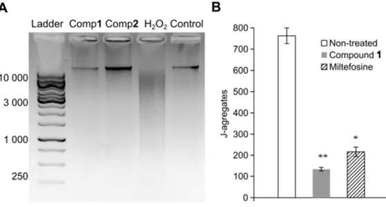
202
2.7. Promastigotes DNA integrity
203
To detect whether the compounds induced fragmentation on L. infantum nuclear DNA, 204
promastigote forms in stationary phase of growth (2 x 10⁸ cells) were incubated with IC50
205
concentrations of compounds 1 (44.9 µM), 2 (94.4 µM) and hydrogen peroxide (6.2 µM) as 206
an inductor of DNA damage in parasites (Das et al., 2001) for 24 h at 25 ºC. Non-treated cells 207
were used as control. After incubation, plates were centrifuged at 1479 × g for 10 min at 4 ºC, 208
and the supernatants discarded. Parasites pellets were extracted with a Macherey-Nagel 209
nucleoSpin® Blood kit according with the manufacturer recommendations and ran on a 2% 210
agarose gel, 100 V for 90 min. 211
212
2.8. Promastigote transmembrane mitochondrial potential
213
In order to evaluate the influence of compound 1 on the promastigote mitochondrial 214
membrane potential (∆Ψm), parasites in the stationary phase (2×10⁶ parasites/mL) were 215
incubated with compound 1 and miltefosine at their IC50 values (44.9 and 23.1 µM,
216
respectively) for 24h on 96-well plates. Mitochondrial membrane potential was evaluated 217
M
AN
US
CR
IP
T
AC
CE
PT
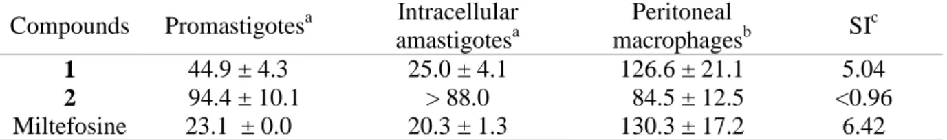
ED
ACCEPTED MANUSCRIPT
using the widefield automated microscope Mitoscreen Kit (BD Biosciences) according to the 218
manufacturer’s recommendations (Levy et al., 2014; Yamamoto et al., 2015). Briefly, cells 219
were incubated with working solution, containing the JC-1 (5,5,6,6-tetrachloro-1,1,3,3-220
tetraethylbenzimidazolylcarbocyanine iodide) fluorochrome, for 15 min at 37 °C in an 221
atmosphere of 5% CO2. ∆Ψm induces the uptake of JC-1 monomers into the functional
222
mitochondria. Once inside the organelle, JC-1 monomers aggregate, exhibiting high levels of 223
red fluorescence and ∆Ψm is assessed through the determination of the presence of JC-1 224
fluorochrome inside the mitochondria. ImageXpress® Micro XLS Widefield High-Content 225
Analysis System and transfluor MetaXpress software were used to determine the presence of 226
J-aggregates in nine sites per well and three wells per treatment. ∆Ψm was expressed as a 227
percentage of J-aggregates per cell. 228
229
2.9. Cytotoxicity against murine macrophages
230
To determine the compounds toxicity in vitro, murine peritoneal macrophages, were 231
seeded in RPMI-1640 at a density of 106 cells/mL and incubated overnight at 37 ºC in 232
humidified atmosphere with 5% CO2, allowing the cells to adhere to the plate background.
233
Compounds 1 and 2 were tested for 24h at concentrations ranging from 0.9 to 227.0 and 0.9 234
to 220.0 µM, respectively. Miltefosine control cells were incubated with RPMI-1640 medium 235
at concentrations from 3.8 up to 490.7 µM. Cell viability was evaluated by the MTT 236
colorimetric assay (Ferrari et al., 1990; Dal Picolo et al., 2014), as described above, for the 237
determination of the activity against Leishmania promastigotes. Absorbance was measured at 238
590 nm using a Thermo Scientific Multiskan™ FC Microplate Photometer. Results were 239
expressed in terms of the cytotoxic concentration causing a 50% decrease in cell viability 240
(CC50; µM) relative to non-treated cells (100 %).
241
242
2.10. Activity against Leishmania intracellular amastigotes and NO production
243
Peritoneal macrophages of BALB/c mice were collected by intraperitoneal lavage, seeded 244
on 24-well plates (105 cells/mL) and incubated at 37ºC with 5% CO2 during 2h for cell
245
attachment. Afterwards, L. infantum promastigotes in stationary phase were added to each 246
well at an infection ratio of 10 promastigotes per cell, being further incubated at 37 ºC for 247
24h. Infected macrophages were treated with compounds 1 and 2 at concentrations ranging 248
from 7 to 90 µM to determine the corresponding IC50. Supernatants were collected for nitric
249
oxide (NO) determination after 24h and intracellular amastigote burden was microscopically 250
M
AN
US
CR
IP
T
AC
CE
PT
ED
ACCEPTED MANUSCRIPT
assessed upon Giemsa staining for determination of the infection index [% of infected 251
macrophages × internalized amastigote forms / macrophage)] (Passero et al., 2015) and the 252
inhibitory concentration allowing 50% reduction of the infection index (IC50) was estimated.
253
Miltefosine was used as positive control. Culture supernatants of treated and control 254
macrophages were used for NO determination that was performed using the Measure-iTTM 255
High-Sensitivity Nitrite Assay Kit in accordance with the manufacturer's recommendations 256
(Life Technologies). The NO concentration was determined using a calibration curve 257
prepared with several known concentrations (2.75, 5.5, 11, 22, 33, 44 and 55 µM) of nitrite as 258
standard. Results were expressed as NO production (µM) and compared with untreated 259
infected and non-infected macrophages. The selectivity index (SI) was obtained by 260
calculating the ratio of the CC50 of the macrophage by the IC50 of the intracellular
261 amastigotes. 262 263 2.11. Statistical analysis 264
Bioassays results were expressed as mean ± standard error of the mean (SEM) of 265
replicates samples from at least two independent assays. The IC50 values were calculated
266
fitting the data as a non-linear regression using a dose-response inhibitory model, in the 267
GraphPad Prism V 5.0 program. Student’s t-test was used to determine whether differences 268
between means were significant at different levels (p < 0.05 and p < 0.01). 269
3. Results and Discussion
270
The hexane extract from the C. baccata was incubated with promastigote forms of L. 271
infantum for 24h, and cell viability was determined by means of the MTT assay. As this
272
extract decreased the viability of the parasite by 74% at a concentration of 250 µg/mL, it was 273
selected for further study. Bioactivity-guided fractionation afforded compounds 1 and 2 274
(Fig. 1). 275
Compound 1 was obtained as an optically active oil [α]D = + 17.8° (CHCl3, c 2.7).
276
Structural evidence was obtained by analysis of NMR (1H, 13C and DEPT 135°), HREIMS 277
spectra and comparison with those data previously reported in the literature to (3R)-(1a) and 278
(3S)-(1b) tetraprenyltoluquinol, previously isolated from C. baccata (Valls et al., 1993). In 279
addition, some corrections in the attributions of chemical shifts of C-18 and C-19 in 13C 280
NMR spectrum were carried out, based on the HMBC spectral analysis (Table 1). Compound 281
2, also obtained as an optically active colourless oil [α]D = + 0.06° (CHCl3, c 0.15), appeared
M
AN
US
CR
IP
T
AC
CE
PT
ED
ACCEPTED MANUSCRIPT
to be homogeneous on the TLC chromatograms, revealing that it is a mixture of closely 283
related derivatives. The 1H NMR spectrum of compound 2 revealed some similarities with 284
compound 1 - two peaks assigned to hydrogens of aromatic ring at δH 7.15 (d, J = 3.0 Hz,
H-285
3’) and 7.00 (d, J = 3.0 Hz, H-5’), one methoxyl group at δH 3.78 (s) as well as five singlets
286
assigned to methyl groups at δH 1.20 (H-20), 1.25/1.26 (H-17), 1.13/1.11 (H-16), 1.09/1.04
287
(H-18), and 0.91/0.83 (H-19). 13C and DEPT 135° NMR spectra confirmed the presence of 288
aromatic ring due the peaks at range δC 151.9 – 114.6 (C-1’ – C-6’), and one methoxyl group
289
at δC 55.7. Additionally, peaks assigned to a carbonyl group at δC 192.2/192.1 (C-1), to
290
carbinolic carbons at δC 81.3/81.2 (C-3) and 71.0 (C-15) as well as an α,β-unsaturated
291
carbonyl carbon at δC 153.3/154.3 (C-5), 133.5/134.0 (C-13) and 208.0/208.1 (C-12) were
292
observed. Finally, HRESIMS showed the [M+H]+ and [M + Na]+ quasi-molecular ion peaks 293
at m/z 455.2776 and 477.2604, respectively, indicating the molecular formula C28H38O5. The
294
connectivity between hydrogens and carbon atoms was revealed by analysis of the HMBC 295
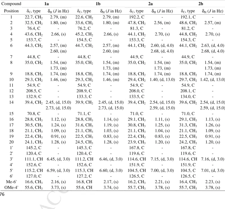
spectrum as showed in Fig. 2. The correlations between signals at δH 7.15 (H-3’) and
296
2.56/2.57 (H-2) with δC 192.2/192.1 (C-1) as well as between δH 2.70 (H-4) with δC 81.3/81.2
297
(C-3) and 133.5/134.0 (C-13) indicated that compound 2 contained one additional carbonyl 298
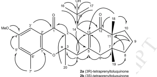
group at C-1. Based on these results, it was possible to identify 2 as epimers of (3R)-(2a) and 299
(3S)-(2b) tetraprenyltoluquinones. 300
In vitro antiparasitic activity and cytotoxic studies of the compounds 1 and 2 were
301
evaluated by the colorimetric MTT method against promastigote forms of L. infantum and 302
murine macrophages, respectively (Table 2). Compound 1 displayed an IC50 value of 44.9 ±
303
4.3 µM against promastigote forms of L. infantum. The cytotoxicity against mouse peritoneal 304
macrophages (CC50 = 126.6 ± 21.1 µM) was similar to that of the reference drug, miltefosine
305
(130.3 ± 17.2 µM). Compound 2 showed lower activity against the promastigote forms (IC50
306
= 94.4 ± 10.1 µM), and higher toxicity to the mouse peritoneal macrophages (CC50 = 84.5 ±
307
12.5 µM). 308
To assess the alterations induced by the compounds on the promastigotes forms of L. 309
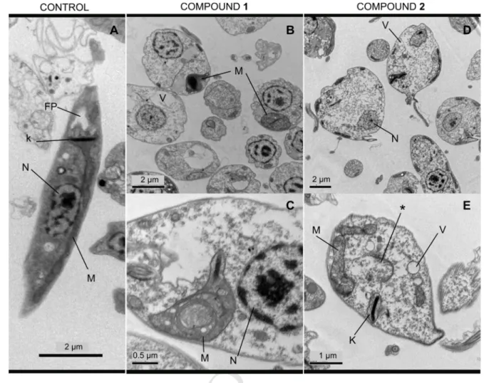
infantum, transmission electron microscopy images were acquired (Fig. 3). Important
310
changes were observed with both treatments, including loss of the typical fusiform shape (Fig. 311
3A). Ultrastructural analysis revealed morphologic changes in parasites treated with the IC50
312
concentrations of both compounds 1 (Figs. 3B and 3C) and 2 (Figs. 3D and 3E). Moreover, 313
cellular vacuolization was observed, which might be a consequence of cytoplasmic organelle 314
disruption (Figs. 3B and 3D). When treated with compound 1, parasites presented coiled 315
M
AN
US
CR
IP
T
AC
CE
PT
ED
ACCEPTED MANUSCRIPT
multilamellar structures within the mitochondria (Fig. 3C). These structures have been shown 316
to be a consequence of starvation processes caused by deficient mitochondrial activity or 317
autophagic mechanisms caused by the action of chemical compounds on these organelles 318
(Lockshin and Zakeri, 2004). If left unchecked, both processes may result in the removal of 319
the damaged organelles as well as cell death (Nishikawa et al., 2010). Previous studies have 320
described similar structures in promastigotes of different Leishmania species treated with 321
distinct natural products (Monte Neto et al., 2011). Compound 2 induced noticeable changes 322
in the ultrastructure of the cell, in particular the occurrence of pyknotic nuclei, which was 323
accompanied by the disappearance of the chromatin associated with the nuclear inner 324
membrane (Fig. 3D). 325
Overall, these compounds seem to induce parasite death through different mechanisms. 326
Other reports have shown that Leishmania apoptosis occurs in response to different drugs 327
(Holzmuller et al., 2002). In order to evaluate if the alterations observed in the nuclei were 328
associated with DNA fragmentation and consequently with programmed cell death, 329
promastigote DNA was analysed through horizontal electrophoresis. This analysis did not 330
reveal any fragmentation of the genomic DNA when promastigote forms of L. infantum were 331
treated with the IC50 concentrations of compounds 1 and 2 (Fig. 4A), suggesting that the
332
observed cytotoxic effect might not be associated with programmed cell death. Although 333
chromatin condensation culminating in nucleolytic pyknosis is usually accompanied by 334
macronuclear DNA digestion, generating oligonucleosomal fragments of low molecular 335
weight (Kobayashi and Endoh, 2003), non-nucleolytic pyknotic processes have also been 336
described previously (Burgoyne, 1999). 337
As Leishmania cells have a single mitochondrion, the proper functioning of mitochondria, 338
including the stability of their membrane potential, is vital for the survival of the parasite. 339
This organelle is usually considered as a good indicator of cellular dysfunction and therefore 340
is an interesting target for chemotherapeutic studies (Souza et al., 2009). Because the 341
variation of the mitochondrial membrane potential (∆Ψm) in different Leishmania species 342
exposed to various drugs has been reported (Britta et al., 2014) and that changes were 343
observed in the morphology of the mitochondria of promastigotes treated with compound 1, 344
the ∆Ψm in cells incubated with the latter chemical was evaluated. This was carried out in 345
order to elucidate possible mechanisms of cell death induced by the compound displaying the 346
most potent activity against L. infantum promastigotes. This parameter was determined by 347
assessing the presence of JC-1 fluorochrome inside the mitochondria using a widefield 348
automated microscope. ∆Ψm induces the uptake of JC-1 monomers into the functional 349
M
AN
US
CR
IP
T
AC
CE
PT
ED
ACCEPTED MANUSCRIPT
mitochondria. Once inside the organelle, JC-1 monomers aggregate, exhibiting high levels of 350
red fluorescence. At the IC50, compound 1 induced a significant (p ≤ 0.01) decrease in
351
fluorescence-emitting cells (133.3 ± 8.5 J-aggregates/well) as compared to non-treated (762.5 352
± 36.7 J-aggregates/well) promastigotes (Fig. 4B), corresponding to a disruption of 83% of 353
the ∆Ψm. This effect was higher than that observed with miltefosine (216.0 ± 22.6 J-354
aggregates/well), which disrupted the ∆Ψm by only 72%. Interestingly, similar drops in ∆Ψm 355
coupled with changes in the mitochondrial ultrastructure have also been detected when using 356
an iron chelator against L. (V.) braziliensis (Mesquita-Rodrigues et al., 2013). 357
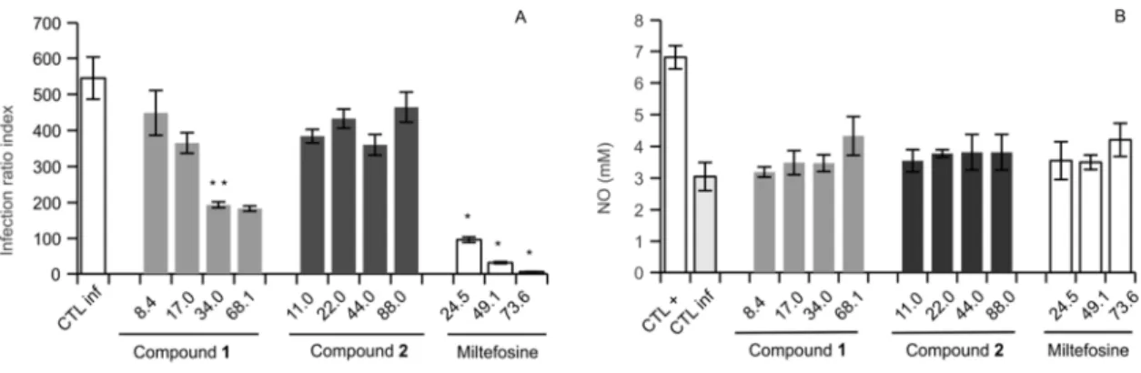
When tested against L. infantum-infected macrophages the tetraprenyltoluquinol (1) 358
applied at concentrations of 34 and 66 µM decreased the infection index by 64.5% and 66.3%, 359
respectively, showing an IC50 of 25.0 ± 4.1 µM and a selectivity index of 5.04 against the
360
peritoneal macrophages (Fig. 5A; Table 2). 361
Only five compounds isolated from marine algae with antileishmanial activity have been 362
reported previously (da Silva Machado et al., 2011; dos Santos et al., 2010, 2011; Soares et 363
al., 2012). However, none of the studies was about Cystoseira macroalgae. Reported 364
sesquiterpenes obtusol (IC50 = 9.4 µM; da Silva Machado et al., 2011) and elatol (IC50 = 13.5
365
µM and 0.45 µM) from the red alga Laurencia dendroidea (daSilva Machado et al., 2011; 366
dos Santos et al., 2010) showed strong activity against L. amazonensis intracellular 367
amastigotes. However, the triquinane sesquiterpene isolated from the same algae was 368
significantly less effective (IC50 = 217.4 µM; da Silva Machado et al., 2011). In addition, 4-369
acetoxydolastane and dolabelladienetriol, isolated from the brown alga Canistrocarpus 370
cervicornis (IC50 = 12.3 µM; dos Santos et al., 2011) and Dictyota pfaffii (IC50 = 44.0 µM;
371
Soares et al., 2012), respectively, were also tested against the same species and form of 372
Leishmania. Therefore, the activity of compound 1 was in the range of that reported for the
373
aforementioned diterpenes. 374
Despite the lower activity of compound 2 against promastigotes (IC50 = 94.4 ± 10.1), it
375
was higher than the effect reported for triquinane (IC50 = 195.5 µM) on promastigotes.
376
However, similarly to what has been reported for triquinane (da Silva Machado et al., 2011), 377
the treatment with the tetraprenyltoluquinone (2) did not decrease the infection index (Fig. 378
5A). 379
During the infection by Leishmania, NO is released by macrophages to eliminate 380
intracellular amastigotes (reviewed by de Almeida et al., 2003). In addition, NO production 381
can be triggered by natural compounds, including those from algae (Robertson et al., 2015). 382
In the present study, infected peritoneal macrophages treated with compounds 1 and 2 383
M
AN
US
CR
IP
T
AC
CE
PT
ED
ACCEPTED MANUSCRIPT
produced low or undetectable amounts of NO as compared to controls. The NO released 384
when the lowest concentrations (8.4 and 17 µM) were applied to the cells was residual, 385
suggesting that the leishmanicidal effect observed for 1 was not related to NO production by 386
the host macrophages (Fig. 5B) and that these compounds did not display an 387
immunomodulatory effect. These results are in agreement with Silva Machado et al. (2011) 388
who observed that triquinane, elatol and obtusol did not promote enhanced NO levels, 389
indicating that leishmanicidal effect of these compounds might be mediated by a mechanism 390
that does not involve the release of this signalling molecule by the host cell. 391
In conclusion, this is the first report describing the identification of compounds from 392
Cystoseira macroalgae displaying activity against Leishmania parasites. In addition, the
393
isolation of tetraprenyltoluquinone (2) as a novel metabolite from algae of the Cystoseira 394
genus is described. Concerning the particular chemical structure of these compounds, our 395
data suggest that the presence of the carbonyl group in C-1 could play a role in the 396
antileishmanial activity of the compounds 1 and 2. Although not as active as miltefosine, 397
tetraprenyltoluquinol (1) displayed significant antileishmanial activity and could be 398
considered as an interesting scaffold for the development of novel chemotherapeutic 399
molecules for canine and human visceral leishmaniases studies. Furthermore, this work 400
reinforces the evidence of the marine environment as source of novel molecules. 401
402
Conflict of interest
403
The authors declare that they have no competing interests. 404
405
Acknowledgments
406
Financial support was provided by Portuguese FCT (projects PTDC/MAR/103957/2008 407
and CCMAR/Multi/04326/2013), from FAPESP (projects 2013/16297-2 and 2015/11936-2) 408
and CNPq (project 470853/2012-3). CBS, CVD were supported by FCT doctoral grants 409
(SFRH/BD/78062/2011 and SFRH/BD/81425/2011, respectively), KNG by a FCT post-410
doctoral grant (SFRH/BPD/81882/2011) and LC by the FCT Investigator Programme 411
(IF/00049/2012). TRM, GAC, JHGL and MU are grateful to CAPES, FAPESP and CNPq. 412
The authors would like to thank Vera Gomes by laboratorial support specific, and Tânia 413
Pereira (Centre of Marine Sciences, University of Algarve) and Dr Javier Cremades 414
M
AN
US
CR
IP
T
AC
CE
PT
ED
ACCEPTED MANUSCRIPT
(Facultade de Ciencias, University of A Coruña, Spain) for their support during the collection 415
and morphological identification of the algal biomass. 416
417
References
418
419
Alvar, J., Velez, I.D., Bern, C., Herrero, M., Desjeux, P., Cano, J., Jannin, J., den Boer, M., et 420
al. 2012. Leishmaniasis worldwide and global estimates of its incidence. PLoS One. 421
7(5), e35671. 422
Amico, V. 1995. Marine Brown algae of family Cystoseiraceae: chemistry and 423
chemotaxonomy. Phytochem. 39, 1257-1279. 424
Britta, E.A., Scariot, D.B., Falzirolli, H., Ueda-Nakamura, T., Silva, C.C., Dias Filho, B.P., 425
Borsali, R., Nakamura, V. 2014. Cell death and ultrastructural alterations in Leishmania 426
amazonensis caused by new compound 4-Nitrobenzaldehyde thiosemicarbazone
427
derived from S-limonene.BMC Microbiol. 14, 236. 428
Burgoyne, L.A. 1999. The Mechanisms of Pyknosis: Hypercondensation and Death. Exp. 429
Cell. Res. 248(1), 214-222. 430
Cardellina II. 1983. Step gradient elution in gel permeation chromatography. A new approach 431
to natural products separation. J. Nat. Prod. 46(2), 196-199. 432
da Silva Machado, F.L., Pacienza-Lima, W., Rossi-Bergmann, B., de Souza Gestinari, L.M., 433
Fujii, M.T., Campos de Paula, J., Costa, S.S., Lopes, N.P., Kaiser, C.R., Soares, A.R. 434
2011. Antileishmanial sesquiterpenes from the Brazilian red alga Laurencia dendroidea. 435
Planta Med. 77(7), 733-5. 436
Dal Picolo, C.R., Bezerra, M.P., Gomes, K.S., Passero, L.F., Laurenti, M.D., Martins, E.G., 437
Sartorelli, P., Lago, J. H. 2014. Antileishmanial activity evaluation of adunchalcone, a 438
new prenylated dihydrochalcone from Piper aduncum L. Fitoterapia. 97, 28-33. 439
Das, M., Mukherjee, S.B., Shaham C. 2001. Hydrogen peroxide induces apoptosis-like death 440
in Leishmania donovani promastigotes. J Cell Sci. 114(13), 2461–2669. 441
de Almeida, M.C., Vilhena, V., Barral, A., Barral-Netto, M. 2003. Leishmanial infection: 442
analysis of its first Steps. A review. Mem Inst Oswaldo Cruz. 98(7), 861-870. 443
de Los Reyes, C., Zbakh, H., Motilva, V., Zubía, E. 2012. Antioxidant and anti-inflammatory 444
meroterpenoids from the brown alga Cystoseira usneoides. J. Nat. Prod. 76(4), 621-629. 445
Dutta, A., Bandyopadhyay, S., Mandal, C., Chatterjee, M. 2005. Development of a modified 446
MTT assay for screening antimonial resistant field isolates of Indian visceral 447
leishmaniasis. Parasitol Int. 54, 119–122. 448
M
AN
US
CR
IP
T
AC
CE
PT
ED
ACCEPTED MANUSCRIPT
Ferrari, M., Fornasiero, M.C., Isetta, A.M. 1990. MTT colorimetric assay for testing 449
macrophage cytotoxic activity in vitro. J Immunol Methods. 131(2), 165-72. 450
Freitas-Junior, L., Chatelain, E., Andrade Kim, H., Siqueira-Neto, J.L. 2012. Visceral 451
leishmaniasis treatment: What do we have, what do we need and how to deliver it? Int J. 452
Parasitol. Drugs Drug Resist. 2, 11-19. 453
Ghannadi, A., Plubrukarn, A., Zandi, K., Sartavi, K., Yegdaneh, A. 2013. Screening for 454
antimalarial and acetylcholinesterase inhibitory activities of some Iranian seaweeds. 455
Res. Pharm. Sci. 8(2), 113-118. 456
Guiry, M.D., Guiry, G.M. AlgaeBase. World-wide electronic publication, National 457
University of Ireland, Galway. http://www.algaebase.org; searched on 12 November 458
2015. 459
Holzmuller, P., Sereno, D., Cavaleyra, M., Mangot, I., Daulovede, S., Vincendeau, P., 460
Lemesre, J.L. 2002. Nitric oxide-mediated proteasome-dependent oligonucleosomal 461
DNA fragmentation in Leishmania amazonensis amastigotes. Infect. Immul. 70, 3727-462
3735. 463
Ibraheem, I.B.M., Abdel-Raouf, N., Abdel-Hameed, M.S, Kel-yamany, K. 2012. 464
Antimicrobial and antiviral activities against Newcastle disease virus (NDV) from 465
marine algae isolated from Qusier and Marsa-Alam Seashore (Red Sea), Egypt. African 466
J. Biotech. 11(33), 8332-8340. 467
Khanavi, M.; Nabavi, M.; Sadati, N.; Ardekani, M.; Sohrabipour, J.; Nabavi, S.; Ghaeli, P.; 468
Ostad, S. N. 2010. Cytotoxic activity of some marine brown algae against cancer cell 469
lines. Biol. Res. 43, 31-37. 470
Kobayashi, T., Endoh, H. 2003. Caspase-like activity in programmed nuclear death during 471
conjugation of Tetrahymena thermophila. Cell Death Differ. 10, 634-640. 472
Levy, D., Ruiz, J.L.M., Celestino, A.T., Silva, S.F., Ferreira, A.K., Isaac, C., Bydlowski, S.P. 473
2014. Short-term effects of 7-ketocholesterol on human adipose tissue mesenchymal 474
stem cells in vitro.Biochem. Biophys. Res. Commun. 446 (3), 720–725. 475
Lockshin, R.A., Zakeri, Z. 2004. Apoptosis, autophagy, and more. Int J Biochem. Cell Biol. 476
36, 2405–2419. 477
Lopes, G., Pinto, E., Andrade, P., Valentão, P. 2013. Antifungal activity of phlorotannins 478
against dermatophytes and yeasts: approaches to the mechanism of action and influence 479
on Candida albicans virulence factor. PloS One. 8(8), e72203. 480
Mesquita-Rodrigues, C., Menna-Barreton R.F.S., Saboia-Vahia, L., Da-Silva, S.A.G., de 481
Souza, E.M., Waghabi, M.C., Cuervo, P., de Jesus, J.B. 2013. Cellular growth and 482
M
AN
US
CR
IP
T
AC
CE
PT
ED
ACCEPTED MANUSCRIPT
mitochondrial ultrastructure of Leishmania (Viannia) braziliensis promastigotes are 483
affected by the iron chelator 2,2-Dipyridyl. PLoS Negl. Trop. Dis. 7(10), e2481. 484
Mhadhebi, L., Laroche-Clary, A., Robert, J., Bouraoui, A. 2011. Anti-inflammatory, 485
antiproliferative and antioxidant activities of organic extracts from the Mediterranean 486
seaweed, Cystoseira crinita. African J. Biotechnol. 10(73), 16682-16690. 487
Mondal, D., Alvar, J., Hasnain, M.G., Hossain, M.S., Ghosh, D., Huda, M.M., Nabi, S.G., 488
Sundar S., Matlashewski, G., Arana, B. 2014. Efficacy and safety of single-dose 489
liposomal amphotericin B for visceral leishmaniasis in a rural public hospital in 490
Bangladesh: a feasibility study. Lancet Glob. Health. 2(1), e51–e57. 491
Monte Neto, R.L., Sousa, L.M.A., Dias, C.S., Barbosa Filho, J.M., Oliveira, M.R., Figueiredo, 492
R.C.B.Q. 2011. Morphological and physiological changes in Leishmania promastigotes 493
induced by yangambin, a lignan obtained from Ocotea duckei. Exp. Parasitol. 127, 215-494
221. 495
Nishikawa, T., Tsuno, N.H., Okaji, Y., Shuno, Y., Sasaki, K., Hongo, K., Sunami, E., 496
Kitayama, J., Takahashi, K., Nagawa, H. 2010. Inhibition of autophagy potentiates 497
sulforaphane-induced apoptosis in human colon cancer cells. Ann. Surg. Oncol. 17, 498
592–602. 499
Passero, L.F., Assis, R.R., da Silva, T.N., Nogueira, P.M., Macedo, D.H., Pessoa, N.L., 500
Campos, M.A., Laurenti, M.D., Soares, R.P. 2015. Differential modulation of 501
macrophage response elicited by glycoinositolphospholipids and lipophosphoglycan 502
from Leishmania (Viannia) shawi. Parasitol Int. 64(4), 32–35. 503
Robertson, R.C., Guihéneuf, F., Bahar, B., Schmid, M., Stengel, D.B., Fitzgerald, G.F., Ross, 504
R.P., Stanton, C. 2015. The Anti-Inflammatory Effect of Algae-Derived Lipid Extracts 505
on Lipopolysaccharide (LPS)-Stimulated Human THP-1 Macrophages. Mar Drugs. 506
13(8), 5402-24. 507
Santos, A.O., Britta, E., Bianco, E.M., Ueda-Nakamura, T., Dias-Filho, B.P., Pereira R.C., 508
Nakamura, C.V. 2011. 4-Acetoxydolastane diterpene from the Brazilian brown alga 509
Canistrocarpus cervicornis as antileishmanial agent. Mar. Drugs. 9, 2369-2383.
510
Santos, A.O., Veiga-Santos, P., Ueda-Nakamura, T., Dias-Filho, B.P., Sudatti, D.B., Bianco, 511
E.M., Pereira, R.C., Nakamura, C.V. 2010. Effect of elatol, isolated from red seaweed 512
Laurencia dendroidea, on Leishmania amazonensis. Mar. Drugs. 8, 2733-2743.
513
Soares, D.C., Calegari-Silva, T.C, Lopes, U.G., Teixeira, V.L., de Palmer Paixão, I.C., Cirne-514
Santos, C., Bou-Habib, D.C., Saraiva, E.M. 2012. Dolabelladienetriol, a compound 515
M
AN
US
CR
IP
T
AC
CE
PT
ED
ACCEPTED MANUSCRIPT
from Dictyota pfaffii algae, inhibits the infection by Leishmania amazonensis. PLoS 516
Negl. Trop. Dis. 6(9), e1787. 517
Souza, W., Attias, M., Rodrigues, J.C.F. 2009. Particularities of mitochondrial structure in 518
parasitic protists (Apicomplexa and Kinetoplastida). Int. J. Biochem. Cell Biol. 41, 519
2069–2080. 520
Spavieri, J., Allmendinger, A., Kaiser, M., Casey, R., Hingley-Wilson, S., Lalvani, A., Guiry, 521
M.D., Blunden, G., Tasdemir, D. 2010. Antimycobacterial, antiprotozoal and cytotoxic 522
potential of twenty-one brown algae (Phaeophyceae) from British and Irish waters. 523
Phytother. Res. 24, 1724–1729. 524
Tajbakhsh, S.; Ilkhani, M.; Rustaiyan, A.; Larijani, K.; Sartavi, K.; Tahmasebi, R.; Asayesh, 525
G. 2011. Antibacterial effect of the brown alga Cystoseira trinodis. J. Med. Plants Res. 526
5(18), 4654-4657. 527
Tempone, A.G., Martins de Oliveira, C., Berlinck, R.G. 2011. Current approaches to discover 528
marine antileishmanial natural products. Planta Med. 77, 572-585. 529
Valls, R., Piovetti, L., Banaigs, B., Praud, A. 1993. Secondary metabolites from morocco 530
brown algae of the genus Cystoseira. Phytochem. 32(4), 961-966. 531
Yamamoto, E.S., Campos, B.L., Jesus, J.A., Laurenti, M.D., Ribeiro, S.P., Kallás, E.G., 532
Rafael-Fernandes, M., Santos-Gomes, G., Silva, M.S., Sessa, D.P., Lago, J.H., Levy, D., 533
Passero, L.F. 2015. The effect of ursolic acid on Leishmania (Leishmania) amazonensis 534
is related to programed cell death and presents therapeutic potential in experimental 535
cutaneous leishmaniasis. PLoS One. 10(12), e0144946. 536
M
AN
US
CR
IP
T
AC
CE
PT
ED
ACCEPTED MANUSCRIPT
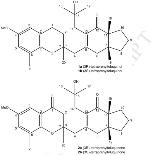
Figures 538 539 540Fig. 1. Structures of the tetraprenyltoluquinols (1a-1b) and tetraprenyltoluquinones (2a-2b)
541
isolated from C. baccata 542
M
AN
US
CR
IP
T
AC
CE
PT
ED
ACCEPTED MANUSCRIPT
544 545Fig. 2. HMBC of the tetraprenyltoluquinones (2a-2b) isolated from C. baccata
546
547
M
AN
US
CR
IP
T
AC
CE
PT
ED
ACCEPTED MANUSCRIPT
549 550Fig. 3. Effect of compounds 1 and 2 on the ultrastructure of L. infantum promastigotes.
551
Parasites were treated with 1a/1b (44.9 µM) and treated with compound 2a/2b (94.4 µM). N 552
– nucleus, FP - flagellar pocket, K – kinetoplast, M – mitochondrion, V – vacuole, * - 553
disappearance of the chromatin associated with the nuclear inner membrane 554
M
AN
US
CR
IP
T
AC
CE
PT
ED
ACCEPTED MANUSCRIPT
556 557Fig. 4. Effects of compounds 1 (Comp1) and 2 (Comp2) on the nuclear DNA fragmentation
558
(A) and mitochondrial membrane potential (B) of L. infantum promastigotes. Parasites were 559
treated with 1a/1b (44.9 µM) and 2a/2b (94.4 µ M). Hydrogen peroxide (6.2 µM) (A) and 560
miltefosine (23.1 µM) (B) and untreated parasites (A and B) were used as controls. *, p < 561
0.05; **, p < 0.01. 562
M
AN
US
CR
IP
T
AC
CE
PT
ED
ACCEPTED MANUSCRIPT
564 565Fig. 5. Effect of compounds 1 and 2 on the L. infantum intracellular amastigotes (A) and on
566
the nitric oxide production (mM) of the infected mouse peritoneal macrophages (B) after a 567
24-h treatment with different concentrations (µM). Untreated non-infected macrophages 568
(CTL+), untreated infected macrophages (CTL inf) and infected macrophages treated with a 569
reference drug, miltefosine, were used as controls. 570
M
AN
US
CR
IP
T
AC
CE
PT
ED
ACCEPTED MANUSCRIPT
Tables 572 573Table 1. 1H and 13C NMR data (500 and 125 MHz, CDCl3, δ/ppm) for compounds 1 (a/b)
574
and 2 (a/b) 575
Compound 1a 1b 2a 2b
Position δC, type δH (J in Hz) δC, type δH (J in Hz) δC, type δΗ (J in Hz) δC, type δH (J in Hz) 1 22.7, CH2 2.79, (m) 22.6, CH2 2.79, (m) 192.2, C - 192.1, C -2 32.5, CH2 1.80, (m) 33.6, CH2 1.80, (m) 47.8, CH2 2.56, (m) 48.6, CH2 2.57, (m) 3 76.4, C - 76.2, C - 81.3, C - 81.2, C -4 43.6, CH2 2.66, (s) 45.2, CH2 2.66, (s) 44.1, CH2 2.70, (s) 44.8, CH2 2.70, (s) 5 153.7, C - 154.5, C - 153.3, C - 154.3, C -6 44.3, CH2 2.57, (m) 44.7, CH2 2.57, (m) 44.1, CH2 2.60, (d, 4.0) 44.1, CH2 2.63, (d, 4.0) 2.60, (m) 2.60, (m) 2.68, (d, 4.0) 2.68, (d, 4.0) 7 44.8, C - 44.8, C - 44.9, C - 44.9, C -8 35.0, CH2 1.54, (m) 35.0, CH2 1.54, (m) 35.0, CH2 1.54, (m) 35.0, CH2 1.54, (m) 1.73, (m) 1.73, (m) 1.73, (m) 1.73, (m) 9 18.8, CH2 1.74, (m) 18.8, CH2 1.74, (m) 18.8, CH2 1.74, (m) 18.8, CH2 1.74, (m) 10 29.3, CH2 1.46, (m) 29.3, CH2 1.46, (m) 29.4, CH2 1.40, (d, 13.0) 29.7, CH2 1.42, (d, 13.0) 11 54.9, C - 54.9, C - 54.9, C - 54.9, C -12 208.5, C - 208.9, C - 208.0, C - 208.1, C -13 132.9, C - 133.3, C - 133.5, C - 134.0, C -14 39.4, CH2 2.45, (d, 15.0) 39.9, CH2 2.45, (d, 15.0) 39.4, CH2 2.54, (d, 15.0) 39.6, CH2 2.54, (d, 15.0) 2.73, (d, 15.0) 2.73, (d, 15.0) 2.59, (d, 15.0) 2.59, (d, 15.0) 15 70.8, C - 71.1, C - 71.0, C - 71.0, C -16 28.8, CH3 1.12, (s) 28.8, CH3 1.14, (s) 29.1, CH3 1.11, (s) 29.1, CH3 1.13, (s) 17 30.5, CH3 1.24, (s) 31.6, CH3 1.19, (s) 30.8, CH3 1.25, (s) 31.3, CH3 1.26, (s) 18 21.1, CH3 1.09, (s) 21.1, CH3 1.03, (s) 21.1, CH3 1.04, (s) 21.1, CH3 1.09, (s) 19 22.4, CH3 0.91, (s) 22.5, CH3 0.83, (s) 22.4, CH3 0.83, (s) 22.5, CH3 0.91, (s) 20 24.1, CH3 1.28, (s) 24.5, CH3 1.28, (s) 23.9, CH3 1.20, (s) 24.2, CH3 1.20, (s) 1' 145.2, C - 145.3, C - 167.8, C - 167.8, C -2' 120.4, C - 120.4, C - 119.6, C - 119.6, C -3' 111.1, CH 6.45, (d, 3.0) 111.2, CH 6.46, (d, 3.0) 114.6, CH 7.15, (d, 3.0) 114.6, CH 7.16, (d, 3.0) 4' 152.6, C - 152.6, C - 151.9, C - 151.9, C -5' 115.2, CH 6.59, (d, 3.0) 115.3, CH 6.60, (d, 3.0) 104.5, CH 7.00, (d, 3.0) 104.5, C 7.01, (d, 3.0) 6' 127.0, C - 127.2, C - 126.5, C - 126.5, C -Me-6' 16.6, CH3 2.16, (s) 16.8, CH 2.17, (s) 16.2, CH3 2.21, (s) 16.4, CH3 2.23, (s) OMe-4' 55.6, CH3 3.73, (s) 55.6, CH 3.74, (s) 55.7, CH3 3.78, (s) 55.7, CH3 3.78, (s) 576
M
AN
US
CR
IP
T
AC
CE
PT
ED
ACCEPTED MANUSCRIPT
Table 2. Effect of the compounds 1 and 2 against L. infantum promastigotes and intracellular
577
amastigotes and mouse peritoneal macrophages 578
579
Compounds Promastigotesa Intracellular amastigotesa Peritoneal macrophagesb SI c 1 44.9 ± 4.3 25.0 ± 4.1 126.6 ± 21.1 5.04 2 94.4 ± 10.1 > 88.0 84.5 ± 12.5 <0.96 Miltefosine 23.1 ± 0.0 20.3 ± 1.3 130.3 ± 17.2 6.42 aIC
50 - Half maximal inhibitory concentration in µM; bCC50 - Cytotoxic concentration that causes the death of 50% of the viable cells in µM; cSI – Selectivity index concerning the activity against the intracellular amastigotes.
M
AN
US
CR
IP
T
AC
CE
PT
ED
ACCEPTED MANUSCRIPT
Biographies of the authors
581
Carolina Bruno-de-Sousa - MSc in Animal Production and Post-graduated in Medical
582
Parasitology. As PhD student of the Center of Marine Sciences at the Algarve University, is 583
currently studding marine algae as source of bioactive molecules against Leishmania 584
parasites. Main interests include animal parasitological studies and genetic characterization of 585
domestic animals and algae populations. Also collaborated in studies of parasitic diseases 586
with public health significance. 587
Katkam N. Gangadhar - Post-doctoral Research Fellow at CCMAR, University of Algarve.
588
Works in synthetic/organic lipid medicinal chemistry and pharmaceutical applications: (i) 589
isolation of wound healing and anti-cancer bioactive compounds from natural products; (ii) 590
synthesis of lipid carriers as drug delivery materials for anti-tuberculosis drug and 591
Amphotericin-B; (iii) chemo-enzymatic synthesis of cetyl myristoleates and diacylglycerol 592
and evaluation of their anti-inflammatory, anti-arthritic and nutritional properties; (iv) 593
development of carbon-based solid acid catalyst from crude glycerol for biodiesel production 594
from microalgae and non-edible oils and (v) its application in organic methodologies. 595
Thiago R. Morais - PhD student of Chemical Biology at UNIFESP, working with Natural
596
Products chemistry, especially with isolation and characterization of micromolecules using 597
NMR and MS data analysis. 598
Geanne A. A. Conserva - MSc student at the Federal University of São Paulo – UNIFESP –
599
working with Chemistry of Natural Products, mainly in the search and characterization of 600
bioactive derivatives in plant species, particularly those with antitumoral activity. 601
Catarina Vizetto-Duarte - PhD student at Centre of Marine Sciences (CCMAR) at the
602
University of Algarve. She has an MSc in Molecular Genetics and Biomedicine from the 603
University of Lisbon in 2009. As a PhD student she is evaluating the biomedical applications 604
(especially antioxidant and antitumoral properties) of brown algae, focusing on finding novel 605
bioactive molecules and studying the molecular mechanisms responsible for the said 606
activities in terms of cellular responses to drug exposure, inflammation, cell death 607
(apoptosis/necrosis) versus cell survival. 608
Hugo Pereira - MSc on Aquaculture and Fisheries, where he worked on the optimization of
609
a novel culture medium for large-scale production of microalgae in photobioreactors at 610
Necton S.A. (Portugal). He is currently a PhD student aiming the development of an algal 611
biorefinery for different biotechnological applications, including the determination of 612
bioactivities to improve the added-value of algal biomass 613
Márcia D. Laurenti - PhD in Veterinary Pathology; full professor and head chief of
614
Laboratory of Pathology of Infectious Diseases, Department of Pathology, Medical School, 615
University of São Paulo; with experience in the immunopathology of human, canine and 616
experimental cutaneous and visceral leishmaniasis. 617
Lenea Campino - Full Professor in Medical Parasitology, at the Institute of Hygiene and
618
Tropical Medicine, Universidade Nova de Lisboa (IHMT/UNL). Main areas of interest are: 619
leishmaniasis and Leishmania-HIV co-infections; molecular epidemiology, parasite diversity, 620
vector/host-parasite interactions; immunology of the infection; natural and experimental 621
M
AN
US
CR
IP
T
AC
CE
PT
ED
ACCEPTED MANUSCRIPT
leishmaniasis models; vaccine and drug candidates; diagnostics on visceral and cutaneous 622
leishmaniasis; environmental changes and emerging parasitic diseases. She led national and 623
International research projects in those areas, supervised several postgraduate degrees, and 624
acted as a consultant for the Portuguese National Directorate of Health. 625
Debora Levy - PhD in medical science at Medical School of Sao Paulo University. Currently
626
is scientific researcher at Laboratory of Genetics and Molecular Hematology, and has 627
experience in hematology, genetics and drug development. 628
Miriam Uemi - Associate Professor at the Federal University of the State of São Paulo - has
629
experience in molecular characterization by nuclear magnetic resonance and mass 630
spectrometry. 631
Luísa Barreira - Assistant Professor of the Chemistry and Pharmacy Department of the
632
Faculty of Sciences and Technology of the University of Algarve since 2007. She has PhD in 633
Environmental Sciences and Technologies and is currently a senior researcher in MarBiotech 634
for I+D+I of biotechnological applications of marine organisms, from the production of 635
biodiesel and other bioproducts (e.g. phospholipids) from microalgae to the search of natural 636
products with biological activities in marine organisms. 637
Luísa Custódio - PhD in Biotechnological Sciences and carried out her post-doctoral
638
research at the University of Algarve and CCMAR. Presently she is a research assistant hired 639
by CCMAR under the frame of the FCT investigator programme and her research has 640
focused on the search for bioactive compounds in marine organisms and halophyte species, 641
and the evaluation of the nutritional profile of edible organisms (e.g. algae, halophytes and 642
sea cucumbers). 643
Felipe Passero - He got PhD in physiopathology at Medical School of Sao Paulo University.
644
Currently is full professor at São Paulo State University, and has experience in Parasitology, 645
mainly with leishmaniasis. 646
Joao Lago - Full professor at Federal University of Sao Paulo - has experience in Chemistry
647
of Natural Products, mainly in the search and characterization of bioactive derivatives in 648
plant species, including those with antiparasitic anti-inflammatory, antimicrobial and 649
antitumoral activities. 650
João Varela - Assistant Professor at the University of Algarve and Group Leader of the
651
MarBiotech (Marine Biotechnology) research group at the Centre of Marine Sciences 652
(CCMAR). MarBiotech, which has the following research lines i) search for novel bioactive 653
compounds in marine organisms, with particular emphasis on microalgae, macroalgae and 654
halophytes; ii) design and implementation of biorefineries for the upgrade of algal biomass 655
for biofuel, food and feed production; and iii) marine organisms (e.g. sea cucumbers and 656
halophytes) as innovative gourmet food. 657
M
AN
US
CR
IP
T
AC
CE
PT
ED
ACCEPTED MANUSCRIPT
Highlights•••• Tetraprenyltoluquinols and tetraprenylquinones from Cystoseira baccata.
•••• Tetraprenyltoluquinols displayed antileishmanial activity
•••• Tetraprenyltoluquinols induce alterations on promastigotes morphology.