Short Communication
Corresponding author: Dra. Ana Maria Roselino. e-mail: amfrosel@fmrp.usp.br
Received 25 May 2017 Accepted 24 August 2017
Anti-phospholipid syndrome in seven leprosy patients with
thrombotic events on corticosteroid and/or thalidomide
regimen: insights on genetic and laboratory profiles
Sebastian Vernal
[1], Maria Jose Franco Brochado
[1], Roberto Bueno-Filho
[1],
Paulo Louzada-Junior
[2]and Ana Maria Roselino
[1][1]. Divisão de Dermatologia, Departamento de Clínica Médica, Faculdade de Medicina de Ribeirão Preto, Universidade de São Paulo, Ribeirão Preto, SP, Brasil.
[2]. Divisão de Imunologia, Departamento de Clínica Médica, Faculdade de Medicina de Ribeirão Preto, Universidade de São Paulo, Ribeirão Preto, SP, Brasil.
Abstract
Introduction: Corticosteroids and/or thalidomides have been associated with thromboembolism events (TBE) in multibacillary
(MB) leprosy. This report aimed to determine genetic and laboratory profiles associated with leprosy and TBE. Methods: Antiphospholipid antibodies (aPL), coagulation-related exams, prothrombin and Leiden’s factor V mutations, and ß2-glycoprotein-I
(ß2GPI) Val247Leupolymorphism were assessed. Results: Six out of seven patients with leprosy were treated with prednisone
and/or thalidomide during TBE and presented at least one positive aPL. All patients presented ß2GPI polymorphism, and one
showed prothrombin mutation. Conclusions: Corticosteroid or thalidomide adverse effects and aPL and ß2GPIpolymorphisms
may cause TBE in patients with MB leprosy.
Keywords: Leprosy. Antiphospholipid antibodies. Beta-2-glycoprotein-I. Factor V Leiden. Prothrombin. Thromboembolism.
Leprosy immune reactions are systemic inflammatory complications that commonly occur when introducing a treatment. However, these reactions may also occur at any time during the disease course and can relapse intermittently over several years1,2. Leprosy immune reactions are classified by the
World Health Organization (WHO) as type 1 (T1R or reverse reaction) and type 2 (T2R or erythema nodosum leprosum)
and are manifested by multibacillary (MB) patients1,2. T1R
typically occurs in patients with borderline spectrum, whereas T2R mainly occurs in patients with borderline lepromatous (BL)
and lepromatous leprosy (LL) (Ridley-Jopling classification)1,2.
Reverse reaction is a delayed-type hypersensitive reaction
caused by increased cell-mediated immunity to Mycobacterium
leprae antigens. T1R may damage nerves and is mainly treated
with corticosteroids1. Erythema nodosum leprosum (ENL),
the main symptom of T2R, is caused by a humoral immune response to M. leprae and results in direct deposits of
antigen-antibody immune complexes in the vessels and tissues1. The
treatment of ENL varies depending on the country and leprosy center treatment due to restrictions and costs implicated in the
use of some drugs2. Corticosteroids and/or thalidomide are
the preferred medications to suppress ENL2; notwithstanding,
both of them are related to several adverse events (AEs)2.
Thromboembolic phenomena (TEP) are well-documented AEs of thalidomide, a drug used as monotherapy to treat myeloma. The risk of TEP is even higher when thalidomide is associated with corticosteroids or chemotherapy2. In the last decade, some
reports have described the occurrence of TEP due to ENL treatment with thalidomide3.
Although the prothrombotic AE of thalidomide is clearly
defined, other intrinsic factors may also cause TEP associated
with leprosy. Indeed, high frequency of antiphospholipid antibodies (aPL), especially anti-cardiolipin (aCL) and anti-ß2-glycoprotein-I (anti-ß2GPI), has been observed in patients with leprosy4-6. The presence of aPL is not necessarily associated with
antiphospholipid syndrome (APS), which is known to manifest thrombotic events. Nevertheless, some authors have debated whether some clinical features of leprosy, such as Lucio’s reaction, TR1 and TR2 immune reactions, and gangrene, may manifest APS6-8.
This report aimed to identify the clinical and laboratory data related to coagulation, including factor V Leiden and
prothrombinG20210A polymorphisms, and therapeutic aspects
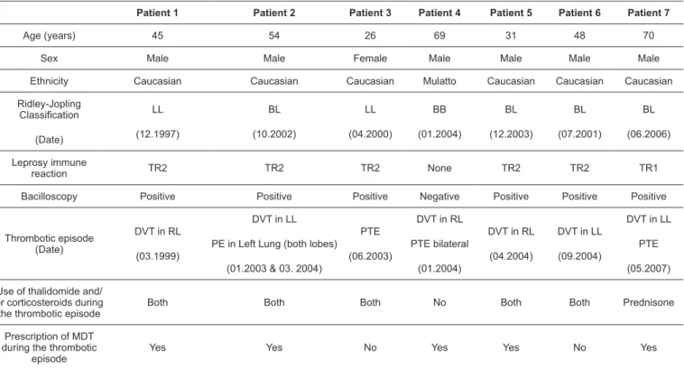
Patient 1 Patient 2 Patient 3 Patient 4 Patient 5 Patient 6 Patient 7
Age (years) 45 54 26 69 31 48 70
Sex Male Male Female Male Male Male Male Ethnicity Caucasian Caucasian Caucasian Mulatto Caucasian Caucasian Caucasian Ridley-Jopling
Classification
(Date)
LL (12.1997)
BL (10.2002)
LL (04.2000)
BB (01.2004)
BL (12.2003)
BL (07.2001)
BL (06.2006) Leprosy immune
reaction TR2 TR2 TR2 None TR2 TR2 TR1 Bacilloscopy Positive Positive Positive Negative Positive Positive Positive
Thrombotic episode (Date)
DVT in RL (03.1999)
DVT in LL PE in Left Lung (both lobes)
(01.2003 & 03. 2004)
PTE (06.2003)
DVT in RL PTE bilateral
(01.2004)
DVT in RL (04.2004)
DVT in LL (09.2004)
DVT in LL PTE (05.2007) Use of thalidomide and/
or corticosteroids during the thrombotic episode
Both Both Both No Both Both Prednisone
Prescription of MDT during the thrombotic
episode
Yes Yes No Yes Yes No Yes
TABLE 1: Clinical data of seven patients with multibacillary leprosy who experienced thrombotic events.
LL: Lepromatous leprosy; BL: borderline leprosy; BB: borderline-borderline leprosy; TR1: leprosy reaction type 1; TR2: leprosy reaction type 2; DVT: deep venous thrombosis; RL: right leg; LL: left leg; PTE: pulmonary thromboembolism; MDT: multi-drug treatment.
events. Additionally, aPL and ß2-glycoprotein-I Val247Leu
polymorphism were investigated to differentiate intrinsic thrombophilia and APS.
Patients were diagnosed at the University Hospital of Ribeirão Preto Medical School, University of São Paulo, Southeastern Brazil, from 1997 to 2006. Data were collected from medical charts and electronic database system of the
University Hospital. Leprosy was diagnosed and confirmed
based on WHO’s recommendations1. Both WHO and
Ridley-Joplin classifications were used to classify leprosy1.
Anti-phenolic glycolipid 1 (Anti-PGL1) IgM was measured using in-house enzyme-linked immunosorbent assay (ELISA) as described previously9; IgM and IgG aCL and anti-ß2P levels
were determined using commercial ELISA kits (QUANTA Lite, INOVA, USA). Lupus anticoagulants (LA) were analyzed using Viperquik LA-Check test and Kaolin clotting time (KCT). The coagulation factors, i.e., anti-thrombin III, proteins C and S,
and fibrinogen, were measured in the peripheral blood using
chromogenic methods during thrombotic episodes.
Factor V Leiden and G20210A mutation of prothrombin gene were analyzed using polymerase chain reaction–restriction fragment length polymorphism (PCR-RFLP). The Val247Leu
polymorphism of the ß2-glycoprotein-Igene was determined
as described previously10.
Informed consent was obtained from all patients. This study was approved by the local Human Ethics Committee (#3605/2006) in accordance with the ethical standards of the
Helsinki Declaration (1964, amended most recently in 2008) of the World Medical Association.
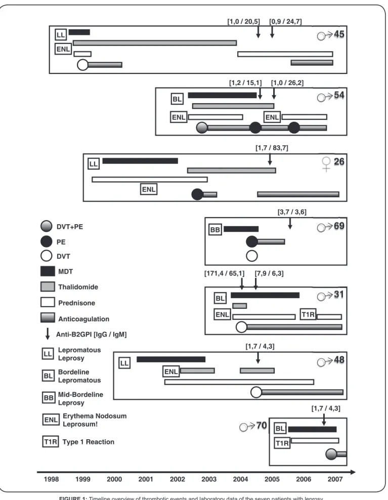
This report included seven leprosy patients with thrombotic events. Tables 1 and 2 and Figure 1 summarize the clinical, laboratory, and therapeutic data. Patients’ median age was 48 years (youngest, 26 years; oldest, 70 years); six of them were males. The seven MB patients corresponded to the BB,
BL, and LL classifications (one, four, and two, respectively).
Five of the patients presented TR2 at the onset of leprosy,
corresponding to BL and LL classifications; another BL patient
was affected by TR1, as commonly described2. The patient
with BB leprosy did not present any associated reaction. The most common thrombotic events were deep venous thrombosis (DVT) that occurred in three patients and DVT associated with pulmonary thromboembolism (PTE) that occurred in the other three patients. One patient presented with PTE only. Among
the six patients with leprosy reaction, five were treated with
thalidomide plus corticosteroids during the thrombotic episode, whereas one patient was treated with corticosteroids alone. None of the patients were active smokers. Except for case 4, tests for thrombophilia did not show any genetic predisposition (Figure 1).
Patient 1 Patient 2 Patient 3 Patient 4 Patient 5 Patient 6 Patient 7
(Date) IgM anti-PGL1 (index) (NV < 1.0)
(09.2000) 0.3 (04.2005) 0.5
(10.2002) 25.0 (03.2004) 1.5 (09.2004) 4.6
(05.2000) 1.7 (04.2002) 1.0 (05.2002) 0.5
(03.2005) 0.8 (01.2004) 1.5
(07.2001) 26.8 (10.2004) 1.8 (11.2004) 1.5
(01.2006) 0.5 (12.2006) 0.4
( D a t e ) IgG aCL (GPL) (NV < 15)
(04.2004) <15 (04.2005) <15
(01.2004) 35.0 (08.2004) 32.0 (09.2004) 34.0
(06.1999) <15 (04.2001) 49.0 (05.2001) <15 (08.2004) <15 (10.2004) <15
(03.2005) <15 (04.2005) 16.3 (07.2005) 18.8
(04.2004) 44.0 (09.2004) <15 (06.2005) <15
(04.2005) <15 (06.2005) <15
(05.2007) <15
( D a t e ) IgM aCL (MPL) (NV < 12.5)
(04.2004) <12.5 (04.2005) <12.5
(01.2004) 125.0 (08.2004) 85.0 (09.2004) 94.0
(01.2001) <12.5 (05.2001) 70.0 (08.2004) 54.0 (10.2004) 68.0
(03.2005) <12.5 (04.2005) <12.5 (07.2005) <12.5
(04.2004) 34.0 (08.2004) <12.5 (06.2005) <12.5
(04.2005) <12.5 (06.2005)
<12.5
(05.2007) <12.5
( D a t e ) Lupus anticoagulants
(04.2004) (-) (06.2005) (+) (12.2005) (+)
Not done (09.2005) (+) (07.2005) (-) Not done
(09.2004) (-) (04.2005) (-) (06.2005) (+) (09.2005) (+)
Not done
(Date) IgG anti - ß2GPI
(SMU) (NV < 10) (09.2005) <10 (09.2005) <10 (09.2005) <10 (09.2005) <10 (09.2005) >100 (09.2005) <10 (05.2007) <10 ( D a t e ) IgM
anti-ß2GPI (NV < 12) (09.2005) <12 (09.2005) <12 (09.2005) >100 (09.2005) <12 (09.2005) >100 (09.2005) <12 (05.2007) <12 Anti-thrombin III (NV
70-130%) 107 73 86 101 Not done 114 Not done Protein C (NV
70-130%) 150 Not done 115 68 Not done 128 Not done Protein S (NV
50-130%) 70 Not done 88 Not done Not done 95 Not done Kaolin clotting time Negative Positive Negative Negative Not done Positive Not done Fibrinogen (mg/dL)
(NV 200-400) Not done 394 326 800 Not done 454 Not done Factor V Leiden (NV
+/+) +/+ +/+ +/+ +/+ +/+ +/+ +/+
Prothrombin G20210A
mutation (NV -/-) -/- -/- -/- +/- -/- -/-
-/-ß2GPI Leu247Val Polymorphism
Heterozygote (Haplotype 1&4)
Heterozygote (Haplotype
1&4)
Homozygote Val/Val (Haplotype 4)
Heterozygote (Haplotype
1&4)
Heterozygote (Haplotype
1&4)
Homozygote Val/Val (Haplotype 4)
Homozygote Val/Val (Haplotype
4&7)
TABLE 2: Laboratory data of seven patients with multibacillary leprosy who experienced thrombotic events.
IgM: immunoglobulin M; NV: normal value; PGL1: Phenolic Glycolipid 1; aCL: anti-Cardiolipin; GPL: standard IgG cardiolipin units; MPL: standard IgM cardiolipin units; ß2GPI: ß2-glycoprotein-I; Leu: leucine; Val: valine.
prednisone (60mg/day) and thalidomide (300mg/day), due to ENL. Laboratorial tests were performed 5 years after the DVT episode and yielded negative results for aCL and anti-ß2GPI. LA was negative in 2004, but turned positive in 2005. LA has been a stronger risk factor for thrombosis than positive aCL or anti-ß2GPI in purely obstetrical APS11. Protein C was the
only abnormal laboratory test result related to coagulation. The primary effect of activated protein C is to inactivate the Va and
VIIIa coagulation factors, which are necessary to efficiently generate thrombin and activate factor X. Genetic deficiency of
LL
BL
BB
ENL
T1R
Bordeline Lepromatous
Mid-Bordeline Leprosy
Erythema Nodosum Leprosum!
Lepromatous Leprosy
Type 1 Reaction DVT+PE
DVT PE
MDT
Thalidomide
Prednisone
Anticoagulation
Anti-B2GPI [IgG / IgM]
1998 1999 2000 2001 2002 2003 2004 2005 2006 2007
45
54
26
69
31
48
70
♂
♂
♂
♂
♂
♂
LL
LL
LL
BL
BL!
BL!
T1R!
T1R! ENL
ENL
ENL ENL!
ENL!
ENL
[1,0 / 20,5] [0,9 / 24,7]
[1,2 / 15,1] [1,0 / 26,2]
[1,7 / 83,7]
BB
[3,7 / 3,6]
[1,7 / 4,3] [171,4 / 65,1] [7,9 / 6,3]
[1,7 / 4,3]
Case 2 was a 54-year-old Caucasian male patient diagnosed as having BL in 2002. He had been presenting multiform bullous erythema and ENL outbreaks since the diagnosis. In 2003 and 2004, he presented DVT in the left limb associated with PTE in the upper and lower lobes of the left lung, respectively. Both episodes occurred during the use of MDT plus prednisone (10–60mg/day) and thalidomide (100–200 mg/day). During the PTE episode in
2004, IgM and IgG aCL increased.In infectious diseases, aPL
antibodies tend to be IgM type because the infectious agent might
triggertheautoimmune response. However, IgG aCL has also
been detected in patients with leprosy12. The only laboratory test
related to abnormal coagulation was the KCT. This was expected from the positive aPL result, which would interfere with the accumulation of prothrombinase complex11.
The third patient was a 26-year-old Caucasian female diagnosed as having LL in 2001. In the last two years, she had been chronically using prednisone due to misdiagnosis of lupus erythematosus. In 2001, ENL outbreaks occurred when she used prednisone and an alternative MDT scheme. Thalidomide was introduced in May 2003. In July 2003, she presented PTE while using prednisone (40mg/day) and thalidomide (200mg/ day). She was not using any other medications at the time, not even contraceptive pills. aPL screening during PTE revealed high IgM and IgG aCL and anti-ß2GPI levels, and LA tests yielded positive results.
The fourth patient was a 69-year-old mulatto male who had been diagnosed as having BB leprosy in 2003. In January 2004, he experienced a DVT in the right limb and bilateral PTE while using MDT and hydrochlorothiazide. High IgG aCL (18.8 GPL)
was confirmed 18 months after the thrombotic event. He also presented high levels of fibrinogen (800 mg/dL), which may be elevated in response to inflammation or tissue injury13 during leprosy.
Heterozygosis for the prothrombin G20210A gene was demonstrated. The fifth patient was a 31-year-old Caucasian male diagnosed as having BL in 2003 and who had been experiencing ENL outbreaks since the diagnosis. In 2004, he had DVT in the right limb while using MDT associated with prednisone (30 mg/ day) and thalidomide (300 mg/day). High IgM and IgG aCL
were confirmed at the time of DVT. Six months later, IgM and
IgG aCL turned and remained negative in 2005. High levels of
IgM and IgG anti-β2GPI antibodies were also observed 1.5 years
after the DVT episode. Unfortunately, laboratory tests related to coagulation were not performed. Assessment for thrombophilia did not show any genetic predisposition.
The sixth patient was a 48-year-old Caucasian male who was diagnosed as having BL and ENL in 2001. In 2004, he developed DVT in the left limb while using thalidomide
(100mg/day). IgM and IgG aCL and anti-β2GPI levels were
within the normal limits and LA was negative during and after the thrombotic episode, but LA became positive 1 year later.
Like the fourth case, this patient had high fibrinogen levels as expected in an inflammatory scenario13. KCT was also positive
due the presence of aPL, as expected11.
The last patient was a 70-year-old Caucasian male diagnosed as having BL in 2006, with T1R. In 2007, he developed DVT in the left limb and PTE during an alternative MDT scheme
associated with prednisone (60 mg/day). IgM and IgG aCL and aPL were normal. Regrettably, LA and laboratory tests related to coagulation were not performed.
Regarding the β2GPIgenetic polymorphism evaluated in haplotypes 1 and 4, three patients presented Val/Val homozygosis (cases 4, 6, and 7), and the other three presented Leu247Val heterozygosis (cases 1, 2, 3, and 5). High frequency
of β2GPI Val/Val homozygosis has been described in Brazilian
leprosy patients as compared to controls. Furthermore, MB
patients with high IgM anti-β2GPI levels presented higher
β2GPI Val/Val homozygosis frequency than the controls6,10.
β2GPI Val/Val homozygosis seems to represent a higher risk
factor than Leu247Val heterozygosis for the development of APS in the general population6,10. However, this has not been
confirmed yet, and larger studies attributing higher risk of APS
to Val/Val homozygosis should be conducted.
Only one patient (case 4) had mutated prothrombin G20210A gene, which independently conferred a 2.8-fold increased risk of DVT in both sexes and in all age groups14.
This patient presented a thrombotic event without leprosy reaction outbreaks even though he was not treated with thalidomide and/or corticosteroid. Interestingly, he was the only patient in this case series who had genetic polymorphism for thrombophilia. Therefore, genetic thrombophilia could explain the development of a thrombotic event even without using thalidomide and/or corticosteroids. Moreover, the presence of
aCL and β2GPI Leu247Val heterozygosis may have contributed
to the development of DVT in this patient.
Some reports of thrombotic events due to the use of thalidomide and/or corticosteroids in MB leprosy patients with ENL are notorious5-9. However, serum tests or assessment of
patients’ genetic background regarding the diagnosis of intrinsic thrombophilia was not conducted in these studies. Here, we analyzed several pro-thrombotic factors in seven MB patients from Southeastern Brazil, not only to determine acquired risk factors due to leprosy and AEs of the drugs but also the manifestation of
APS and β2GPI Leu247Val polymorphism. Based on our results,
the Brazilian Ministry of Health has recommended the use of aspirin (100 mg/day) during the treatment of ENL15, which could
also be recommended in other countries where thalidomide and/
or corticosteroids are the first-line therapy for T2R.
Acknowledgments
We thank the biologists Flavia Vieira and Sandra Silva Rodrigues Santos for their technical laboratory support.
Conflict of interest
All authors declared to have no conflicts of interest.
Financial support
This research did not receive any specific grant from funding agencies in the public, commercial, or not-for-profit sectors. SV received PhD scholarship from
REFERENCES
1. World Health Organization. WHO Expert Committee on Leprosy. World Health Organ Tech Rep Ser. 2012(968):1-61, 1 p following 61.
2. Walker SL, Balagon M, Darlong J, Doni SN, Hagge DA, Halwai V, et al. ENLIST 1: An International Multi-centre Cross-sectional Study of the Clinical Features of Erythema Nodosum Leprosum. PLoS Negl Trop Dis. 2015;9(9):e0004065.
3. Petiti-Martin GH, Villar-Buill M, de la Hera I, Fuertes L, Burgués-Calderón M, Rivera-Díaz R, et al. Deep vein thrombosis in a patient with lepromatous leprosy receiving thalidomide to treat leprosy
reaction. Actas Dermosifiliogr. 2013;104(1):67-70.
4. Hojnik M, Gilburd B, Ziporen L, Blank M, Tomer Y, Scheinberg MA, et al. Anticardiolipin antibodies in infections are heterogenous in their dependency on beta 2-glycoprotein I: analysis of anticardiolipin antibodies in leprosy. Lupus. 1994;3(6):515-21.
5. Ribeiro SL, Pereira HL, Silva NP, Souza AW, Sato EI. Anti-β2-glycoprotein I antibodies are highly prevalent in a large number of Brazilian leprosy patients. Acta Reumatol Port. 2011;36(1):30-7.
6. Brochado MJ, Figueiredo JF, Mendes-Junior CT, Louzada-Junior P, Kim OM, Roselino AM. Correlation between beta-2-glycoprotein I gene polymorphism and anti-beta-2 glycoprotein I antibodies in patients with multibacillary leprosy. Arch Dermatol Res. 2010;302(8):583-91.
7. Bakos L, Correa CC, Bergmann L, Bonamigo RR, Muller LF. Antiphospholipid antibodies thrombotic syndrome misdiagnosed as Lucio's phenomenon. Int J Lepr Other Mycobact Dis. 1996;64(3):320-3.
8. Akerkar SM, Bichile LS. Leprosy & gangrene: a rare association; role of anti phospholipid antibodies. BMC Infect Dis. 2005;5:74.
9. Moura RS, Calado KL, Oliveira ML, Bührer-Sékula S. Leprosy serology using PGL-I: a systematic review. Rev Soc Bras Med Trop. 2008;41(suppl 2):11-8.
10. Brochado MJ, Nascimento MM, Louzada Junior P, Figueiredo JF, Roselino AM. Val247Leu polymorphism of beta2 glycoprotein
1 gene may justify the genesis of anti beta2GP1 antibodies and
antiphospholipid syndrome in multibacillary leprosy. An Bras Dermatol. 2009;84(4):355-9.
11. Amin NM. Antiphospholipid syndromes in infectious diseases. Hematol Oncol Clin North Am. 2008;22(1):131-43.
12. Giannakopoulos B, Krilis SA. The pathogenesis of the antiphospholipid syndrome. N Engl J Med. 2013;368(11):1033-44.
13. Green F, Humphries S. Control of plasma fibrinogen levels. Baillieres Clin Haematol. 1989;2(4):945-59.
14. Margaglione M, Brancaccio V, Giuliani N, D'Andrea G, Cappucci G, Iannaccone L, et al. Increased risk for venous thrombosis in
carriers of the prothrombin G→A20210 gene variant. Ann Intern
Med. 1998;129(2):89-93.