Escola de Ciências
Andreia Maria Azevedo da Silva
Production of silk-elastin-like
composite materials with antimicrobial
properties
Tese de Mestrado em Bioquímica Aplicada
Trabalho efetuado sob a orientação de
Doutor Raul Machado
Doutor Vitor Sencadas
Professora Doutora Margarida Casal
ii
Nome: Andreia Maria Azevedo da Silva
Ende reço electrónico: amazevedosilva@gmail.com Telefone: 911895757
Número do cartão de Cidadão: 13538504
Título da Tese de Mestrado:
Production of silk-elastin- like composite materials with antimicrobial properties
Orientadores:
Doutor Raul Machado Doutor Vitor Sencadas
Professora Doutora Margarida Casal
Instituições de Acolhimento:
Centro de Biologia Molecular Ambiental (CBMA)
Ano de Conclusão: 2015 Designação do Mestrado:
Mestrado em Bioquímica Aplicada
1. É AUTORIZADA A REPRODUÇÃO INTEGRAL DES TA TES E, APENAS PARA EFEITOS DE INVES TIGAÇÃO, MEDIANTE DECLARAÇÃO ES CRITA DO INTER ESSADO, QUE A TAL S E COMPROMETE.
Uni versi dade do Minho, 31 de Janeiro de 2015
_____________________________________________ Andrei a Mari a Aze ve do da Silva
iii
Acknowledgments/Agradecimentos
Este trabalho não é apenas resultado de um empenho individual, mas sim de um conjunto de esforços que tornaram possível a finalização desta etapa ao longo da qual adquiri competências pessoais e profissionais com todos aqueles que me acompanharam ao longo deste percurso. Desta forma, expresso a minha gratidão a todos pelos ensinamentos, ajuda, compreensão e pelo apoio nos momentos mais difíceis.
Aos meus orientadores, Doutor Raul Machado, Doutor Vitor Sencadas e Professora Doutora Margarida Casal, agradeço a oportunidade de realização do presente trabalho, toda a disponibilidade e conhecimentos transmitidos. Em primeiro lugar, agradeço ao Doutor Raul Machado pela dedicação na orientação, pelas conversas produtivas, amizade, incentivo, boa disposição e constante disponibilidade. Ao Doutor Vitor Sencadas pela simpatia, incentivo, paciência, ajuda e atenção prestada. À Professora Doutora Margarida Casal, pela amabilidade, por me ter acolhido no seu grupo de investigação e ter tornado a realização deste trabalho possível.
Ao Departamento de Biologia, particularmente ao Laboratório de Biotecnologia Molecular. Aos meus colegas do LBM pela amizade, ajuda, incentivo e pelo excelente ambiente de trabalho. Um agradecimento especial ao André pelo ensinamento, amizade, ajuda, paciência, apoio e por estar sempre presente. À Margarida, obrigada pela amizade, pelos conselhos e pelas boas conversas nos momentos certos.
Ao Laboratório de Biologia Animal, particularmente à Professora Andreia Gomes e ao André Costa por ter proporcionado a realização dos ensaios de citotoxicidade.
Queria agradecer ao Departamento de Física, pela disponibilidade, simpatia e pela disponibilização de equipamentos necessários para a realização deste trabalho.
Aos funcionários do Centro de Biologia Molecular e Ambiental pela simpatia e por toda a ajuda prestada.
Aos meus Pais, por serem os melhores pais do mundo, pelo apoio incondicional, por nunca deixarem de acreditar em mim, pela amizade, carinho e por possibilitarem a realização desta etapa, que representa um importante marco na minha vida.
iv
Ao meu irmão por todo o apoio, carinho, simplesmente por ser quem é. À minha cunhada pelo carinho. Ao meu afilhado por me fazer sorrir nos momentos mais difíceis e por ser um dos Homenzinhos mais importantes da minha vida.
Aos meus amigos de infância, especialmente Elisabete e Ana, verdadeiros amigos, pela amizade incondicional, pelos bons conselhos e pelo apoio. Estiveram sempre presentes nos bons momentos e, o mais importante, estiveram comigo nos momentos mais difíceis.
Quero também agradecer a todos os meus amigos com os quais criei laços durante o meu percurso académico. Obrigada pelo apoio e pelos momentos inesquecíveis que me proporcionaram. Em especial a Andreia e a Cidália que se tornaram indispensáveis na minha vida. Um agradecimento especial também ao meu padrinho Marco por toda a ajuda, apoio e amizade nesta fase.
v
Production of silk-elastin-like composite materials with
antimicrobial properties
Abstract
Pathogenic microorganisms can cause several infectious diseases in humans and, in a more dangerous situation, these microorganisms can acquire resistance over time due to excessive and inappropriate use of the pharmaceutical drugs used to cure such infections.
Currently, there is a great interest in the research of new antimicrobial agents to overcome the increasing problem associated with antibiotic resistance and therefore, the discovery of new effective antimicrobial agents will contribute to improve life quality. Antimicrobials of natural origin have attracted special interest for various biomedical applications due to its biocompatib ility, availability and broad spectrum of action in combating microbial infections.
Considering the therapeutic potential shown by silver nitrate (AgNO3) and bovine lactoferrin (bLF), this work is devoted to the production of novel biopolymer composites based on recombinant Silk-Elastin- Like Proteins (SELPs) and silver nitrate and lactoferrin as active fillers.
Initially, SELPs were produced and purified, and used for the fabrication of composite materials by solvent casting and electrospinning techniques. The materials were then characterized by analytical techniques and evaluated for their antimicrobial performance against bacteria and fungi. Furthermore, the cytotoxicity of the SELP/Ag materials was also evaluated using normal human skin fibroblasts.
vii
Produção de copolímeros do tipo seda-elastina com
propriedades antimicrobianas
Resumo
Os microrganismos patogênicos podem causar diversas doenças infeciosas em seres humanos e, numa situação mais perigosa, esses microrganismos podem adquirir resistência ao longo do tempo devido ao uso excessivo e inadequado dos medicamentos usados para tratar essas infeções.
Atualmente, existe um grande interesse na pesquisa de novos agentes antimicrobianos para superar o problema associado com o aumento da resistência aos antibióticos pelo que, a descoberta de novos agentes antimicrobianos mais eficazes poderá contribuir para melhorar a qualidade de vida. Os antimicrobianos de origem natural têm despertado especial interesse para várias aplicações biomédicas devido à sua biocompatibilidade, disponibilidade e largo espectro de ação no combate a infeções microbianas.
Considerando o potencial terapêutico demonstrado pelo nitrato de prata (AgNO3) e lactoferrina bovina (bLF), este trabalho é dedicado à produção de novos compósitos de biopolímeros com base em Proteína Recombinante do Tipo Seda-Elastina (SELPs) e os agentes ativos nitrato de prata e lactoferrina.
Inicialmente, SELPs foram produzidos e purificados, e usados para a fabricação de materiais compósitos por técnicas de evaporação do solvente e eletrofiação. Os materiais foram depois caracterizados por técnicas analíticas e avaliados quanto ao seu desempenho antimicrobiano contra bactérias e fungos. Além disso, a citotoxicidade dos materiais de SELP/Ag também foi avaliada utilizando fibroblastos de pele humana normais.
ix
Index
Acknowledgments/Agradecimentos ... iii
Abstract ...v
Resumo ...vii
List of figures... xiii
List of tables ...xx
1. INTRODUCTION ...1
1.1. Antibiotic resistance ...3
1.2. Antifungal resistance ...5
1.3. Antimicrobial agents ...8
1.3.1. Silver as an antimicrobial agent ...8
1.3.2. Lactoferrin as an antimicrobial agent ...12
1.4. Silk-elastin-like proteins (SELPs) ...16
1.5. Fabrication of composite materials with antimicrobial properties ...17
2. OBJECTIVES...23
3. MATERIAL AND METHODS...27
3.1. Protein production and purification ...29
3.1.1. Production screening ...30
3.1.2. Protein purification...30
3.2. Materials processing ...31
3.2.1. Production of films by solvent casting ...31
3.2.2. Production of fibres by electrospinning ...31
3.2.3. Post-processing treatment ...32
3.3. Materials characterization ...32
3.3.1. Scanning electron microscopy (SEM) ...32
3.3.2. Fourier transform infrared spectroscopy (FTIR) ...33
3.3.3. Ultraviolet-visible spectroscopy ...34
3.3.4. X-ray diffraction (XRD) ...34
3.3.5. Degree of swelling ...35
3.3.6. Hydrolytic degradation ...35
3.4. Evaluation of antimicrobial activity ...36
3.4.1. Biological material ...36
3.4.2. Antimicrobial assays...37
x
4. RESULTS AND DISCUSSION ...41
4.1. Protein production ...43
4.1.1. Protein purification...47
4.2. Fabrication and morphological characterization of free standing films ...49
4.2.1. Production of films by solvent casting ...49
4.2.2. Scanning electron microscopy (SEM) of free standing films ...50
4.2.3. X-ray diffraction (XRD) of silver-containing films ...53
4.2.4. Ultraviolet-visible spectroscopy (UV-vis) of free standing films ...56
4.3. Fabrication and morphological characterization of electrospun fibre mats...59
4.3.1. Production of fibres by electrospinning ...59
4.3.2. Scanning electron microscopy (SEM) of electrospun fibre mats...59
4.4. Fourier transform infrared spectroscopy...67
4.4.1. Degree of swelling and hydrolytic degradation ...71
4.5. Evaluation of antimicrobial activity ...75
4.5.1. Growth curves...75
4.5.2. Antimicrobial assays for silver composites ...76
4.5.3. Antimicrobial assays for lactoferrin composites...85
4.5. Cytotoxicity evaluation ...87
5. FINAL REMARKS AND FUTURE PERSPECTIVES ...91
6. REFERENCES ...95
xi
List of abbreviations
[(NH4)2SO4]- ammonium sulphate
A- L-alanine
AgNO3- silver nitrate
AGNPs- silver nanoparticles Al- aluminum
Au- gold
B. subtilis- Bacillus subtilis
BJ-5ta- telomerase- immortalized
normal human skin fibroblasts cell line
bLF- bovine lactoferrin C- carbon
C. albicans- Candida albicans C. glabrata- Candida glabratta
CDR1 and CDR2- Candida drug
resistance 1 and 2
CFUs- Colony forming units
DMEM- Dulbecco’s modified Eagle’s
medium
DMSO- Dimethyl sulfoxide DNA- Deoxyribonucleic acid
E. coli- Escherichia coli
EBS- Electron Backscattering
EDS- Energy Dispersive Spectrometer EDTA- Ethylenediamine tetraacetic
acid disodium salt dihydrate
ERG11- gene encoding the target
enzyme for azoles
ESEM- Environmental Scanning
Electron Microscope
FA- formic acid
FdUMP- 5- fluoro-deoxyuridine
monophosphate
FKS1- the gene encoding for the major
and presumed catalytic subunit of 1,3-β-D-glucan synthase
FTIR- Fourier Transform Infrared
Spectroscopy
FUMP- 5-fluorouridine monophosphate FUTP- fluorouridine triphosphate G- glycine
H2O or dH2O- deionized water
HCl- hydrochloric acid IPTG- Isopropyl
β-D-1-thiogalactopyranoside
LB + lac- LB medium supplemented
with lactose
LB- Lysogeny Broth LF- Lactoferrin
LPS- lipopolysaccharides MDR1- MultiDrug Resistance 1 MFS- major facilitator superfamily MTS- Tetrazolium salt assay
(3-(4,5- dimethylthiazol-2-yl)-5-(3-carboxymethoxyphenyl)-2-(4-
NB- Nutrient broth O- oxygen
OD- Optical density
OD600- Optical density measured at a
xii
ODf – Final OD (0.05, value optimized
for staining copper)
ODi – OD of the culture cells
P- L-proline
P. aeruginosa- Pseudomonas
aeruginosa
PBS- Phosphate Buffered Saline RNA- Ribonucleic acid
rPBPs- recombinant protein-based
polymers
S- L-serine
S. aureus- Staphylococcus aureus
SDS-PAGE- Sodium dodecyl
sulphate-polyacrilamide gel electrophoresis
SELPs- Silk-elastin- like proteins SEM- Scanning electron microscopy SPR- surface plasmon resonance
sulfophenyl)-2H-tetrazolium)
TB + lac- TB medium supplemented
with lactose
TB- Terrific Broth TE- Tris EDTA UPRTase- uracil phosphoribosyltransferase UV-vis- Ultraviolet-visible spectroscopy V- L-valine XRD- x-ray diffraction
xiii
List of figures
Figure 1.1- Mechanisms of antibiotic resistance by gene transfer mechanisms. DNA
from the biosphere containing an antibiotic resistance gene (pink) can be transmitted by horizontal transfer through several mechanisms: conjugation, transformation and transduction. Resistance can also appear by de novo mutation (red cross) (Andersson and Hughes, 2010).
Figure 1.2- Mechanisms of action of silver ions in bacteria (Mijnendonckx et al., 2013). Figure 1.3- Schematic representation of the mechanisms for antibacterial action of
silver nanoparticles. Grey circles indicate silver nanoparticles and Ag+ are silver ions released from the nanoparticles (Reidy et al., 2013).
Figure 1.4- Structure of Lactoferrin bonded to iron (Fe2LF) (Baker and Baker, 2012).
Figure 1.5- Mechanism of antibacterial action lactoferrin (González-Chávez et al.,
2009).
Figure 1.6- Schematic representation of the electrospinning equipment configuration.
The syringe is mounted in a flow pump (P) and connected to a needle by a capillary tube which in turn is connected to a high voltage source (V). The collector (C), where the polymer fibres are collected is grounded (Machado, 2012).
Figure 3.1- Treatment the materials with methanol.
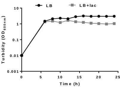
Figure 4.1- Cell growth of E. coli BL21(DE3) transformed with pCM13(S5E9) in LB
medium without (LB) and with lactose (LB+lac) at different time periods for 24 h at 37 ºC, 1:4 volume ratio and 200 rpm.
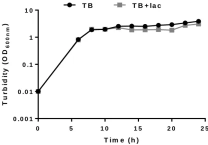
Figure 4.2- Cell growth of E.coli BL21(DE3) transformed with pCM13(S5E9) in TB
medium without (TB) and with lactose (TB+lac) at different time periods for 24 h at 37 ºC, 1:4 volume ratio and 200 rpm.
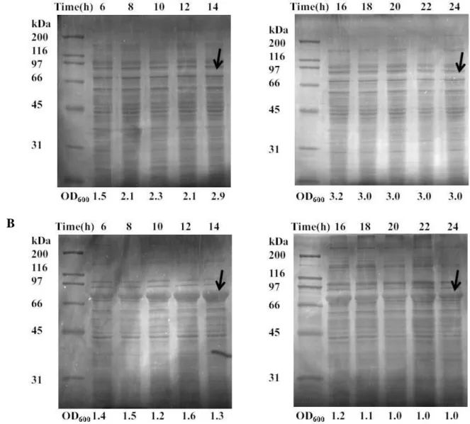
Figure 4.3- Protein production analyzed by SDS-PAGE (Copper staining) of soluble
fractions of SELP-59-A at different time points. (A) Growth in LB, (B) Growth in LB+ lactose. The optical density is indicated below each fraction, and the arrow indicates the band correspondent to the recombinant protein.
xiv
Figure 4.4- Protein production analyzed by SDS-PAGE (Copper staining) of soluble
fractions of SELP-59-A in different time points. (A) Growth in TB, (B) Growth in TB+ lactose. The optical density is indicated below each fraction, and the arrow indicates the band correspondent to the recombinant protein.
Figure 4.5- SDS-PAGE analysis of the expression levels of SELP-59-A in cell cultures
induced with 1mM IPTG in LB medium at different times points (indicated on top of the gel). The optical density is indicated below each fraction. The arrow indicates the band correspondent to the recombinant protein.
Figure 4.6- Purification of SELP-59-A by ammonium sulphate precipitation observed
by SDS-PAGE (cooper staining). 1- Molecular Weight Marker (Broad Range, Bio-Rad); 2- Without production (cells transformed with empty vector); 3- Crude cell extract after induction; 4- Culture medium; 5- Crude cell lysate after sonication; 6- Supernatant of acidification. The acidified fraction was saturated with increasing concentrations of ammonium sulphate (indicated abo ve the gel). The arrow indicates the recombinant protein band.
Figure 4.7- SDS-PAGE showing highly pure polymer fractions after resuspension of
the precipitated copolymer in ddH2O followed by centrifugation to remove insoluble debris. The different percentages of saturation are indicated above the gel.
Figure 4.8- Pure lyophilized SELP-59-A.
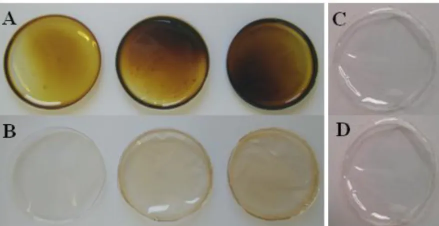
Figure 4.9- Photograph showing colour changing of the films. (A) FA-SELP/Ag films
(1, 3 and 5 wt %); (B) H2O-SELP/Ag films (1, 3 and 5 wt %); (C) FA-SELP films; (D) FA-SELP/LF(5 wt%).
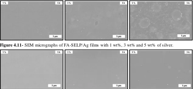
Figure 4.10- SEM micrographs of H2O-SELP/Ag films with 1 wt%, 3 wt% and 5 wt%
of silver.
Figure 4.11- SEM micrographs of FA-SELP/Ag films with 1 wt%, 3 wt% and 5 wt% of
silver.
Figure 4.12- SEM micrographs of FA-SELP/LF films with 1 wt%, 3 wt% and 5 wt% of
xv
Figure 4.13- Representative micrographs of FA-SELP films without (SELP) and with 5
wt% of silver nitrate (5%).
Figure 4.14- Mechanism of silver reduction by formic acid (Shi et al., 2011).
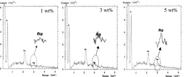
Figure 4.15- EDS analysis of FA-SELP/Ag films with different concentrations of silver
showing characteristic elemental peaks.
Figure 4.16- EDS analysis of H2O-SELP/Ag films with different concentrations of
silver showing characteristic elemental peaks.
Figure 4.17- XRD spectra of (A) FA-SELP/Ag (0 and 5 wt%) films and (B) H2
O-SELP/Ag (0 and 5 wt%) films.
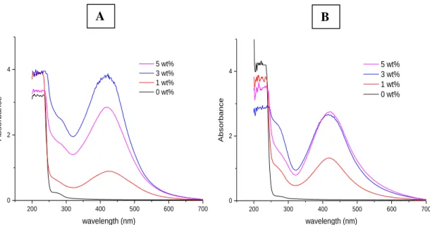
Figure 4.18- UV-vis absorption spectra of FA-SELP/Ag films with different silver
nitrate content (0, 1, 3 and 5 wt%). (A) non-treated FA-SELP/Ag films. (B) methanol-treated FA-SELP/Ag films.
Figure 4.19- UV- vis absorption spectra of H2O-SELP/Ag films with different silver
nitrate content (0, 1, 3 and 5 wt%). (A) non-treated H2O-SELP/Ag films. (B) methanol-treated H2O-SELP/Ag films.
Figure 4.20- UV-vis absorption spectra of FA-SELP/LF films with different lactoferrin
content (0, 1, 3 and 5 wt%). (A) non-treated FA-SELP/LF films. (B) methanol-treated FA-SELP/LF films.
Figure 4.21- UV-vis absorption spectra of H2O-SELP/LF films with different
lactoferrin content (0, 1, 3 and 5 wt%). (A) non-treated H2O-SELP/LF films. (B) methanol-treated H2O-SELP/LF films.
Figure 4.22- Images showing the colour change of SELP-59-A fibres. (A) SELP/Ag
fibres (from left to right) with 0, 1, 3 and 5 wt %); (B) SELP/LF fibres with 5 wt% bLF.
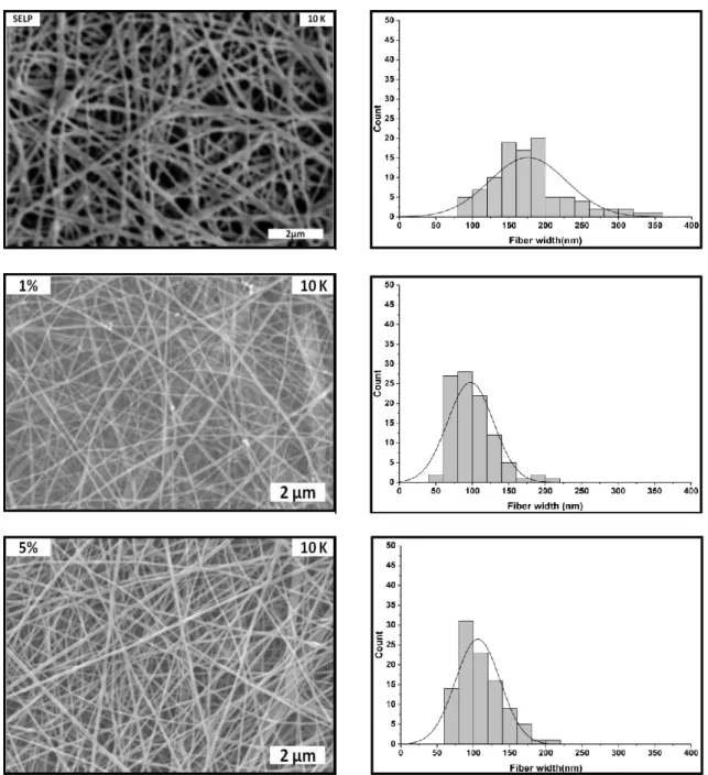
Figure 4.23- SEM micrographs of SELP/Ag electrospun fibres with different
concentrations of silver nitrate: 0, 1, 3 and 5 wt%. The histograms represent fibre size distribution for each corresponding concentration. A normal distribution curve was applied for all histograms.
xvi
Figure 4.24- Graphical representation correlating the average fibre diameter with silver
content.
Figure 4.25- Representative SEM micrographs and histograms of fibre size distribution
for methanol-treated SELP/Ag fibre mats with 0 wt% (SELP), 1 wt% (1%) and 5 wt% (5%).
Figure 4.26- Electron Backscattering images of SELP/Ag electrospun fibres with
different silver concentrations: 1 wt%, 3 wt% and 5 wt%.
Figure 4.27- Representative micrographs of the FA-SELP/Ag (5 wt%) fibres obtained
after treatment with methanol-saturated air.
Figure 4.28- EDS analysis of fibres with different concentrations of silver showing
characteristic elemental peaks.
Figure 4.29- Representative SEM micrographs and histograms of fibre size distribution
for SELP/LF fibre mats with 1 wt%, 3 wt% and 5 wt% of lactoferrin.
Figure 4.30- Graphical representation correlating the average fibre diameter with
lactoferrin and silver content.
Figure 4.31- Representative SEM micrographs and histograms of fibre size distribution
for methanol-treated SELP/LF fibres with 1 wt% and 5 wt% of lactoferrin.
Figure 4.32- (A) FTIR spectra of SELP/Ag composite materials. (B) FTIR spectra of
methanol-treated SELP/Ag composite materials.
Figure 4.33- FTIR spectra in the amide I region of non-treated (normal line) and
methanol-treated (intense line) SELP/Ag with different silver concentrations (0, 1, 3, 5 wt%).
Figure 4.34- (A) FTIR spectra of SELP/LF composite materials. (B) FTIR spectra of
methanol-treated SELP/LF composite materials.
Figure 4.35- FTIR spectra in the amide I region of non-treated (normal line) and
methanol-treated (intense line) for the different lactoferrin concentrations (0, 1, 3, 5 wt%).
Figure 4.36- (A) Degree of swelling obtained for SELP, SELP/Ag (3 wt%) and
xvii
temperature. (B) Inset graphic represents the degree of swelling determined during the first 5 min of immersion.
Figure 4.37- (A) Degree of swelling obtained for H2O-SELP, H2O-SELP/Ag (5 wt%)
and H2O-SELP/LF (5 wt%) films for a total of 60 min of immersion in deionized water at room temperature. (B) Inset graphic represents the degree of swelling determined during the first 5 min of immersion.
Figure 4.38- Hydrolytic degradation profile of SELP, SELP/Ag (3 wt%) and SELP/LF
(5 wt%) fibres in PBS at different time periods. (B) SELP/Ag membranes removed from the PBS after 15 days of incubation. (C) SELP/LF membranes removed from the PBS after 15 days of incubation.
Figure 4.39- Hydrolytic degradation profile of SELP, SELP/Ag (5 wt%) and SELP/LF
(5 wt%) films in PBS at different time periods.
Figure 4.40- Growth curves of Gram-negative bacteria (Escherichia coli and
Pseudomonas aeruginosa) (blue), Gram-positive bacteria (Staphylococcus aureus and Bacillus subtilis) (black) and yeast (Candida glabrata and Candida albicans) (red).
Figure 4.41- Growth inhibition against E .coli for (A) FA-SELP/Ag films, (B) H2
O-SELP/Ag films and (C) O-SELP/Ag fibre mats. Bars represent means ± SD. **** p<0.0001.
Figure 4.42- Growth inhibition rates against E. coli and P. aeruginosa by SELP/Ag
fibre mats. Bars represent means ± SD. **** p<0.0001.
Figure 4.43- Halo of inhibition of FA-SELP/Ag films (1- 0 wt%; 2- 1 wt%); 3- 3 wt%);
4- 5 wt%) against E. coli, P. aeruginosa, S. aureus and B. subtilis.
Figure 4.44- Graphical representation comparing the diameter of the halos of inhibition
found for FA-SELP/Ag films against different bacteria. Results are expressed by calculating the diameter of zones of inhibition (mm).
Figure 4.45- Halo of inhibition of H2O-SELP/Ag films (1- 0 wt%; 2- 1 wt%); 3- 3
xviii
Figure 4.46- Graphical representation comparing the diameter of the halos of inhibition
found for H2O-SELP/Ag films against different bacteria. Results are expressed by calculating the diameter of zones of inhibition (mm).
Figure 4.47- Halo of inhibition of SELP/Ag fibres (1- 0 wt%; 2- 1 wt%); 3- 3 wt%); 4-
5 wt%) against E. coli, P .aeruginosa, S. aureus and B. subtilis.
Figure 4.48- Graphical representation comparing the diameter of the halos of inhibition
found for SELP/Ag fibres against different bacteria. Results are expressed by calculating the diameter of zones of inhibition (mm).
Figure 4.49- Halo of inhibition of FA-SELP/Ag films (1- 0 wt%; 2- 1 wt%); 3- 3 wt%);
4- 5 wt%) against C. albicans and C. glabrata.
Figure 4.50- Graphical representation comparing the diameter of the halos of inhibition
found for FA-SELP/Ag films against two different yeast. Results are expressed by calculating the diameter of zones of inhibition (mm).
Figure 4.51- Halo of inhibition of H2O-SELP/Ag films (1- 0 wt%; 2- 1 wt%); 3- 3
wt%); 4- 5 wt%) against C. albicans and C. glabrata.
Figure 4.52- Graphical representation comparing the diameter of the halos of inhibition
found for H2O-SELP/Ag films against two different yeast species. Results are expressed by calculating the diameter of zones of inhibition (mm)
Figure 4.53- Halo of inhibition of SELP/Ag fibres (1- 0 wt%; 2- 1 wt%); 3- 3 wt%); 4-
5 wt%) against C. albicans and C. glabrata.
Figure 4.54- Assay of direct contact of H2O-SELP/Ag films against Aspergillus
nidulans. (A) Two days at 37 ° C after performing these assays (B) Three days at 37 ° C after performing these assays. 1-H2O-SELP ;2- H2O-SELP/Ag (1 wt%);3- H2 O-SELP/Ag (5 wt%) ,4- sterile disc ;5- itraconazole and 6- positive control.
Figure 4.55– Diffusion assay of SELP/Ag materials against Aspergillus nidulans. (A)
FA-SELP/Ag films (B) SELP/Ag fibres 1- SELP (0 wt%) ;2- SELP/Ag (1 wt%);3- SELP/Ag (3 wt%) ,4- SELP/Ag (5 wt%) and 5- itraconazole.
xix
Figure 4.56- Growth inhibition against E.coli of (A) FA-SELP/LF films, (B) H2
O-SELP/LF films and (C) O-SELP/LF fibre mats. Bars represent means ± SD. ** p<0.01and **** p<0.0001.
Figure 4.57- Halo of inhibition of SELP/LF fibres (1- 0 wt%; 2- 1 wt%); 3- 3 wt%); 4-
5 wt%) against E. coli, P. aeruginosa, S. aureus, and B. subtilis.
Figure 4.58- Halo of inhibition of FA-SELP/LF films (1- 0 wt%; 2- 1 wt%); 3- 3 wt%);
4- 5 wt%) against four E. coli, P. aeruginosa, S. aureus and B. subtilis.
Figure 4.59- Halo of inhibition of H2O-SELP/LF films (1- 0 wt%; 2- 1 wt%); 3- 3
wt%); 4- 5 wt%) against four E. coli, P. aeruginosa, S. aureus and B. subtilis.
Figure 4.60- Indirect contact cytotoxicity test of pristine FA-SELP and FA-SELP/Ag
films on normal human skin fibroblasts (BJ-5ta cell line) with positive (control) and negative controls (30% DMSO) for cell viability after 24 and 72 h of cell culture. Results are expressed as % of viability compared to the control. Bars represent means ± SD. ns- non-significant,* p<0.05,** p<0.01, *** p<0.001 and **** p<0.0001.
Figure 4.61- Indirect contact cytotoxicity test of pristine H2O-SELP and H2O-SELP/Ag
films on normal human skin fibroblasts (BJ-5ta cell line) with positive (control) and negative controls (30% DMSO) for cell viability after 24 and 72 h of cell culture. Results are expressed as % of viability compared to the control. Bars represent means ± SD. ns- non-significant,* p<0.05,** p<0.01, *** p<0.001 and **** p<0.0001.
Figure 4.62- Indirect contact cytotoxicity test of pristine SELP and SELP/Ag fibres on
normal human skin fibroblasts (BJ-5ta cell line) with positive (control) and negative controls (30% DMSO) for cell viability after 24 and 72 h of cell culture. Results are expressed as % of viability compared to the control. Bars represent means ± SD. ns- non-significant,* p<0.05,** p<0.01, *** p<0.001 and **** p<0.0001.
xx
List of tables
Table 3.1- Culture media used and composition.
Table 3.2- List of bacterial and fungal species and strains used in the work Table 4.1- Volumetric productivities of SELP-59-A using different media.
3
The infectious diseases are a major cause of mortality in the world with more than 15 million deaths per year due to infections (Spížek et al., 2010). For some years, chemical antibiotics have demonstrated to be part of the "solution" for the treatment of infectious diseases. However, bacteria and fungi have developed antibiotic and antifungal resistance, making the medical treatments very difficult and expensive. Thus, it is extremely important to improve the medical care as well as to find alternatives in order to overcome this resistance (Rai et al., 2012; Spížek et al., 2010).
1.1. Antibiotic resistance
Pathogens can be found everywhere i.e., in the air, soil and water; and since the beginning of history, humans have handled a war against microorganisms that caused infections and diseases. Regardless of the great discoveries in the 20th century that contributed to infection control such as the discovery and commercialization of penicillin in the 40’s, bacteria quickly demonstrated various forms of resistance. In fact, despite the development of new antimicrobial agents, microorganisms continue to develop new mechanisms of resistance (Harbottle et al., 2006; Tenover, 2006). Presently, we are susceptible to various microorganisms that acquired resistance to different drugs (Alekshun and Levy, 2007) and therefore, the study of antibiotic resistance has had priority due to the following main factors: i) bacteria are mainly responsible for community and nosocomial infections and ii) the development of several classes of antibiotics often results in diverse mechanisms of resistance (Ghannoum and Rice, 1999).
The antimicrobial therapy used against bacterial infections may be grouped, according to their principal mechanism of action, in i) cell wall perturbation, ii) inhibition of protein synthesis, iii) interference with nucleic acid synthesis, and iv) inhibition of a metabolic pathway (Tenover, 2006).
Microorganisms may express resistance by several mechanisms which are associated to intrinsic or acquired resistance (Harbottle et al., 2006; Kenneth and Ray, 2004; Tenover, 2006). The intrinsic resistance is considered a natural trait, associated with the genome of a bacterial specie and thus is present in all the elements of a specie (Harbottle et al., 2006; Shanks and Peteroy-Kelly, 2009). Examples of such intrinsic
4
mechanism include the multi-drug resistant (MDR) phenotype of Gram- negative bacteria and the production of β- lactamases that are responsible for innate resistant against β-lactam antibiotics (Alekshun and Levy, 2007).
Of greater concern are the cases of acquired resistance (Tenover, 2006). This resistance mechanism arises from the mutation of regulatory or structural genes or from the horizontal acquisition of new genes that confers defense to a particular antimicrobial agent (Hogan and Kolter, 2002). This phenotype is not common to all elements of the specie, but rather is present in the individuals of a bacterial strain derived from a previously susceptible organism (Harbottle et al., 2006; Shanks and Peteroy-Kelly, 2009).
The mutations associated with antibiotic resistance may include i) modifications of the target protein to which the antibacterial agent binds. For example, resistance to erythromycin, a ribosome inhibitor, is caused when adenosine in the bacterium's rRNA is methylated which results in alterations in the binding site (Shanks and Peteroy-Kelly, 2009); ii) downregulation or alteration of a membrane protein channel required by the antimicrobial agent to enter the cell. For example, decreased permeability via porin alterations (eg, OmpF in E. coli), can develop resistance to several antibiotics (Harbottle
et al., 2006; Tenover, 2006); iii) upregulation of the production of enzymes that
inactivate the drug such as β- lactamases. These enzymes hydrolyze the ring structure of β-lactam antibiotics such as penicillins and cephalosporins (Shanks and Peteroy-Kelly, 2009; Tenover, 2006); or iv) upregulation of pumps that expel the antimicrobial agent out of the bacterial cell. For example, the efflux of tetracyclines in both Gram-positive and Gram- negative bacteria (Poole, 2002; Tenover, 2006).
Regarding horizontal gene transfer the main mechanisms are conjugation, transformation and transduction (Figure 1.1). Conjugation is the most frequent mechanism and is mediated by circular plasmids (DNA fragments) and conjunctive transposons. The plasmids are transferred between bacteria through a structure denominated "pilus" formed by the proximity of bacteria, connecting them and allowing the passage of the genetic material. Transformation is another transmission mechanis m of bacterial resistance and occurs when there is a direct passage of free DNA from one cell to another. The receiving bacterium then incorporates the free DNA in its own genome, spreading the resistance. The third mechanism is named transduction and
5
arises through bacteriophages. The virus containing the bacterial antibiotic resistance gene infects a new cell by introducing the genetic material (Alanis, 2005).
Figure 1.1- Mechanis ms of antibiotic resistance by gene transfer mechanis ms. DNA fro m the biosphere
containing an antibiotic resistance gene (pink) can be transmitted by horizontal transfer through several mechanis ms: conjugation, transformat ion and transduction. Resistance can also appear by de novo mutation (red cross) (Andersson and Hughes , 2010).
1.2. Antifungal resistance
Presently, the fungal infections represent a global health threat (Vandeputte et al., 2012). In the last decades, the incidence of fungal infections has increased dramatically due to the increase in the number of vulnerable immunocompromised individuals to fungal infection (LaFayette et al., 2010; Mishra et al., 2007).
The antifungal treatment and the selection of drug are determined by various fundamental aspects such as the status of the host immune system, the location of the infection, evaluation of characteristics of the infection (fungi species present and their different susceptibility to antifungal drugs ) and p harmacokinetic properties of the drug (e.g. absorption, excretion and toxicity) (Loeffler and Stevens, 2003).
The different classes of antifungal use different means to kill or inhibit the growth of fungi (Pfaller, 2012). The mode of action for the principal antifungals can be distributed in four classes namely, i) polyenes that result in modification of membrane functions; ii) azoles that inhibit the 14α- lanosterol demethylase, a fundamental enzyme
6
in ergosterol biosynthesis; iii) DNA and RNA synthesis inhibitors (flucytosine); and iv) 1,3-β-glucan synthase inhibitors (echinocandins) (Loeffler and Stevens, 2003).
The fungi can be intrinsically resistant to antifungal agents prior to drug exposure (primary resistance) or can acquire resistance when exposed to the antifungal drug during treatment (secondary resistance) (Perea and Patterson, 2002). The fungal pathogens develop mechanisms to neutralize the fungicidal or fungistatic action of the antifungal drugs based on the following principal mechanisms: i) reduction in the accumulation of the antifungal inside the fungal cell; ii) decrease the affinity of the drug for its target and iii) changes in the metabolism to nullify the effect of the antifungal agents (Vandeputte et al., 2012).
Accordingly, the resistance mechanisms of the main antifungals are described.
Azole Resistance
Several mechanisms related to resistance to azoles in Candida species have been described. The first mechanism is associated with the induction of efflux pumps leading to a decrease in the accumulation of the antifungal inside the fungal cell (Pfaller, 2012). The over-expression of efflux pumps belonging to the family of ATP-binding cassette (ABC) encoded by the CDR1 and CDR2 genes, and the major facilitator superfamily (MFS) encoded by MDR1gene have been associated with azole resistance in Candida
albicans (Lewis et al., 2012). A second mechanism relates with point mutations in the ERG11 gene that codes for the target enzyme lanosterol 14α-demethylase, inducing
modifications in the target such as decreased affinity or the inability to bind to azoles (Pfaller, 2012).
Polyene Resistance
The polyenes binds to sterol, preferably ergosterol, in cell membrane causing alterations in the permeability of the membrane and cell death (Prasad and Kapoor, 2004). Resistance to polyenes arises as a consequence of considerable modifications in the lipid composition of the plasma membrane (Loeffler and Stevens, 2003). One example can be a decrease of ergosterol content due to a lack of the Δ8,7
-sterol isomerase. Thus, this condition results in a lower affinity of the antifungal (amphotericin B) to the plasma membrane, possibly the result of a lack of the binding site (Ghannoum and Rice, 1999; Loeffler and Stevens, 2003). Another reason for
7
amphotericin B resistance may be a modified content β-1,3 glucans in the fungal cell wall. These constituents that influence the stability of the cell wall, induce access of large molecules such as amphotericin B to the plasma membra ne (Loeffler and Stevens, 2003).
Echinocandins Resistance
This antifungal class inhibits 1,3-β-D-glucan synthase and thereby disrupts the biosynthesis of 1,3-β-D-glucan, a fundamental component of the fungal cell wall. This results in the formation of a defective cell wall that relates with cellular instability and lysis in yeasts (Pfaller, 2012). Mutations in the glucan synthase gene FKS1 are able to drastically reduce the susceptibility to echinocandins in yeast and molds. In fact, mutations in the FSK1 resulting in a single amino acid change in the protein, of
Candida parapsilosis, Candida orthopsilosis, and Candida metapsilosis shows up as a
possible cause of intrinsic reduced susceptibility to echinocandins in these species (Marie and White, 2009).
5-fluorocytosine Resistance
The 5-fluorocytosine enters cells through cytosine permease and is deaminated to the active form 5- fluorouracil, by cytosine deaminase. In turn, 5-fluorouracil is then
converted to 5-fluorouridine monophosphate (FUMP) by uracil
phosphoribosyltransferase (UPRTase). FUMP can be converted into fluorouridine triphosphate (FUTP) or 5-fluoro-deoxyuridine monophosphate (FdUMP), which inhibits protein synthesis or DNA synthesis, respectively (Mishra et al., 2007; Prasad and Kapoor, 2004). The 5-fluorocytosine or flucytosine resistance is associated with loss or mutation of enzymes (cytosine permease) involved in absorption of drugs (Bossche, 1997) or loss of enzymatic activity responsible for conversion to FUMP. The interruption in the formation of FUMP by the absence of cytosine deaminase activity or loss of UPRTase activity provides 5-fluorocytosine resistance (Ghannoum and Rice 1999).
In opposition to bacteria, fungi do not appear to have ability to transfer resistance genes from one organism to another. Therefore, development of resistance is remarkably slower than that verified for bacteria, and is related to the antifungal
8
pressure to propel mutations or reveal previously suppressed resistance genes within a given fungal strain (Klepser et al., 1997).
1.3. Antimicrobial agents
Commonly, pathogenic microorganisms represent a public health problem therefore, a way of preventing this problem is essential (Azam et al., 2012). The use of antimicrobial agents plays a significant role in b iomedicine wherein the fundamental prerequisite of these materials is based on the capacity to perform a specific application associated to a specific and reproducible response. The antimicrobial agents can be of chemical or biological origin and one can find several examples of these. However, and considering the scope of this work, only silver (chemical origin) and bovine lacto ferrin (biological origin) will be described.
1.3.1. Silver as an antimicrobial agent
Noble metals have attracted interest not only due to its versatile reactivity, but also to the compatibility and non-toxicity towards eukaryotic cells. From this group, silver is the preferred choice for application in the biomedical field owing to its attractive physicochemical properties, lower cost and particularly low toxicity to humans when present in lower concentrations (Saengmee-Anupharb et al., 2013).
Silver is a basic element, very ductile and malleable, displaying higher electrical and thermal conductivity with minimum contact resistance (Rai et al., 2012). The antimicrobial activity of silver is known since ancient times and its use remained until nowadays. In fact, Egyptians, Romans and Greeks used silver salts to clean wounds and silver threads in sutures to promote healing. Silver was (and still is) used in wound dressings to treat open infected wounds, skin ulcers, compound fractures, and burn injuries (Knetsch and Koole 2011). Silver was also the most widely used therapy for the treatment of infections until the discovery of antibiotics; although it is still applied due the increase of bacterial resistance to antibiotics (Knetsch and Koole, 2011; Monteiro et
al., 2009; Prabhu and Poulose, 2012). Silver presents antimicrobial effects of large
9
to antibiotics, fungi, protozoa and certain viruses, even at lower concentrations (Monteiro et al., 2009).
Silver mechanism of action
This chemical element is widely used due to its bacteriostatic and bactericidal ability towards microorganisms however, its mechanisms of action are not fully understood and several theories were developed to explain such behavior (Egger et al., 2009).
Silver immediately interacts with sulfhydryl groups (-SH) on the surface of the bacteria, replacing the hydrogen atoms and giving origin to an S-Ag bond. This interrupts respiration and electron transfer, which hinders the induction of rescue mechanisms (Mijnendonckx et al., 2013). Furthermore, interruption of respiration and electron transfer induces a collapse of the proton motive force, leading to the de-energization of the membrane and consequently cell death (Cao and Liu, 2010; Mijnendonckx et al., 2013). The disruption of the cell membrane allows the entrance of silver ions in the cytoplasm, where it may affect many steps. The silver ions interact with nucleic acids, particularly with the nucleosides, rather than with phosphate groups (Jung et al., 2008). Binding to the guanine base (N7 atom), which is perturbed by methylation, enhances pyrimidine dimerization and affects the DNA replication (Mijnendonckx et al., 2013). The suspension of replication for bacteria proliferation results in a decrease or absence of cells or promotes the formation of reactive oxygen species (ROS) within the cell, causing cellular damage and subsequent death of the bacterial structures (Cao and Liu, 2010). However, the effect which is considered most effective of silver ions is their interaction with thiol groups. Since the thiol group of cysteine residues is essential for the function of various enzymes, interaction leads to conformational changes and to inactivation of enzymatic functions (Mijnendonckx et
10
Figure 1.2- Mechanisms of action of silver ions in bacteria (M ijnendonckx et al., 2013).
Silver nanoparticles show great antimicrobial activity due to the large surface area available, which promotes a better interaction between Ag+ and the microorganisms. The nanoparticles can bind to the cell membrane and are also able to penetrate into the bacteria (Rai et al., 2009).
The action of silver nanoparticles has been described by the following mechanisms: (1) Adhesion of silver nanoparticles onto the surface of the bacterium, which induces an increase in membrane permeability, causing structural alterations and thereby cell death. The small size and large surface area of the nanoparticles provides a strong contact with the bacterium surface;
(2) Formation of free radicals, which are derived from the surface of the silver nanoparticles that interact with bacteria causing damage to the cell membrane, which becomes permeable leading to cell death;
(3) Decomposition of silver nanop articles releases Ag+ ions which can interact with sulphur-containing proteins in the bacterial cell walls inactivating them. The contact between the bacterial cells and silver interferes with the normal functions of the cell, resulting in the production of reactive oxygen species;
11
(4) Silver nanoparticles penetrate inside the bacterial cell and attack proteins that contain sulfur as well as phosphorus-containing compounds such as DNA. Nanoparticles cause DNA damage that will induce cell death. The interaction of the nanoparticles with DNA may hinder replication resulting in cell death (Egger et al., 2009; Prabhu and Poulose, 2012; Reidy et al., 2013).
The mechanisms for antibacterial action of silver nanoparticles as reported above are summarized in Figure 1.3.
Figure 1.3- Schemat ic representation of the mechanisms for antibacterial action of silver
nanoparticles. Grey circles indicate silver nanoparticles and Ag+ are silver ions released fro m the nanoparticles (Reidy et al., 2013).
Antifungal action of silver
Silver action mechanism in fungi has not been the subject of much study and its potential is not totally clear. Silver nanoparticles acts on fungi, targeting and disrupting the cell membrane, which results in formation of pores and subsequently causing cell death (Rai and Bai, 2011). It has been proposed that the silver nanoparticles affect transport systems of the cell, leading to accumulation of silver ions, causing complications in cellular processes such as metabolism and respiration. The silver ions
12
produce reactive oxygen species that are harmful to cell, causing damage to proteins, lipids and nucleic acids (Gavanji, 2013).
Silver applications
As mentioned, in small concentrations, silver is a compound having low toxicity, high thermal stability and low volatility to human cells and high antimicrobial activity against a broad spectrum of microorganisms (Rai et al., 2009; Raja et al., 2012). Nowadays, silver ions are used to control/inhibit bacterial growth in various medical applications, such as catheters, dental practice, wound healing, coating of medical devices and even in cosmetics (eg. hydrating creams, antiperspirants) (Jung et al., 2008).
Silver dressings play an important role in wound treatment. These make use of delivery systems that release silver in different concentrations. Other factors, such as the silver disposal in the dressing, its chemical and physical state form, affinity of the dressing to moisture are also involved the killing of microorganisms (Rai et al., 2009). Silver is also used in topical creams for the treatment of burn wounds, in dental amalgam, eye care and in silk impregnated polymers to inhibit the growth of microorganisms on medical devices, including catheters and heart valves. The use of silver has also been used in water systems acting as a disinfectant such as in swimming pools, hot water equipment in hospitals and potable water apparatus (Mijnendonckx et
al., 2013).
1.3.2. Lactofe rrin as an antimicrobial agent
Lactoferrin (LF) was firstly reported in 1960 and is a glycoprotein that belongs to the transferrin family with a molecular weight of ~80 kDa showing a high affinity for iron (Adlerova et al., 2008). Structurally, lactoferrin is a polypeptide with approximately 700 amino acids, folded into two globular lobes, each situated in one of the terminals, denominated N and C-lobe; each lobe consists of two subunits (N1, N2, C1 and C2) connected by an α-helix (Figure 1.4). Each lobe can bind a metal atom, in cooperation with the carbonate ion, which is fundamental for the binding of iron
13
(Adlerova et al., 2008; García-Montoya et al., 2012; Strate et al., 2001). This glycoprotein is found in several mammalian mucosal surfaces including man, cattle, goats, dogs, among others. Lactoferrin is present in the mucosal secretions including saliva, tears, vaginal secretions, semen, bronchial and nasal secretions, gastrointestinal fluids, urine and more intensely in milk and colostrum, designating it as the second most abundant protein in milk after casein (García-Montoya et al., 2012; González-Chávez et al., 2009; Strate et al., 2001).
Lactoferrin is considered a multifunctional protein presenting a diversity of physiological functions including the control of the amount of iron absorbed from the intestine; immune response; antioxidants, anticancer and anti- inflammatory properties. However, until the present moment, their antimicrobial capability is the most studied subject (García-Montoya et al., 2012; González-Chávez et al., 2009).
Figure 1.4- Structure of Lactoferrin bonded to iron (Fe2LF) (Baker and Ba ker, 2012).
Antimicrobial activity
The presence on the mucosal surface makes lactoferrin a major form of defense against microorganisms that invade the body through mucosal tissues. Lactoferrin inhibits the growth of a broad spectrum of microorganisms capable of attacking humans and promote the appearance of diseases, including Gram-positive and Gram-negative
14
bacteria, viruses, protozoa, or fungi (Adlerova et al., 2008; Jenssen and Hancock, 2009; Legrand et al., 2008).
The antimicrobial activity of lactoferrin is primarily related with two mechanisms: the first is based on the ability of lactoferrin to remove the iron, essential nutrient for the growth of potential pathogens at sites of infection, acting as a bacteriostatic agent and the second mechanism is the direct effect of lactoferrin against a variety of microorganisms (García-Montoya et al., 2012).
Antibacterial activity
The bacteriostatic effect of LF is due to its ability to sequester the Fe3+ ion resulting in the inhibition of microorganism growth (González-Chávez et al., 2009). However, various bacteria can develop resistance mechanisms to overcome the sequestration of iron. Some Gram-negative bacteria synthesize siderophores, which function is to remove the iron from lactoferrin and transport it to zones of interest in the bacterium (Ward, 2002).
The bactericidal effect has been demonstrated by direct interaction of lactoferrin with the bacterial surface (Figure 1.5). In Gram-negative bacteria, LF damages the outer membrane by interacting with anionic lipid A, a constituent of the lipopolysaccharides (LPS) or porins. The positively charged N-terminal of lactoferrin prevents the interaction between LPS and bacterial cations (Ca2+ and Mg2+), leading to the release of LPS from the cell wall, triggering the membrane permeabilization and consequential bacterial damage. The interaction of LF and LPS develops the action of natural antibacterial agents, lysozyme is one of the agents is secreted from the mucosa in parallel with LF. On the other hand, in Gram-positive bacteria, LF binds to positively charged anionic molecules located on the surface of bacteria, namely lipoteichoic acid, contributing in a reduction of negative charge on the cell wall and subsequently promoting the action of antibacterial agents, namely the contact between lysozyme and underlying peptidoglycan over which it exerts an enzymatic action (González-Chávez et
al., 2009).
Besides the aforementioned antimicrobial actions. LF contains a N-terminal domain with serine protease activity which inactivates proteins of the outer membrane of
15
bacteria that are essential for the adherence to host cells. Thus, LF has several antimicrobial actions for the protection of mucosal surfaces against infections caused by infectious agents (Jenssen and Hancock, 2009; Ward, 2002).
Figure 1.5- Mechanism of antibacteria l action lactofe rrin (Gon zá lez-Chávez et al., 2009).
Antifungal activity
The antifungal activity of lactoferrin has been attributed to its ability to trap Fe3+ ions leading to changes in cell surface permeability (García-Montoya et al., 2012). Due to its direct interaction with surface glycoproteins, lactoferrin induces changes in the fungal cell wall with the occurrence of surface bubbles that lead to swelling and failure (Jenssen and Hancock, 2009).
Lactoferrin applications
The various LF properties make this protein promising for clinical and industrial applications. The most widely used application is as supplement of infant formula, such as drinks and milk. Presently, it has been used in cosmetics, toothpaste, pet food supplements and drinks (García-Montoya et al., 2012; Legrand et al., 2008; Wakabayashi et al., 2006). These products are reported as anti- inflammatory, antioxidant and anti- infection, and have the ability to improve oro-gastrointestinal microflora.
16
Lactoferrin has also been used in the maintenance and food protection by retarding lipid oxidation or reducing the growth of microorganisms. One example, is to obtain edible films or sprays with lactoferrin, providing a physical barrier or acting as an antimicrobial agent. In addition, clinical studies have sho wn good efficacy of LF for the treatment of infections and inflammatory diseases (García-Montoya et al., 2012; Legrand et al., 2008). Lactoferrin may also be used as a clinical marker of inflammatory diseases, once its concentration in blood and fluids may increase considerably in septicemia or during a very severe acute respiratory syndrome (García-Montoya et al., 2012; Legrand et al., 2008).
1.4. Silk-elastin-like proteins (SELPs)
The search for effective antimicrobial compounds with capacity to reduce and/or avoid infections has become a priority area. Due to their intrinsic properties, polymers are interesting candidates as substrates for the development of antimicrobial materials devoted for biomedical applications, either by incorporation or functio nalization with antimicrobial agents. Characteristics such mechanical properties, hydrophilicity or molecular weight are important factors for antimicrobial activity, particularly in aspects such as the release rate of the biocide (Muñoz-Bonilla and Fernández-García, 2012).
The recombinant protein-based polymers (rPBPs) are a class of macromolecules obtained by recombinant DNA technology. These polymers mimic the characteristics of natural proteins, but can also display functions and properties that are not prese nt in the natural counterparts. The silk-elastin- like proteins are an example of such macromolecules which composition is based on tandem repeats of silk- like (GAGAGS; G: glycine, A: alanine, S: serine) and elastin- like (VPGVG; V: valine, P: L-proline) blocks (Machado et al., 2013a). The size of each block as well as the silk:elastin block ratio are crucial to the physicochemical properties of SELP copolymers (Machado, 2012).
Elastin is present in the extracellular matrix, providing strength and elasticity to mammalian tissues and organs, in particular in blood vessels, cartilage, ligaments, skin, and lungs. The combination of silk with elastin confers unique mechanical and biological properties to the macromolecule. The silk blocks provide strength, chemical
17
and thermal stability due to the formation of antiparallel beta-sheet stabilized by hydrogen bonds; whereas the periodic insertion of elastin blocks reduces the overall crystallinity of the polymeric molecule and increases its flexibility and water solubility (Machado et al., 2013a). Indeed, SELPs with large silk blocks and small elastin blocks precipitate in aqueous solution even at relatively low concentrations, while the inclusion of elastin blocks with adequate size renders SELPs soluble in water (Machado et al., 2013a). This characteristic is fundamental for protein purification as well as for the conception of the polymer when directed for biomedical applications.
Due to their biocompatibility and ability to self-assemble into hydrogels, SELPs have been explored as matrix- mediated delivery systems for gene therapy (Gustafson et
al., 2010; Gustafson et al., 2009; Greish et al., 2010; Megeed et al., 2002; Price et al.,
2012), drug delivery systems (Cappello et al., 1998; Dinerman et al., 2002) and as scaffolds for encapsulation and chondrogenesis of human mesenchymal stem cells (Haider et al., 2008). The SELP-59-A (composition S5E9, where S represents the silk block and E the elastin block) used in this study differs from the most frequent SELPs as the sequence of the elastin- like block is altered to VPAVG. This simple substitution of the central glycine by an L-alanine demonstrated to dramatically change the thermal properties of elastin- like polypeptides and have been suggested to change the mechanical response from elastic to plastic (Machado et al., 2009; Urry, 2006). Furthermore, electrospun fibre mats produced from SELP-59-A showed promising properties for application in skin tissue engineering namely, for wound healing (Machado et al., 2013b). In this sense, the production of SELP fibre membranes can be explored for several applications, such as in the development of artificial muscles, biosensors, dressings, protective materials and drug administration systems (Ner et al., 2009).
1.5. Fabrication of composite materials with antimicrobial properties
A composite material is defined when two or more different materials are combined
together, normally with different properties that, when combined, produce a material
with unique characteristics. The individual components remain separate and distinct within the composite structure (Sahay et al., 2012). In recent years, composite materials have been shown to be effective candidates for technological and research applications,
18
especially those with advantageous characteristics such as good mechanical and electrical properties, and chemical and thermal stability (Sahay et al., 2012).
Solvent casting
Solvent casting is a commonly used technique for the production of films and porous membranes. This technique improved the hydrophilicity of a surface forming netted pours inside membranes which are generally used for biomaterials and tissue engineering applications (Lin et al., 2012). This technique involves the solubilization, in a solvent, casting and drying whereas the solvent used confers different properties to the dried films (Byun et al., 2011). The procedure to obtain films is simple: the lyophilized compound (polymer) is dissolved in a solvent, cast into a mold and usually evaporated at room temperature. The concentration and volume of the solution determines the thickness of the film (Machado, 2012).
Electrospinning
The most common methods for the production of fibrous membranes used in biomedical applications are the techniques of electrospinning, phase separation and self-assembly (Beachley and Wen, 2010). Electrospinning is a very versatile technique that allows a controlled process for the production of nano and micro fibres from different polymer solutions at laboratory scale; nevertheless, this process also allows an up-scale of materials production. The hydrodynamic stability of a liquid jet when submitted to an applied electric field was studied in 1897 by Rayleigh (Bhardwaj and Kundu, 2010). However, the fundamental idea of electrospinning dates back to 1934 when Anton and Formhals published a series of patents related with the production of fibres using an electrostatic force (Garg and Bowlin, 2011). Despite its antiquity, this technique is still used in research and industry due to its versatility and potential for applications in many areas such as nanoscience and nanotechnology. The electrospinning technique consists in the production of fibres with different diameters ranging between a few tens of nanometers to a few micrometres, through application of electrostatic forces between a capillary and a collector (Bhardwaj and Kundu, 2010; Rutledge and Fridrikh, 2007).
19
The fibres resulting from this process are removed in the form of a porous membrane with an elevated specific surface area (Bhardwaj and Kundu, 2010).
Regarding the nature of the polymers used in this processing technique, natural or synthetic polymers, or a mixture of both, involving nucleic acids, proteins, polysaccharides, have been processed into electrospun nanofibres (Bhardwaj and Kundu, 2010). Considering biomedical applications, natural polymers are often most appropriate as they show a better biocompatibility and low immunogenicity when compared to synthetic ones. However, the natural polymers have some disadvantages such as delicate mechanical properties and its difficult processing (Kundu et al., 2013; Bhardwaj and Kundu, 2010). Characteristic natural polymers include collagen, chitosan and silk protein (fibroin) (Bhardwaj and Kundu, 2010). Among others, electrospun nanofibres have been used in biomedical applications as scaffolds for tissue engineering, drug delivery systems, wound healing and prosthetic vascular implants (Bhardwaj and Kundu, 2010).
Basic principles of the processing
In a general way, the electrospinning apparatus is composed of a high voltage source which generates a high potential difference between a capillary (metallic needle) and a grounded metal collector, a pump to control polymer flow and a deposition place (grounded metal collector) where the as-spun fibres are deposited (McCann et al., 2005).
Initially, a polymer is dissolved in a suitable solvent and after complete dissolution, the solution is transferred into a syringe and mounted in a flow pump (Bhardwaj and Kundu, 2010) that connects to the high voltage source. In the electrospinning process, mutual repulsion of charge induced by the electric field causes a n opposite force to the surface tension of the polymer fluid. As the electric field intensity is increased, the hemispherical surface of the fluid at the tip of the capillary tube elongates to obtain a conical shape designated as "Taylor Cone." With the increasing electrostatic field, the electrostatic repulsive force exceeds the surface tension of the polymer solution and a jet- loaded fluid is ejected from the tip of the "Taylor cone" (Huang et al., 2003; Martins
20
The polymer solution jet is in this way directed from the capillary to the metal collector, and along the way, the external electric field forces accelerates and stretch the polymer chains. With the stretch and the fast evaporation of the solvent, the jet radius becomes smaller and the radial load forces become large enough to overcome the cohesive forces of the fiber dividing it into two or more smaller fiber. This process occurs several times in rapid succession and produces a large number of small electrically charged fibres moving towards the collector (Martins et al., 2008). A schematic of the electrospinning technique is represented in Figure 1.6.
Figure 1.6- Schemat ic representation of the electrospinning equipment configuration. The syringe is
mounted in a flow pump (P) and connected to a needle by a capillary tube which in turn is connected to a high voltage source (V). The collector (C), where the polymer fibres are collected is grounded (Machado, 2012).
During the electrospinning process, several parameters must be taken in account as they can interfere with the fibre morphology. These include sample properties such as viscosity, conductivity, molecular weight and surface tension of the polymeric solution; parameters inherent to the process such as applied electric field, the capillary diameter and the distance between the tip of the capillary and the collector ; as well as environmental parameters such as humidity and temperature (Bhardwaj and Kundu, 2010; Huang et al., 2003).
21
Important aspects of the technique
The material used is an essential component and there is a wide variety of polymers that can be processed (Bhardwaj and Kundu, 2010). The polymer concentration is also an essential variable as it is directly related to fiber diameter and inversely related to the existence and number of beads or particles which lead to the deposition of defective fibres (Huang et al., 2003).
The solvent used also plays a fundamental role in this process as it must provide an adequate dissolution of the polymer while maintaining the integrity of the polymer sample. Furthermore, other characteristics include good volatility and vapor pressure (Bhardwaj and Kundu, 2010). The vapor pressure requires a determination of the rate of evaporation and the crystallization time for the materials. The volatility is fundamental in the formation of structures due to interference demonstrated in the process of phase separation. In summary, the solvent has two main functions in electrospinning, first to dissolve the polymer molecules for the formation of an electrified jet and secondly, to transport the dissolved polymer molecules towards the collector (Bhardwaj and Kundu, 2010).
25
The present work was developed in the scope of the 2nd year of the Master Course in Applied Biochemistry, in the Department of Biology of University of Minho, under the supervision of Doctor Raul Machado (CBMA, Department of Biology), Doctor Vitor Sencadas (CFUM, Physics Department) and Prof. Doctor Margarida Casal (CBMA, Department of Biology). Experimental work was performed in the Laboratories of Molecular Biotechnology and Animal Biology from the Department of Biology, as well as in the laboratory of Electroactive smart materials group from the Physics Department, all from University of Minho.
The main objective of this project was to obtain materials displaying antimicrobial properties through the use of electrospinning and solvent cast techniques, aiming at applications directed for skin tissue engineering, namely wound healing. Due to the enormous potential of SELPs such as biocompatibility, biodegradability (as they are composed of amino acids), the ability to be fully customized for specific applications as well as the high versatility of processing into different types of materials, these recombinant biomolecules were used as base material. Antimicrobial functionality was achieved by adding two antimicrobial agents during material processing namely, bovine lactoferrin and silver nitrate.
The main objectives for this work comprised:
1. Production and purification of SELP copolymers using genetic constructions previously obtained in Margarida Casal’s research group and established methodologies.
2. Production of SELP composites with antimicrobial agents ( lactoferrin and silver nitrate) by electrospinning and solvent casting techniques
3. Characterization of the materials by analytical techniques 4. Evaluation of antimicrobial activity of the processed material 5. Evaluation of cytotoxicity using normal human skin fibroblasts