R E S E A R C H A R T I C L E
Open Access
Comparative genomics of the major fungal agents
of human and animal Sporotrichosis:
Sporothrix
schenckii
and
Sporothrix brasiliensis
Marcus M Teixeira
1, Luiz GP de Almeida
2, Paula Kubitschek-Barreira
3, Fernanda L Alves
4,5, Érika S Kioshima
1,6,
Ana KR Abadio
1, Larissa Fernandes
7, Lorena S Derengowski
1, Karen S Ferreira
8, Rangel C Souza
2, Jeronimo C Ruiz
5,
Nathalia C de Andrade
3, Hugo C Paes
1, André M Nicola
9,10, Patrícia Albuquerque
1,10, Alexandra L Gerber
2,
Vicente P Martins
1, Luisa DF Peconick
1, Alan Viggiano Neto
1, Claudia B Chaucanez
1, Patrícia A Silva
1,
Oberdan L Cunha
2, Fabiana FM de Oliveira
1, Tayná C dos Santos
1, Amanda LN Barros
1, Marco A Soares
4,
Luciana M de Oliveira
4,11, Marjorie M Marini
12, Héctor Villalobos-Duno
13, Marcel ML Cunha
3, Sybren de Hoog
14,
José F da Silveira
12, Bernard Henrissat
15, Gustavo A Niño-Vega
13, Patrícia S Cisalpino
5, Héctor M Mora-Montes
16,
Sandro R Almeida
17, Jason E Stajich
18, Leila M Lopes-Bezerra
3, Ana TR Vasconcelos
2and Maria SS Felipe
1,9*Abstract
Background:The fungal genusSporothrixincludes at least four human pathogenic species. One of these species,
S. brasiliensis, is the causal agent of a major ongoing zoonotic outbreak of sporotrichosis in Brazil. Elsewhere, sapronoses are caused byS. schenckiiandS. globosa.The major aims on this comparative genomic study are: 1) to explore the presence of virulence factors inS. schenckii and S. brasiliensis; 2) to compareS. brasiliensis,which is cat-transmitted and infects both humans and cats withS. schenckii, mainly a human pathogen; 3) to compare these two species to other human pathogens (Onygenales) with similar thermo-dimorphic behavior and to other plant-associated Sordariomycetes.
Results:The genomes ofS. schenckii and S. brasiliensiswere pyrosequenced to 17x and 20x coverage comprising a total of 32.3 Mb and 33.2 Mb, respectively. Pair-wise genome alignments revealed that the two species are highly syntenic showing 97.5% average sequence identity. Phylogenomic analysis reveals that both species diverged about 3.8-4.9 MYA suggesting a recent event of speciation. Transposable elements comprise respectively 0.34% and 0.62% of theS. schenckii
andS. brasiliensisgenomes and expansions ofGypsy-like elements was observed reflecting the accumulation of repetitive elements in theS. brasiliensisgenome. Mitochondrial genomic comparisons showed the presence of group-I intron encoding homing endonucleases (HE’s) exclusively inS. brasiliensis. Analysis of protein family expansions and contractions in theSporothrixlineage revealed expansion of LysM domain-containing proteins, small GTPases, PKS type1 and leucin-rich proteins. In contrast, a lack of polysaccharide lyase genes that are associated with decay of plants was observed when compared to other Sordariomycetes and dimorphic fungal pathogens, suggesting evolutionary adaptations from a plant pathogenic or saprobic to an animal pathogenic life style.
(Continued on next page)
* Correspondence:msueliunb@gmail.com
1Departamento de Biologia Celular, Universidade de Brasília, Brasília, DF, Brazil 9Pós-Graduação em Ciências Genômicas e Biotecnologia, Universidade Católica de Brasília, Brasília, DF, Brazil
Full list of author information is available at the end of the article
(Continued from previous page)
Conclusions:Comparative genomic data suggest a unique ecological shift in theSporothrixlineage from plant-association to mammalian parasitism, which contributes to the understanding of how environmental interactions may shape fungal virulence. . Moreover, the striking differences found in comparison with other dimorphic fungi revealed that dimorphism in these close relatives of plant-associated Sordariomycetes is a case of convergent evo-lution, stressing the importance of this morphogenetic change in fungal pathogenesis.
Keywords:Sporothrix schenckii,Sporothrix brasiliensis, Comparative genomics, Fungal evolution
Background
The fungal genus Sporothrix includes about 60 species found on all inhabited continents mainly occurring as environmental saprobes, living in association with plants or decaying matter. One lineage within the genus is composed of at least four pathogenic species associated with human and animal sporotrichosis:Sporothrix schenckii sensu stricto, S. brasiliensis, S. globosa, and S. luriei[1-6]. Subcutaneous infections caused byS. schenckiiare globally endemic [1,2]. Additionally, outbreaks have been described from South Africa, Australia, China and India [7-10]. During the last two decades, an ongoing zoonotic out-break of sporotrichosis has been observed in Brazil. Initially thought to be caused by Sporothrix schenckii, detailed studies demonstrated that most outbreak iso-lates were actuallyS. brasiliensis[6].
Sporothricosis is classically associated with rural activ-ities such as agriculture, floriculture or hunting, but more recently felines have emerged as source of human infection. The most common clinical form is a chronic subcutaneous/lymphocutaneous disease acquired after inoculation of fungal material into the skin. Extracuta-neous and disseminated forms secondary to cutaExtracuta-neous infection have been described in patients who are im-munocompromised as a result of AIDS, chronic alcohol-ism and diabetes [11]. Rarely, severe cases involving pulmonary infection are noted [3,4].
The pathogens of the genusSporothrixexhibit a thermo-dimorphic phenotype: in its saprophytic stage or inin vitro culture at 25°C the fungus grows with its filamentous form characterized by hyaline, septate hyphae with sympodial conidiogenous cells that produce two types of spores: hyaline conidia that form clusters and brown, thick-walled spores that are distributed perpendicularly along-side the hyphae. During the parasitic stage, the fungus is found as cigar-shaped yeast cells that can also be obtained in vitro by switching the temperature to 37°C [12]. This dimorphism is essential for virulence in the mammalian host [13,14] and is also found in other human patho-genic fungi such asBlastomyces dermatitidis,B. gilchristii, Histoplasma capsulatum, Paracoccidioides brasiliensis, P. lutzii,Coccidioides immitisand C. posadasi. However, all of these other dimorphic fungi are members of the order Onygenales, phylogenetically distant from Sporothrix in
the Ophiostomatales [15]. The long genetic distance be-tween these two orders suggests that thermo-dimorphism is a convergent phenotype shared by only a few members of these two orders. Genes such as histidine kinase (drk1) regulates the transition of mycelium to yeast and conse-quently the maintenance of virulence inB. dermatitidis and H. capsulatum [16]. This gene, also identified in S. schenckii, shows 65% of identity with its ortholog in B.dermatitidisand seems to be highly expressed during the yeast stage [17].
Besides dimorphism and thermo-tolerance, current knowledge about virulence factors ofSporothrixremains scant. The cell surface of pathogenic fungi plays a key role in the host-fungus interplay, mediating various pro-cesses associated with pathogenesis. The fungal cell wall is mainly composed of glycoconjugates: structural poly-saccharides such as chitin and β-glucans, and cell wall glycoproteins [18]. Few proteins and glycoconjugates have been identified so far in the S. schenckii cell wall and their relevance for the host-fungus interaction and stimulation of the host immune system was reinforced by recent studies [19,20]. However, the identity of the enzymes involved in biosynthetic pathways of cell wall components is still lacking. Another cell wall virulence factor, melanin, was found in S. schenckii conidia and yeast cells being produced in vitro or in vivo during infection [21]. Melanin pigments protect the fungus from the mammalian host’s innate immune responses providing resistance to oxidizing agents and fungal cell death during phagocytosis [22,23].
Members of the pathogenic lineage inSporothrixseem to behave in the host remarkably different from Ophios-toma species, suggesting a fundamental habitat shift from a plant- to a mammal-associated life style [24]. Re-markably, most fungi from the order Ophiostomatales live in association with bark beetles in woody plants, dis-playing adaptation strategies for insect transmission that are very different from those ofS. schenckiiand their rel-atives [2,25]. The main biological questions of this work revolve around the dimorphic and pathogenic status of the two Sporothrix species, which are phenotypically similar to human/animal pathogenic Onygenales but philogenetically closely related to plant-associated Sor-dariomycetes. To address these questions, we performed
Teixeiraet al. BMC Genomics2014,15:943 Page 2 of 22
a comparative genomic analysis of the pathogens with 14 other fungi, either dimorphic pathogens or plant-associated Sordariomycetes. Of these, we chose the closest relative to the Sporothrix lineage, Grosmannia clavigera, for more detailed comparison. G. clavigerais a tree-pathogenic and insect-associated fungus from a related genus from the Ophiostomatales order [26]. It is a haploid filamentous Ascomycete and a symbiont of the bark beetle Dendrocto-nus ponderosae, which affects commercial conifer forests, parks, protected areas and urban forests across North America [27]. These genomic analyses allowed us to identify the core genes for general and secondary me-tabolism as well genes related to autophagy, adhesion, cell wall assembly and melanin biosynthetic processes. We have also shown that genomic adaptation in the Sporothrixlineage has led to expansion of some protein domains and lack of genes associated with plant bio-mass decay when compared to other Sordariomycetes, which can be interpreted as an adaptation from plant to an animal associated life style.
Results and discussion
Genomes features, assemblies and synteny
The S. schenckii and S. brasiliensis genomes were each pyrosequenced to ~20x coverage. The S. schenckii gen-ome (strain 1099–18) yielded 16 scaffolds with N50 of 4.3 Mb, containing 237 contigs comprising a total size of 32.4 Mb. TheS. brasiliensisgenome (strain 5110) yielded 13 scaffolds with N50 of 3.8 Mb, containing 601 contigs, had a total genome size of 33.2 Mb, and shared similar genomic characteristics with G. clavigera[26] (Table 1).
Telomeric repeats (TTAGGG/CCCTAA)n were found at
5’ or 3’ terminal ends of 5 out of 13 scaffolds in the S. schenckiiand 7 out of 13 scaffolds in theS. brasiliensis genome. Terminal repeats were found in both ends of 1 and 3 scaffolds of S. schenckii and S. brasiliensis respectively,
revealing the presence of complete linear chromosomes. Pair-wise genome alignments showed that both Sporo-thrix species are highly syntenic sharing 97.5% average sequence identity (Figure 1A). According to the gen-omic alignments long inverted segments were found in the twoSporothrixgenomes (Figure 1A-C). S. schenckii andS. brasiliensiswere predicted to have 10,293 and 9,091 protein coding genes respectively, similar to other Eurotio-mycetes and SordarioEurotio-mycetes, and slightly higher than G. clavigera(Table 1). The G + C content in S. schenckii andS. brasiliensisgenomes is one of the highest in Asco-mycota. S. schenckii and S. brasiliensis genomes display 62% of G + C contents in both species, which is consider-ably higher thanG. clavigera(53.4%) [26] and 50–52% in most other fungi in Pezizomycotina [28]. We detected similar distributions of transcript lengths, but we found less introns per gene inSporothrixgenomes than in those of G. clavigera.The tRNA contents revealed a great dis-crepancy among the analyzed fungi; G. clavigera harbors at least 2-fold more tRNAs than Sporothrix genomes (Table 1). We analyzed the homology relationships among fungi from the Ophiostomataceae family, com-paring the gene content of S. schenckii, S. brasiliensis andG. clavigeraby Bidirectional-best Blast Hits (BBH). A total of 4,788 genes were found in all three genomes and 2,001 were found to beSporothrix-restricted genes, indicating a high content of specific genes in the Sporo-thrix lineage. A total of 1,549 and 508 genes were considered orphan sequences inS. schenckiiandS. bra-siliensis, respectively (Figure 1B). We have performed the comparative analysis of core genes involved in general and secondary metabolism, as well genes involved in transport and catabolism showing a high degree of conservation when compared to those present in other Ascomycetes (Additional file 1: Text 1). Genomes from S. schenckii andS. brasiliensiswere deposited in the Gen-bank under respectively accession numbers: AXCR00000000 and AWTV00000000.
Phylogenomic analysis
A total of 395 orthologous protein clusters were identi-fied by BBH after searching 25 fungal genomes, includ-ing Ascomycetes, Basidiomycetes and Chytridiomycetes (Additional file 2: Table S1). A Maximum Likelihood phylogenomic tree was generated using a 153,436 amino acids position alignment and calibrated with the origin of Ascomycota clade around 500–650 MYA. The phy-logenomic tree, as expected, placed S. schenckii and S. brasiliensisin a monophyletic clade closest toG. clavi-gera being apart from other Sordariomycetes (Figure 2). According to the phylogenomic tree, S. schenckii and S. brasiliensisdiverged about 3.8-4.9 MYA suggesting a recent event of speciation in the genus Sporothrix. Additionally, evolutionary origin of the ophiostomatoid
Table 1Sporothrixgenome characteristics
Characteristic S. schenckii S. brasiliensis G. clavigera*
Genome size 32.4 Mb 33.2 Mb 29.8 Mb
Coverage 17X 20X 64X
Supercontig number 16 13 18
N50 supercontig 4.3 Mb 3.8 Mb 1.2 Mb
G + C content 62% 62% 53.4%
Protein coding genes 10,293 9,091 8,314 Median Transcript length 1,522 bp 1,602 bp 1,641 bp
Introns per gene 1.0 1.1 1.9
Median Intron length 91.2 bp 123.4 bp 70 bp Median Intergenic distance 1,530 bp 1,913 bp 1,466 bp
tRNA 139 140 268
*Genomic information collected according previously publishedG. clavirera
fungi was dated to 69.1-89.9 MYA, being highly diver-gent from the plant pathogen G. clavigera (Figure 2). The divergence time varied across sister species of fun-gal pathogens along the Ascomycota phylum, such asC. immitis vs. C. posadasii diverged about 5.1 Mya [29] andP. brasiliensisvs.P. lutziiabout 11 to 32 Mya [30].
Mitochondrial genomic comparisons
The mitochondrial genome assembly ofS. schenckiistrain 1099–18 is 26.5 Kb in size and shares 99-100% average sequence identity and 97-100% coverage in comparison to that of previously published S. schenckii strains ATCC 10268 (AB568599) and KMU2052 (AB568600) (data not shown). The mitochondrial genome assembly of S. brasi-liensisstrain 5110 spans 36 Kb but covers only 71-75% of the threeS. schenckiimentioned genomes before. Despite the high similarity between the two analyzed mitochon-drial genomes (99% of identity), S. brasiliensis harbors parasitic group-I intron encoding homing endonucleases (HE’s) which is responsible for the higher mitochondrial genome size in this species. Those elements were detected in the cytochrome C oxidase 1, ATP synthase subunit 6 and between NADH dehydrogenase subunits 2 and 3
ORF’s (Figure 3). These HE’s found in theS. brasiliensis mitochondrial genome were classified into two families according Interpro domain screening: LAGLIDADG and GIY-YIG. Mitochondrial LAGLIDADG HE’s fromS. brasiliensis (SPBR09268, SPBR09281 and SPBR09282)
shared 75%, 84% and 78% of identity to Madurella
mycetomatis (YP_006576197), Fusarium graminearum (YP_001249331) andF. solani(YP_005088115), respectively. The S. brasiliensismitochondrial GIY-YIG HE (SPBR09426 and SPBR09429) is highly conserved among other Sordarimycetes, sharing 72% and 77% of identity to Podos-pora anserina (NP_074919) and Ceratocystis cacofunesta (YP_007507073), respectively .C. cacofunestacontains 37 intronic ORFs, thus being responsible for one of the lar-gest mitochondrial genomes among Sordariomycetes [31]. Fungal mitochondrial genomes present a constant genetic mobility, probably due to the activity of group-I intron encoding homing endonucleases. Mitochondrial introns and their ORFs have been associated with mitochondrial parasitism and genomic size changes thus causing gen-omic instability, which was reported before inS. cerevi-seae, P. anserina, Neurospora crassa, Ophiostoma and Aspergillus[32-36]. According to the phylogenetic tree,
B
A
C
Figure 1Genomic alignments, synteny and homology ofS. schenckiiandS. brasiliensis. A)Dot-plot ofS. schenckiiandS. brasiliensisusing ordered scaffold sequences.B)Predicted proteins inS. schenckiiandS. brasiliensiswere compared with the predicted proteins ofG. clavigera. The Venn diagram was built using minimum query/subject coverage of 50% and e-value of E≤1×10−20.C)Genomic alignments ofS. schenckii(bottom) andS. brasiliensis(top) showing chromosomal inversions in the genomes of these pathogens.
Teixeiraet al. BMC Genomics2014,15:943 Page 4 of 22
no common ancestor was found in Sordariomycetes class, suggesting an independent or convergent evolu-tion of group-I intron encoding LAGLIDADG and GIY-YIG elements in Ascomycota (Figure 3).
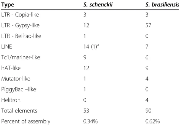
Transposable elements expansions inS. brasiliensis
genome
Transposable elements (TEs) comprise 0.34% and 0.62% of the S. schenckii and S. brasiliensisgenomes, respect-ively (Table 2). Fungal genomes contain substantially different amounts of repetitive DNA sequences. The assembled genome ofMagnaporthe oryzaecontains 10.8% of repetitive DNA sequences, inM. griseait is 4.2%, inN. crassais 10% and inS. cerevisiae almost 6% [37-40]. Dif-ferences in the TE contents are observed even between closely related species, e.g. in the genusParacoccidioides. InP. brasiliensis TEs correspond to 8-9% of the genome and twice this amount in P. lutzii (16%) [41]. Although less common, lower TE contents have been described in other fungi, for example 0.48% ofTrichodermaand 0.1% ofFusarium graminearumgenomes assemblies [39,42].
repetitive elements in microorganisms genomes [44,45]. Assexual propagation could lead extinction due inability to control proliferation of vertically transmitted TEs, as accumulation occurs due to the inefficiency of purifying selection in clonal species [46].
Equivalent numbers of DNA transposons were found in S. schenckiiandS. brasiliensis genomes.hAT-likeelements were the most prevalent in both species (Table 2). Trans-posons fromTc1/mariner andMutatorsuperfamilies were
also found in both genomes. Four copies ofHelitronswere identified in S. brasiliensis while a PiggyBac-like element was detected only inS. schenckii. Almost all TEs identified in Sporothrix genomes corresponded to defective and truncated copies, with exception of oneLINE-like element found inS. schenckii, suggesting thatSporothrixspecies are able to control their proliferation. This finding suggests that TEs are present in Sporothrix but with low transposition activity. We discuss why this could be below.
A
B
Figure 3Comparative analysis of mitochondrial genomes ofS. schenckiiandS. brasiliensis. (A)Gene content and order in mitochondrial genomes ofS. schenckiivs.S. brasiliensisshowing high synteny despite the considerable difference in size. Insertions of LAGLIDADG and GIY-YIG intron I type homing endonucleases inS. brasiliensisare present inside cytochrome C oxidase 1, ATP synthase subunit 6 and NADH dehydrogenase subunits 2 and 3 genes.(B)Phylogenetic analysis was performed using the five LAGLIDADG or GIY-YIG elements found inS. brasiliensisshowing no pattern of ancestry with other close related species.
Teixeiraet al. BMC Genomics2014,15:943 Page 6 of 22
Protein family expansion and contraction in the
Sporothrixlineages
Gene duplications are an important source of evolution-ary innovation and new gene copies can evolve new adaptive functions shaping an organism’s gene content. The differences among gene families have been related to emerging processes due to differential degrees of genetic drift, and thus the effectiveness of selection, acting on genomes [47]. The pathogenic phenotypes of S. schenckii and S. brasiliensiscould result of expansion in specific gene families that confer advantages in the
interaction with human/animal hosts. On the other hand, gene families that ate necessary for the plant-associated lifestyles of other Sordariomycetes could be contracted. To test these hypotheses, we have compared the genomes from closely related Sordariomycetes and dimorphic fungal pathogens. Changes in S. schenckii and S. brasi-liensis gene families were inferred based on domain expansions or contractions assigned by Interpro, Pfam and SMART databases and statistically tested by hyper-geometric comparisons (P < 0.05 - Figure 5, Additional file 3: Figure S2, Additional file 4: Figure S3 and Additional file 5: Figure S4) and the reported p-values were used for multiple testing using q-value.
We have not observed the enrichment of peptidases genes in Sporothrix lineage, specifically the MEROPS families M35 or M36, which are expanded in dimorphic fungal pathogens as adaptation to mammalian hosts [29,41]. On the other hand, we observe a lack of polysac-charide lyase genes which are associated with decay of plants (CAZy PL family) when compared to other Sor-dariomycetes (Figure 6A, Additional file 2: Table S5). The subfamilies PL1, PL3 and PL4 are broadly distrib-uted among Sordariomycetes, but were not observed in theSporothrixspecies. Interestingly loss of plant degrad-ing enzymes were also observed in other dimorphic fun-gal pathogens such as H. capsulatum and C. immitis (Figure 6A, Additional file 2: Table S5), which has also been previously interpreted as adaptation from plants to animals [29,41]. As an alternative for the absence of PL
Table 2 Transposable element composition inSporothrix genomes
Type S. schenckii S. brasiliensis
LTR - Copia-like 3 3
LTR - Gypsy-like 12 57
LTR - BelPao-like 1 0
LINE 14 (1)a 7
Tc1/mariner-like 9 6
hAT-like 12 9
Mutator-like 1 4
PiggyBac–like 1 0
Helitron 0 4
Total elements 53 90
Percent of assembly 0.34% 0.62%
aThe number of potentially functional elements is shown in parentheses.
proteins, it may be hypothesized that Sporothrix may digest pectin by using polygalactouronase (CAZy GH28) as a replacement strategy. Additionally, we didn’t de-tect any GH72 genes in Sporothrix genomes although
those genes were found in all remaining analyzed fungi (Additional file 2: Table S5).
LysM domain-containing proteins display carbohydrate binding modules, usually 42–48 amino acids residues in Figure 5Interpro domains most enriched (A) or depleted (B) inSporothrixlineages compared to other thermo dimorphic fungi and close related Sordariomycetes.SBRA -S. brasiliensis, SSCH–S. schenckii, GCLA–G. clavigera, MORY–M. oryzae, PANS–P. anserina, NCRA–N. crassa, VDAH -V. dahliae, FGRA–F. graminearum, AFUM–A. fumigatus, PMAR–P. marneffei, CIM–C. immitis, PBRA–P. brasiliensis, PLUT–P. lutzii, BDER–B. dermatitidis, HCAP–H. capsulatum. The reported p-values were used for multiple testing using q-value.
Teixeiraet al. BMC Genomics2014,15:943 Page 8 of 22
length, found in prokaryotes and eukaryotes. These pro-teins are classified in the CAZy database as CBM family 50 and harbor N-acetylglucosamine (GlcNAc) binding characteristics. According to PFAM and CAZy counts, a marked expansion of the LysM domain PF01476 (CAZy CBM family 50) was detected in theSporothrixlineage. The phylogenetic tree clearly showed highly expanded branches in the Sporothrix lineage when compared to other Sordariomycetes and thermo dimorphic fungal pathogens (Figure 7). Moreover, the comparative ana-lysis of CAZy CBM family 50 also revealed a high ex-pansion of this domain (Figure 6B, Additional file 2: Table S5). Recent events of duplications of LysM homo-logs subsequent to a speciation event were detected in the Sporothrix lineage using reconciled tree approach (Additional file 6: Figure S1). The first duplication (Figure 7,
clade I) is related to genes containing single or multiple repetitions of LysM domains, chitin-binding module type 1 (CBM18) plus glycoside hydrolase (GH18). The second event of duplication was observed in genes har-boring multiple copies of LysM, providing evidence of recent intergene duplication of this domain within para-logues (Figure 7, clade II). The structure of Sporothrix LysM domain-containing genes are presented separately in Additional file 3: Figure S2.
Chitin is a linear polymer of β-(1,4)-linked GlcNAc, and is one of the major components of fungal cell wall. The absence of chitin in mammalian cells makes this polymer a potential targets for the innate immune sys-tem [48]. Chitin can be recognized by mammalian cells, and is bound and degraded by chitin-binding proteins GH18, playing an important role in inflammation and
A
B
innate and adaptive immunity based on their modulation on various disease states [49]. Chitin can mask immune recognition by blocking dectin-1-mediated interaction with fungal cell walls [50]. Also, chitin modulates epi-thelial immunity of the skin expressing high levels of cytokine and chemokine and increases TLR4 expres-sion on keratinocytes [51]. Possibly these proteins are
present to bind the own Sporothrix chitin exposed
upon cell damage, and in this way it may protect itself against recognition of this polymer by keratinocytes.
LysM effector (Ecp6) from Cladosporium fulvum was
characterized as a virulence factor of this phytopatho-genic fungus on tomato plants. Carbohydrate binding assays have shown that Ecp6 specifically binds to chitin, the major constituent of the fungal cell wall, acting as a PAMP upon recognition by the plant during fungal invasion. The presence of chitin-binding effector Ecp6 in the apoplast masks the perception of chitin by plant receptors preventing the activation of defense responses [52]. In addition significant expansion of the LysM domain
was detected in dermatophytes, compared to the thermo-dimorphic fungi [53]. Dermatophytosis and sporotrichosis are characterized as cutaneous mycoses and the infection is acquired after contact and/or trauma of skin. LysM domain containing proteins have not been characterized as virulence factors in human or animal fungal patho-gens so far, and the role of those proteins requires further investigation and could be a novel mechanism for fungal evasion in mammalian host tissue.
Small GTPases are an independent superfamily of GTP-binding proteins, sharing a common enzymatic activity and producing GDP by the hydrolysis of GTP, and play pivotal roles in cell division and signaling, vesicle fusion and protein synthesis [54]. These pro-teins are also involved in filamentation, mating, growth at 37°C and virulence inC. neoformans [55,56]. Signifi-cant expansions in Ras, Rho and Rab Small GTPase superfamilies (IPR020849, IPR003578 and IPR003579) were observed in the Sporothrix lineage when com-pared to other Ascomycetes (Figure 5, Additional file 2: Figure 7Unrooted maximum likelihood tree revealing LysM expansions in theSporothrixlineage.Two major expansions were detected: The first duplication is related to genes containing single or multiple repetitions of LysM domains (CBM50), chitin binding module type 1 (CBM18) plus glycoside hydrolase (GH18)–clade I. The second event of duplication was observed in genes presenting multiple copies of LysM (CBM50), and is evidence of recent intergene duplication of this domain within paralogous - clade II.
Teixeiraet al. BMC Genomics2014,15:943 Page 10 of 22
Table S4). Judging from the phylogenetic trees of small GTPases, the majority of branches harboring Sporo-thrixhomologues suggests a higher diversity among the Ascomycetes we analyzed (Additional file 4: Figures S3, Additional file 5: Figure S4, Additional file 7: Figure S5). Reconciliation of small GTPases gene trees and species tree do not indicate Sporothrix-specific duplications, that instead independent gene losses in other species explain the increased copy number inSporothrix(Additional file 8: Figure S6, Additional file 9: Figure S7, Additional file 10: Figure S8). An alternative hypothesis is that the SporothrixRas, Rho and Rab genes have duplicated re-cently and rapidly diverged creating the observed long branch lengths. In addition we detected highly sup-ported clades containing Sporothrix and other patho-genic dimorphic fungi, suggesting convergent evolution of small GTPases, reinforcing the high plasticity of sig-nal transduction in the Sporothrix lineage (Additional file 4: Figure S3, Additional file 5: Figure S4, Additional file 7: Figure S5). To date, such a high diversity of Small GTPase proteins as found in theSporothrixlineage has not been reported from any other class of fungi.
Another group that was expanded by INTERPRO and SMART domain counts in the Sporothrix lineage is the polyketide synthase (PKS), enoylreductase (IPR020843) family (Figure 5, Additional file 2: Tables S3, S4). Polyke-tides comprises diverse fungal secondary metabolites such as antibiotics, pigments, and mycotoxins that are formed from simple carbon precursor acids catalyzed by polyketide synthases (PKSs) [57]. Filamentous fungi are producers of polyketide metabolites, several of which of pharmacological or agricultural interest [58,59]. Fungal PKSs are in general a linear succession of ketosynthetase (KS), acyltransferase (AT), dehydratase (DH), enoyl reductase (ER), ketoreductase (KR), acyl carrier protein (ACP), and thioesterase (TE) domains [60]. ER do-mains reduce enoyl groups to alkyl groups (saturated) during production of secondary metabolites. Among fungal genomes, few potential PKS orthologous genes are shared, even between closely related taxa [61]. We identified various paralogous duplications in the phylo-genetic analysis of PKS-containing protein, and the Sporothrix lineage appears to have of PKS-encoding genes that is at least 3-fold larger than that of the other species analyzed (Additional file 2: Table S4, Figure 8). This was confirmed using the reconciled approach of PKS gene tree with the species tree analyzed (Additional file 10: Figure S8). Convergent branches linking Sporo-thrixand other pathogenic dimorphic fungi were also observed. We identified discontinuous distributions of PKS homologs among the analyzed fungal species, with low bootstrap values, that can be explained by gene duplication, divergence, and gene loss [61]. We also identified expanded clades harboring Sporothrix
and dimorphic fungi with high branch support values, suggesting a great diversity of this protein family.
Another large expansion found in theSporothrixlineage is a fungal trichothecene efflux pump (Pfam PF06609), an evidence of detoxification via mycotoxin pump [62]. Apart from that, fungal genomes generally harbor a lower content of Leucine-rich repeat (LRR) proteins than other ophistokonts [63]. Gene expansions of several LRR superfamilies were identified in the Sporothrix lineage (Additional file 2: Tables S3, S4). LRR proteins expan-sions are commonly found in eukaryotic parasites such as Trypanosoma and Giardia [64,65]. These domains consist of 2–45 motifs of 20–30 amino acids in length providing a structural framework for protein-protein interactions. LRR proteins are involved in a variety of biological processes and are source of genetic variation for the ongoing process of antigenic variability in patho-gens [66,67]. In addition expansion of LRR proteins was described forCandidaspecies as a virulence factor [68]. The role of these proteins should be further investigated in the genusSporothrix.
Cellular processes and dimorphism
The basic mechanisms of DNA repair in Sporothrixdo not deviate from the eukaryotic consensus, as expected, but neitherS. brasiliensisnorS. schenckiiseem to have a homolog to the Neurospora crassa RIP-defective (RID) methyl-transferase [69]. This suggests that RIP-like muta-tion patterns found in transposable elements in this fun-gus are not generated by the classical, RID-dependent mechanism. This is not a surprise, given that at least one previous report [70] pointed to the possibility of alterna-tive pathways for repetialterna-tive DNA quelling via RIP. This is in contrast to other Sordariomycetes and even more distantly related members of the Ascomycota, such as P. brasiliensis and H. capsulatum, for which the tBLASTn tool yielded hits with high homology. Since the number of degenerate TEs and overall evidence of transposon activity inSporothrix spp.genomes is low, it would be interesting to identify which pathway is responsible for TE suppression in this genus instead of RIP. We also note the absence of a mus-18homologue for the alternative, UV damage-related nucleotide exci-sion repair pathway [71], found inNeurospora crassa.
latter [16], it seems reasonable to speculate that both also have been coopted by this genus to coordinate di-morphic transition.
Cell wall assembly
The cell wall of fungi is a dynamic organelle, which is constantly adjusted depending on environmental insults. Fungal cell wall components are considered relevant for virulence, have antigenic properties and participate in the modulation of the host immune response by being recognized by innate immunity receptors [14]. Current models propose an arrangement of several stratified layers composed of structural polysaccharides, mainlyβ-glucans and chitin, proteins and glycoproteins, generally known as mannoproteins, and other minor components. Cell wall proteins (CWP) are either covalently linked to the cell wall β (1,6)-glucans by a glycophosphatidylinositol (GPI) moiety or linked via an alkali sensitive linkage to
β(1,3)-glucans. The sugar moieties in CWP areN–and/or O- linked to the protein core [74]. Despite the importance
of those components, little is known about the surface protein composition ofS. schenckiiandS. brasiliensis.
a) Adhesins and/or cell surface proteins
Previous studies searching for adhesins and antigens in cell extracts of Sporothrixdemonstrated the presence of a main antigen on both species, known as Gp70, a secreted antigen that is also present on the cell surface act-ing as an adhesin [75-77]. The genomes ofS. schenckiiand S. brasiliensis were investigated with different predictors to ascertain the presence of these cell wall components.
An in silico comparative analysis was performed in order to determine the putative adhesins and/or cell surface proteins bearing a GPI-anchor. According to ProFASTA and FungalRV,S. schenckiiharbors 68 and 61 surface proteins, respectively, that have adhesin properties (n = 129). Of these, 12 were found by both predictors (Additional file 11: Figure S9A, Additional file 2: Table S6) totalizing 117 unique predicted proteins in the cell wall of S. schenckii. ForS. brasiliensis we have identified 54 and Figure 8Unrooted maximum likelihood tree of Polyketide synthase (PKS) enoylreductase (IPR020843) family showing expansions in theSporothrixlineage.Paralogous duplications are displayed in the related branches suggesting vast genomic apparatus of PKS containing genes in theSporothrixlineage. Clades harboringSporothrixand dimorphic fungi are displayed in red.
Teixeiraet al. BMC Genomics2014,15:943 Page 12 of 22
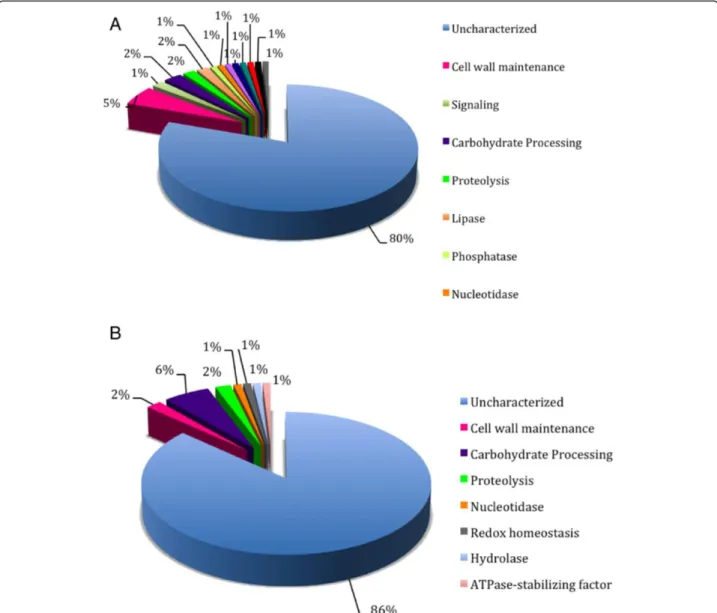
63 cell wall proteins (n = 117), respectively, 11 of which were predicted by both algorithms. For this species, a total of 106 unique predicted proteins (Additional file 11: Figure S9B, Additional file 2: Table S7). The protein sequence relative to the previously proven cell surface/ adhesin Gp70 was not predicted to possess this function or to be present in the surface location by any of the pre-dictors used [77]. Previous studies described several other important, non-classical surface proteins present in other fungi but which were not recognized in Sporothrix by FungalRV and ProFASTA [78-80]. The major classes of proteins predicted in S. schenckii and S. brasiliensis, by both ProFASTA and FungalRV, are currently annotated either as hypothetical proteins or belonging to a protein family with unknown function (Figure 9). This indicates
that proteomic studies are needed to validate the expres-sion of such proteins and biochemical functional studies are necessary to clarify their role on the fungal cell surface. The cell wall proteins and/or adhesins predicted for these species were blasted against 11 fungi for comparative ana-lysis, and each protein was blasted against the two Sporo-thrixspecies (Additional file 10: Table S8). Eight proteins were found exclusively in S. brasiliensis and eight pro-teins were present only in S. schenckii. Sixteen Sporo-thrix-specific proteins were annotated as hypothetical proteins. None of the proteins described are specific to the group of human pathogenic fungi, but interestingly there are some proteins putatively present in the cell wall of Sporothrix that share homology with those of plant and insect-associated fungi.
b) Glucan and chitin metabolism
In S. schenckii, β-glucans are major components of the cell wall [81,82], and are present as alkali-insoluble and alkali-soluble glucans, containing predominantlyβ -(1,3)-linkages in both cases [82]. In S. schenckii, no genes related to the synthesis or degradation of β -glucans have been reported to date. Genomic data ana-lysis revealed a single FKS ortholog in S. schenckiiand S. brasiliensis genomes, as well as single orthologs in the genomes of the other 14 fungal species studied here (Additional file 11: Table S9). No genes related to the synthesis of either β-1,6- orβ-1,4-glucans were identi-fied, although hydrolase orthologs for the three types of β-glucan linkages in the S. schenckii cell wall, are present in both genomes (Additional file 11: Table S9).
Chitin synthesis in fungi is a rather complex process, regulated by multigene families encoding chitin synthase isoenzymes, whose activities may be spatially regulated to fulfill the multitude of roles ascribed to them [83]. Based on differences in regions of high sequence conser-vation, chitin synthases have been attributed to seven classes [83,84], whose functional implications are not yet clear in all cases. Despite the low chitin content reported for S. schenckii [81], genomic analysis showed the pres-ence of seven CHS genes, in S. schenckii as well as in S. brasiliensis genomes. A cluster analysis of their puta-tive products, including 36 fungal chitin synthases, revealed that each of the translation products of the sevenCHS Sporothrixgenes identified in genomic data-bases (CHS1toCHS7), could be ascribed to each of the seven chitin synthase classes known (I to VII) (Additional file 12: Figure S10, Additional file 2: Table S10). It is worth noting that class III chitin synthases were thought to occurr exclusively in filamentous fungi [85]. In our ana-lysis, genes for class III chitin synthases were found in theSporothrixgenomes as well as the genomes of other dimorphic fungi (Additional file 2: Table S10), an indi-cation that class III fungal chitin synthases might be more widespread in fungi. Another interesting finding was the head-to-head arrangement in the S. schenckii and S. brasiliensis genomes of the CHS4 and CHS5 genes, whose putative translated products are class V and VII chitin synthases (Additional file 8: Figure S6, Additional file 2: Table S10). A similar arrangement was reported for genes coding for classes V and VII of chitin synthases in P. brasiliensis, A. nidulans, C. posadasii and F. oxysporum [84,86-88]. The meaning of such arrangement is unclear, although a common transcrip-tional regulation for these genes has been suggested for A. nidulansandP. brasiliensis[84,88,89]. In agreement with the large number of CHS genes in the Sporothrix genomes, genomic analysis showed the presence of ten and nine chitinase genes, respectively, in the S. schenckii andS. brasiliensisgenomes (Additional file 2: Table S10).
Polysaccharide synthesis and hydrolysis-related genes identified in S. schenckii and S. brasiliensis genomes correlate with the biochemical composition of the cell wall as reported by Previato et al. [81,82]. It remains to be determined which individual synthase and/or hydrolase gene might be involved in shaping the yeast, mycelial and spore (conidial) walls of Sporothrix spe-cies, or even whether any of them might have any role in survival and would provide potential targets for the development of specific antifungal drugs.
c) Protein glycosylation
Glycoproteins are key components of the S. schenckii cell wall, but thus far little is known about their biosyn-thetic pathways [23,24]. The genomes ofS. schenckiiand S. brasiliensiscontained the orthologs involved in elab-oration of the N-linked glycan core, its transference to proteins and in early trimming. These genes are also known to be involved in glycoprotein endoplasmic reticulum-associated degradation, a quality control sys-tem for proteins synthesized within the secretory path-way [90-92] (Additional file 2: Table S11). Furthermore, the genomes contain the putative orthologs encoding Golgi-resident glycosidases and glycosyltransferases that further modify N-linked glycans, generating both hybrid and complexN-linked glycans. The presence of a gene with significant similarity to those encoding the N-acetylglucosaminidase III (Additional file 2: Table S11), which adds the bisecting GlcNAc residue found in both hybrid and complex N-linked glycans [93], suggests an ability to elaborate more complex oligosaccharides than those found inS. cerevisiae[94]. Moreover, our analysis revealed genes encoding putative Golgi UDP-galactose and CMP-sialic acid transporters, suggesting the ability of these fungi to add these sugars to their glycans. S. schenckii andS. brasiliensis also contain an ortholog of A. nidulans ugmA, whose product generates the galactomannan-building sugar donor [95], and some puta-tive galactosyltransferases (Additional file 2: Table S12). However, it remains to be addressed whether these enzymes participate in elaboration of glycoproteins and/ or glycolipids. Sialic acid has previously been reported as a component ofS. schenckiicell wall glycolipids [96], so it is likely that the putative Golgi CMP-sialic acid transporter is involved in modification of such lipids.
The biosynthetic pathway for O-linked glycans can also be predicted from the analysis of S. schenckii and S. brasiliensis genomes (Additional file 2: Table S13). Optimal characterization ofO-linked glycans is via isola-tion from peptide-rhamnomannans [97]. They contain an
α1,2.mannobiose core, an α1,2-glucuronic acid unit, and one or two rhamnose residues. The S. schenckii and S. brasiliensisgenomes contain three putative glucurono-syl transferases that might participate in the elaboration of
Teixeiraet al. BMC Genomics2014,15:943 Page 14 of 22
this O-linked glycan (Additional file 2: Table S13). Our genomic analysis could not find any obvious ortholog for rhamnosyl transferases, but Sporothrix contains all the required genes for synthesis of UDP-L-rhamnose (Additional file 2: Table S13) [98,99], the sugar donor in the enzyme reaction catalyzed by rhamnosyltransferases [100]. Synthesis of GPI is quite conserved in eukaryotic cells and the Sporothrix genome contains all genes to elaborate this glycolipid (Additional file 2: Table S14).
Melanin metabolism
Melanins are dark pigments formed by phenolic and indo-lic oxidation. These biopolymers are produced by a wide range of organisms, possibly contributing to the mainten-ance of several species throughout evolution [101]. In fungi, the expression of these pigments has been associ-ated with virulence [102]. Fungi may synthesize melanin by several pathways: in pathogenic fungi, most commonly from endogenous substrate via the 1,8-dihydroxynaphtha-lene (DHN) pathway or the L-3,4-dihydroxyphenylalanine (L-DOPA) pathways [102]. The latter type is prevalent in Basidiomycetes. However, evidence of both pathways has been found inS. schenckiiby means of specific substrate supplementation or drug-related inhibition of the respect-ive pathways [22,23,103,104].
Melanins are found inS. schenckiispores and yeast cells and are produced in vitro and during infection using hamsters as host model. Its detection has also been confirmed by immunofluorescence with monoclonal antibodies raised againstS. schenckiimelanin [21,22]. In S. schenckii, melanin pigments can protect the fungus from the mammalian host’s innate immune responses providing resistance to killing by phagocytosis and oxidizing agents [22,105]. Recently it was reported that S. schenckii and S. brasiliensis also produce pyomelanin, a melanoid pig-ment derived from the degradation of L-tyrosine via a 4-hydroxyphenylpyruvate dioxygenase [103].
Genomic comparison showed that both S. schenckii and S. brasiliensispossess enzymes with central roles in melanin synthesis via DHN and DOPA pathways, and also in pyomelanin synthesis. Sporothrix schenckii and S. brasiliensisloci which are postulated to be involved in the melanin biosynthesis pathway are described in Additional file 2: Table S15. We found 19 loci related to melanin biosynthesis inS. schenckiiand 17 inS. brasi-liensis. Homology with previously described melanin-related enzymes found in the sequence analyses are: pigment biosynthesis protein yellowish-green 1, polyketide synthase I and III, tetrahydroxynaphthalene/trihydroxi-naphtalene reductase, scytalone dehydratase, laccase, tyrosinase and 4-hydroxyphenylpyruvate dioxygenase, as illustrated in Additional file 13: Figure S11. The mul-tiple functions of melanin in a cell and, especially, the resistance to antifungal drugs and survival of the host
immune system, are a strong motivation for the study of the genetic characteristics of melanin biosythesis.
Conclusions
In this study we provide high quality genomic sequence assemblies and annotations forS. schenckiiand S. brasi-liensis. Genomic analyses showed a convergent evolu-tionary fate compared to other dimorphic fungi, even though Sporothrix is a close relative of plant-associated Sordariomycetes. Similar to other dimorphic fungal patho-gens we have observed a lack of polysaccharide lyase genes which are associated with decay of plants, suggesting evolutionary adaptations from a plant pathogenic or saprobic to an animal pathogenic life style. In addition, convergent branches linkingSporothrix and other patho-genic dimorphic fungi were also observed in genes in-volved in signal transduction and secondary metabolism which suggest similar evolutionary traits. The recent hypothesis of habitat shift from a saprobic life style in fermented plant material to mammal transmission may explain numerous plant/related atavisms. Comparative genomics reveals a certain degree of specialization in the Sporothrix lineage which may contribute to our understanding of how fungal-environment-human in-teractions lead to the selection of pathogenic pheno-types of these species. TheSporothrixsystem may bring new opportunities for functional studies in order to understand the biology of fungi and infection.
Methods
Fungal strains and DNA extraction
Sporothrix schenckii strain 1099–18 (ATCC MYA-4821) was originally obtained from the Mycology Section, De-partment of Dermatology, Columbia University, New York, isolated from a patient manifesting subcutaneous sporotrichosis, and has been widely used in experiments of cell wall composition and virulence studies in mice models [97,106].Sporothrix brasiliensisstrain 5110 (ATCC MYA-4823) was isolated from a feline skin lesion in the epidemic area of sporotrichosis in Rio de Janeiro, Brazil, presenting high virulence in mouse model [77]. Mycelial cells were cultivated in Sabouraud broth at 25°C, with shaking (150 rpm) for 14 days, collected by centrifugation and washed 3 times with Phosphate-buffered saline (PBS) solution. Cells were disrupted using the Precellys®24-Dual (Bioamerica) with help of CK28 hard tissue hom-ogenizing tubes. DNA extraction was performed using Qiagen DNeasy Plant Mini Kit, according to manufac-turer’s protocols.
Genome sequencing and assembly
were constructed and sequenced in the 454 GS FLX plat-form according to Roche’s protocols at the Computational Genomics Unity of the National Laboratory for Scientific Computing (LNCC, Petrópolis, RJ, Brazil). Genomic assemblies were carried out using Newbler and Celera Assembler. Sequence gap filling and the removal of contigs corresponding to rDNA genes were manually done, decreasing the numbers of scaffolds and contigs. The assembled scaffolds generated by the two species were aligned and oriented using MAUVE [107]. Simi-larity scores and dot-plot graphs were generated using LALING/PLALING (http://fasta.bioch.virginia.edu/fasta_ www2/fasta_www.cgi?rm=lalign).
Ab initioGene prediction, annotation and protein family classification
Gene predictions were performed using three different approaches: SNAP [108], AUGUSTUS [109] and EXON-ERATE [110] using ORF’s identified in the G. clavigera strain kw1407/UAMH 11150 [26] as reference and for training and genomic comparisons. Proteins deduced for G. clavigera proteome were aligned to theS. brasiliensis and S. schenckii assembled genomes using Exonerate (percent threshold equal 50) with the model protein2-genome. Gene predictions (SNAP and AUGUSTUS) and protein (EXONERATE) alignments were used as input in order to identify consensus gene structures using EVi-denceModeler [111]. Consensus ORF’s were subjected to Blast searches against NCBI refseq_protein, KEGG and SwissProt databases. Automatic annotations were per-formed using SABIA - upgraded for eukaryotic organ-isms [112] and validated ORF’s were considered with minimum query/subject coverage of 60% and minimum positive 50%. In addition, gene categories according KEGG were inspected manually in order to re-assemble the metabolic pathways ofS. brasiliensisandS. schenckii. Alignments were made by Blastp and the lowest e-value was used to consider homologous sequences. Next, loci identified in S. schenckii and S. brasiliensis genomes were blasted against the genome libraries of 14 selected fungi (Neurospora crassa, Aspergillus nidulans, A. fumi-gatus, Talaromyces marneffei, Paracoccidioides lutzii, P. brasiliensis,Coccidioides immitis,Blastomyces dermati-tidis, Histoplasma capsulatum, Fusarium graminearum, Magnaporthe oryzae, Sordaria macrospora, Verticillium dahliae, Grosmannia clavigera) to infer the putative orthologues. Gene products were categorized according to biological process, cellular component and molecular function using GeneOntology (GO) using Blast2GO. Secreted proteins were identified using SignalP3.0 (http://www.cbs.dtu.dk/services/SignalP/) using hidden Markov model.
The prediction of mobile genetic entities was performed by similarity searches using the following approaches and
databases: a) Nucleotide Blast against Repbase version 17.10 (http://www.girinst.org/repbase/) [113], Dfam data-base version 1.1 [114] and Gypsy datadata-base version 2.0 (GyDB) [115]; b) PSI-BLAST (Position-Specific Iterated BLAST) using profiles of proteins corresponding to major clades/families of Transposable Elements (TEs) imple-mented with TransposonPSI tool (http://transposonpsi. sourceforge.net/); c) reverse position-specific BLAST algo-rithm (RPSBLAST) against Conserved Domain Database (CDD) version March 2013 [116]; and d) tblastn taking specific protein subsets against the Sporothrix genome. These subsets were built from NCBI Non Redundant (NR) database version March 2011 using particular description terms related to transposable elements including (apurinic/ apyrimidinic endonuclease, aspartic proteinase, ATPase, endonuclease, envelope, GAG protein, helicase, integrase, polymerase B, replication protein A, reverse transcriptase, RNase, transposase, tyrosine transposase/recombinase). In addition, the Tandem repeat finder (TRF) algorithm was used for finding tandem repeats [117]. Transposable ele-ments were classified accordingly [118]. All results were obtained using locally compiled databases. Perl scripts were built for automation of genome scans, report generation and data integration. Artemis sequence visualization and annotation tool [119] was used for manual curation and annotation of transposable elements.
Whole genome gene families were identified using InterproScan combined with Pfam domain assignments. Annotation of carbohydrate-active enzymes was performed in a two-step procedure where the translated protein sequences were compared to the full length sequences derived from the Carbohydrate-Active enZymes (CAZy) database (www.cazy.org; [120]) using BLAST [121]. The query sequences that had an e-value <0.1 were subjected to a BLAST search against sequence fragments corresponding to individual catalytic and carbohydrate-binding mod-ules described in CAZy, along with a HMMer search [122] using hidden Markov models corresponding to each CAZy module family. A family assignment was considered reliable when the two methods gave the same result. Borderline cases were resolved by inspec-tion of conserved features such as the presence of known catalytic residues.
Gene family expansion and contractions
Gene families were determined using OrthoMCL ap-proach comparing with other 13 fungi (Additional file 2: Table S1). Domains were annotated for each orthologous cluster using Interpro [123], Pfam [124] and SMART [125] databases. Significant enrichment or depletion of domains in the Sporothrix lineage were calculated based on hypergeometric comparisons (P < 0.05) and the reported p-values are adjusted for multiple testing using q-value [126,127]. The expanded families with
Teixeiraet al. BMC Genomics2014,15:943 Page 16 of 22
highest discrepancies between Sporothrixand other com-pared fungi were individually analyzed. Protein sequences of LysM, PKS enoyl reductase, Ras, Rab and Rho small gtpases we individually aligned using ClustalW [128] and the domains were manually checked. Uninformative positions of the alignment were eliminated using trimal [129] and the best-fit protein substitution model was inferred based on likelihood values under AIC criteria, implemented in ProtTest [130]. Phylogenetic analysis of expanded gene families were carried out using Max-imum likelihood methods implemented in PhyML 3.0 software and 1.000 of non-parametric bootstraps were tested for branch support [131]. Gene duplications/losses for the given family trees were inferred using Notung 2.6 software [132].
Blast reciprocal best hit and phylogenomic analysis
Bidirectinoal-best Blast Hit (BBH) were performed using two different datasets: first we performed the compari-sons betweenS. schenckii,S. brasiliensisandG. clavigera genomes in order to identify the unique genes in Sporo-thrix lineage. In addition, Blast reciprocal best hits were performed to identify common orthologues in 25 fungal genomes (Additional file 2: Table S1) using minimum query/subject coverage of 50% and e-value of E≤1×10⁻²⁰
. A total of 395 orthologs were found in all species analyzed and were aligned using MAFFT [133] and retrieved alignments were trimmed using Trimal [129] in order to exclude spurious sequences or poorly aligned regions. Phylogenomic analysis was performed using RAxML [134] and the Dayhoff aminoacid substitution model was selected according ProtTest [130]. Divergence time between species was calculated with help of r8s v 1.8 [135] program using Langley-Fitch model [136] consid-ering the origin of the Ascomycota at 500 to 650 MYA (Millions Years Ago) [137].
Cell wall protein/adhesin analysis
Two programs were used for the prediction of cell wall proteins/adhesins: ProFASTA [138] and FungalRV [139]. Analysis using the fasta files of the complete genomes of S. schenckii and S. brasiliensis were performed for pre-diction of GPI-anchor secretion signal and transmembrane domain identification. The SignalP 4.0 server (http://www. cbs.dtu.dk/services/SignalP/) was applied for prediction of the presence and location of signal peptide cleavage sites in amino acid sequences, using the method of Input sequences, which do not include TM regions. Then, the TMHMM Server v. 2.0 (http://www.cbs.dtu. dk/services/TMHMM/) was used for prediction of trans-membrane helices in proteins, and finally the Big-PI fungal predictor [140] was used for GPI modification sites. The ProFASTA requires the combination of these three ana-lyses to prospect cell wall proteins and adhesins, with the
following parameters: SignalIP 4.0 positive; TMHMM 2.0 < 1 helices and number of AA to exclude as 45 from N-terminus and 35 for C-terminus; Big-PI positive. The FungalRV validates only proteins with score up to 0.5 for adhesin or adhesin-like features.
Autophagy, peroxisome and endocytosis
The initial tool used for this annotation was the KEGG automatic classification, which adequately identified genes involved in peroxisome biogenesis. However, the automatic annotation algorithms only picked up a few genes involved in autophagosome biogenesis and endo-cytosis, so different approaches were necessary. For the genes involved in autophagy, we started by collecting on the SGD all protein sequences annotated as involved in autophagy and autophagosome biogenesis in Saccharomy-ces cerevisiae. All of these sequences were blasted against the S. schenckii and S. brasiliensis databases in order to correctly identify homologous protein sequences. To narrow down the list, we focused the analysis on 17 genes that are necessary for autophagosome biogenesis in yeast [141] plus those that are shown on KEGG. Regarding endocytosis, the KEGG table only showed two genes involved in the process itself and several genes involved in vacuolar degradation. To overcome this limitation, the genes that were used for annotation were those listed in a review article as being involved in clathrin-mediated endocytosis inS. cerevisiae [142]. All protein sequences encoded by these genes were blasted against theS. schenckiiandS. brasiliensisdatabases.
Availability of supporting data
The data sets supporting the results of this article are in-cluded within the article and its additional files.
Additional files
Additional file 1:Core genes for general and secondary metabolism.
Additional file 2:Table S1.Information about the 25 genomes retrieved for phylogenomic inference.Table S2.Pfam domain expansions/contractions ofSporothrix schenckiiandS. brasiliensis.Table S3.
SMART domain expansions/contractions ofSporothrix schenckiiand S. brasiliensis.Table S4.INTERPRO domain expansions/contractions of Sporothrix schenckiiandS. brasiliensis.Table S5.Comparative genomic analysis of carbohydrate active enzymes (CAZy) ofSporothrix schenckii andS. brasiliensis.Table S6.Putative adhesins ofS. schenckii.Table S7.
Putative adhesins ofS. brasiliensis.Table S8.Cell wall and adhesin-related genes characterized inSporothrix schenckiiandS. brasiliensis.Table S9.
Glucan synthase and glucanase genes identified inSporothrix schenckii andS. brasiliensis.Table S10.Chitin synthase and chitinase genes identified inSporothrix schenckiiandS. brasiliensis.Table S11.Genes involved in N-linked glycosylation inAspergillus nidulansandNeurospora crassa,and their putative orthologs inSporothrix schenckii and S. brasiliensis.Table S12.
GPI-anchor elaboration inAspergillus nidulansandNeurospora crassa, and their putative orthologs inSporothrix schenckiiandS. brasiliensis.
Table S15.Melanin biosynthesis pathway and putative orthologs in Sporothrix schenckiiandS. brasiliensis.Table S16.Comparative analysis of core genes related to amino acids, Secondary, Energy, Cofactor and Vitamin metabolisms ofSporothrix schenckiiandS. brasiliensis.Table S17.
Genomic identification and classification of phospholipases A, C, and D enzyme families inSporothrix schenckiiandS. brasiliensisgenomes.
Table S18.Homologs of vitamin and cofactor genes presented analyzed by FUNGIpath in bothSporothrixspecies.Table S19.Orthologous genes related to catabolism and transport inSporothrix schenckiiandS. brasiliensis. Additional file 3: Figure S2.Phylogenetic distribution of LysM domains containing proteins inSporothrix. The LysM domains are displayed along the taxa (red bars), chitin binding module type 1 (CB1) (blue bars) plus catalytic sites identified (glycoside hydrolase - GH or Pectin Lyase–PL). Gene paralogous duplications are highlighted by red boxes.
Additional file 4: Figure S3.Unrooted maximum likelihood tree of Ras Small GTPase proteins (IPR020849) family shows high diversification in theSporothrixlineage. Clades harboringSporothrixand dimorphic fungi are highlighted in red.
Additional file 5: Figure S4.Unrooted maximum likelihood tree of Rho Small GTPase proteins (IPR003578) family shows high diversification in theSporothrixlineage. Clades harboringSporothrixand dimorphic fungi are highlighted in red.
Additional file 6: Figure S1.Gene tree and species tree reconciliation of LysM domain-containing genes showing specificSporothrixduplications (blue boxes).
Additional file 7: Figure S5.Unrooted maximum likelihood tree of Rab Small GTPase proteins (IPR003579) family shows high diversification in theSporothrixlineage. Clades harboringSporothrixand dimorphic fungi are highlighted in red.
Additional file 8: Figure S6.Gene tree and species tree reconciliation of small GTPase Ras gene family showing independent gene losses in other species and the increased copy number inSporothrix(blue boxes). Additional file 9: Figure S7.Gene tree and species tree reconciliation of small GTPase Rho gene family showing independent gene losses in other species and the increased copy number inSporothrix(blue boxes). Additional file 10: Figure S8.Gene tree and species tree reconciliation of small GTPase Rab gene family showing independent gene losses in other species and the increased copy number inSporothrix(blue boxes). Additional file 11: Figure S9.Chart pies showing the efficiency of the algorithms used to predict the putative adhesins and/or cell wall GPI-anchored proteins of (A)S. schenckii(n = 118) and (B)S. brasiliensis (n = 106). The relative percentage of putative adhesins and/or GPI- anchored proteins, predicted by either ProFASTA or Fungal RV, is shown as well as the proteins in common by both predictors. Additional file 12: Figure S10.Phylogenetic tree of relatedness of Sporothrixspp. chitin synthases. The Mega 4 software package was employed, using ClustalW for sequence alignment. Construction of the phylogenetic tree was done by the neighbor-joining method using 1000 replications. The seven chitin synthases identified for both,Sporothrix brasiliensisandS. schenckii, cluster within the seven chitin synthase classes (I to VII) previously reported [83]. GenBank accession numbers of sequences, and names of fungal species used for construction of the tree are displayed in Additional file 12: Table S10.
Additional file 13: Figure S11.Melanin biosynthesis pathways for DHN-Melanin, DOPA-melanin and pyomelanin proposed forS. schenckii andS. brasiliensisbased on melanin biosynthetic pathways described in other pathogenic fungi. The putative enzymes identified in the genomes ofS. schenckiiandS. brasiliensisare indicated in circles. Locus tags, Gene products, numbers of exons, size of transcripts, estimated protein sizes and current annotations are listed.
Competing interests
The authors declare that they have no competing interests.
Authors’contributions
Designed the experiments: MMT LGPA RCS JCR JFS BH HMMM SRA JES GAN PSC LMLB ATRV MSSF; Sequencing, assembling and annotation: MMT LGPA PK FLA ESK AKRA LF LSD KSF RCS JCR NCA HCP AMN PA ALG VPM LDFP AV CBC PAS OLC FFMO TCS ALNB MAS LMO MMM HV MMLC BH HMMM JES; Comparative genomics analysis: MMT LGPA PK FLA ESK AKRA LF LSD KSF RCS JCR NCA HCP AMN PA ALG VPM LDFP AV CBC PAS OLC FFMO TCS ALNB MAS LMO MMM HV MMLC SH BH HMMM JES; Contributed to analysis tools: MMT BH GAN PSC HMMM SRA JES LMLB ATRV MSSF; Wrote the manuscript: MMT MSSF. All authors read and approved the final manuscript.
Acknowledgments
We are grateful to FAP-DF, CNPq and Capes for the financial support and fellowships of the projects Pronex (grant number 193000569/2009) and Genoprot (grant number 559572/2009-3). HMMM is supported by CONACyT, México (grant number CB2011-166860). BH is an Honorary Professor of Glycomics at the Faculty of Health and Medical Sciences, University of Copenhagen, Denmark.
Author details
1Departamento de Biologia Celular, Universidade de Brasília, Brasília, DF, Brazil.2Laboratório Nacional de Computação Científica, Petrópolis, RJ, Brazil. 3Departamento de Biologia Celular, Instituto de Biologia Roberto Alcântara Gomes, Universidade do Estado do Rio de Janeiro, Rio de Janeiro, RJ, Brazil. 4Departamento de Microbiologia, Universidade Federal de Minas Gerais, Belo Horizonte, MG, Brazil.5Grupo Informática de Biossistemas, Centro de Pesquisas René Rachou, FIOCRUZ, Minas, Belo Horizonte, MG, Brazil. 6Departamento de Análises Clínicas, Universidade Estadual de Maringá, Maringá, PR, Brazil.7Programa de Pós-Graduação em Ciências e Tecnologias em Saúde, Universidade de Brasília, Ceilândia, Brasília, DF, Brazil.8Instituto de Ciências Ambientais, Químicas e Farmacêuticas, Universidade Federal de São Paulo, Campus Diadema, São Paulo, SP, Brazil.9Pós-Graduação em Ciências Genômicas e Biotecnologia, Universidade Católica de Brasília, Brasília, DF, Brazil.10Programa de pós-graduação em Medicina Tropical, Universidade de Brasília, Brasília, DF, Brazil.11Programa de pós-graduação em Bioinformática, Universidade Federal de Minas Gerais, Minas Gerais, Brazil.12Departamento de Microbiologia Imunobiologia e Parasitologia, Universidade Federal de São Paulo, São Paulo, SP, Brazil.13Centro de Microbiología y Biología Celular, Instituto Venezolano de Investigaciones Cientificas, Caracas, Venezuela. 14CBS-KNAW Fungal Biodiversity Centre, Utrecht, The Netherlands.15Centre National de la Recherche Scientifique, Aix-Marseille, Université, CNRS, Marseille, France.16Departamento de Biología, Universidad de Guanajuato, Guanajuato, Mexico.17Departamento de Análises Clínicas e Toxicológicas, Universidade de São Paulo, São Paulo, SP, Brazil.18Department of Plant Pathology & Microbiology, University of California, Riverside, CA, USA.
Received: 11 February 2014 Accepted: 25 September 2014 Published: 29 October 2014
References
1. Marimon R, Cano J, Gene J, Sutton DA, Kawasaki M, Guarro J:Sporothrix brasiliensis, S. globosa, and S. mexicana, three new Sporothrix species of clinical interest.J Clin Microbiol2007,45(10):3198–3206.
2. Rodrigues AM, de Hoog S, de Camargo ZP:Emergence of pathogenicity in the Sporothrix schenckii complex.Med Mycol: Offic Publ Int Soc Hum Anim Mycol2013,51(4):405–412.
3. Silva-Vergara ML, de Camargo ZP, Silva PF, Abdalla MR, Sgarbieri RN, Rodrigues AM, dos Santos KC, Barata CH, Ferreira-Paim K:Disseminated Sporothrix brasiliensis infection with endocardial and ocular involvement in an HIV-infected patient.Am J Trop Med Hyg2012,86(3):477–480. 4. Rosane Orofino-Costa NU, Alexandre Carlos G, de Macedo PM, Arles B,
Emylli D, de Melo Teixeira M, Maria Sueli F, Bernardes-Engemann AR, Leila Maria L-B:Pulmonary cavitation and skin lesions mimicking tuberculosis in a HIV negative patient caused by Sporothrix brasiliensis.Med Mycol Case Rep2013,2:7.
5. Oliveira MM, Almeida-Paes R, Muniz MM, Gutierrez-Galhardo MC, Zancope-Oliveira RM:Phenotypic and molecular identification of Sporothrix isolates from an epidemic area of sporotrichosis in Brazil.Mycopathologia2011,
172(4):257–267.
6. Rodrigues AM, de Melo TM, de Hoog GS, Schubach TM, Pereira SA, Fernandes GF, Bezerra LM, Felipe MS, de Camargo ZP:Phylogenetic
Teixeiraet al. BMC Genomics2014,15:943 Page 18 of 22