A Retrospective Survey Comparing Suture
Techniques Regarding the Risk of Permanent
Epicardial Pacemaker Implantation After
Ventricular Septal Defect Closure
Mehmet Fatih Ayık
1, MD; Emrah Şişli
1, MD; Münevver Dereli
1, MD; Yasemin Özdemir Şahan
2, MD; Hatice Şahin
3, MD;
Reşit Ertürk Levent
2, MD; Yüksel Atay
1, MD
Abstract
Objective: The aim of this study is to compare the continuous and combined suturing techniques in regards to the needing epicardial pacing at the time of weaning from cardiopulmonary bypass (EP-CPB) and to evaluate permanent epicardial pacemaker (PEP) implantation in patients who had undergone surgical ventricular septal defect (VSD) closure.
Methods: This single-centre retrospective survey includes 365 patients who had consecutively undergone VSD closure between January 2006 and October 2015.
Results: The median age and weight of the patients were 15 months (range 27 days – 56.9 years) and 10 kg (range 3.5 – 100 kg), respectively. Continuous and combined suturing techniques were utilised in 302 (82.7%) and 63 (17.3%) patients, respectively. While 25 (6.8%) patients required EP-CPB, PEP was implanted in eight (2.2%) patients. Comparison of the continuous and combined
suturing techniques regarding the need for EP-CPB (72% vs.
28%, P=0.231) and PEP implantation (87.5% vs. 12.5%, P=1.0) were not statistically significant. The rate of PEP implantation in patients with perimembraneous VSD without extension and perimembraneous VSD with inlet extension did not reveal significant difference between the suture techniques (P=1.0 and
P=0.16, respectively). In both univariate and multivariate analyses, large VSD (P=0.001; OR 8.63; P=0.011) and perimembraneous VSD with inlet extension (P<0.001; OR 9.02; P=0.005) had a significant
influence on PEP implantation.
Conclusion: Both suturing techniques were comparable regarding the need for EP-CPB or PEP implantation. Caution should be exercised when closing a large perimembraneous VSD with inlet extension.
Keywords: Heart Defects, Congenital. Heart Septal Defects, Ventricular. Suture Technique. Heart Block. Pacemaker, Artificial.
DOI: 10.21470/1678-9741-2018-0010
1Department of Cardiovascular Surgery, Faculty of Medicine, Ege University, Izmir, Turkey. 2Pediatric Cardiology, Faculty of Medicine, Ege University, Izmir, Turkey. 3Medical Education, Faculty of Medicine, Ege University, Izmir, Turkey.
This study was carried out at Department of Cardiovascular Surgery, Faculty of Medicine, Ege University, Izmir, Turkey.
No financial support. No conflict of interest.
Correspondence Address: Mehmet Fatih Ayık
Department of Cardiovascular Surgery Faculty of Medicine, Ege University
Kazım Dirik district, Üniversite street, 35040, Bornova, İzmir, Turkey E-mail: [email protected]
Article received on January 12nd, 2018. Article accepted on March 13rd, 2018.
Abbreviations, acronyms & symbols
ACC CHB CPB PAB PEP TVD VSD
= Aortic cross-clamp = Complete heart block = Cardiopulmonary bypass = Pulmonary artery banding = Permanent epicardial pacemaker = Tricuspid valve detachment = Ventricular septal defect
INTRODUCTION
Ventricular septal defect (VSD) is the most frequent congenital heart pathology faced by congenital cardiac surgeons[1-3]. Despite increased theoretical and technical
improvements, the bundle of His and its branches are almost always at risk of damage during surgical correction of VSD. The trajectory of the conduction system, which lies close to the posteroinferior rim of the VSD, may be damaged either by direct suturing or applying traction while exploring the VSD rim. While the reported incidence of surgically induced permanent complete heart block (CHB) after VSD closure remains between 0 and 8%[1,2,4-11], the occurrence of iatrogenic CHB is expected to be less than 1% in which the occurrence has been mainly attributed to biological variations and lack of awareness of the disposition of the atrioventricular conduction axis[1,9,12,13].
tricuspid septal leaflet in a continuous mattress fashion. In patients with pulmonary artery stenosis or previous PAB, pulmonary artery debanding and pulmonary artery reconstructions were performed while the aortic cross-clamp (ACC) was in place.
Outcome Variables
PEP implantation was the primary outcome of the current study. The occurrence of CHB necessitating epicardial pacing at the time of weaning from CPB (EP-CPB) was also noted. Dexamethasone (0.5 mg/kg/day) was given to patients who required EP-CPB for at least 7 days. During in-hospital course, the postoperative day that the sinus rhythm was restored in patients who needed EP-CPB was also recorded.
Statistical Analysis
Statistical analyses were performed using Statistical Package for Social Sciences (SPSS; IBM Corp., Armonk, NY, USA) version 19. None of the continuous variables showed a normal distribution, thus they were presented as median (range) values. For comparison, Mann–Whitney U-test, Pearson chi-square test or Fisher’s exact test were used. A multivariable binary forward logistic regression analysis was performed to determine the covariates for EP-CPB and PEP implantation. A P-value of <0.05 was considered significant.
RESULTS
Population
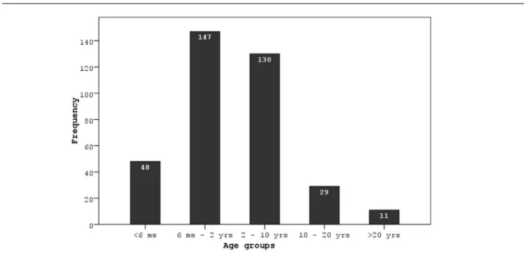
The median age and weight of the patients at the time of VSD closure were 15 months (range 27 days – 56.9 years) and 10 kg (range 3.5 – 100 kg), respectively. As shown in Figure 1, most of the patients (n=147, 40.3%) were aged between 6 months and 2 years. One hundred seventy-nine (49%) patients were male. There were 12 (3.3%) patients with chromosomal abnormality. The demographic and clinical characteristics of the patients are presented in Table 1. Apart from 10 (2.7%) patients with doubly-committed VSD, all VSDs were perimembraneous either with or without extension to the other segments of the interventricular septum. While perimembraneous VSD without extension was the most common (n=285, 78.1%) VSD characteristic, perimembraneous VSD with inlet extension (PM-I) was present in 44 (12.1%) patients. Eight (2.2%) patients had multiple VSDs. Excluding the patients with a history of PAB, there were 20 (5.5%) patients with pulmonary stenosis at various levels.
There were 140 (38.4%) patients who were preoperatively evaluated by catheterisation to estimate PVR and assess pulmonary artery stenosis, for which the median age was 42.6 months (range 6.1 months – 56.9 years). The PVR was severely elevated (≥8 Wood) in five (3.6%) patients in whom the pulmonary vascular reactivity test was positive, thus none of the patients were considered inoperable.
Surgery
VSD closure was accomplished through transatrial approach in the majority of the patients (95.1%). Other VSD approaches and combined suturing techniques in regards to the risk of
permanent epicardial pacemaker (PEP) implantation in patients who underwent surgical VSD closure.
METHODS
Study Design and Population
This single centre survey was retrospectively designed. Patients who had consecutively undergone VSD closure between January 2006 and October 2015 were recorded in a database. Patients with an atrial septal defect, patent foramen ovale, pulmonary infundibular, valvar or supravalvar stenosis, patent ductus arteriosus and/or previous pulmonary artery banding (PAB) were included. Patients with an associated major cardiac anomaly (discordant atrioventricular or ventriculoarterial connection, double outlet right ventricle, tetralogy of Fallot, coarctation of the aorta, VSD with pulmonary atresia and atrioventricular septal defect) were excluded from the study. A total of 365 patients were included in the data analysis. The ethical committee of non-invasive clinical research of Ege University Faculty of Medicine (protocol: 15/9-4) approved the current retrospective survey on October 12, 2015.
Study Variables
The demographic, clinical and operative characteristics of the patients were obtained from medical records. The type of VSD was determined according to the Congenital Heart Surgery Nomenclature and Database Project[16]. The size of the VSD was designated as large, moderate or small in accordance with the diameter of the aortic annulus. While transthoracic echocardiography was the primary diagnostic tool, catheterisation was utilised in select number of patients to estimate pulmonary vascular resistance (PVR) and perform pulmonary vascular reactivity test.
Surgical Procedure
Additionally, the rate of combined suture technique was higher in moderate and large VSDs. The ACC and CPB times, along with the duration of intensive care unit and hospital stay, were higher in patients who received combined suture technique.
Risk of Complete Heart Block
Overall, there were 25 (6.8%) patients with advanced second degree or CHB who needed EP-CPB. While the heart block recovered to sinus rhythm in 17 (68%) patients at a median of postoperative 5 days (range 1 – 11 days), PEP was implanted in eight (32%) patients (2.2% of all patients) at a median of postoperative 8 days (range 7 – 12 days). Neither EP-CPB nor PEP were required in patients with doubly-committed VSD, PM-O or PM-IO.
The univariate analysis for the need of EP-CPB and PEP is presented in Table 3. In patients who received EP-CPB, the proportion with either a small or large VSD was lower than those who did not receive EP-CPB (24% vs. 76%, P=0.012; 40% vs.60%,
P=0.013, respectively). In contrast, the proportion of patients with large VSD was higher in those who received PEP (75% vs. 25%,
P=0.001). The proportion of patients with perimembraneous VSD without extension was higher in those who needed EP-CPB (60% vs. 40%, P=0.024) but in contrast, it was lower in patients who received PEP implantation (37.5% vs. 62.5%, P=0.018). On the other hand, while the rate of patients with PM-I was significantly lower than that of patients who needed EP-CPB (40% vs. 60%, P<0.001), it was substantially higher in patients who received PEP (62.5% vs. 37.5%, P<0.001). As presented in Table 4, the comparison of the suturing techniques according to the need for EP-CPB and PEP implantation among patients with perimembraneous VSD without extension and in patients with PM-I revealed no statistically significant difference.
were performed through transatrial in five (1.4%) patients, transpulmonary in four (1.1%) and combined transatrial and transventricular in nine (2.5%). Continuous suturing (n=302, 82.7%) was the most frequently used technique. Fifty (13.7%) patients received tricuspid valve detachment (TVD), four of which required anteroseptal commissuroplasty to correct moderate tricuspid regurgitation following reattachment of the septal leaflet. While ASD/PFO closure (n=189, 51.8%) was the most frequent concurrent procedure, the other concurrent procedures were pulmonary artery debanding in 63 (17.3%) patients, PDA ligation/division in 27 (7.4%), pulmonary valvotomy in nine (2.5%), and right pulmonary artery reconstruction in three (0.8%). Pulmonary artery debanding in 63 (17.3%) patients was performed through supra-annular patch plasty of the main pulmonary artery using a Dacron patch. Interatrial communication in 12 patients was left open due to high PVR.
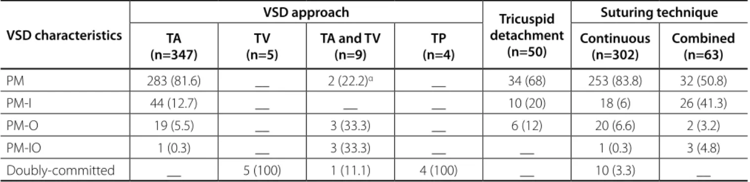
Because of the characteristics of the VSD, 12 patients required right ventriculotomy in which the VSD was doubly-committed in six, perimembraneous with outlet extension (PM-O) in three, and perimembraneous with both inlet and outlet extension (PM-IO) in three patients. In eight (2.2%) patients with multiple VSD, while three were closed with a separate patch, three were incorporated into the principal VSD patch, and two were closed via left ventriculotomy. The distribution of the VSD closure technique in terms of VSD characteristics are presented in Table 2. None of the patients were operated for recurrent VSD in the corresponding hospital admission. As revealed in Table 1, the rate of continuous suture technique increased significantly with increasing age and weight. While the proportion of continuous suturing was higher in perimembraneous VSD without extension, the rate of combined suture technique increased considerably with PM-I.
Table 1. The demographic and clinical characteristics of the patients.
Characteristics
Overall (n=365) n (%)
Continuous suturing (n=302)
n (%)
Combined suturing (n=63)
n (%) P
-value
Age, months 15
(27 days–56.9 years)*
22.3 (48 days–56.2 years)
12.7
(27 days–16.3 years) 0.002α Weight, kg 10 (3.5–100)* 10.1 (4–100) 7.2 (3.5–46) <0.001α
Male 179 (49) 155 (51.3) 24 (38.1) 0.056β
Previous pulmonary artery banding 63 (17.3) 46 (15.2) 17 (27) 0.025β VSD type and characteristics
PM 285 (78.1) 253 (83.8) 32 (50.8) <0.001β
PM-I 44 (12.1) 18 (6) 26 (41.3) <0.001β
PM-O 22 (6) 20 (6.6) 2 (3.2) 0.450γ
PM-IO 4 (1.1) 1 (0.3) 3 (4.8) 0.016γ
Doubly-committed 10 (2.7) 10 (3.3) __ 0.298γ
Multiple VSDb 8 (2.2) 5 (1.7) 3 (4.8) 0.290γ
VSD size
Small 176 (48.2) 161 (53.3) 15 (23.8) <0.001β
Moderate 114 (31.2) 85 (28.1) 29 (46) 0.005β
Large 75 (20.5) 56 (18.5) 19 (30.2) 0.038β
Catheterisation 140 (38.4)
Mean pulmonary artery pressure, mmHg 35.5 (18–75)* 32 (18–75) 35 (19–70) 0.222α Pulmonary vascular resistance, Wood 3.8 (1.8–9.0)* 4 (1.8–9) 3.3 (2–8) 0.682α Qp/Qs 2.0 (1.6–3.0)* 2.1 (1.6–3.1) 2.1 (1.6–2.8) 0.971α Tricuspid valve detachment 50 (13.7) 43 (14.2) 7 (11.1) 0.511β ACC time, min* 36 (18–85) 35 (18–85) 45 (21–75) <0.001α CPB time, min* 50 (26–101) 47.5 (26–101) 58 (34–90) <0.001α ICU stay, days* 1 (1–20) 1 (1–16) 1 (1–20) <0.001α Hospital stay, days* 7 (2–34) 7 (2–34) 8 (4–25) 0.001α ACC=aortic cross clamp; CPB=cardiopulmonary bypass; ICU=intensive care unit; PM=perimembraneous; PM-I=perimembraneous with inlet extension; PM-O=perimembraneous with inlet and outlet extension; PM-O=perimembraneous with outlet extension. * Data are presented as the median (range). α Patients with previous pulmonary artery banding were not included. β Indicates an additional muscular VSD. α Mann–Whitney U-test, β Pearson chi-square test, γ Fisher’s exact test.
Table 2. The distribution of surgical characteristics of ventricular septal defect closure according to VSD characteristics.
VSD characteristics
VSD approach Tricuspid
detachment (n=50)
Suturing technique
TA (n=347)
TV (n=5)
TA and TV (n=9)
TP (n=4)
Continuous (n=302)
Combined (n=63)
PM 283 (81.6) __ 2 (22.2)α __ 34 (68) 253 (83.8) 32 (50.8) PM-I 44 (12.7) __ __ __ 10 (20) 18 (6) 26 (41.3) PM-O 19 (5.5) __ 3 (33.3) __ 6 (12) 20 (6.6) 2 (3.2) PM-IO 1 (0.3) __ 3 (33.3) __ __ 1 (0.3) 3 (4.8) Doubly-committed __ 5 (100) 1 (11.1) 4 (100) __ 10 (3.3) __ TA=transatrial; TP=transpulmonary; TV=transventricular
the rhythm returned to sinus in 48 patients, 16 (1.9%) patients required PEP implantation. Furthermore, a body weight less than 4 kg and inlet VSD were identified as risk factors for the development of permanent CHB. In the current study, PM-I (P<0.001) was also found to be a risk factor for the development of CHB, both in the univariate and multivariate analyses. On the other hand, weight was not found to have an influence on the need for either EP-CPB or PEP implantation. The main reason for this lack of association may be related to the low number of patients weighing less than 4 kg in the current study, because PAB has been still the procedure of choice in some centres to avoid the consequences of open heart surgery in low-weight patients[17].
Table 3. Univariate analysis of the influence of independent variables on requirement for epicardial pacing at the time of weaning from cardiopulmonary bypass and permanent epicardial pacing.
Variable
EP-CPB PEP
Yes (n = 25) n (%)
No (n = 340)
n (%) P-value
Yes (n = 8) n (%)
No (n = 357)
n (%) P-value
Age, days* 401 (27–7011) 575.5 (48–
20518) 0.052α
649.5 (180– 2754)
551 (27–
20518) 0.959α Weight, kg* 8.5 (4–69) 10 (3.5–100) 0.052α 9 (7–18) 10 (3.5–100) 0.945α Male 12 (48) 167 (49.1) 0.914β 2 (25) 177 (49.6) 0.309γ VSD size
Small Yes 6 (24) 170 (50) 0.012β 1 (12.5) 175 (49) 0.092γ No 19 (76) 170 (50) 7 (87.5) 182 (51)
Moderate Yes 9 (36) 105 (30.9) 0.594β 1 (12.5) 113 (31.7) 0.441γ No 16 (64) 235 (69.1) 7 (87.5) 244 (68.3)
Large Yes 10 (40) 65 (19.1) 0.013β 6 (75) 69 (19.3) 0.001γ No 15 (60) 275(80.9) 2 (25) 288 (80.7)
VSD characteristics
PM Yes 15 (60) 270 (79.4) 0.024β 3 (37.5) 282 (79) 0.018γ No 10 (40) 70 (20.6) 5 (62.5) 75 (21)
PM-I Yes 10 (40) 34 (10) <0.001γ 5 (62.5) 39 (10.9) <0.001γ No 15 (60) 306 (90) 3 (37.5) 318 (89.1)
Multiple VSD Yes 2 (8) 6 (1.8) 0.178γ __ 8 (2.2) 1.0γ No 23 (92) 334 (98.2) 8 (100) 349 (97.8)
Previous PAB 5 (20) 58 (17.1) 0.919γ 2 (25) 61 (17.1) 0.910γ VSD approach
Transatrial 25 (100) 322 (94.7) 0.483γ 8 (100) 339 (95) 1.0γ Transventricular __ 5 (1.5) 1.0γ __ 5 (1.4) 1.0γ Transatrial and transventricular __ 9 (2.6) 0.876γ __ 9 (2.5) 1.0γ Tricuspid valve detachment 5 (20) 45 (13.2) 0.517γ 1 (12.5) 49 (13.7) 1.0γ Suturing technique
Continuous 18 (72) 284 (83.5)
0.231γ 7 (87.5) 295 (82.6) 1.0γ Combined 7 (28) 56 (16.5) 1 (12.5) 62 (17.4)
PAB=pulmonary artery banding. * Data are presented as themedian (range). α Mann–Whitney U-test, β Pearson chi-square test, γ Fisher’s exact test.
In multivariate analysis, PM-I was found to be the significant covariate associated with the need for both EP-CPB (OR=6.0; 95% CI [2.5, 14.4]; P<0.001) and PEP implantation (OR=9.02; 95% CI [1.97, 41.36]; P=0.005) (Table 5). The large VSD was another significant covariate for PEP implantation (OR=8.63; 95% CI [1.63, 45.8]; P<0.011).
DISCUSSION
extension, which, in our opinion, was a strong indicator of the importance of the tractions and manipulations applied to obtain better exposure of the VSD rims. Another important point of the current study was the importance of the inlet extension of a VSD for the development of heart block, and neither of the suturing techniques applied were found to have a significant influence in either the univariate or multivariate analyses. Among 4432 patients with surgical perimembraneous VSD closure enrolled in the Pediatric Cardiac Care Consortium Database, Down syndrome was reported to be the most significant risk factor associated with PEP implantation, indicating the importance of inlet extension of a perimembraneous VSD[7]. From this point of view, the application of continuous suturing in patients with inlet extension is the most striking finding of the current study, which, in our opinion, was the reason underlying the higher prevalence of CHB necessitating PEP implantation when compared to that described in the literature. Because the bundle of His runs through the thin rim of the muscle separating the VSD from the tricuspid valve annulus[3,21]. On the other hand, CHB was not expected to occur in a VSD with inlet extension when the combined suture technique was applied. From our point of view, this issue could have been addressed using a larger patch which may have been able to keep sufficiently away from the conduction system.
When compared to the interrupted suture technique, the ACC and CPB times have been reported to be significantly reduced with continuous suture closure of a VSD[15]. As revealed in Table 1, the ACC and CPB times were considerably lower in patients in whom the continuous suture technique was utilised. Biventricular performance diminishes soon after congenital
heart surgery due to either the impact of CPB, changes in the loading conditions of the ventricles, or attachment of an akinetic patch to the ventricular septum itself during VSD closure[17-20]. Quinn et al.[19] reported a decrease in fractional segmental shortening localised to the interventricular septum in patients who underwent VSD closure surgery. During the interrupted suture technique, with the use of a patch larger than VSD size results in a larger segment of the septum to be compromised[19,20]. While not evaluated in the current study, the avoidance of ventricular dysfunction soon after VSD closure was the main reason why interrupted suture closure surrounding a VSD was not utilised.
Shallow stitching at the posteroinferior rim of the VSD during the continuous suture technique (<1.5 mm depth and <4 mm away from the rim) was found to be an important factor for avoiding postoperative CHB[8,21], which was supported in the current series. To the best of our knowledge, only few articles in the literature have compared the type of suture technique in regards to the development of CHB[15]. Continuous and interrupted suturing techniques were compared in a cohort of 231 patients, and CHB developed in five patients who received the continuous suture technique[15]. An interesting finding of the present study was the shift in significance in terms of the need for EP-CPB. While it was higher in patients with perimembraneous VSD without extension, the rate of PEP was greater in patients with PM-I (Tables 3 and 4). These inverted changes in the rates were strongly related to the recovery of the sinus rhythm in patients with perimembraneous VSD without
Table 5. Multivariate analysis for the need for permanent epicardial pacemaker implantation.
Variable EP-CPB PEP
Exp(B) 95% CI P-value Exp(B) 95% CI P-value
PM-I 6.0 2.50, 14.40 <0.001 9.02 1.97, 41.36 0.005
Large VSD - - - 8.63 1.63, 45.80 0.011
The independent variables included the suture technique, large VSD, multiple VSD, tricuspid valve detachment, perimembraneous VSD without extension, perimembraneous VSD with inlet extension, transatrial approach, aortic cross-clamp and cardiopulmonary bypass time
Table 4. Comparison of suturing techniques according to the need for epicardial pacing at the time of weaning from cardiopulmonary bypass and permanent epicardial pacing in perimembraneous VSD and perimembraneous VSD with inlet extension.
VSD type
EP-CPB (n=25) n (%)
PEP (n=8) n (%)
Yes No P-value Yes No P-value
PM Continuous 11 (73.3) 242 (89.6) 0.127γ 3 (100) 250 (88.7) 1.0γ Combined 4 (26.7) 28 (10.4) __ 32 (11.3)
PM-I Continuous 7 (70) 11 (32.4) 0.078γ 4 (80) 14 (35.9) 0.160γ Combined 3 (30) 23 (67.6) 1 (20) 25 (64.1)
ACKNOWLEDGEMENTS
The authors thank the Department of Biostatistics for supervising the statistical analyses.
The reduction in ACC and CPB times gained through applying continuous suture closure of a VSD may represent the most favourable advantage of this technique.
Additionally, continuous suture closure of a VSD was reported to be a risk factor for the development of a residual VSD[15]. The durability of the posteroinferior rim where the shallow continuous sutures were placed was the primary concern for the development of a residual VSD. The current study demonstrated that neither the continuous nor the combined suture closure had an unfavourable influence on the development of residual VSD necessitating reoperation.
With the aim of improving visualisation of a VSD, TVD was performed in a considerable proportion of the patients who underwent VSD closure operation[6,12,14,22]. Additionally, TVD has not been identified as a risk factor for the development of CHB[6,12,14]. TVD was utilised in 13.7% of patients in the current study. In support of the literature, TVD in the current series was not found to be a risk factor for EP-CPB or PEP implantation.
Aside from the retrospective design of this study, the major limitation was that it did not assess VSD closure by the interrupted suture technique completely surrounding the VSD[15]. Thus, this commonly utilised technique could not be compared with the techniques applied in the current study. The literature describes a significant number of patients who received PEP implantation in whom the rhythm returned to sinus over a variety of periods[7,11,13]. Thus, another important limitation of the current study was that the out-hospital course of the patients was not assessed.
CONCLUSION
In conclusion, based on the improved theoretical and technical advancements, VSD closure can be safely and efficiently performed with a very low rate of CHB. The current study showed that there was no difference between the suture techniques regarding the development of permanent CHB, which put forward the biological variations and the manoeuvres applied to obtain better exposure during surgery as the primary cause of the development of heart block. Development of CHB after VSD closure occurred independent of the performance of TVD, which can be safely utilised in patients with multiple chordal attachments that interrupt the visibility of the VSD rims. Additionally, although they seem to have a favourable influence on reducing ACC and CPB times, surgeons should be more cautious or avoid the continuous suturing technique when closing a PM-I.
REFERENCES
1. Andersen HØ, Leval MR, Tsang VT, Elliott MJ, Anderson RH, Cook AC. Is complete heart block after surgical closure of ventricular septum defects still an issue? Ann Thorac Surg. 2006;82(3):948-56.
2. Anderson BR, Stevens KN, Nicolson SC, Gruber SB, Spray TL, Wernovsky G, et al. Contemporary outcomes of surgical ventricular septal defect closure. J Thorac Cardiovasc Surg. 2013;145(3):641-7.
3. van Doorn C, Leval MR. Ventricular septal defects. In: Stark JF, Leval MR, Tsang VT, eds. Surgery for congenital heart defects. West Sussex: Wiley; 2006. p.355-72.
Authors’ roles & responsibilities
MFA
EŞ
MD
YÖŞ
HŞ
REL
YA
Substantial contributions to the conception or design of the work; agreement to be accountable for all aspects of the work in ensuring that questions related to the accuracy or integrity of any part of the work are appropriately investigated and resolved; final approval of the version to be published
Substantial contributions to the conception or design of the work; drafting the work or revising it critically for important intellectual content; agreement to be accountable for all aspects of the work in ensuring that questions related to the accuracy or integrity of any part of the work are appropriately investigated and resolved; final approval of the version to be published
Substantial contributions to the conception or design of the work; agreement to be accountable for all aspects of the work in ensuring that questions related to the accuracy or integrity of any part of the work are appropriately investigated and resolved; final approval of the version to be published
Substantial contributions to the conception or design of the work; drafting the work or revising it critically for important intellectual content; final approval of the version to be published
Substantial contributions to the conception or design of the work; drafting the work or revising it critically for important intellectual content; final approval of the version to be published
4. Nygren A, Sunnegardh J, Berggren H. Preoperative evaluation and surgery in isolated ventricular septal defects: a 21 year perspective. Heart. 2000;83(2):198-204.
5. Scully BB, Morales DL, Zafar F, McKenzie ED, Fraser CD Jr, Heinle JS. Current expectations for surgical repair of isolated ventricular septal defects. Ann Thorac Surg. 2010;89(2):544-9.
6. Gaynor JW, O’Brien JE Jr, Rychik J, Sanchez GR, DeCampli WM, Spray TL. Outcome following tricuspid valve detachment for ventricular septal defects closure. Eur J Cardiothorac Surg. 2001;19(3):279-82.
disorders in continuous versus interrupted suturing technique in ventricular septal defect surgical repair. Res Cardiovasc Med. 2016;5(1):e28735. 16. Jacobs JP, Burke RP, Quintessenza JA, Mavroudis C. Congenital Heart Surgery
Nomenclature and Database Project: ventricular septal defect. Ann Thorac Surg. 2000;69(4 Supl):S25-35.
17. Berkowitz DH, Gaynor JW. Management of pediatric cardiopulmonary bypass. In: Mavroudis C, Backer CL, eds. Pediatric cardiac surgery. Sussex: Wiley-Blackwell; 2013. p.169-213.
18. Klitsie LM, Hazekamp MG, Roest AA, Van der Hulst AE, Gesink-van der Veer BJ, Kuipers IM, et al. Tissue Doppler imaging detects impaired biventricular performance shortly after congenital heart defect surgery. Pediatr Cardiol. 2013;34(3):630-8.
19. Quinn TA, Cabreriza SE, Blumenthal BF, Printz BF, Altmann K, Glickstein JS, et al. Regional functional depression immediately after ventricular septal defect closure. J Am Soc Echocardiogr. 2004;17(10):1066-72.
20. Vassalos A, Lilley S, Young D, Peng E, MacArthur K, Pollock J, et al. Tissue Doppler imaging following paediatric cardiac surgery: early patterns of change and relationship to outcome. Interact Cardiovasc Thorac Surg. 2009;9(2):173-7.
21. Anderson RH, Becker AE. The anatomy of ventricular septal defects and their conduction tissues. In: Stark JF, Leval MR, Tsang VT, eds. Surgery for congenital heart defects. Sussex: Wiley; 2006. p.121-43.
22. Sasson L, Katz MG, Ezri T, Tamir A, Herman A, Bove EL, et al. Indications for tricuspid valve detachment in closure of ventricular septal defect in children. Ann Thorac Surg. 2006;82(3):958-63.
atrioventricular conduction block after operative repair of perimembranous ventricular septal defect. J Am Coll Cardiol. 2007;50(12):1196-200. 8. Fukuda T, Suzuki T, Kashima I, Sato M, Morikawa Y. Shallow stitching close
to the rim of the ventricular septal defect eliminates injury to the right bundle branch. Ann Thorac Surg. 2002;74(2):550-5.
9. Shi G, Chen H, Sun Q, Zhang H, Zheng J. Mattress stitch: a modified shallow stitching in the surgical closure of large perimembranous ventricular septal defect in infants. Ann Thorac Cardiovasc Surg. 2015;21(3):282-8. 10. Aydemir NA, Harmandar B, Karaci AR, Sasmazel A, Bolukcu A, Saritas T, et al.
Results for surgical closure of isolated ventricular septal defects in patients under one year of age. J Card Surg. 2013;28(2):174-9.
11. Siehr SL, Hanley FL, Reddy VM, Miyake CY, Dubin AM. Incidence and risk factors of complete atrioventricular block after operative ventricular septal defect repair. Congenit Heart Dis. 2014;9(3):211-5.
12. Bang JH, Park CS, Park JJ, Yun TJ, Baek JS, Yu JJ, et al. Detachment of the tricuspid valve for ventricular septal defect closure in infants younger than 3 months. J Thorac Cardiovasc Surg. 2016;152(2):491-6.
13. van Geldorp IE, Vanagt WY, Vugts G, Willems R, Rega F, Gewillig M, et al. Late recovery of atrioventricular conduction after postsurgical chronic atrioventricular block is not exceptional. J Thorac Cardiovasc Surg. 2013;145(4):1028-32.
14. Bol-Raap G, Weerheim J, Kappetein AP, Witsenburg M, Bogers AJ. Follow-up after surgical closure of congenital ventricular septal defect. Eur J Cardiothorac Surg. 2003;24(4):511-5.
15. Gholampour-Dehaki M, Zareh A, Babaki S, Javadikasgari H. Conduction