Annexin II mRNA expression in bovine oocytes during follicular development
Luis Fabiano Santos da Costa, Márcia Silveira Netto Machado, João Francisco Coelho de Oliveira,
Gustavo Zamberlan, and Paulo Bayard Dias Gonçalves
Universidade Federal de Santa Maria, Departamento de Clínica de Grandes Animais, Santa Maria,
RS, Brazil.
Abstract
We investigated the expression of calcium-dependent phospholipid binding protein annexin-II (Ann-II) messenger RNA (mRNA) during preantral follicle development and in oocytes from antral follicles of different diameters (< 3 mm, 5 to 8 mm and > 8 mm). The action of retinol on Ann-II mRNA expression in mature oocytes was also examined. Only oocytes from secondary preantral follicles expressed Ann-II mRNA and at the germinal vesicle stage expression by oocytes from follicles larger than 8 mm was significantly higher (p < 0.05) compared with oocytes from follicles smaller than 3 mm or between 5 and 8 mm. Ann-II mRNA expression by metaphase II oocytes from follicles larger than 8 mm was significantly higher (p < 0.05) than that from oocytes from follicles smaller than 3 mm, with oocytes from both these size-classes showing similar levels of Ann-II mRNA expression as oocytes recovered from 5-8 mm follicles. In the presence of retinol, Ann-II mRNA expression was higher than when retinol was absent (p < 0.05). Our data indicate that Ann-II mRNA expression is highest in competent oocytes and that retinol increases Ann-II mRNA and may be involved in the regulation of oocyte competence by decreasing the translation and/or degradation of Ann-II mRNA.
Key words:bovine oocytes, annexin II, retinol.
Received: December 22, 2004; Accepted: August 31, 2005.
Introduction
The kinetics of the nuclear maturation of bovine oocytes have been extensively studied (Sirardet al.1989; Süsset al.1988). Bovine oocytes acquire the competence for resuming and completing meiosis after the follicle reaches between 1.7 and 2.0 mm in diameter, a stage in which the oocyte has fully developed and measures 150µm (Motlik and Kubelka, 1990). The key regulator in the cell cycle for both meiosis and mitosis is maturation-promoting factor, a serine/threonine kinase protein composed of one catalytic sub-unit (p34cdc2) and one regulator sub-unit (B cyclin; Lohka et al. 1988). The active form of matura-tion-promoting factor induces the disintegration of the nu-clear membrane, directing several events such as germinal vesicle breakdown, chromosome condensation, and reorga-nization of the cytoplasm during meiosis and mitosis (Parrish et al. 1992). Oocytes have very low levels of p34cdc2during development and are unable to progress from the G2 to the M phase of meiosis but at the end of the devel-opmental stage the concentration and activity of p34cdc2are
high and the oocyte then acquires meiotic competence (Wu et al.1997). Greater maturation-promoting factor expres-sion and activity during follicle growth suggests that the proteins involved in the regulation of meiosis have similar expression profiles.
Annexin-II (Ann-II) is a calcium-dependent phos-pholipid binding protein, significantly expressed in various epithelial tissues (intestine, lungs, vascular endothelium, endometrium and ovaries) (Dreieret al.1998). This protein plays a regulatory role in exocytosis, endocytosis, plasminogen activation, cell adhesion (Brownsteinet al. 2004; Fitzpatricket al.2000), immunoglobulin transport (Kristoffersen and Matre, 1996) and ion channel activity (Burger et al. 1996). Ann-II forms an active tetrameric complex with S-100 protein sub-type A10 (S-100A10, also known as protein p11), the S-100 protein being known to be involved in cell-cycle regulation (Cajone and Sherbet, 1999).
Costa (1999) used immunohistochemical analyses to detected the S-100 protein in preantral and antral follicle oocytes from cows and cow fetuses and although the spe-cific S-100 subtype was not studied these results imply that protein p11 could be present, suggesting that Ann-II may
Send correspondence to Paulo Bayard Dias Gonçalves. Universi-dade Federal de Santa Maria, Departamento de Clínica de Gran-des Animais, Sala 416, 97105-900 Santa Maria, RS, Brazil. E-mail: [email protected].
regulate growth and maturation in oocytes during follicular development.
Retinol (vitamin A) participates in the normal devel-opment of young animal tissues, acting via growth factors such as epidermal growth factor (Wolf, 1984). Ann-II gene expression can be regulated by retinol, suggesting that genes that are activated by retinol-binding retinoic acid re-ceptors may play an important role in the survival of the oogonium and the oocyte and the prevention of apoptosis (Wolf, 1984).
During the study described in this paper we investi-gated the expression of Ann-II mRNA in preantral follicles and oocytes from antral follicles at different developmental stages. We also assessed the effects of retinol on Ann-II mRNA expression in mature oocytes.
Material and Methods
Oocyte collection and preparation
For collection of oocytes from primordial, primary or secondary follicles ovaries were collected from bovine fe-tuses obtained from cows (Bos taurus taurus) slaughtered at an abattoir in the town of Santa Maria in the Brazilian state of Rio Grande do Sul, Brazil. The fetuses were calculated to be 210 to 240 days old based on the method of Rüsse (1983), which correlates the cranio-caudal length of the fe-tus to pregnancy time in days. The ovaries were transported to our laboratory in TCM199 culture medium (Gibco, United States) at 30 °C in an isothermic container and preantral follicles isolated by mechanical dissociation (Figueiredoet al. 1993 as modified by Carámbulaet al., 1999). Follicles were designated as primordial, primary or secondary according to their size as measured using an in-verted microscope equipped with a micrometer (Hulshofet al., 1994). One hundred follicles from each size-class were pooled for messenger RNA (mRNA) extraction and PCR detection of complementary (cDNA).
Tertiary follicle oocytes were collected from ovaries obtained from the same abattoir and were transferred to our laboratory in the same manner as the other ovaries except that in this case the transport medium was 0.9% NaCl. In the laboratory, the ovaries were washed twice in 0.9% NaCl and then kept in a double bath at 30 °C until needed. Folli-cles of different sizes (less than 3 mm, between 5 and 8 mm and larger than 8 mm in diameter) were individually as-pired to collect grade I (Leibfried and First 1979) oocyte-cumulus complexes (COCs,) which were characterized as compact cumulus, complete cumulus, and oocyte with ho-mogeneous cytoplasm.
Non-matured COCs of each follicle size-category were mechanically agitated to disrupt the cumulus cells and the denuded oocytes washed twice with TCM199 medium supplemented with Earle salts, L-glutamine, 25 mM Hepes (Sigma), 0.2 mM sodium pyruvate, 100 IU penicillin mL-1, 50 µg streptomycin mL-1, and 1.0 mg polyvinyl alcohol
(PVA) mL-1. A set of 10 oocytes per follicle category was used for maturation in three replicates.
Immature COCs from different follicle categories were cultured in TCM199 medium supplemented with Earle salts, L-glutamine, 25 mM Hepes (Sigma), 0.2 mM sodium pyruvate, 26.19 mM sodium bicarbonate, 100 IU penicillin mL-1, 50µg streptomycin mL-1and 1.0 mg PVA mL-1(TCM-PVA) for 24 h at 39 °C in a 5% CO2saturated humidity atmosphere before being mechanically agitated to disrupt the cumulus cells, the denuded oocytes being washed twice in TCM199/(TCM-PVA). The state and cate-gory of the mature oocytes was checked by microscopy by mounting a set of oocytes from each of the follicle catego-ries on slides, fixing them with acetic acid:methanol (1:3 v/v) for 24 h and staining them with 1% (w/v) Lacmoid in 45% acetic acid diluted with phosphate-buffered saline (PBS) (Vignolaet al.1994).
Retinol addition during maturation
Immature COCs from tertiary follicle were obtained and cultured in the same conditions of the experiment de-scribed above. The COCs were randomly divided into con-trol and treatment group. In the concon-trol group, the oocytes were matured in TCM199 medium supplemented with Earle salts, L-glutamine, 25 mM Hepes (Sigma), 0.2 mM sodium pyruvate, 26.19 mM sodium bicarbonate, 100 IU penicillin mL-1, 50µg streptomycin mL-1and 1.0 mg PVA mL-1(TCM-PVA maturation medium) for 24 h at 39 °C in a 5% CO2saturated humidity atmosphere. In the treatment group, the oocytes were incubated under the same condi-tions, but the TCM-PVA maturation medium was supple-mented with 1 ng of retinol/mL-1. Ten oocytes per group were used for maturation in three replicates. After matura-tion, the oocytes were submitted to a semiquantitative anal-ysis for Ann-II mRNA expression.
Extraction of mRNA
tube containing 40µL of ultrapure water and the tube cen-trifuged for 1 min at 8000 g to dilute the mRNA.
Complementary DNA (cDNA) synthesis and PCR amplification
We produced complementary DNA (cDNA) using a commercial kit (SensiscriptTMReverse Transcriptase (RT) kit, QIAGEN Inc., Chatsworth, CA, USA), each RT-PCR mixture containing 2.0µL buffer, 2.0µL dNTP, 2.0 µL oligoDT-primer, 0.25µRnase inhibitor, 1.0µL Sensiscript enzyme, and 12.5µL of the mRNA extraction product. The reaction was carried out in a thermocycler at 37 °C for 1 h and the reaction product containing cDNA was stored at -18 °C until use.
Ann-II cDNA was amplified by PCR using a primer (forward 5’CCTCCAAGTGCA TACGGG3’ and reverse 5’TCATACTGAGCAGGTGTTTT3’) produced by Invi-trogen Life Technologies (USA) and a primer (forward 5’TGTTCCAGTATGA TTCCACCC3’ and reverse 5’TC CACCACCCTGTTGCTGTA3’) for the constitutive en-zyme glyceraldehyde-3-phosphate dehydrogenase (GAPDH) as a positive control to ensure that cRNA had in-deed been extracted. The PCR cDNA amplification mix-ture contained 17.5µL ultrapure water, 1.5 mM MgCl2, 50 mM KCl, 10 mM Tris-HCl, pH 8.3, 250µM of each dNTP, 0.4µM of each primer, 2.5 U Taq DNA polymerase and 3µL of cDNA in a final volume of 25µL. Amplifica-tion was performed in a thermocycler under the following conditions: step 1, 94 °C for 5 min; step 2, 94 °C for 1 min; step 3, 60 °C for 1 min for the Ann-II primers or 54 °C for 45 s for the GAPDH primers; step 4, 72 °C for 1 min; step 5, a final extension at 72 °C for 5 min. Steps 2-4 were repeated for 34 cycles for Ann-II and 31 cycles for GAPDH. The rel-ative amount of transcript was compared between groups based on the amplification results by comparing the consti-tutive GAPDH gene and the Ann-II gene. The PCR prod-ucts were separated on 2% (w/v) agarose gel in pH 8.0 Tris, boric acid EDTA buffer (TBE, containing 0.09 M Tris, 0.09 M boric acid and 0.5 M EDTA), stained with ethidium bromide and visualized under ultraviolet light. The data for semi-quantitative analysis of gene expression were ob-tained by capturing the gel images with a digital camera and recording them with the Alpha DigiDoc 1000 program to obtain arbitrary values. The arbitrary values were obtained using GAPDH as an internal control in each group of oocytes from different class-size.
To ensure that mRNA extraction and cDNA amplifi-cation were effective only samples in which GAPDH mRNA was detected were used in the analyses. Expression of Ann II mRNA in preantral follicles at different stages of development was assessed only as presence or absence of expression and no statistical analysis was performed. The semi-quantitative expression of Ann II mRNA based on
ar-bitrary values were subjected to analysis of variance (ANOVA).
Results and Discussion
We observed expression of Ann-II in secondary folli-cles but not primordial and primary follifolli-cles, this being the first report of Ann-II expression in bovine preantral folli-cles. Preantral follicles express several proteins important for follicle and oocyte development (van Wezel and Rog-ers, 1996) but the factors and underlying mechanism in-volved in primordial follicle activation are not yet completely understood. Our data indicates that Ann-II is probably not involved in the activation of primordial folli-cles because it was not detected in the early stages of follicular development. We detected GAPDH mRNA, indi-cating that mRNA was indeed being detected, and this was used as the control in the assessment of the Ann-II gene ex-pression in oocytes from different follicle sizes.
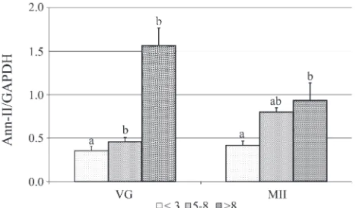
At the germinal vesicle stage, oocytes from different follicle sizes were mutually different (p < 0.05) regarding the expression of Ann-II mRNA. Oocytes recovered from follicles smaller than 3 mm or between 5 and 8 mm showed significantly (p < 0.05) lower Ann-II mRNA expression as compared with oocytes from follicles larger than 8 mm (Figure 1), suggesting that Ann-II plays a positive role in the processes of oocyte competence and capacity. It is known that oocytes remain in the first meiotic division prophase up until a little before ovulation and that the pro-teins responsible for the increase in size during this phase are synthesized during the oocyte growth phase (Mermillod et al., 1996). Oocyte development is completed soon after the follicular antrum forms when oocytes acquire the com-petence necessary to restart the first meiotic division but not the complete capacity for embryo development (Sirard and First, 1988). Our results showing significant expres-sion of Ann-II mRNA by oocytes from follicles larger than 8 mm as compared to the smaller oocytes points to Ann-II as an important protein in oocyte capacity.
Metaphase II oocytes from follicles larger than 8 mm in diameter expressed significantly more (p < 0.05) Ann-II mRNA than oocytes from follicles smaller than 3 mm, with oocytes from both these size-classes showing similar levels of Ann-II mRNA expression as oocytes recovered from 5-8 mm follicles (Figure 1).
These data reinforce the evidence that Ann-II may be involved in the process of oocytes capacity, since oocytes from follicles larger than 8 mm are more capable of reach-ing the blastocyst stage as compared with oocytes from smaller follicles.
matura-tion, our results indicate that retinol may either have in-creased transcription at the beginning of maturation or lessened the degradation of Ann-II mRNA.
Although retinol does not affect the kinetics of oocyte maturation, it has been confirmed that it can significantly increase the rate of cell cleavage in a chemically defined environment (Bortolottoet al.2001). It is also possible that retinol is capable of modifying gene expression or transla-tion during maturatransla-tion, which could influence fertilizatransla-tion capacity and embryo development. The resumption and progression of meiosis depends on intracellular oscillations in calcium levels (Kline, 1996) which induces the degrada-tion of maturadegrada-tion-promoting factor, a single Ca+ oscilla-tion being enough to bring about germinal vesicle breakdown (Wuet al.1997). Ann-II is a protein that takes part in the activity of oocyte calcium channels (Dreieret al. 1998) and may be an important factor in oocyte compe-tence and capacity.
In summary, we detected higher levels of Ann-II mRNA in capacitated oocytes as compared to
uncapa-citated oocytes. We also found that retinol increased Ann-II mRNA levels and may be involved in the regulation of oocyte competence by increasing translation or preventing degradation.
Acknowledgments
We thank the Silva abattoir for providing the bovine ovaries for the study. We also thank CNPq for a fellowship and financial support.
References
Bortolotto EB, Gonçalves PBD, Neves JP, Costa LFS, Maciel MN, Montagner MM, Farias AM and Stranieri P (2001) Fator de crescimento derivado das plaquetas, retinol e insu-lina na regulação da maturação nuclear de oócitos bovinos e suas conseqüências no desenvolvimento embrionário. Arq Bras Med Vet Zootec 53:191-197.
Brownstein C, Deora AB, Jacovina AT, Weintraub R, Gertler M, Khan KMF, Falcone DJ and Hajjar KA (2004) Annexin II mediates plasminogen-dependent matrix invasion by human monocytes: Enhanced expression by macrophages. Blood 103:317-324.
Burger A, Berendes R, Liemann S, Benz J, Hofmann A, Gottig P, Huber R, Gerke V, Thiel C, Romisch J and Weber K (1996) Identification of binding domains in the sodium channel NaV1.8 intracellular N-terminal region and annexin II light chain p11. J Mol Biol 257:839-847.
Cajone F and Sherbet GV (1999) Stathmin is involved in S100A4-mediated regulation of cell cycle progression. Clin Exp Metastasis 17:865-871.
Carámbula SF, Gonçalves PBD, Costa LFS, Figueiredo JR, Wheeler MB, Neves JP and Mondadori RG (1999) Effect of fetal age and method of recovery on isolation of preantral follicles from bovine ovaries. Theriogenology 52:563-571. Costa LFS (1999) Desenvolvimento de folículos pré-antrais e
imunohistoquímica de células ovarianasin vitroein situ. MSc Thesis, Universidade Federal de Santa Maria, Santa Maria.
Dreier R, Schmid KW, Gerke V and Riehemann K (1998) Diffential expression of annexins I, II and IV in human tis-sues: An immunohistochemical study. Histochem Cell Biol 110:137-148.
Figueiredo JR, Hulshof SCJ, Van Den Hurk R, Ectors FJ, Fontes RS, Nusgens B, Bevers MM and Beckers JF (1993) Devel-opment of a combined new mechanical and enzymatic method for the isolation of intact preantral follocles from fe-tal, calf and adult bovine ovaries. Theriogenology 40:789-799.
Fitzpatrick SL, Kassam G, Choi KS, Kang HM, Fogg DK and Waisman DM (2000) Regulation of plasmin activity by annexin II tetramer. Biochemistry 39:1021-1028.
Hulshof SC, Figueiredo JR, Beckers JF and Bevers MM (1994) Isolation and characterization of preantral follicles from fe-tal bovine ovaries. The Veterinary Quarterly 16:78-80. Kline D (1996) Activation of the mouse egg. Theriogenology
45:81-90.
Kristoffersen EK and Matre R (1996) Surface annexin II on pla-cental membranes of the fetomaternal interface. Am J Reprod Immunol 36:141-149.
Figure 1- Semi-quantitative analysis of the Ann-II mRNA expression in oocytes from different follicle sizes (< 3 mm, 5 to 8 mm, > 8 mm) at the germinal vesicle (VG) and metaphase II (MII) stages. Data are shown as mean and standard error of the mean. Different letters at the same stage in-dicate statistical difference as determined by an ANOVA (p < 0.05).
Figure 2- Semi-quantitative Ann-II mRNA expression in oocytes ma-tured for 24 h without retinol (control group) and supplemented with 1 ng retinol mL-1of medium (experimental group). The data are shown as mean
Leibfried L and First NL (1979) Characterization of bovine folli-cular oocytes and their ability to maturein vitro. J Anim Sci 48:76-86.
Lohka MJ, Hayes MK and Maller JL (1988) Purification of matu-ration-promoting factor, an intracellular regulator of early mitotic events. Proc Natl Acad Sci USA 85:3009-3013. Mermillod P, Lonergan P, Carolan C, Khatir H, Poulin N and
Cognie Y (1996)In vitromaturation of oocytes from domes-tic ruminants. Contraception Fertilite Sexualite 24:552-558. Motlik J and Kubelka M (1990) Cell cycle aspects of growth and
maturation of mammalian oocytes. Mol Reprod Dev 27:366-375.
Parrish JJ, Kim CL and Bae IH (1992) Current concepts of cell-cycle regulation and its relationship to oocyte matura-tion, fertilization and embryo development. Theriogenology 38:277-296.
Russe I (1983) Oogenesis in cattle and sheep. Biblthca Anat 24:77-92.
Sirard MA and First NL (1988)In vitroinhibition of oocyte nu-clear maturation in the bovine. Biology of Reproduction 39:229-234.
Sirard MA, Florman HM, Leibfried-Rutledge ML, Barnes FL, Sims ML and First NL (1989) Timing of nuclear progression and protein synthesis necessary for meiotic maturation of bovine oocytes. Biol Reprod 40:1257-1263.
Suss U, Wuthrich, K and Stranzinger G (1988) Chromosome con-figurations and time sequence of first meiotic division in bo-vine oocytes maturedin vitro.Biol Reprod 38:871-880. van Wezel I and Rodgers RJ (1996) Morphological
characteriza-tion of bovine primordial follicles and their environmentin vivo. Biol Reprod 55:1003-1011.
Vignola AGH, Prado AD, Valente A, Rubim MIB and Gonçalves PBD (1994) Técnicas de coloração cromossômica para está-gios específicos da maturação nuclear de oócitos bovinos. Ciência Rural 24:583-589.
Wolf G (1984) Multiple functions of vitamin A. Physiol Rev 64:873-936.
Wu B, Ignotz G, Currie WB and Yang X (1997) Dynamics of mat-uration-promoting factor and its constituent proteins during
in vitromaturation of bovine oocytes. Biol Reprod 56:253-259.