(Annals of the Brazilian Academy of Sciences)
Printed version ISSN 0001-3765 / Online version ISSN 1678-2690 www.scielo.br/aabc
Epigallocatechin-3-gallate combined with alpha lipoic acid attenuates high glucose-induced receptor for advanced glycation end products
(RAGE) expression in human embryonic kidney cells
JYH-GANG LEU1,2, CHIN-YAO LIN2, JHIN-HAO JIAN3, CHIN-YU SHIH4 and YAO-JEN LIANG3 1Fu-Jen Catholic University School of Medicine, Nº. 510 Zhongzheng Road, Xinzhuang Dist, New Taipei City 24205, Taiwan
2
Division of Nephrology, Department of Internal Medicine,
Shin Kong Wu Ho-Su Memorial Hospital, Nº. 95 Wen Chang Road, Shih Lin District, Taipei 11101, Taiwan 3
Department and Institute of Life Science, Fu-Jen Catholic University, Nº. 510 Zhongzheng Road, Xinzhuang Dist, New Taipei City 24205, Taiwan 4Department of Animal Pharmacology, Development Center for Biotechnology,
Nº. 101, Lane 169, Kanging St. Xizhi District, New Taipei City 22160, Taiwan
Manuscript received on march 8, 2012; accepted for publication on December 17, 2012
ABSTRACT
The anti-oxidant effects of epigallocatechin gallate (EGCG) and alpha lipoic acid (ALA) have been demonstrated in previous studies. The kidney protection effects of EGCG and ALA in patients with kidney
injury are still under investigation. The purpose of this study is to investigate the anti-infl ammatory and
anti-oxidant effects of EGCG and ALA on high glucose-induced human kidney cell damage. EGCG
inhibited high glucose(HG)-induced TNF-α and IL-6 production in human embryonic kidney (HEK)
cells. Both EGCG and ALA decreased HG-induced receptor of advanced glycation end products (RAGE) mRNA and protein expressions in HEK cells. EGCG and ALA also recovered HG-inhibited superoxide dismutase production and decreased ROS expressions in HEK cells. The synergism of EGCG and ALA was also studied. The effect of EGCG combined with ALA is greater than the effect of EGCG alone in all
anti-infl ammation and anti-oxidant experiments. Our studies provide a potential therapeutic application of
EGCG and ALA in preventing progression of diabetic nephropathy.
Key words: diabetic nephropathy, receptor of advanced glycation end products, epigallocatechin gallate, alpha lipoic acid, anti-oxidant.
Correspondence to: Yao-Jen Liang E-mail: 071558@mail.fju.edu.tw
INTRODUCTION
Diabetic nephropathy is the leading cause of end-stage renal disease in most developed countries (Ansari et al. 2003). The clinical manifestations
include increased glomerular fi ltration rate,
micro-albuminuria, macromicro-albuminuria, and renal failure. The pathological changes include glo merular basement membrane thickness, mesangial
expan-sion, glomerular sclerosis, and interstitial fi brosis
(Mauer et al. 1984). High serum glucose levels in diabetic patients may stimulate angiotensin II
(AT-II), various infl ammatory substances, and
superoxide production in kidney cells; augment inflammatory reaction; destroy normal kidney
cells and induce fi brosis formation (Jiao et al. 2011).
Oxidative stress has recently been proposed as the major damaging factor in hyperglycemia (Giugliano et al. 1996). Apoptotic cell death associated with increased oxidative stress in organs of diabetic patients has been well documented (Yang et al. 2011). However, the effect of anti-oxidant therapy in diabetic patients is still under investigation.
Advanced glycation end products (AGEs) is the products of nonenzymatic glycation and oxidation of proteins and lipids in diabetic patients. AGEs has been speculated to play a key role in the pathogenesis of diabetic nephropathy (Bohlender et al. 2005). The cellular effects of AGEs are mediated
by specifi c receptors, one of which is the receptor
for AGE (RAGE). RAGE is a transmembrane receptor of the immunoglobulin superfamily, expressed on the cell surface of several circulating
and organ-specifi c cells, including monocytes,
macrophages, proximal tubular cells, podocytes, and mesangial cells (Sourris et al. 2010). Recent studies showed that RAGE is a multiple ligand receptor. The ligand families of RAGE includes AGEs, advanced oxidation protein products, S100/calgranulins,
high-mobility group box-1, amyloid-β peptide, and β-sheet fi brils. In diabetic patients, hyperglycemia
may stimulates the production of these ligands and activate RAGE, the consequent signaling pathways,
and infl ammatory reaction (Yan et al. 2010). Oxidative
stress and NF-κκκB have been shown to play major B have been shown to play major
roles in kidney and vascular damage induced by RAGE activation (Yamagishi 2011, Yan et al. 1994).
Epigallocatechin-3-gallate (EGCG) is the main and most significant polyphenol in green tea. Numerous health-promoting functions of EGCG,
including oxidant, infl ammatory,
anti-atherogenic, and anti-cancer effects have been demonstrated. (Mereles and Hunstein 2011). Previous studies revealed that EGCG could inhibited various intracellular signal transduction pathways mediated by NF-κκκB, epidermal growth factor B, epidermal growth factor
receptor (EGFR), insulin-like growth factor-I (IGF-I) and mitogen-activated protein kinases
(MAPKs) (Khan et al. 2006). EGCG has also shown therapeutic potential in diabetic nephropathy. EGCG has attenuated AGE-induced cytokine synthesis in human embryonic kidney cells (Liang et al. 2010), and decreased RAGE and NF-κκκB expression and B expression and
pathological changes in the kidney tissue of rats with subtotal nephrectomy plus streptozotocin (STZ) injection (Yamabe et al. 2006).
Alpha-lipoic acid (ALA) is a natural occurring dithiol compound that can be found in animal food such as meat and liver (Kataoka 1998). ALA was discovered in 1951 as a molecule that assists in acyl-group transfer and acts as a coenzyme in the Krebs cycle. In addition to eliminating peroxide, ALA is a powerful lipophilic anti-oxidant both in vitro and in vivo (Packer et al. 1997). ALA can interact with other anti-oxidants, such as vitamins C and E, and strengthen their anti-oxidation abilities. ALA is now used as a dietary supplement for treating aging, diabetes mellitus, and vascular, and neurodegenerative diseases (Yilmaz et al. 2002).
This study will examine the effects of ALA and
EGCG in hyperglycemia-induced infl ammation
and oxidative stress in kidney cells. Furthermore, the hypothesis that ALA augments the anti-oxidant effects of EGCG will be tested.
MATERIALS AND METHODS
CELL CULTURE AND REAGENTS
Human embryonic kidney 293 (HEK293) cells
were cultured in Dulbecco’s modifi ed eagle medium
(DMEM) from Gibco (Grand Island, NY) containing 10% fetal bovine serum, 100 µg/mL penicillin, and 100 µg/mL streptomycin. Cultures were subcultured
on reaching 80% confl uence, using 0.05% trypsin/
MEASUREMENT OF TUMOR NECROSIS FACTOR-α(TNF-α)
AND INTERLEUKIN-6 (IL-6)
The cells were treated with high glucose for 18
h with or without EGCG and ALA. The TNF-α
and IL-6 levels in cell supernatants were measured by enzyme-linked immunosorbent assay (ELISA) kits (Peprotech Inc., Rocky Hill, NJ). Supernatants were diluted in the ratio of 1:30 before assay. The reaction products were measured at 450 nm with a microplate reader.
RNA ISOLATION AND REVERSE TRANSCRIPTION(RT)
Total cellular RNA was isolated from HEK293 cells using the single-step acid guanidinium thiocyanate/phenol/chloroform extraction method. For reverse transcription, 1 µg of RNA was incubated with 200 U of HiScript I reverse transcriptase (Bionovas Biotechnology, Toronto,
Canada) in a buffer contai ning a fi nal concentration
of 20 mmol/L Tris/HCl (pH 7.8), 100 mmol/L NaCl, 0.1 mmol/L EDTA, 1 mmol/L DTT, 50% glycerol, 2.5 µmol/L poly (dT)12-18 oligomer, and 0.5 mmol/L of each dNTP at a fi nal volume of 20
µL. The reaction mixture was incubated at 45°C for 1 h and then at 70°C for 15 min to inactivate the enzyme. The produced cDNA was used to generate DNA product by polymerase chain reaction (PCR).
REAL-TIME PCR
The cDNA had a 10-fold dilution in nuclease-free water and was used for the Smart Quant Green Master Mix (Protech Technology Enterprise Co.,
Taipei, Taiwan): 2 μL of cDNA solution, 0.5 μmol/L primers, 5 mmol/L magnesium chloride, and 2 μL of Master SYBR-Green in nuclease-free water with a fi nal volume of 20 μL. The primers
used for PCR were:
RAGE: forward, 5’AAGCCCCTGGTGCCTAATGAG3’; reverse, 5’CACCAATTGGACCTCCTCCA3’. GAPDH: forward, 5’CGACCACTTTGTCAAGCTCA3’;
reverse, 5’AGGGGTCTACATGGCAACTG3’.
The initial denaturizing phase was 5 min
at 95 °C followed by an amplifi cation phase as
detailed below: denaturation at 95 °C for 10 sec; annealing at 55 °C for 10 sec; elongation at 72 °C for 15 sec and detection at 79 °C for 45 cycles.
Amplifi cation, fl uorescence detection, and
post-processing calculation were performed using the ABI step1 apparatus. Individual PCR product was
analyzed for DNA sequence to confi rm the purity
of the product.
WESTERN BLOT ANANLYSIS
Total protein samples were mixed with sample buffer, boiled for 5 minutes, separated by 10% SDS-PAGE under denaturing conditions, and electro-blotted to nitrocellulose membranes (Amersham Pharmacia Biotech, CB, UK). The nitrocellulose membranes were blocked in blocking buffer, incubated with human anti-RAGE (Santa Cruz Biotechnology Inc., CA) antibodies, washed, and incubated with horseradish peroxidase-conjugated secondary antibodies. Signals were visualized by enhance chemiluminescent detection.
OXIDATIVE STRESS TEST
The cells were treated with high glucose concen-trations for 18 h with or without EGCG and ALA. The levels of superoxide dismutase (SOD) activity in cell supernatants were measured using SOD assay kit (catalog no. 706002, Cayman Chemical, Ann Arbor, MI). Supernatants were diluted in the ratio of 1:30 before assay. The reaction products were measured at 440-460 nm with a microplate reader.
STATISTICAL ANALYSIS
The data were expressed as mean ± SEM. A Tukey test was used for comparing parametric variables between the two groups, while ANOVA with repeat measurement design was used for
time-course changes. Statistical signifi cance was
San Diego, CA). A p-value of less than 0.05 was
considered statistically signifi cant.
RESULT
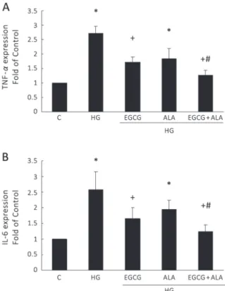
EGCG AND ALA INHIBITS TNF-α AND IL-6 PRODUCTION
High concentration glucose has been found to
stimu-late infl ammatory reaction in glomerular cells. In our
experiments, high-concentration glucose increased
TNF-α (Figure. 1A) and IL-6 (Figure. 1B) production
in HEK293T cells. Both EGCG alone and EGCG
combined with ALA (EA) signifi cantly inhibited high glucose-induced TNF-α and IL-6 secretion
in the culture medium. The combination of EGCG and ALA showed greater effect than EGCG alone.
However, the effect of ALA alone was not signifi cant.
ANTI-OXIDANTS INHIBIT THE EXPRESSION OF RRRAGE
RAGE is a key factor in diabetic nephropathy. We examined the effects of EGCG, ALA, and EA against glucose-induced RAGE up-regulation. As shown
in Figure 2, high-concentration glucose signifi cantly
induced RAGE mRNA expression in HEK293T cells.
Both EGCG and ALA signifi cantly attenuated high
glucose-induced RAGE mRNA expression. The combination of EGCG and ALA showed greater effect than EGCG alone. RAGE protein expression in HEK
cells was also signifi cantly attenuated by being treated
with EGCG, ALA, and EA (Figure. 3). Similar to the
fi ndings in RAGE mRNA experiments, the
combi-nation of EGCG and ALA showed greater effect than
EGCG alone. These fi ndings indicate that both EGCG and ALA may inhibit glucose-induced infl ammation
in kidney cells through decreasing RAGE expression and that ALA augments the effect of EGCG.
EFFECTS OF EGCG ON GLUCOSE-INDUCED INCREASE IN
OXIDATIVE STRESS
To evaluate the effects of EGCG and ALA on hyper glycemia-induced oxidative stress, we examined the activity of SOD and ROS at various levels in HEK293T cells with or without
treatment. As shown in Figure 4, HEK293T cells exposed to high concentration glucose had decreased SOD activity. However, EGCG, ALA,
and EA signifi cantly recovered SOD activity in
HEK293T cells in a high glucose environment.
High-glucose incubation signifi cantly increased ROS, EGCG, ALA, and EA signifi cantly decreased
high glucose-induced ROS (Figure. 4). The combination of EGCG and ALA showed greater
effect than EGCG alone. These fi ndings suggest
that EGCG, ALA, and EA inhibit hyperglycemia-induced oxidative stress in HEK293T cells and that ALA augments the effect of EGCG.
Figure 1 - EGCG and EGCG combined with ALA attenuated high glucose(HG)-induced TNF-α and IL-6 secretion from HEK cells. ELISA was used to measure the TNF-α(A) and IL-6
DISCUSSION
Diabetic nephropathy is the most common cause of end-stage renal disease. Various hyperglycemia-induced metabolic substances, including AGEs, protein kinase C (PKC), and AT-II were considered to contribute to the progression of diabetic nephropathy (Yamagishi and Imaizumi 2005). However, recent studies showed that intensive blood glucose control may better prevent long-term kidney damage (de Boer et al. 2011, Holman et al. 2008). Although the Figure 2 - EGCG and ALA attenuated high
glucose(HG)-induced expression of RAGE mRNA in HEK cells. Pre-treatment with EGCG (2.2 mM) and/or ALA (8 mM) signifi cantly attenuated high glucose-induced RAGE mRNA expression. The combination of EGCG and ALA showed more effect than EGCG alone. Total RNA was extracted and real-time PCR was performed. *p < 0.01 versus control; +p < 0.05 when compared to high glucose group; #p < 0.05 when compared to EGCG group.
Figure 3 - EGCG and ALA attenuated high glucose(HG)-induced RAGE protein expression in HEK cells. Pre-treatment with EGCG (2.2 mM) and/or ALA (8 mM) signifi cantly attenuated high glucose(HG)-induced RAGE protein expression. The combination of EGCG and ALA showed more effect than EGCG alone. *p < 0.01 versus control; +p < 0.05 when compared to high glucose group ; #p < 0.05 when compared to EGCG group.
Figure 4 - (A) Effects of EGCG and ALA on high glucose-decreased superoxide dismutase (SOD) activity in HEK cells. Cellular SOD activity was revealed by SOD assay kit. Both EGCG (2.2 mM) and ALA (8 mM) signifi cantly reversed high glucose-decreased SOD expression. The combination of EGCG and ALA showed more effect than EGCG alone. (B) The inhibitory effect of EGCG and ALA on high glucose-induced ROS expression. Both EGCG (2.2 mM) and ALA (8 mM) signifi cantly decreased the high-glucose-induced ROS expression in HEK cells. The combination of EGCG and ALA showed more effect than EGCG alone. *p < 0.01 versus
exact mechanism responsible for hyperglycemia-induced kidney damage is still under investigation, accumulated evidence indicates that AGE-RAGE oxidative stress may be the main pathologic pathway for diabetic nephropathy (Yamagishi 2011). The possible mechanism of RAGE-mediated
infl ammation was evaluated in previous studies
(Yan et al. 2010). Hyperglycemia causes AGE accumulation and RAGE activation, followed by increased production of extracellular ROS. Increased ROS stimulates the production of AGEs and non-AGE ligands of RAGE. These ligands and ROS stimulate RAGE, increase intracellular NF-κκκB
production, and induce production of infl ammatory
substances. In our study, EGCG and ALA decreased high glucose-induced RAGE expression. ALA also augmented the RAGE-inhibiting effect of EGCG in HEK cells. Previous studies showed that ALA regenerated vitamins C and E from their oxidized forms and augmented their anti-oxidant activity
(Kozlov et al. 1999). This is the fi rst study to identify
the synergism of EGCG and ALA in kidney cells.
The role of ROS in RAGE-mediated infl
am-mation was also shown in other studies (Yao and Brownlee 2010). Hyperglycemia induces intracellular ROS production by the mitochondrial electron transport chain, and increases expression of RAGE and endogenous RAGE ligands in human aortic endothelial cells. This increase in RAGE can be suppressed by overexpression of superoxide dismutase 2 (SOD2) or by the application of a superoxide dismutase mimetic compound. Oxidative stress and superoxide are causes of diabetic complications. SOD is the major anti-oxidant enzyme for superoxide removal, which converts superoxide into hydrogen peroxide and molecular oxygen (Miao and St Clair 2009). In a study of non-insulin-dependent diabetes, the SOD activity in erythrocytes was higher in patients without nephropathy than in patients with nephropathy (Kedziora-Kornatowska et al. 1998). Overexpression of SOD genes in transgenic mice attenuated the pathological progression of diabetic
nephropathy (DeRubertis et al. 2004). Recent studies also showed that kidney SOD was lower in diabetic mice than in control mice (Fujita et al. 2009). AT-II was thought to increase oxidative stress by enhancing production of reactive oxygen species. An AT-II receptor blocker, telmisartan, was shown to increase left ventricle SOD levels in diabetic rats (Goyal et al. 2011). These results indicated that oxidative stress is a major pathological factor for kidney damage in patients with diabetic nephropathy. Anti-oxidative agents are going to be the new research targets for drug development. This study demonstrated that EGCG
and ALA signifi cantly decreased the oxidative stress.
Few papers have studied the effect of EGCG on RAGE expression and oxidative stress activation. EGCG was shown to attenuate AGEs-induced RAGE expression in human kidney (Liang et al. 2010) and human neuroblastoma cells (Lee and Lee 2007), decrease oxidative stress and serum creatinine in a mouse model of immune-mediated glomerulo-nephritis (Peng et al. 2011), and reduce
chemotherapy-induced TNF-α up-regulation and oxidant stress
signals in mouse kidneys (El-Mowafy et al. 2010). Oxygen free radicals are important mediators in the pathogenesis of arthritis as they increase
pro-infl ammatory cytokine expression. EGCG was
shown to remove ROS and increase anti-oxidant enzymes, such as catalase, superoxide dismutase, and glutathione peroxidase (Singh et al. 2010). In our study, EGCG was shown to inhibit
hyperglycemia-induced RAGE, ROS, TNF-α, and IL-6 expression.
Furthermore, ALA augmented the anti-oxida oxidant effects of EGCG in kidney cells. Anti-oxidants may serve as a potential therapy for attenuating kidney function decline in diabetic patients.
metastatic prostate carcinoma, six grams of green tea were administrated orally per day. Adverse effects were observed in 69% of patients and included nausea, emesis, insomnia, fatigue, diarrhea, abdominal pain, and confusion (Jatoi et al. 2003).
The therapeutic effect of ALA supplementation in diabetic patients was also studied in many previous
trials (Singh and Jialal 2008). ALA effi ciently
decreased endothelial damage, vascular dysfunction, oxidative stress, and polyneuropathies in patients with type 1 and 2 diabetes. ALA also increased insulin sensitivity and improved glycemic control. The usual daily oral dosages of ALA were between 600 and 1,800 mg (Golbidi et al. 2011). Few adverse effects were noted in previous trials. At higher doses, gastrointestinal symptoms, such as abdominal pain, nausea, vomiting, and diarrhea, and skin allergic reactions had been reported (Ziegler et al. 2006).
The synergistic effects of hypoglycemic agents and anti-oxidants in preventing the progress of diabetic nephropathy in human subjects need further evaluation. Our study may provide cues for designing new anti-oxidant-based drugs for therapeutic purposes.
ACKNOWLEDGMENTS
This study was partially supported with grants from the Shin Kong Wu Ho-Su Memorial Hospital, Taipei, Taiwan (SKH-8302-101-DR-04).
RESUMO
Os efeitos antioxidantes de galato de epigalocatequina (EGCG) e ácido alfa lipóico (ALA) foram demonstrados
em estudos anteriores. Os efeitos renais da proteção de
EGCG e ALA em pacientes com lesão renal ainda estão
sob investigação. A fi nalidade deste estudo é investigar os efeitos anti-infl amatórios e antioxidantes de EGCG e
ALA em lesão de células de rim humano induzida pela
alta glicose. EGCG inibiu a produção de TNF-α e IL-6
induzida por HG em células de rim embrionário humano (HEK). Ambos EGCG e ALA diminuíram o mRNA do
receptor de produtos fi nais de glicação avançada (RAGE)
induzida por HG e a expressão de proteínas em células
HEK. EGCG e ALA também recuperaram a produção
de superóxido dismutase inibida por HG e diminuíram a expressão de ROS em células HEK. O sinergismo de EGCG e ALA também foi estudado. O efeito de EGCG combinado com ALA é maior do que o efeito de EGCG
sozinho em todos os experimentos anti-infl amatórios e
antioxidantes. Os nossos estudos fornecem uma potencial
aplicação terapêutica do EGCG e ALA na prevenção da
progressão de nefropatia diabética.
Palavras-chave: nefropatia diabética, receptores de
produtos fi nais da glicação avançada, epigalocatequina
galato, ácido alfa-lipóico, antioxidante.
REFERENCES
ANSARI A
A A, THOMAS S AND GOLDSMITH D. 2003. Assessing glycemic control in patients with diabetes and end-stage renal failure. Am J Kidney Dis 41: 523-531.
BOHLENDER JM, FRANKE S, STEIN G AND WOLF G. 2005. Advanced glycation end products and the kidney. Am J Physiol Renal Physiol 289: F645-659.
CHEN D, WAN SB, YANG H, YUAN J, CHAN TH AND DOU QP. 2011. EGCG, green tea polyphenols and their synthetic analogs and prodrugs for human cancer prevention and treatment. Adv Clin Chem 53: 155-177.
DE BOER IH, SUN W, CLEARY PA, LACHIN JM, MOLITCH ME, STEFFES MW AND ZINMAN B. 2011. Intensive diabetes therapy and glomerular fi ltration rate in type 1 diabetes. N Engl J Med 365: 2366-2376.
DERRRUBERTIS FR, CRAVEN PA, MELHEM MF AND SALAH EM. 2004. Attenuation of renal injury in db/db mice over-expressing superoxide dismutase: evidence for reduced superoxide-nitric oxide interaction. Diabetes 53: 762-768. EL-MOWAFY AM, AL-GAYYAR MM, SALEM HA, EL-MESERY ME AND DARWEISH MM. 2010. Novel chemotherapeutic and renal protective effects for the green tea (EGCG): role of oxidative stress and infl ammatory-cytokine signaling. Phytomedicine 17: 1067-1075.
FUJITA H, FUJISHIMA H, CHIDA S, TAKAHASHI K, QI Z, KANETSUNA
K
K Y, BREYER MD, HARRIS RC, YAMADA Y AND TAKAHASHI T. 2009. Reduction of renal superoxide dismutase in progressive diabetic nephropathy. J Am Soc Nephrol 20: 1303-1313.
GIUGLIANO D, CERIELLO A AND PAOLISSO G. 1996. Oxidative stress and diabetic vascular complications. Diabetes Care 19: 257-267.
GOLBIDI S, BADRAN AND LAHER I. 2011. Diabetes and alpha lipoic Acid. Front Pharmacol 2: 69.
GOYAL BR, PARMAR K, GOYAL RKRK AND AND MEHTA AA. 2011.
HOLMAN RR, PAUL SK, BETHEL MA, MATTHEWS DRDR AND AND
NEIL HA. 2008. 10-year follow-up of intensive glucose control in type 2 diabetes. N Engl J Med 359: 1577-1589. JATOI A, ELLISON N, BURCH PA, SLOAN JA, DAKHIL SR, NOVOYNY P, TAN W, FITCH TR, ROWLAND KM, YOUNG CY AND FLYNN PJ. 2003. A phase II trial of green tea in the treatment of patients with androgen independent metastatic prostate carcinoma. Cancer 97: 1442-1446. JIAO B, WANG YS, CHENG YN, GAO JJJJ AND AND ZHANG QZ.
2011. Valsartan attenuated oxidative stress, decreased MCP-1 and TGF-beta1 expression in glomerular mesangial and epithelial cells induced by high-glucose levels. Biosci Trends 5: 173-181.
KATAOKA K
K H. 1998. Chromatographic analysis of lipoic acid and related compounds. J Chromatogr B Biomed Sci Appl 717: 247-262.
K EDZIORA-K
K KORNATOWSKA KZ, LUCIAK M, BLASZCZYK J AND PAWLAK W. 1998. Lipid peroxidation and activities of antioxidant enzymes in erythrocytes of patients with non-insulin dependent diabetes with or without diabetic nephropathy. Nephrol Dial Transplant 13: 2829-2832. KHAN
K
K N, AFAQ F, SALEEM M, AHMAD N AND MUKHTAR H. 2006. Targeting multiple signaling pathways by green tea polyphenol (-)-epigallocatechin-3-gallate. Cancer Res 66: 2500-2505.
KOZLOV AV, GILLE L, STANIEK KK AND AND NOHL H. 1999. Dihydrolipoic acid maintains ubiquinone in the antioxidant active form by two-electron reduction of ubiquinone and one-electron reduction of ubisemiquinone. Arch Biochem Biophys 363:148-154.
LEE SJSJ AND AND LEE KW. 2007. Protective effect of (-)-epigallo-catechin gallate against advanced glycation endproducts-induced injury in neuronal cells. Biol Pharm Bull 30: 1369-1373.
LIANG YJ, JIAN JH, LIU YC, JUANG SJ, SHYU KG, LAI LP, WANG BWBW AND AND LEU JG. 2010. Advanced glycation end products-induced apoptosis attenuated by PPARdelta activation and epigallocatechin gallate through NF-kappaB pathway in human embryonic kidney cells and human mesangial cells. Diabetes Metab Res Rev 26: 406-416. MAUER SM, STEFFES MW, ELLIS EN, SUTHERLAND DE,
BROWN DM AND GOETZ FC. 1984. Structural-functional relationships in diabetic nephropathy. J Clin Invest 74: 1143-1155.
MERELES D AND HUNSTEIN W. 2011. Epigallocatechin-3-gallate (EGCG) for Clinical Trials: More Pitfalls than Promises? Int J Mol Sci 12: 5592-5603.
MIAO L AND ST CLAIR DK. 2009. Regulation of superoxide dismutase genes: implications in disease. Free Radic Biol Med 47: 344-356.
PACKER L, TRITSCHLER HJHJ AND AND WESSEL K. 1997. Neuro-protection by the metabolic antioxidant alpha-lipoic acid. Free Radic Biol Med 22: 359-378.
PENG A, YE T, RRRAKHEJA D, TU Y, WANG T, DU Y, ZHOU JK, VAZIRI ND, HU Z, MOHAN C AND ZHOU XJ. 2011. The green tea polyphenol (-)-epigallocatechin-3-gallate ameliorates experimental immune-mediated glomerulonephritis. Kidney Int 80: 601-611.
SINGH BN, SHANKAR S AND SRIVASTAVA RK. 2011. Green tea catechin, epigallocatechin-3-gallate (EGCG): mecha-nisms, perspectives and clinical applications. Biochem Pharmacol 82: 1807-1821.
SINGH R, AKHTAR N AND HAQQI TM. 2010. Green tea polyphenol epigallocatechin-3-gallate: infl ammation and arthritis. [corrected]. Life Sci 86: 907-918.
SINGH U AND JIALAL I. 2008. Alpha-lipoic acid supplemen-tation and diabetes. Nutr Rev 66: 646-657.
SOURRIS KC, HARCOURT BE, PENFOLD SA, YAP FY, MORLEY AL, MORGAN PE, DAVIES MJ, BAKER ST, JERUMS G AND FORBES JM. 2010. Modulation of the cellular expression of circulating advanced glycation end-product receptors in type 2 diabetic nephropathy. Exp Diabetes Res 2010: 974681. WATANABE KK ET AL. ET AL. 2010. Role of differential signaling
pathways and oxidative stress in diabetic cardiomyopathy. Curr Cardiol Rev 6: 280-290.
WOLFRAM S. 2007. Effects of green tea and EGCG on cardio-vascular and metabolic health. J Am Coll Nutr 26: 373S-388S. YAMABE N, YOKOZAWA T, OYA T AND KIM M. 2006. Therapeutic potential of (-)-epigallocatechin 3-O-gallate on renal damage in diabetic nephropathy model rats. J Pharmacol Exp Ther 319: 228-236.
YAMAGISHI S. 2011. Role of advanced glycation end products (AGEs) and receptor for AGEs (RAGE) in vascular damage in diabetes. Exp Gerontol 46: 217-224.
YAMAGISHI S AND IMAIZUMI T. 2005. Diabetic vascular complications: pathophysiology, biochemical basis and potential therapeutic strategy. Curr Pharm Des 11: 2279-2299.
YAMAGISHI S AND MATSUI T. 2010. Advanced glycation end products, oxidative stress and diabetic nephropathy. Oxid Med Cell Longev 3: 101-108.
YAN SD, SCHMIDT AM, AAANDERSON GM, ZHANG J, BRETT J, ZOU YS, PINSKY D AND STERN D. 1994. Enhanced cellular oxidant stress by the interaction of advanced glycation end products with their receptors/binding proteins. J Biol Chem 269: 9889-9897.
YAN SF, RRRAMASAMY RR AND AND SCHMIDT AM. 2010. The RAGE axis: a fundamental mechanism signaling danger to the vulnerable vasculature. Circ Res 106: 842-853.
YANG H, JIN X, KKKEI LAM CW AND YAN SK. 2011. Oxidative stress and diabetes mellitus. Clin Chem Lab Med 49: 1773-1782.
YAO D AND BROWNLEE M. 2010. Hyperglycemia-induced reactive oxygen species increase expression of the receptor for advanced glycation end products (RAGE) and RAGE ligands. Diabetes 59: 249-255.
YILMAZ O, OZKAN Y, YILDIRIM M, OZTURK AI AND ERSAN Y. 2002. Effects of alpha lipoic acid, ascorbic acid-6-palmitate, and fi sh oil on the glutathione, malonaldehyde, and fatty acids levels in erythrocytes of streptozotocin induced diabetic male rats. J Cell Biochem 86: 530-539. ZIEGLER D ET AL. 2006. Oral treatment with alpha-lipoic acid