UNIVERSIDADE DO ALGARVE
Lymphotoxin-beta receptor and RANK signaling
in TEL-JAK2-induced T-cell leukemia
Mónica Alexandra Teotónio Fernandes
Tese para obtenção do grau de Doutor em Ciências Biomédicas
Trabalho efetuado sob a orientação de:
Doutor Nuno Rodrigues dos Santos
UNIVERSIDADE DO ALGARVE
Lymphotoxin-beta receptor and RANK signaling
in TEL-JAK2-induced T-cell leukemia
Mónica Alexandra Teotónio Fernandes
Tese para obtenção do grau de Doutor em Ciências Biomédicas
Trabalho efetuado sob a orientação de:
Doutor Nuno Rodrigues dos Santos
Lymphotoxin-beta receptor and RANK signaling
in TEL-JAK2-induced T-cell leukemia
Declaração de autoria de trabalho
Declaro ser a autora deste trabalho, que é original e inédito. Autores e trabalhos consultados estão devidamente citados no texto e constam da listagem de referências incluída.
______________________________________________________________
Copyright – Mónica Alexandra Teotónio Fernandes. Universidade do Algarve. Departamento de Ciências Biomédicas e Medicina.
A Universidade do Algarve tem o direito, perpétuo e sem limites geográficos, de arquivar e publicar este trabalho através de exemplares impressos reproduzidos em papel ou de forma digital, ou por qualquer outro meio conhecido ou que venha a ser inventado, de o divulgar através de repositórios científicos e de admitir a sua cópia e distribuição com objetivos educacionais ou de investigação, não comerciais, desde que seja dado crédito ao autor e editor.
First of all, I would like to thank my supervisor, Dr. Nuno Rodrigues dos Santos, for allowing me to work in this project and for assistance in reviewing this thesis. I am also grateful to my lab colleagues that accompanied me all the way through or just part of it: Marinella (my lab soulmate), Teresa, Patrícia, Fábio, Ricardo, Faiza, and others.
I have also to acknowledge the financial support provided by the Portuguese Foundation for Science and Technology (FCT) through a 4-year individual PhD Studentship.
In addition, I would like to express my gratitude to the University of Algarve and the Department of Biomedical Sciences and Medicine for accepting me in the PhD Program in Biomedical Sciences. The support and consideration provided specially by the Director Professor Leonor Cancela and the secretary Conceição José have helped me to comply with all the Program requirements. Also, I thank my Thesis Committee, including Dr. Álvaro Tavares and Dr. João T. Barata, for productive discussions and the interest demonstrated. I am also grateful to former CBME and current CBMR members, especially Dra Gabriela Silva, Dr. José Belo, Dr. Guilherme Ferreira, and their lab teams for their priceless support. Carol and Rubi, I am really thankful that you were there to help me make it through the working weekends and holidays and for showing me how to prepare MEFs. Vanessa, thank you for your companionship and help. I thank also Dr. Paulo Martel and Lina Lopes for helping me to deal with bureaucracies.
I would like to thank Márcio Simão, Dr. Paulo Gavaia, and Dr. Ravi Kalathur for technical support. I also appreciated the technical assistance provided by technicians from core facilities at Department of Biomedical Sciences and Medicine namely, André Mozes, Cláudia Florindo, Sara Marques, Neuza Miguel, and Maurícia Vinhas
To our collaborators, Dr. João T. Barata, Dr. José A. Yunes, Dr. Jacques Ghysdael, Dr. Nuno L. Alves and their teams, thank you for your collaboration and intellectual input. In addition, I thank Dr. Emmanuel Dejardin for having me in his lab, and the people that helped me to adapt, namely Caroline, Khalid, Emilie, Raji in LMIST lab, and many others. My dear lucky Xana, thank you for you friendship and for borrowing me your colleagues. Anna, thank you so much for “being there” and for your illustration. Cátia, my WB guru, I will never forget your kindness and comforting food. Also, my sincere thanks to the University of Liege and GIGA Research Centre.
My final words go to my family and friends. Aníbal, Maria, Luísa, Sílvia, Bruno, Graciana, Décio, Andreia, … I really appreciated your support and all your efforts to encourage me to keep going. I also thank someone that is not among us anymore but remains my greatest influence, António Duarte.
Finally, I would like to thank all the other people that made this thesis possible even though they were not mentioned.
A leucemia linfoblástica aguda de linfócitos T (LLA-T) é uma patologia hematológica maligna que afeta essencialmente crianças e adultos jovens e é fatal na ausência de um diagnóstico precoce e uma terapêutica apropriada. É do conhecimento geral que a LLA-T tem origem em precursores dos linfócitos T, também designados por timócitos, que sofrem um bloqueio da diferenciação e expansão clonal. As células imaturas transformadas disseminam-se pela corrente sanguínea e invadem a medula ósdisseminam-sea e vários órgãos como o baço, fígado, gânglios linfáticos e, por vezes, o sistema nervoso central. Apesar das melhorias observadas em termos de prognóstico nas últimas décadas, vários problemas clínicos persistem incluindo o prognóstico reservado nas recidivas e no subgrupo de LLA-T caracterizado pela presença de células leucémicas mais imaturas, o crescente número de casos resistentes à terapêutica e as graves sequelas derivadas da terapêutica agressiva com recurso a quimioterapia em altas doses e/ou radiação. Logo, a identificação de novos alvos terapêuticos para usar possivelmente em terapêutica combinada, o que permitiria reduzir as doses de fármacos quimioterapêuticos, constitui um objetivo prioritário.
Na origem do desenvolvimento da LLA-T encontram-se sucessivas alterações genéticas em timócitos como, por exemplo, translocações cromossómicas que levam à sobre-expressão de fatores de transcrição oncogénicos ou criam genes de fusão aberrantes, deleções, duplicações e mutações específicas em proto-oncogenes ou em genes supressores de tumores. Estas alterações acabam por desregular processos cujo controlo tem uma importância extrema e que acabam por conduzir as células transformadas a um bloqueio da sua diferenciação, a adquirir capacidades de auto-renovação, de subverter os controlos da proliferação e de resistir a sinais pro-apoptóticos.
Apesar da grande maioria dos conhecimentos adquiridos até à data sobre o desenvolvimento da LLA-T referirem-se a alterações genéticas acumuladas nas células leucémicas, a importância dos fatores do microambiente onde estas células se desenvolvem tornou-se evidente nas últimas décadas. Acredita-se que as células neoplásicas podem interagir com células do estroma modificando a composição do microambiente onde se desenvolvem, em termos celulares e expressão de fatores, de modo a favorecer o desenvolvimento das primeiras. Como se pensa que a LLA-T poderá ter origem no timo e este órgão é caracterizado por um microambiente dinâmico, rico em fatores de crescimento, citoquinas e contactos linfo-estromais diretos indispensáveis ao desenvolvimento dos
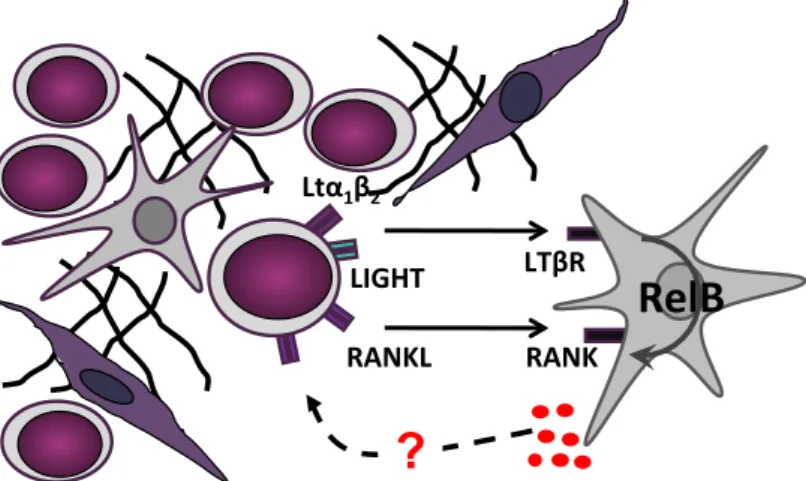
doença em questão. Recentemente, estudos realizados utilizando um modelo murino de LLA-T demonstraram que a proteína RelB, pertencente à família de fatores de transcrição NF-κB, quando expressa em células do estroma, é importante para o desenvolvimento desta doença. Esta descoberta apoia a hipótese de que sinais moleculares produzidos por timócitos transformados podem, através da ativação do fator de transcrição RelB em células do estroma tímico, levar à constituição de um microambiente favorável ao desenvolvimento de leucemia. Contudo, a identidade das células do estroma e/ou os sinais provenientes do estroma tímico que favorecem o desenvolvimento da LLA-T constituem ainda uma incógnita.
No timo, o fator de transcrição RelB é ativado em células do estroma por membros da superfamília de recetores TNF como, por exemplo, o recetor da linfotoxina beta (LTβR) ou o recetor ativador do NF-κB (RANK) e, tal como no caso da proteína RelB, a sua ausência em células do estroma tímico leva a defeitos na microestruturais. Estes defeitos, em parte caracterizados por uma diminuição do número e maturação das células epiteliais medulares do timo, são causados pela ausência de comunicação bidirecional entre timócitos que expressam os ligandos do LTβR e RANK (i.e., LTα1β2, LIGHT e RANKL) e as células do estroma que expressam os respetivos recetores. Para além de afetar a homeostasia de células que fazem parte do estroma, a ativação dos referidos recetores induz a ativação das vias clássica e alternativa do NF-κB, conduzindo a diferentes padrões de expressão génica. A ativação dos heterodímeros p50/RelA (via clássica) resulta essencialmente na expressão de citoquinas pro-inflamatórias. Por outro lado, a ativação dos heterodímeros p52/RelB (via alternativa) resulta por exemplo, na expressão de quimiocinas como CCL19, CCL21, CXCL12 ou CXCL13. Destas, as quimiocinas CCL19 e CCL21 e o seu recetor CCR7 que é expresso por timócitos, foram previamente implicadas na disseminação de células leucémicas do tipo LLA-T. Estudos recentes sugeriram também o envolvimento da via de sinalização LTα1β2/LTβR no desenvolvimento de leucemia e linfoma com origem em linfócitos B, nomeadamente na indução de nichos pro-tumorigénicos que suportam a viabilidade das células malignas.
É portanto possível que a interação dos ligandos, expressos em timócitos transformados, com o LTβR em células do estroma tímico promova a ativação do fator de transcrição RelB e consequente expansão das células do estroma que compõem o microambiente do timo e/ou a expressão de genes-alvo que podem eventualmente favorecer o
desenvolvimento da LLA-T. Para este fim, recorreu-se ao murganho transgénico TEL-JAK2 como modelo pois desenvolve LLA-T a partir de timócitos que expressam a referida proteína de fusão. Estes animais desenvolvem LLA-T com características histológicas e patológicas bastante similares à doença em humanos e períodos de latência variáveis que resultam da acumulação de alterações genéticas secundárias e do maior ou menor suporte de fatores do microambiente no qual as células leucémicas se desenvolvem.
Ao estudar a expressão de RNA mensageiro em linfomas tímicos de murganhos transgénicos TEL-JAK2, verificou-se expressão do gene Ltbr. Por outro lado, detetou-se expressão aumentada dos genes que codificam os ligandos LTα1β2 (i.e., Lta e Ltb) e LIGHT nas células T leucémicas quando comparadas com timócitos normais. Recorrendo ao uso de inibidores farmacológicos, identificou-se a via de sinalização NF-κB como sendo a principal responsável pela indução da expressão de Lta, Ltb e Light nas células leucémicas, provavelmente através da ativação dos recetores de células T, i.e., pre-TCR/TCR. Para além disso, a expressão de Lta parece ser também induzível pela via JAK/STAT. É portanto possível que a sinalização pelo LTβR esteja ativa em linfomas tímicos de ratinhos TEL-JAK2, tal como acontece no timo normal. Todavia, apenas a expressão de LTα1β2, em detrimento de LIGHT, foi detetada à superfície de células T leucémicas cultivadas ex vivo ou estimuladas com mitogénios, i.e., PMA e ionomicina. Além disso, verificou-se diminuição da expressão de LTα1β2 à superfície in vivo quando estas células contactam células do estroma que expressam o LTβR, pois aquele ligando é facilmente detetado quando as células permanecem em ambientes desprovidos do recetor. Portanto, as dificuldades encontradas em termos de deteção dos ligandos em microambientes com expressão do LTβR serão consequência de um controlo apertado em termos de expressão superficial do ligando e/ou sinalização continuada seguida por clivagem do mesmo.
De forma a avaliar se o desenvolvimento da leucemia induzida pela proteína de fusão TEL-JAK2 é comprometido na ausência do LTβR, cruzaram-se murganhos transgénicos TEL-JAK2 com murganhos nos quais a expressão do gene Ltbr foi geneticamente eliminada, de forma a gerar 2 coortes: TEL-JAK2;Ltbr-/- e TEL-JAK2;Ltbr+/- (controlo). Neste contexto, verificou-se que a inativação do LTβR levou a um atraso significativo no desenvolvimento da leucemia apesar da carga tumoral em órgãos linfóides e o fenótipo celular de superfície das células leucémicas não serem significativamente diferentes entre os dois grupos na fase terminal da doença. Curiosamente, numa fase precoce, quando os animais ainda não
JAK2;Ltbr-/- em relação a TEL-JAK2;Ltbr+/-, o que implica o eixo de sinalização LTα1β2/LTβR na fase inicial do desenvolvimento da leucemia. Além disto, a expressão de RANKL por células T leucémicas e do seu recetor RANK em linfomas tímicos, abrem caminho à possibilidade de os dois receptores, LTβR e RANK, colaborarem na indução do nicho adequado para suportar o desenvolvimento de células leucémicas no murganho transgénico TEL-JAK2.
De forma a verificar se as observações descritas poderão ser transpostas para LLA-T humana, procedeu-se inicialmente ao estudo de linhas celulares humanas. Várias semelhanças foram verificadas no que diz respeito ao padrão de expressão de LTα1β2 e à sua regulação por NF-κB. Para além disso, detetou-se a expressão dos genes que codificam LTα e LTβ em amostras de doentes com LLA-T, encontrando-se significativamente aumentada no subtipo molecular TAL/LMO.
Assim, pode-se concluir que a ativação do recetor LTβR no microambiente por células T leucémicas que expressam o ligando LTα1β2 contribui para o desenvolvimento de LLA-T numa fase precoce da doença. É portanto essencial o desenvolvimento de mais estudos visando clarificar os mecanismos pelos quais a comunicação entre células leucémicas e células do estroma através do eixo de sinalização LTα1β2/LTβR poderá apoiar a LLA-T e determinar a utilidade da inibição desta via como nova estratégia terapêutica.
PALAVRAS-CHAVE: Leucemia linfoblástica aguda de linfócitos T, modelo murino transgénico TEL-JAK2, timo, microambiente tumoral, recetor da linfotoxina beta, fator nuclear κB.
T-cell acute lymphoblastic leukemia (T-ALL) is an aggressive hematopoietic malignancy that arises from the combination of genetic and epigenetic alterations in thymic T-cell precursors and extracellular signals provided by the microenvironment. It was previously found that RelB expression in non-hematopoietic stromal cells promoted T-cell leukemogenesis in the EµSRalpha-TEL-JAK2 transgenic (TJ2-Tg) mouse model. In thymic stromal cells, RelB is a transcription mediator of lymphotoxin-beta receptor (LTβR). Lymphotoxin-mediated activation of LTβR has been implicated in physiological crosstalk between T cells and lymphoid organ stromal cells, but also pathological processes, including carcinogenesis. Since its role in T-ALL has remained elusive, we aimed to determine whether LTβR signaling is activated and playing a role in TEL-JAK2-induced leukemogenesis. In TJ2-Tg thymic lymphomas, activation of LTα1β2-LTβR signaling axis was supported by LTβR-encoding gene expression, while the genes LTβR-encoding its cognate ligand, lymphotoxin (LT)-α and LTβ, were found to be expressed by leukemic T cells, in an NF-κB-dependent manner. LTα1β2 protein was detected at the surface of TJ2-Tg leukemic cells only upon ex vivo culture or mitogenic stimulation. Moreover, we found that cell-surface LTα1β2 is downmodulated in vivo, indicating ongoing signaling. Further supporting a role for lymphotoxin signaling, LTβR genetic deficiency delayed TEL-JAK2-induced leukemia onset, but the tumor load in lymphoid organs and leukemia cell surface phenotype were comparable in end-stage disease. In accordance, the detection of reduced proportions of malignant thymocytes in TJ2-Tg;Ltbr-/- mice with no signs of disease implicated LTβR in early stages of leukemia development. Together, these data indicate that T-ALL-derived lymphotoxin activates LTβR signaling in thymic stromal cells, promoting leukemogenesis. Importantly, lymphotoxin-encoding genes were expressed in T-ALL patient samples, indicating that these may be also involved in human disease. Thus, future studies should provide a better understanding on how cellular crosstalk mediated by the lymphotoxin-LTβR axis supports T-ALL and assess the utility of blocking LTβR signaling as a novel therapeutic approach.
KEYWORDS: T-cell acute lymphoblastic leukemia, TEL-JAK2 transgenic mouse model, thymus, tumor microenvironment, lymphotoxin-beta receptor, nuclear factor kappaB.
ACKNOWLEDGEMENTS ... vii
RESUMO ... ix
ABSTRACT ... xiii
LIST OF FIGURES ... xix
LIST OF TABLES ... xxi
LIST OF ABBREVIATIONS, ACRONYMS AND SYMBOLS ... xxiii
CHAPTER 1 - INTRODUCTION ... 1
1.1. HEMATOPOIESIS AND T LYMPHOCYTE DEVELOPMENT ... 3
1.2. T-CELL ACUTE LYMPHOBLASTIC LEUKEMIA ... 8
1.2.1. Cell-intrinsic mechanisms ... 9
1.2.2. Microenvironmental factors ... 13
1.2.3. Contribution of cell-autonomous and noncell-autonomous factors to T-ALL development ... 17
1.2.4. T-ALL therapeutics and potential new targets ... 18
1.2.5. Models to study T-ALL ... 20
1.3. THE LTα1β2/LIGHT TO LTβR SIGNALING AXIS ... 22
1.3.1. The lymphotoxin-β receptor ... 22
1.3.2. The LTβR ligands: LTα1β2 and LIGHT ... 24
1.3.3. LTβR activation, signal transduction and target genes ... 29
1.3.4. Biological functions of the LTβR signaling pathway ... 34
1.4. LTβR SIGNALING IN CANCER DEVELOPMENT ... 37
1.4.1. LTβR cell-intrinsic pro-carcinogenic roles ... 39
1.4.2. LTβR pro-carcinogenic roles mediated by interactions with the tumor microenvironment ... 40
1.4.2.1. LTβR-induced angiogenesis ... 40
1.4.2.2. LTβR-induced chronic inflammation ... 41
1.4.2.3. Immune evasion mediated by LTβR ... 43
1.4.2.4. Induction of a pro-tumorigenic niche supported by LTβR-expressing stromal cells ... 44
1.4.3. Determinants of LTβR anti- and pro-carcinogenic roles ... 45
1.5. THE RANKL TO RANK SIGNALING AXIS ... 48
1.5.1. The receptor activator of NF-κB ... 48
1.5.2. The RANK ligand ... 49
1.5.3. RANK activation, signal transduction and molecular effects ... 50
1.5.4. Biological functions of the RANK signaling pathway ... 51
1.6. THESIS RATIONALE AND OBJECTIVES ... 54
2.2. MOUSE PROCEDURES ... 59
2.2.1. Mice ... 59
2.2.2. Mouse breeding and survival ... 60
2.2.3. Leukemic cell transplantation ... 60
2.2.4. LTβR-Fc treatment of mice ... 60
2.2.5. Sacrifice, necropsy and sample collection ... 60
2.3. CELL CULTURE ... 61
2.3.1. Cell lines ... 61
2.3.1.1. Maintenance and treatments ... 62
2.3.1.2. Freezing and thawing ... 62
2.3.2. Primary TJ2-Tg leukemic T cells ... 62
2.3.2.1. Isolation of TJ2-Tg leukemic T cells ... 62
2.3.2.2. Maintenance and treatments ... 63
2.3.2.3. Freezing and thawing ... 63
2.3.3. Primary mouse embryonic fibroblasts (MEFs) ... 63
2.3.3.1. Isolation of primary MEFs ... 63
2.3.3.2. Maintenance ... 64
2.3.3.3. Freezing and thawing ... 64
2.3.4. Co-cultures ... 64
2.3.5. Cell counts ... 65
2.4. FLOW CYTOMETRY AND CELL SORTING ... 65
2.4.1. Immunostaining for detection of T-cell markers and RANKL ... 66
2.4.2. Detection of intracellular lymphotoxin-β (LTβ) ... 66
2.4.3. Detection of membrane-bound LTβR ligands ... 67
2.5. GENOTYPING ... 69
2.5.1. DNA extraction ... 69
2.5.2. Polymerase Chain Reaction (PCR) ... 69
2.6. mRNA EXPRESSION ANALYSIS ... 70
2.6.1. RNA extraction ... 70
2.6.2. Complementary DNA (cDNA) synthesis ... 71
2.6.3. Semi-quantitative Reverse Transcription (RT)-PCR ... 72
2.6.4. Quantitative RT-PCR (RT-qPCR) ... 72
2.6.5. Oligonucleotide array analysis ... 73
2.7. PROTEIN EXPRESSION ANALYSIS ... 75
2.7.1. Protein extraction ... 75
2.7.2. Measurement of protein concentration ... 76
2.7.3. SDS-polyacrylamide gel electrophoresis (SDS-PAGE) ... 76
2.7.4. Western Blotting ... 76
2.8. HISTOLOGY AND HISTOCHEMISTRY METHODS ... 77
2.8.1. Histology ... 77
2.8.2. Immunohistochemistry (IHC) ... 78
2.9. STATISTICS ... 79
THYMI ... 85
3.1.1. Leukemic T cells from TEL-JAK2 transgenic mice express lymphotoxin-β receptor ligands ... 85
3.1.2. LTβR ligands are undetectable at the surface of TJ2-Tg leukemic in vivo ... 86
3.1.3. Lymphotoxin expression in TJ2-Tg leukemic T cells is inducible by pre-TCR/TCRαβ activation ... 88
3.1.4. Lymphotoxin expression in TJ2-Tg leukemic T cells depends on JAK/STAT and IKK/NF-κB signaling pathway activity ... 89
3.2. MODULATION OF LYMPHOTOXINS AT THE SURFACE OF TJ2-TG LEUKEMIC CELLS ... 92
3.2.1. Lymphotoxin cell surface induction on TJ2-Tg leukemic T cells depends on de novo protein synthesis ... 92
3.2.2. Microenvironmental LTβR modulates lymphotoxin expression at the surface of TJ2-Tg leukemic cells ... 94
3.3. IMPACT OF LTβR INACTIVATION ON TEL-JAK2-INDUCED LEUKEMOGENESIS ... 99
3.3.1. LTβR inactivation delays leukemia onset in TEL-JAK2 transgenic mice ... 99
3.3.2. Delayed leukemia onset in TEL-JAK2 transgenic mice is not due to developmental defects in the thymus of LTβR-deficient mice ... 100
3.3.3. LTβR inactivation does not affect the malignant phenotype of end-stage diseased TEL-JAK2 transgenic mice ... 101
3.3.4. Lymphotoxin signaling fosters the early stages of leukemogenesis in the thymus . 103 3.3.5. LTβR is not required for TJ2-Tg leukemic cell engraftment and dissemination ... 105
3.3.6. LTβR pharmacological block impacts on TJ2-Tg leukemic cell export from the thymus and dissemination ... 107
3.4. LYMPHOTOXIN EXPRESSION AND REGULATION IN HUMAN T-ALL ... 108
3.4.1. Human T-ALL cell lines express lymphotoxin through an NF-κB-dependent mechanism ... 108
3.4.2. Human primary patient samples express lymphotoxin ... 111
3.4.3. TEL-JAK2 transgenic mice model human T-ALL with cortical/mature immunophenotype ... 115
3.5. ROLE OF RANK SIGNALING IN T-ALL: PRELIMINARY FINDINGS ... 117
CHAPTER 4 - DISCUSSION & FUTURE PERSPECTIVES ... 121
4.1. TNFSF SIGNALING IN THE THYMUS OF TEL-JAK2 TRANSGENIC MICE ... 123
4.2. MECHANISM OF TNFSF LIGAND EXPRESSION IN TJ2-TG LEUKEMIC CELLS ... 127
4.3. IMPACT OF LTβR SIGNALING ABROGATION ON T-CELL LEUKEMOGENESIS ... 129
4.4. TIMING OF LTβR SIGNALING ACTION IN TEL-JAK2-INDUCED LEUKEMOGENESIS ... 131
4.7. LTβR AS A POSSIBLE THERAPEUTIC TARGET IN HUMAN T-ALL ... 136
CHAPTER 1
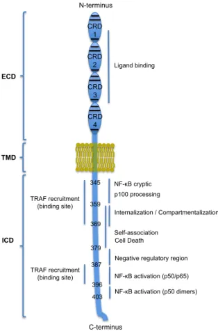
Figure 1.1. Thymus Structure. ... 5 Figure 1.2. Scheme depicting T-cell developmental stages in the thymus, and murine and human thymocyte markers. ... 5 Figure 1.3. Schematic representation of human LTβR structure. ... 24 Figure 1.4. LTβR as part of a network of shared ligand-receptor interactions within the TNF superfamily. ... 29 Figure 1.5. Summary of LTβR-mediated signal transduction for target-gene expression and cell death. ... 33 Figure 1.6. LTβR pro-tumorigenic roles in the tumor microenvironment. ... 46 Figure 1.7. Working hypothesis. ... 55 CHAPTER 3
Figure 3.1. LTβR and LTβR ligand expression in TJ2-Tg mice. ... 86 Figure 3.2. LTβR ligand expression at the surface of TJ2-Tg leukemic cells. ... 87 Figure 3.3. Lymphotoxin cell surface expression in thymocytes and TJ2-Tg leukemic
cells is induced through pre-TCR/TCRαβ activation. ... 89 Figure 3.4. LTβR ligand gene expression in TJ2-Tg leukemic cells depends on IKK and JAK kinase activity. ... 91 Figure 3.5. Lymphotoxin expression on TJ2-Tg leukemic cell surface is upregulated
upon ex vivo culture. ... 93 Figure 3.6. Lymphotoxin expression on TJ2-Tg leukemic cell surface depends on de novo protein synthesis. ... 94 Figure 3.7. Cell surface LTα1β2 expression on TJ2-Tg leukemic cells depends on LTβR expression in microenvironmental cells. ... 96 Figure 3.8. Lymphotoxin downmodulation from the surface of TJ2-Tg leukemic cells
requires direct contact with LTβR-expressing MS5 stromal cells. ... 97 Figure 3.9. Lymphotoxin downmodulation from the surface of TJ2-Tg leukemic cells
requires direct contact specifically with LTβR expressed at the surface of stromal cells. ... 98 Figure 3.10. LTβR inactivation delayed TEL-JAK2-induced leukemogenesis. ... 99 Figure 3.11. CD8+CD25+ leukemic cells originate in the thymus of TJ2-Tg young mice with no signs of disease. ... 100 Figure 3.12. Ltbr deficiency does not cause major thymic alterations regarding
thymocyte subpopulations in 4-5 month-old mice. ... 101 Figure 3.13. The malignant phenotype of end-stage diseased TJ2-Tg;Ltbr-/- leukemic
mice is comparable to that of control TJ2-Tg;Ltbr+/- mice. ... 102 Figure 3.14. Lta and Ltb expression in TJ2-Tg thymocytes correlates with the
emergence of malignant thymocytes. ... 104 Figure 3.15. LTβR is not required for TJ2-Tg leukemia engraftment and dissemination in
recipient mice. ... 106 Figure 3.16. Pharmacological inhibition of LTβR signaling impairs leukemogensis in
TJ2-Tg mice. ... 108 Figure 3.17. LTβR ligand expression in human T-ALL cell lines is induced by PMA
stimulation and inhibited by IKK inhibitor treatment. ... 110 Figure 3.18. T-ALL patient samples express lymphotoxin genes. ... 114
Figure 3.20. RANK and RANK ligand expression in TJ2-Tg mice. ... 118 Figure 3.21. RANK ligand gene expression in TJ2-Tg leukemic cells depends on IKK
activity. ... 119 Figure 3.22. RANK ligand cell surface expression in TJ2-Tg leukemic cells is induced through NF-κB activation and depends on de novo protein synthesis. ... 120
CHAPTER 1
Table 1.1. Common type A genetic abnormalities detected in T-ALL. ... 10 Table 1.2. Common type B genetic abnormalities detected in T-ALL. ... 12 Table 1.3. Microenvironmental factors involved in T-ALL development. ... 18 Table 1.4. LTβR-induced pro-carcinogenic effects in different cellular contexts. ... 47 CHAPTER 2
Table 2.1. Antibodies used for flow cytometry analyses of T-cell markers and RANKL. .... 68 Table 2.2. Primers used for genotyping and expected PCR results. ... 70 Table 2.3. Primer sequences used for RT-PCR analysis (murine). ... 74 Table 2.4. Primer sequences used for RT-PCR analysis (human). ... 75 Table 2.5. Commonly used solutions and gels. ... 80 CHAPTER 3
Table 3. 1. Clinical and immunophenotypical characteristics of primary T-ALL samples (Portuguese cohort). ... 112 Table 3.2. Clinical, immunophenotypical and genetic characteristics of primary T-ALL
2D – two dimensional 3D – three dimentional ºC – degree Celsius ×g – times gravity
CREB – cAMP response element-binding protein
c-Src – C-terminal Src kinase D
A
7-AAD – 7-aminoactinomycin D
ABL1 – Abelson murine leukemia viral
oncogene homolog 1 ActD – actinomycin D Aire – autoimmune regulator
ALL – acute lymphoblastic leukemia AML – acute myeloid leukemia AP-1 – activator protein 1 AP2 – adaptor protein 2 APC – allophycocyanin
ASK1 - apoptosis signal-regulating kinase 1
DAMP – damage-associated molecular patterns
DC – dendritic cell DcR3 – decoy receptor 3 DEPC – diethylpyrocarbonate DLL – delta-like ligand
DMEM– Dulbecco’s modified eagle medium DMSO – dimethyl sulfoxide
DN – double negative
DNA - deoxyribonucleic acid dNTP - deoxyribonucleotide DP – double positive
B
BAFF – B-cell activating factor
BCL11B – B-cell leukemia/lymphoma 11B
BCR – B-cell receptor
BLC – B lymphocyte chemoattractant BM – bone marrow
BSA – bovine serum albumin C
CaP – prostate cancer
CCND2 – cyclin D2
CD (number) – cluster of differentiation CD – cytoplasmic domain
CDKN2A – cyclin-dependent kinase inhibitor
2A
cDNA – complementary DNA
c-fos – FBJ murine osteosarcoma viral oncogene homolog
Chr – chromosome CHX – cycloheximide
cIAP1/2 – cellular inhibitor of apoptosis 1/2 CLL – chronic lymphocytic leukemia CLP – common lymphoid progenitor CMP – common myeloid progenitor CMR – cortico-medullary region CNS – central nervous system CO2 – carbon dioxide
CRD – cysteine-rich domain
E
ECD – extracellular domain
ECL – enhanced chemoluminescence EDTA – ethylenediamine tetraacetic acid EGIL – European Group for Immunological Characterization of Leukemias
Egr-1 – early growth response protein 1 ELC – EBl1-ligand chemokine
ETP – early T-cell precursor Ets – E-twenty six
EZH2 – enhancer of zeste homolog 2
F
FAA – formaldehyde-acetic acid-alcohol FACS – fluorescence-activated cell sorting FBS – fetal bovine serum
FBXW7 – F-box/WD domain-containing 7
FDC – follicular dendritic cell FITC – fluorescein isothiocyanate
FLT3 – Fms-like tyrosine kinase 3
FRC – fibroblastic reticular cell FVB mice – Friend virus B-type G
GALT – gut-associated lymphoid tissues
GAPDH – glyceraldehyde 3-phosphate
HBV – hepatitis B virus
HCC – hepatocellular carcinoma HCV- hepatitis C virus
HEV – high endothelial venules
HMGB1 – high-mobility group protein B1
HOX – homeobox gene HPRT1 – hypoxanthine
phosphoribosyltransferase 1 HPV – human papilloma virus HRP – horseradish peroxidase HSC – hematopoietic stem cell HVEM – herpes virus entry mediator
LTβR – lymphotoxin-beta receptor LYL1 – lymphoblastic leukemia-derived sequence 1
M
MAPK – mitogen-activated protein kinase mBD14 – mouse beta-defensin 14
M-CSF – macrophage colony-stimulating factor
MEF – mouse embryonic fibroblast
MEF2C – myocyte-specific enhancer factor
2C
MEMα – minimum essential medium alpha I
ICAM-1 – intercellular adhesion molecule 1 ICD – intracellular domain
ICN1 – intracellular NOTCH1 IFNγ – interferon gamma
IGF-1 – insulin-like growth factor 1 IKK – IkappaB kinase
IL – interleukin Inr – initiator element Iono – ionomycin
MHC – major histocompatibility complex MIP – macrophage inflammatory protein MITF – microphthalmia-associated transcription factor
MKK – MAP kinase kinase
MLL – mixed-lineage leukemia
MMP – matrix metalloproteinase MPP – multipotent progenitor mRNA – messenger RNA miRNA – micro RNA
mTOR – mammalian target of rapamycin J
JAK – Janus kinase
JNK – c-Jun N-terminal kinase
MYB – myeloblastosis oncogene MYC – myelocytomatosis oncogene
MyoD – myogenic differentiation
K
KLH – keyhole limpet hemocyanin Krt – keratin
N
NaN3 – sodium azide NEC – non-enzyme control
NF1 – nuclear factor 1
L
LBL – lymphoblastic lymphoma
LCK – lymphocyte-specific tyrosine kinase
LEF1 – lymphoid enhancer-binding factor 1 LFA-1 – lymphocyte function-associated antigen
LIC – leukemia-initiating cell
LIGHT – lymphotoxins, exhibits inducible expression, and competes with HSV glycoprotein D for HVEM, a receptor expressed by T lymphocytes
LMO – LIM domain only LN – lymph node
NFAT – nuclear factor of activated T cells NF-κB – nuclear factor-kappa B
NIK – NF-κB-inducing kinase NK – natural killer
NKX2-1 – NK2 homeobox 1
NMS – normal mouse serum NPC – nasopharyngeal carcinoma NS5B – nonctructural protein 5B NTC – non-template control
ODF – osteoclast differentiation factor OPG – osteoprotegerin
SCL – stem cell leukemia SDS – sodium dodecyl sulfate
SLC – secondary lymphoid tissue chemokine P
PAGE – polyacrylamide gel electrophoresis PB – peripheral blood
PBMC – peripheral blood mononuclear cell PBS – phosphate-buffered saline
PCR – polymerase chain reaction PDC – pancreatic ductal carcinoma PE - phycoerythrin
PE-Cy5 – phycoerythrin-cyanine 5 PGE2 – prostaglandin E2
PHF6 – PHD finger protein 6
PI – propidium iodide
PICALM – phosphatidylinositol binding
clathrin assembly protein
PI3K – phosphoinositide 3-kinase
PMA – phorbol myristate acetate
PTEN – phosphatase and tensin homolog
Socs2 – suppressor of cytokine signaling 2
SP – single positive
Sp1 – stimulating protein 1 SRE – skeletal-related effects
STAT – signal transducer and activator of transcription
T
TAB1 – TAK1-binding protein
TACE – TNF alpha-converting enzyme TAE – Tris-acetate
TAK1 – TGF-beta activated kinase
TAL1 – T-cell acute lymphoblastic leukemia protein 1
TCR – T-cell receptor TEC – thymic epithelial cell TEL – translocated ets leukemia
TET1 – ten-eleven translocation-1
Q
qPCR – quantitative PCR
TJ2-Tg – TEL-JAK2 transgenic TLX – T-cell leukemia homeobox TMD – transmembrane domain TNFα – tumor necrosis factor alpha R
Rag2 – recombination activating gene 2 RAS – rat sarcoma gene
RasGRP1 – Ras activator guanine nucleotide exchange factor
RANK – receptor activator of NF-κB RANKL – RANK ligand
RB – retinoblastoma gene
RBC – red blood cell
RelA – v-Rel reticuloendotheliosis viral oncogene homolog A
RelB – v-Rel reticuloendotheliosis viral oncogene homolog B
RIPA – radioimmunoprecipitation assay RNA – ribonucleic acid
ROS – reactive oxygen species rRNA – ribosomal RNA RT – reverse transcription
RUNX1 – runt-related transcription factor 1
TNFSF – TNF superfamily
TNFRSF – TNF receptor superfamily TRANCE – TNF-related activation-induced cytokine
TSC – thymic stromal cell TRA – tissue-restricted antigen
TRAF – TNF receptor-associated factor TRAMP – transgenic adenocarcinoma mouse prostate
U
UTR – untranslated region V
VCAM-1 - vascular-cell adhesion molecule 1 W
WBC – white blood cell
CHAPTER 1
INTRODUCTION
“To study the abnormal is the best way to understanding the normal.”
Although this famous statement from William James (American philosopher and psychologist) addressed character studies, it may be applied in the context of malignancy to state that there is no sharp line drawn between healthy/good or unhealthy/bad players in cancer development. Also, studying the normal context where a specific type of cancer develops may reveal hints on how cells become malignant/abnormal. In the specific case of T-cell acute lymphoblastic leukemia (T-ALL), several studies support the notion that in order to become transformed, T-ALL cells undergo cell-intrinsinc alterations and evolve by taking advantage of normal developmental mechanisms.
In this first chapter, a review of the literature on normal T-cell development and the determinants of transformation leading to T-ALL is presented. Possible therapeutic targeting approaches and experimental models used currently to study T-ALL are also revisited. In addition, important notions on signaling and functions attributed to two TNF superfamily receptors, lymphotoxin-beta receptor (LTβR) and receptor activator of NF-κB (RANK) are provided.
1.1. HEMATOPOIESIS AND T LYMPHOCYTE DEVELOPMENT
All mature blood cell types are derived from bone marrow (BM) resident hematopoietic stem cells (HSCs), through a developmental process called hematopoiesis. Throughout this process, HSCs experience progressive loss of self-renewal properties as they differentiate to multipotent progenitors (MPPs), which lack the ability to self-renew. In addition, HSC multilineage differentiation capabilities are also progressively restrained while the cell commits to a particular lineage (Lai and Kondo, 2008).
Two conflicting lines of evidence defend that the first step towards lineage restriction occurs in different phases of hematopoiesis. The so-called classic model states that MPPs originate symmetrically either a common myeloid progenitor (CMP) or a common lymphoid progenitor (CLP) (Akashi et al., 2000; Kondo et al., 1997). These common progenitors have
differentiation potentials restricted to all cell types within their respective lineage: megakaryocytes, erythrocytes, granulocytes, and macrophages are originated by CMPs, while T, B and NK lymphocytes derive from CLPs. Challenging this model, an alternative version in which common progenitors are generated asymmetrically from MPPs was put forward. Accordingly, myeloid cells were shown to potentially diverge from different subsets of MPPs, while lymphoid differentiation appeared to surpass multiple obligatory lineage restriction steps in such a way that CLPs are generated when the cells lose all abilities to generate myeloid cells (Adolfsson et al., 2005; Lai and Kondo, 2006). Furthermore, the existence of CLPs in the BM is also controversial. In fact, some studies demonstrated that the B lineage branch is segregated from the T cell pathway before a branch point for the T versus myeloid lineage (Bell and Bhandoola, 2008; Porritt et al., 2004; Wada et al., 2008). In spite of these debatable issues concerning hematopoiesis, the notion that in normal conditions BM-derived progenitors have to migrate to the thymus in order to originate self-tolerant, functional T lymphocytes (also known as T cells) is widely accepted (Sitnicka, 2009).
Like the BM, the thymus is a primary lymphoid organ but its only known function is to support the development of T lymphocytes from T-cell progenitors (Heinonen and Perreault, 2008). In humans, the thymus is located in the upper anterior mediastinum and lower part of the neck and is most active during childhood, reaching a peak weight at puberty, after which it undergoes slow involution (Gray et al., 2008). It is composed by two lobes and invested externally by a loose collagenous capsule. Each lobe is further divided in lobules by incomplete septa, which contain a peripheral dark cortical area densely populated by lymphoid cells, and a light medullary area composed of many voluminous pale cells and less abundant lymphoid cells (Milićević et al., 2008; Rezzani et al., 2008). A transitional area rich in blood vessels, known as cortico-medullary region (CMR) separates the thymic cortex and medulla (Figure 1.1). This is the entry site for early T cell precursors (ETPs), which derive from progenitors that emigrate from the BM to the thymus (Lind et al., 2001), and also the exit site for the end-product of thymopoiesis, naïve mature T lymphocytes (Jin et al., 2006). T-cell precursors developing in the thymus, also known as thymocytes, make up approximately 90% of the thymic cellular compartment and form a heterogeneous population composed of cells in different stages of differentiation. According to the cell phenotypes defined by expression of CD4 and CD8 coreceptors, thymocytes can be divided into four major subsets that represent consecutive steps in development namely, CD4-CD8- double negative (DN), CD4+CD8+ double positive (DP), CD4+CD8- single positive (CD4 SP), and
to DN4 based on different profiles of CD117 (also known as c-kit), CD25 and CD44 expression (Godfrey et al., 1993) (Figure 1.2).
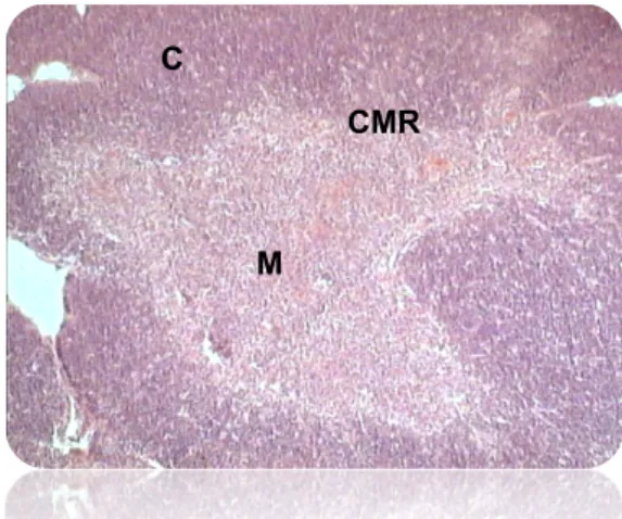
Figure 1.1. Thymus Structure. Hematoxilin-eosin staining of a thymus section showing the thymic structure, composed of an external region, the cortex (C), an internal region, the medulla (M), separated by the cortico-medullary region (CMR).
Figure 1.2. Scheme depicting T-cell developmental stages in the thymus, and murine and human thymocyte markers. The different thymocyte subpopulations defined by CD4/CD8 expression are shown along with the thymic region where they are located, their immunophenotypes, and the main checkpoints found during T-cell development. Differences between murine and human markers are shown.
C M CMR Immunophenotype CD4 - - - - + (Human) + + CD4 SP CD8 - - - - + (Mouse) + + CD8 SP sCD3 - - - - - -/+ + MU R IN E CD117 + + - - - - - CD25 - + + - - - - CD44 + + - - - - - CD24 + + + + + +/- - CD69 - - - -/+ + H U MAN TdT + + + + + + - CD34 + + + + +/- - - CD38 - + + + + + + CD1a - -/+ + + + +/- -
THYMIC CORTEX MEDULLA
DN1 ISP DP CD4 SP CD8 SP DN2 DN3 DN4 T-cell commitment αβ TCR checkpoint Pre-TCR checkpoint
In the thymus, T cell precursors interact with stromal cells, mainly dendritic cells (DC), fibroblasts, macrophages, endothelial cells, and epithelial cells, which make up a three-dimensional network and distinct microenvironments characterized by the production of diverse sets of cytokines, growth factors, and adhesion molecules (Petrie and Zúñiga-Pflücker, 2007; Takahama, 2006). Recent thymic immigrants acquire T-lineage potential and progress through distinct developmental stages by migrating through diverse inductive microenvironments of the thymus guided by factors produced by stromal cells. In such locations, they interact with stromal cells or are simply exposed to factors that control defined steps in T-cell development, such as proliferation, differentiation, T-cell receptor (TCR) gene rearrangements, and selection (Takahama, 2006).
For the initial TCR-independent proliferation and differentiation until the DN3 stage, thymocytes are exposed to signals derived from cortical stromal cells as they migrate to the subcapsular region and begin to rearrange their Tcr loci (Capone et al., 1998; Lind et al., 2001). The complementary expression patterns of both cytokines and their receptors determine their function at every stage of T-cell maturation. For example, gradients of chemokines like chemokine (C-X-C motif) ligand 12 (CXCL12), chemokine (C-C motif) ligand 25 (CCL25), CCL19 or CCL21 were shown to attract and mediate migration of thymocytes expressing their cognate receptors (i.e., CXCR4, CCR9 and CCR7, respectively) (Fu and Chen, 2004; Sitnicka, 2009; Takahama, 2006). Furthermore, expansion and proliferation of immature progenitors are dependent on stem cell factor (SCF) binding to c-kit in thymocytes and on signals delivered by interleukin-7 (IL-7) (Peschon et al., 1994; Rodewald et al., 1997), whereas Notch-mediated signals through binding of Delta-like 1 (DLL1) and Delta-like 4 (DLL4) ligands are also required for T lineage specification at the expense of B lineage (Pui et al., 1999; Radtke et al., 1999; Wilson et al., 2001). After T lineage commitment, DN thymocytes rearrange the genes encoding β, γ, and δ TCR chains. A small proportion of thymocytes productively rearrange γ and δ chains, express TCR γδ at their surface, and are named accordingly as γδ T cells. These cells do not differentiate further in the thymus and have functions related to innate immunity. On the other hand, the vast majority of thymocytes rearrange the TCRβ loci and express a functional TCRβ chain that associates with pre-TCRα (pTα), generating the pre-TCR complex at the cell surface. Through the pre-TCR complex, thymocytes receive specific survival and proliferation signals required for further differentiation to immature CD4+ single positive (ISP) (CD8+ISP in mice) and DP thymocytes (Figure 1.2). In addition, DP thymocytes that rearrange the TCRα loci to
form the TCRαβ complex, undergo TCR-mediated positive and negative selection through interaction with major histocompatibility (MHC) complexes expressed by cortical TECs (cTECs) and DCs (Goldrath and Bevan, 1999). Low-affinity recognition of self-MHC, promotes thymocyte survival and CCR7 expression, which drives migration to CCL19 and CCL21 produced by medullary TECs (mTECs) (Ueno et al., 2004), and determines commitment to either the CD4 or CD8 SP lineage. In the medulla, SP thymocytes that are reactive to tissue-specific antigens promiscuously presented by mTECs are deleted, a process termed negative selection, which is essential for the establishment of central tolerance (Chen, 2004; Takahama, 2006). Finally, the egress of mature T cells from the thymus was also shown to rely on thymic stromal-derived elements either by chemorepulsion or loss of responsiveness to thymic retention signals. In addition, a third mechanism was also described involving chemoattraction to peripheral signals (Jin et al., 2006).
In addition to the multiple roles attributed to stromal cells in thymocyte development, SP thymocytes reciprocally support mTEC organization and differentiation mainly through the action of members of the tumor necrosis factor (TNF) superfamily of ligands and receptors, such as LTα1β2/LTβR, RANKL/RANK, and CD40L/CD40 (Boehm et al., 2003; van Ewijk et al., 2000; Hikosaka et al., 2008; Klug et al., 1998). In general, these lympho-stromal bidirectional exchanges are referred to as thymic crosstalk. In normal conditions, hematopoiesis is tightly regulated by an exquisite balance between expression and extinction of transcription factor action in various combinations and their associated chromatin remodeling factors. Furthermore, it is controlled by the diverse array of factors that mediate complex bidirectional interactions between the microenvironment and hematopoietic stem cells or progenitors. Importantly, these two cell-intrinsic and microenvironmental factors are closely linked since certain transcription factors are inducible by microenvironmental cues (Kittipatarin and Khaled, 2007; Mohtashami et al., 2013; Peschon et al., 1994; Schmitt et al., 2004). When the regulation of proliferation, differentiation and/or survival fails, hematopoietic malignancies such as leukemia and lymphoma may arise. These are designated after the affected cell lineage as myeloid or lymphoid and, when untreated, can range from being rapidly fatal (acute) to slowly growing (chronic).
1.2. T-CELL ACUTE LYMPHOBLASTIC LEUKEMIA
The predominant types of pediatric cancers are leukemia, lymphoma, and cancers of the brain and central nervous system (CNS). Acute lymphoblastic leukemia (ALL) is the most common childhood malignancy and represents about 80% of all leukemia cases in children and 56% in adolescents. This percentage is significantly lower in adults (American Cancer Society, 2015). ALL consists of a group of malignancies of lymphoid cells that morphologically and immunophenotypically resemble B-lineage (B-ALL) and T-lineage precursor cells (T-ALL). These neoplasms may present predominantly with primary involvement of the bone marrow and peripheral blood (ALL) or may be limited to tissue infiltration, with absent or only limited bone marrow involvement, being in this case designated as lymphoblastic lymphomas (LBL). A combination of the two presentations often coexists. In this case, the disease is classified as leukemia when 25% or more lymphoblasts are detected in the bone marrow, even in the presence of tumor masses in organs such as lymph nodes or thymus (Vardiman et al., 2009).
T-cell acute lymphoblastic leukemia (T-ALL) is an aggressive hematologic malignancy that originates from T-cell precursors. In comparison with B-ALL, T-ALL is relatively rare and, importantly, is overall characterized by an inferior treatment outcome (Pui et al., 1990). This disease affects mainly children and adolescents but also adults and accounts for about 15% and 25% of ALL in pediatric and adult cohorts, respectively (Pui et al., 2004). T-ALL is characterized by high white blood cell counts, higher or lower replacement of the bone marrow cellular population with the malignant clone, which interferes with normal development of blood cells, and variable infiltration of organs like the lymph nodes, spleen, liver and the CNS. In some cases, a mediastinal mass is present with or without pleural effusions, which may lead to respiratory distress (Longo, 2012; Pui et al., 1990). Therefore, although this neoplastic disorder originates either in the BM or in the thymus, leukemic cells metastasize throughout the body and it is rapidly fatal without appropriate therapy. It is though a heterogeneous disease regarding clinical presentation.
The precise cell-of-origin is still debated but T-ALL cells share numerous cellular, immunophenotypic, and molecular properties with thymocytes at different stages of differentiation. Therefore, transformation events most likely occur in crucial stages of T-cell development, and thymocytes are considered the normal counterparts of T-ALL (Asnafi et
al., 2003; Crist et al., 1988; Ferrando and Look, 2003; Reinherz et al., 1980). Furthermore, it was previously shown that altering the cell-of-origin in mice produces leukemic cells that model the intrinsic genetic heterogeneity of human T-ALL (Berquam-Vrieze et al., 2011). Due to the heterogeneity found in leukemic cells from different patients, T-ALL is normally classified in different subgroups relying either on the immunophenotype, or on the gene expression profile. Each subgroup has in turn been associated with a particular disease prognosis, yet not sufficiently compelling to justify its use to determine the use of different treatment protocols (Ferrando et al., 2002; Homminga et al., 2011; Soulier et al., 2005).
Three main classifications segregate T-ALL in different subtypes based on the presence or absence of immunophenotypic markers. The European Group for the immunological characterization of leukemias (EGIL) classifies T-ALL as pro-T or T-I (CD7+, CD2-, CD5-), pre-T or T-II (CD7+, CD2+, CD5+/-), cortical T or T-III (CD1a+), and mature T or T-IV (mCD3+, CD1a-) (Bene et al., 1995), from the most immature to the mature stage. In addition, the more recently proposed TCR-based classification system, divides T-ALL in four stages: immature stage (cytoplasmic (c) TCRβ-, surface (s) CD3-), pre-αβ stage (cTCRβ+, sCD3-, pTα+), TCRαβ (sCD3+, TCRαβ+), and TCRγδ (sCD3+, TCRγδ+) (Asnafi et al., 2003). T-ALL can also be classified in genetic subgroups (i.e., immature, HOXA, proliferative, TLX1/3, and TAL/LMO) according to different gene expression profiles or signatures, which are associated with the underlying expression of particular oncogenes (Ferrando et al., 2002; Homminga et al., 2011; Soulier et al., 2005).
Similarly to other types of cancer, T-ALL genesis and progression are driven by a combination of intrinsic genetic lesions and extrinsic events that are dependent on the interaction with the stroma. The cooperating effects of these alterations culminate in differentiation arrest, uncontrolled cell growth, and clonal expansion of T-cell precursors.
1.2.1. Cell-intrinsic mechanisms
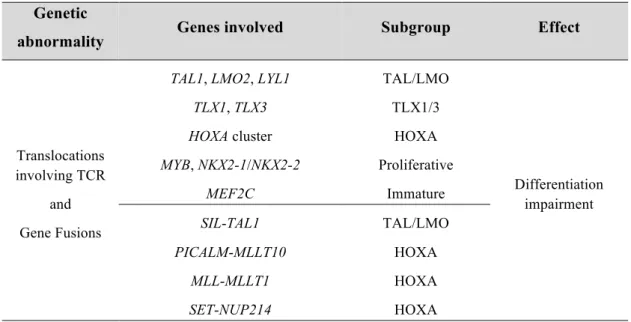
Although T-ALL presents often with a normal diploid karyotype, numerical and structural chromosomal abnormalities may also occur. Indeed, many studies have identified several structural chromosomal abnormalities that contribute to leukemogenesis in addition to other genetic alterations. These genetic abnormalities are variably present and were classified in type A and type B (Meijerink, 2010; Van Vlierberghe et al., 2008). Type A abnormalities usually involve chromosomal translocations, large genomic deletions or amplifications that are essentially mutually exclusive and define T-ALL subgroups (Table 1.1). Chromosomal
translocations involving TCR gene rearrangements and gene fusions are a common consequence of physiologic TCR rearrangements that occur in normal T cell development. Frequently, these lead to ectopic expression of oncogenic transcription factors in developing thymocytes and differentiation arrest (Ferrando and Look, 2003; Ferrando et al., 2002; Reinherz et al., 1980). Therefore, type A abnormalities are generally considered driving chromosomal abnormalities or initiating events. Although the oncogenes involved are usually activated by chromosomal rearrangements, they can also be overexpressed by other means, such as activating deletions or other mutations, epigenetic mechanisms, or upstream regulatory molecules (Ferrando and Look, 2003; Ferrando et al., 2002; de Leval et al., 2009; Van Vlierberghe et al., 2008). T-ALL oncogenic transcription factors include basic helix-loop-helix (bHLH) family members such as TAL1/SCL, TAL2, LYL1; LIM-only domain (LMO) genes like LMO1 and LMO2; homeobox family members including TLX1/HOX11, TLX3/HOX11L2, HOXA, and NKX2-1; and also MYB proto-oncogene and MEF2C (Homminga et al., 2011; Van Vlierberghe et al., 2008). Frequently, chromosomal translocations or inversions result in gene fusions that are expressed as constitutively activated chimeric oncoproteins (e.g., SIL-TAL1, PICALM-MLLT10, and MLL-MLLT1) (Van Vlierberghe et al., 2008).
Table 1.1. Common type A genetic abnormalities detected in T-ALL. Genetic
abnormality Genes involved Subgroup Effect
Translocations involving TCR
and Gene Fusions
TAL1, LMO2, LYL1 TLX1, TLX3 HOXA cluster MYB, NKX2-1/NKX2-2 MEF2C TAL/LMO TLX1/3 HOXA Proliferative Immature Differentiation impairment SIL-TAL1 PICALM-MLLT10 MLL-MLLT1 SET-NUP214 TAL/LMO HOXA HOXA HOXA
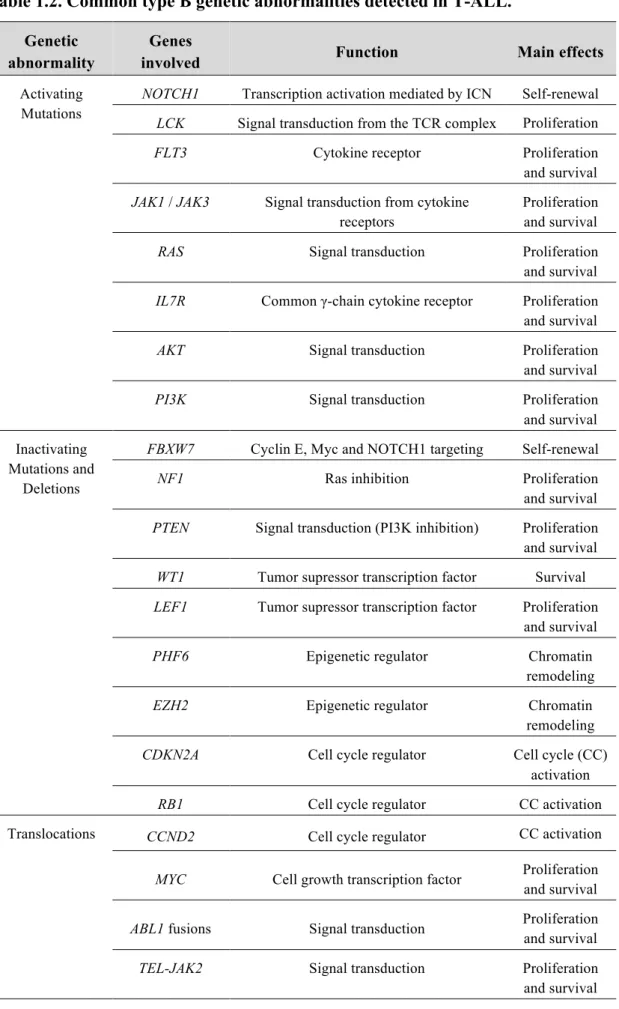
Type B abnormalities include gene-specific small deletions, translocations, duplications and point mutations in proto-oncogenes or tumor suppressor genes (Table 1.2). These are found in different combinations throughout all major T-ALL subgroups and seem
to synergize with type A abnormalities during T-cell leukemogenesis. Several genes encoding members of signaling pathways are affected by activating point mutations and include RAS, JAK1, JAK3, FLT3, IL7R (Zhang et al., 2012), PI3K, and AKT (Gutierrez et al., 2009). Similarly, and besides being involved in low frequency T-ALL translocations, the
NOTCH1 proto-oncogene can also carry activating mutations, as reported for more than 50%
of T-ALL patients (Palomero et al., 2007; Weng et al., 2004). The Notch signaling pathway is also activated in T-ALL due to inactivating point mutations in its negative regulator
FBXW7 (Thompson et al., 2007). Other inactivating mutations or deletions occur in the Ras
inhibitor NF1 (Balgobind et al., 2008) and in the PI3K negative regulator PTEN (Gutierrez et al., 2009), leading to activation of Ras and PI3K/AKT/mTOR signaling pathways, respectively. Gene fusions like ETV6/TEL-JAK2 or BCR-ABL1 are translated as constitutively active kinases and lead to aberrant signaling activation (Carron et al., 2000; De Keersmaecker et al., 2005). In addition, deletions in CDKN2A cell cycle regulator lead to disruption of p53-induced apoptosis pathway (Hebert et al., 1994). Cell cycle defects are also originated due to inactivating deletions in RB1, which encodes the tumor suppressor retinoblastoma (Rb) protein, and to translocations in CCND2 that lead to the activation of cyclin-D2, and consequent inactivation of Rb (Van Vlierberghe and Ferrando, 2012). Tumor suppressor transcription factors have also been shown to be inactivated in T-ALL including WT1, LEF1, GATA3, RUNX1 or BCL11B (Gutierrez et al., 2010, 2011; Tosello et al., 2009; Zhang et al., 2012). Recent studies have also shown high frequency of somatic mutations in epigenetic regulators. Inactivating mutations in PHF6 and EZH2 leading to chromatin remodeling alterations were reported in addition to alterations in genes involved in DNA methylation/demethylation such as TET1 (Peirs et al., 2015). Finally, mutations affecting ribosomal proteins, which are essential components of the translational machinery, were also recently revealed (De Keersmaecker et al., 2013).
When reviewing the current list of oncogenes and tumor suppressor genes involved in T-ALL development, it becomes clear that these are linked to thymocyte differentiation and self-renewal or general cellular functions like proliferation and survival, cell cycle control, and DNA repair (De Keersmaecker et al., 2005).
Table 1.2. Common type B genetic abnormalities detected in T-ALL. Genetic
abnormality
Genes
involved Function Main effects
Activating Mutations
NOTCH1 Transcription activation mediated by ICN Self-renewal
LCK Signal transduction from the TCR complex Proliferation
FLT3 Cytokine receptor Proliferation
and survival
JAK1 / JAK3 Signal transduction from cytokine
receptors
Proliferation and survival
RAS Signal transduction Proliferation and survival
IL7R Common γ-chain cytokine receptor Proliferation
and survival
AKT Signal transduction Proliferation and survival
PI3K Signal transduction Proliferation
and survival Inactivating
Mutations and Deletions
FBXW7 Cyclin E, Myc and NOTCH1 targeting Self-renewal
NF1 Ras inhibition Proliferation
and survival
PTEN Signal transduction (PI3K inhibition) Proliferation
and survival
WT1 Tumor supressor transcription factor Survival
LEF1 Tumor supressor transcription factor Proliferation
and survival
PHF6 Epigenetic regulator Chromatin
remodeling
EZH2 Epigenetic regulator Chromatin
remodeling
CDKN2A Cell cycle regulator Cell cycle (CC)
activation
RB1 Cell cycle regulator CC activation Translocations CCND2 Cell cycle regulator CC activation
MYC Cell growth transcription factor Proliferation and survival
ABL1 fusions Signal transduction Proliferation
and survival
TEL-JAK2 Signal transduction Proliferation
1.2.2. Microenvironmental factors
Although the roles of genetic lesions and epigenetic alterations as steps toward T-ALL development have been largely appreciated, the various aspects on the impact of the microenvironment have been mostly overlooked. It is now recognized that cancers develop in complex microenvironments composed of a dynamic and interactive mixture of different cell types and cytokines plus growth factors, which may sustain cancer-cell growth, invasion, and metastization (Tlsty and Coussens, 2006). In lymphoid cancers, the microenvironment often consists of variable numbers of malignant cells intermixed in a matrix of the so-called stromal cells (e.g., endothelial and epithelial cells, fibroblasts, DCs, macrophages), and also often non-malignant lymphocyte infiltration (Herreros et al., 2008). In fact, tumor cells and the surrounding stroma evolve together through continuous communication through paracrine and/or juxtacrine signaling, which culminates in the creation of permissive or selective microenvironments that support malignant cells to survive, grow, and resist to immune recognition and elimination. An altered microenvironment may disrupt the homeostasis and favor the early steps of transformation, or even promote protection against therapeutic intervention (Raaijmakers, 2010; Zhang et al., 2012). In this context, the malignant cells may respond passively to physiological signals or even become unresponsive to deleterious signals provided by their local microenvironment. Alternatively, cancer cells may more actively modulate stromal cells to change their gene expression profile and thus create more appropriate microenvironments for cancer cell growth and survival (Hanahan and Weinberg, 2000). Importantly, there is reliable evidence that neoplastic cells may interact with the surrounding environment in a bidirectional manner resulting in the acquisition of a competitive advantage by the malignant cells during oncogenesis (Colmone et al., 2008; Scupoli et al., 2003, 2007).
Notably, the thymus, an organ specialized in T cell development, contains a dynamic microenvironment with an high concentration of growth factors, cytokines and stromal cells necessary for thymopoiesis, making it also a potential permissive location for leukemogenesis and tumor progression. Normal thymocytes arise in the thymus where they interact with thymic stromal cells (TSCs) and thus promote T-cell development process and to maintenance of the stromal cell pool and its three-dimensional organization (Boehm et al., 2003). Normal thymocytes divide, differentiate and egress the thymus whereas leukemic T cells undergo a differentiation block and continue to grow possibly under the support of TSCs until some of them eventually acquire the capacity of microenvironment-independent
growth (Hiai et al., 1981). The absolute requirement for the presence of the thymus in murine leukemogenesis is illustrated by results showing that thymectomy impairs T-cell leukemia development induced by γ-irradiation (Kaplan, 1950), murine leukemia viruses (McEndy et al., 1944) or Ikaros protein deficiency (Dumortier et al., 2006). Moreover, a recent study has shown that lack of competition between bone marrow-derived progenitors and T-cell precursors developing in the thymus causes T-cell leukemia that shares many properties with human T-ALL (Martins et al., 2014). Cell competition was thus considered a tumor suppressor mechanism because it acts to eliminate older precursors in the thymus that may display upregulated or mutated genes responsible for stemness and thus become genomically unstable during the ageing process, favoring transformation. Also, the age-related gradual loss of thymic structure and function (termed involution), possibly in combination with other factors, was shown to impact on T-ALL development. In a study, each T-ALL subtype was shown to correlate closely with the stage of thymocyte maturation arrest regardless of age, but the incidence of the different subtypes differed markedly (Asnafi et al., 2004), probably reflecting age-related alterations in the thymocytes at risk of oncogenic transformation, alterations in stroma composition, or variable latency. In addition, a recent genome-wide sequencing study reported a correlation between age and number of somatic mutations in T-ALL, showing that particular genes are preferentially affected in adults versus children (De Keersmaecker et al., 2013).
In addition to their role in normal T-cell development, the establishment of adhesive contacts and the release of cytokines have crucial roles in regulating growth and survival of leukemic cells within the microenvironments where T-ALL develops (Balkwill, 2004). For instance, stromal cells expressing ICAM-1 were reported to favor the survival of T-ALL cells expressing lymphocyte function-associated antigen 1 (LFA-1) integrin (Winter et al., 2001). Likewise, chemokines produced by the bone marrow, thymic stromal cells, and other organs are important signaling molecules involved in T-ALL survival, proliferation, but mostly infiltration. For example, T-ALL cells with activated Notch signaling were shown to express CCR7, which promoted leukemic cell migration to CCL19-expressing CNS and infiltration (Buonamici et al., 2009; Ma et al., 2014). Moreover, the chemokine receptors CXCR4 and CCR9 expressed by leukemic cells were shown to mediate extramedullary organ infiltration in response to their ligands (Crazzolara et al., 2001; Qiuping et al., 2004). In addition, CCL25 to CCR9 signaling was reported to mediate T-ALL resistance to apoptosis and proliferation (Qiuping et al., 2004). In another study, CCR9 expression was shown to be induced by
NOTCH1 in T-ALL cells and to regulate proliferation and chemotaxis towards its ligand (Mirandola et al., 2012).
Additional reports have provided experimental evidence implicating stromal cell-induced activation of signaling in T cells during T-ALL leukemogenesis. Thymic and BM epithelial cells have been shown to promote T-ALL cell survival and proliferation in vitro by producing interleukin (IL)-7 (Scupoli et al., 2003, 2007). IL-7 was also reported to be involved in T-ALL progression and dissemination in vivo (Silva et al., 2011). Another cytokine, IL-18 produced by BM-derived stromal cells in inflammatory microenvironments, was also shown to support T-ALL progression in vivo probably due to enhanced proliferation (Uzan et al., 2014).
Cytokines and growth factors provided by stromal cells exert a protective effect not only on the bulk of the leukemic cell population, but also on leukemia initiating cells (LICs). LICs constitute a rare population of leukemic cells that are enriched in its ability to initiate leukemia in serial transplantation in immunodeficient mice (Cox et al., 2007). In T-ALL, Notch signaling activation induced by stromal cells is required for LIC activity mainly through the maintenance of self-renewal and engraftment abilities of these cells (Armstrong et al., 2009). Moreover, NOTCH1-induced high-level insulin-like growth factor (IGF)-1 receptor (IGF1R) expression was proved to be required for LIC activity in T-ALL. Moreover, inhibition of IGF1R signaling in response to IGF1/2 in the microenvironment halts T-ALL cell growth, survival, and interferes with disease establishment and progression (Medyouf et al., 2011).
In addition, sustained calcineurin activity in leukemic cells has been detected in mouse models of T-ALL induced by TEL-JAK2 or ICN1. In vitro culture of leukemic cells in the absence of stromal cells showed that calcineurin activity was rapidly lost, suggesting that specific signals from the tumor microenvironment may be essential for its maintenance (Medyouf et al., 2007). A recent study has also shown that calcineurin activation in T-ALL cells was important for survival, proliferation and motility in ex vivo studies. In vivo, it was essential to promote LIC activity but the stromal-derived factors responsible for its activation and consequent effects remain unknown (Gachet et al., 2013).
Notch signaling induction in leukemic cells was also reported to occur by microenvironmental elements. For instance, the escape of human T-ALL cells from dormancy to acquire a tumorigenic phenotype was associated with DLL4 expression by endothelial cells in the tumor microenvironment and increased NOTCH3 signaling in human T-ALL cells. Moreover, neutralization of DLL4 greatly reduced endothelial-cell mediated