Universidade do Algarve
Faculdade de Ciências e Tecnologia (FCT)
Master Thesis Report
Screening for biological activities with application in
aquaculture in halophytes from the Algarve coastline
Screening for biological activities with application in aquaculture in
halophytes from the Algarve coastline
Declaração de autoria de trabalho:
Declaro ser o autor deste trabalho, que é original e inédito. Autores e trabalhos consultados estão devidamente citados no texto e constam na listagem de referências incluída.
Copyrigth © 2013, por
A Universidade do Algarve tem o direito, perpétuo e sem limites geográficos, de arquivar e publicar este trabalho através de exemplares impressos reproduzidos em papel ou de forma digital, ou por qualquer outro meio conhecido ou que venha a ser inventado, de o divulgar através de repositórios científicos e de admitir a sua copia e distribuição com objectivos educacionais ou de investigação, não comerciais, desde que seja dado crédito ao autor e editor.
Index
List of abbreviations and acronyms………...4
Figures and tables Index………5
Resumo………..7
Abstract.………
….………8
1.Introduction.……….………
…..…………10
1.1 Aquaculture and infectious diseases……….……...………..10
1.2 Use of Bioactive compounds in aquaculture……….…….…………...13
1.3 Halophyte Species……….16
1.4 Objectives………..23
2. Materials and methods……….23
2.1 Sample Collection……….23
2.2 Preparation of the extracts……….23
2.2.1 Organic
extracts……….23
2.2.2 Water based plant extracts
……….……24
2.3 Testing of antibiotic activity………
….….24
2.3.1 Pathogen collection, culture and growth curve determination
…..………24
2.6 Data presentation and statistic treatment………...………30
3. Results and Discussion………...……….30
3.1 Antibiotic activity………..………30
3.2 Testing of antioxidant activity………..
.…35
3.2.1 Antioxidant activity screening by DPPH free radical…………...….35
3.2.2 Metal chelating activity for copper………...…
..39
3.2.3 Metal chelating activity for Iron……….43
3.3 Testing of immunostimulant activity………
.…………46
3.3.1 Determination of respiratory burst activity by cytochrome c and
Griess assays………46
4. Conclusion………...………49
5. Acknowledgements……….………49
6. References...………50
List of abbreviations and acronyms
BHT Butylated hydroxytoluene
CFU Colony forming units
DMSO Dimethyl sulfoxide
DPPH 2,2-Diphenyl-1-picrylhydrazyl
EDTA Ethylenediaminetetraacetic acid
FBS Fetal bovine serum
HBSS Hank's buffered salt solution
IC50 Half maximal inhibitory concentration
PMA Phorbol myristate acetate
RPMI
Cell culture media developed at Roswell Park Memorial Institute
Figures and tables Index
Fig. 1. General view of featured halophytes and their reproductive morphology. (A) Glaucous glasswort Arthrocnemum macrostachyum. (B) A. macrostachyum inflorescence. (C) Mediterranean saltbush Atriplex halimus. (D) A. halimus inflorescence. (E) Sour fig Carpobrotus edulis. (F) C. edulis
flower
………
17Fig. 2. General view of featured halophytes and their reproductive morphology (part two). (A) Spiny rush Juncus acutus. (B) J. acutus inflorescence. (C) buck's horn plantain Plantago coronopus. (D) P. coronopus inflorescence. ………...18 Fig. 3. Disposition of paper disks with halophyte extracts, antibiotics, paper and solvent controls, inside the marine agar Petri dishes. (A) A. macrostachyum, (B) A. halimus, (C) C. edulis, (D) J. acutus, (E) P. coronopus, (F) ampiciline, (G) tetracycline , (H) DMSO,
(I) HBSS and (J) paper
……….……….
26Fig. 4. Scheme of sample 96-well plate used for the antioxidant and metal chelating IC50 determination of water based halophyte extracts. A represents trial wells. B
represents colour controls.
……….
27Fig. 5. Marine agar Petri dish with L. anguillarum, control plate after incubation. A) ampiciline, B) tetracycline, C) HBSS, D) filter paper.
………
...….…
31Fig. 6. Marine agar Petri dish with P. d. piscicida, control plate after incubation. A) ampiciline, B) tetracycline, C) HBSS, D) filter paper.
……….…
……32 Fig. 7. Iridoid glycosides present in Plantago spp. (A) aucubin, (B) catalpol and (C)plantarenaloside
………...
34Fig. 8. Antioxidant activity of 1, 5 and 10 mg/mL water based halophyte extracts. The data represents means of 6 trial wells in a multichamber plate. The letter * denotes
statistically significant differences (p < 0.05 ) when compared with negative control
wells
………
..
38Fig. 9. Copper chelating activity of 1, 5 and 10 mg/mL water based halophyte extracts. The data represents means of 6 trial wells in a multichamber plate. The letter * denotes statistically significant differences (p < 0.05 ) when compared with negative control
wells
………..
42Fig. 10. Iron chelating activity of 1, 5 and 10 mg/mL water based halophyte extracts. The data represents means of 6 trial wells in a multichamber plate. The letter * denotes statistically significant differences (p < 0.05 ) when compared with negative control
wells
………..
45Fig 11. -Respiratory burst activity (O.D. 550 nm) of sea bream macrophages (individual fish head-kidney cells kept separated) after incubation for 3 hours in 10mg/ml halophyte extracts. The data are means of 5 different individuals
……….………...
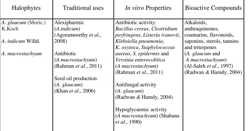
48 Table 1. Traditional uses, in vitro properties and bioactive compounds of medicinal glassworts (Arthrocnemum spp.)……….………...….
19 Table 2. Traditional uses, in vitro properties and bioactive compounds of medicinal saltbushes (Atriplex spp.)
………...…
20Table 3. Traditional uses, in vitro properties and bioactive compounds of medicinal ice plants (Carpobrotus spp.) and spiny rushes (Juncus spp.)
………...…
21Table 6. Inhibition halos of Photobacterium damselae piscicida, caused by antibiotics (positive controls), solvent and paper controls (negative controls)………...…...
….…
32Table 7. Antioxidant activity IC50 values of water based halophyte extracts…
….…
.39Table 8. Copper chelating activity IC50 values of water based halophyte extracts
…
..43Table 9. Iron chelating activity IC50 values of water based halophyte extracts
…
…..
.46Resumo
A produção de organismos aquáticos em sistemas de aquacultura é frequentemente afectada pelo surgimento de doenças infecciosas, sendo a causa de perdas económicas, problemas ambientais e colocando em risco a segurança do consumidor. A forma convencional de controlo de agentes patogénicos era por meio de antibióticos e compostos sintéticos. Este procedimento teve no entanto consequências sob a forma de agentes patogénicos mais resistentes, assim como a contaminação química dos produtos de aquacultura e dos ecossistemas locais. Para evitar os efeitos nefastos do uso de antibióticos, estes foram grandemente substituídos pela vacinação em aquaculturas de países ocidentais. Existe no entanto ainda um uso massivo de antibióticos em sistemas de produção do terceiro mundo, pelo que são necessárias alternativas terapêuticas de baixo custo. Uma forma alternativa de conseguir o controlo de doenças em aquacultura, é pelo uso de compostos orgânicos bioactivos com propriedades antibióticas, antioxidantes e imuno-estimulantes. Estas biomoléculas existem naturalmente em bactérias, algas, fungos, plantas superiores e outros organismos. Ácidos gordos, nucleótidos, monossacarídeos, polissacarídeos, peptídeos, polifenóis e terpenóides são exemplos destas substâncias. Uma fonte promissora de compostos bioactivos são as halófitas ou plantas tolerantes ao sal. Em comparação com outras traqueófitas, as halófitas têm mais defesas moleculares, de forma a lidarem com o stress oxidativo do seu habitat. Muitas halófitas foram tradicionalmente usadas como um recurso alimentar e medicinal por culturas africanas e asiáticas. Este trabalho cientifico avaliou as
propriedades antibióticas, antioxidantes, imuno-estimulantes e quelantes de metais de: Atriplex halimus L., Arthrocnemum macrostachyum Moric., Carpobrotus edulis L., Juncus acutus L. e Plantago coronopus L., provenientes da costa Algarvia. As propriedades antibióticas foram testadas contra: Listonella anguillarum, Photobacterium damselae piscicida e Vibrio fischeri. As propriedades imuno-estimulantes foram testadas com ensaios de cytochrome c e Griess em fagócitos provenientes de Sparus aurata. O extrato de éter de J. acutus inibiu o crescimento de P. damselae piscicida. A. halimus, A. macrostachyum, C. edulis, J. acutus e P. coronopus exibiram propriedades antioxidantes e quelantes para cobre e ferro. Estas halófitas mostram potencial como fontes de compostos bioactivos com aplicação em aquacultura e outras áreas.
Palavras chave: Halófita, Patógenos em aquacultura, Extrato de plantas, Imuno-estimulantes, Vibrio.
Abstract
Infectious diseases often hamper the production of aquatic organisms in aquaculture systems, causing economical losses, environmental problems and consumer safety issues. The conventional way aquaculture producers had to control pathogens was by means of synthetic antibiotics and chemicals. This procedure had consequences in the emergence of more resilient pathogens, drug contamination of seafood products and local ecosystems. To avoid the repercussions of antibiotic use, vaccination has greatly replaced human drugs in western fish farms. However there is still massive unregulated antibiotic use in third world fish farms, so less expensive therapeutic alternatives for
halophytes have been used as a traditional food and medical supply, especially by African and Asian cultures. This scientific work evaluated the antibiotic, antioxidant, immunostimulant and metal chelating properties of Atriplex halimus L., Arthrocnemum macrostachyum Moric., Carpobrotus edulis L., Juncus acutus L. and Plantago coronopus L., from the Algarve coast. The antibiotic properties were tested against Listonella anguillarum, Photobacterium damselae piscicida and Vibrio fischeri. The immunostimulant properties were tested with cytochrome c and Griess assays on Sparus aurata head-kidney phagocytes. J. acutus ether extract inhibited the growth of P. damselae piscicida. A. macrostachyum, A. halimus, C. edulis, Juncus acutus and P. coronopus displayed antioxidant, copper chelating and iron chelating properties. These plants show potential as sources of bioactive compounds with application in aquaculture and in other fields.
1. Introduction
1.1 Aquaculture and infectious diseases
Aquaculture has greatly increased its relevance as colocar resumo em portuges a source of nourishment to satisfy the ever-increasing demand of the world food markets, displaying a fourfold growth in the last twenty years (Naylor et al., 2000). Despite the fact that it reduces pressure on wild stocks and provides aquatic products year round, aquaculture causes environmental problems, which have yet to be solved in a great extent. Examples of ecosystem damage caused by fish farming ranges from nutrient excess in surrounding waters (Boyd & Massaut, 1999) to genetic contamination of wild populations (Crozier, 1993; McGinnity et al., 2003). The need to exploit low commercial value fisheries to use as feed in aquaculture of top predatory species has also attracted criticism (Tidwell & Geoff, 2001).
A major issue facing aquaculture nowadays, present as well in industrial farming, is animal disease control and prevention. The high densities of stock kept in modern aquaculture operations and resulting waste products create a favourable medium for pathogen proliferation and transmission. Because of the stressful conditions to which they are exposed, the immune system of farmed animals is often compromised, further exposing them to infection. High stocking densities, handling, transport and low water quality, are major immunosuppressants (Shoemaker, Evans & Klesius, 2000; Saurabh & Sahoo, 2008). This is particularly relevant in fish and crustaceans. However, opportunistic pathogens can affect cultivated sessile molluscs as well (Ford et al., 2002).
Infectious diseases are the largest single cause of economic loss in all kinds of aquaculture. Gram-negative bacteria of the genera Aeromonas, Listonella,
infections are widely spread in all forms of marine and freshwater aquaculture as well. This is a matter of great concern for the industry of aquaculture because of the associated economical losses, the environmental impacts on local ecosystems, and human consumption safety issues.
Several of these pathogens such as Aeromonas spp. (Gracey, Burke & Robinson, 1982), Mycobacterium marinum (Ghittino et al., 2003), Streptococcus spp. (Appelbaum, 1992) and Vibrio spp. (Blake et al. 1979) can cause potential zoonosis, leading to serious public health questions over the safety of aquaculture products. Diseased stock in an aquaculture facility can have dire consequences for local marine ecosystems as well. Water borne pathogens can be transmitted to wild populations over a wide range of species, and not only to species directly related to the farmed stock. For example, the bacterium Aeromonas salmonicida largely affects aquaculture Salmonids, but it is capable of infecting many other fish groups (Bullock & Stuckey, 1975; Austin, 1993). In addition of captive animals and local wild populations sharing the same waters (except in facilities that are totally isolated like those inland) the problem of disease transmission is increased by escaped livestock. Disease-carrying feral fish have already caused serious declines or even the extinction of isolated wild populations that had little defence against pathogenic strains to which they were never exposed (Heggberget et al., 1993). Under some circumstances, organisms are bred or raised not only for human consumption but also as head-start to boost wild populations. In this case, disease prevention is very important, as testing for the presence of pathogens in apparently and otherwise healthy stock is not completely flawless (Thorburn, 1996).
One of the ways aquaculture producers have to avoid and fight pathogens is by means of antibiotics and chemicals. Massive use of antibiotics was common practice in all forms of fish aquaculture both as a prophylactic and to treat infection. Their use was viewed as a more economic alternative than reducing stock densities and applying additional hygienic measures (Grave, Markestad & Bangen, 1996). However, the unregulated drug use had many harmful effects. Antibiotics, and other compounds used to treat disease, evade the aquaculture confinement via uneaten food, animal waste and lost stock. Commonly used antibiotics like quinolones are not easily biodegradable, remaining active for long periods of time in the sediment or being transported by currents to distant locations (Hansen, Lunestad & Samuelsen; 1992; Coyne, Hiney & Smith, 1997). Also it has been shown that residual amounts of antibiotics, metals and other compounds used to treat infectious diseases in livestock, remain in the tissues of
commercialised fish and invertebrates (Angulo, Nargund & Chiller, 2004; Sapkota et al., 2008).
The selective pressure of antibiotics in fish tissue and in the surroundings of a fish farm has resulted in more virulent strains of bacteria, able to resist most antibiotics. Ever increasing doses and new drugs are needed to keep pathogens in check. Some of the metabolic weaponry acquired by these bacteria has the potential of being transmitted by horizontal gene transfer to terrestrial ecosystems, hypothetically reaching human pathogens (Kruse & Sørum, 1994; Rhodes et al., 2000; Tendencia & De La Peña, 2001; Chelossi et al., 2003). Also as aforementioned, several common pathogenic agents in aquaculture are zoonosis, creating a more direct route for human infection by antibiotic resistant bacteria (Ghittino et al., 2003). Human groups especially at risk from the environmental contaminants and antibiotic resistant pathogen carriers include aquaculture workers, populations living near fish farms and consumers of aquaculture products (Sapkota et al., 2008).
Because of the danger that their use represents to aquaculture, western world fish farms have largely replaced antibiotics by vaccination (Alderman & Hastings, 1998). Any synthetic compound to be used in the treatment of fish destined for human consumption must be on an approved substance list. Also quinolones and several other human prescription drugs have been outlawed for their use in aquaculture or are only available by veterinary order (Grave, Markestad. & Bangen, 1996). However in third world countries, witch contribute to over 90% of the global production, chemicals like chlorine are widely used in all forms of mainstream aquaculture. These compounds are frequently used both as a way to treat infections and as prophylactics (Bakken, 2004; Bondad-Reantaso et al., 2005). It is thus crucial to find alternative ways to control disease in fish farms that do not employ the use of artificial drugs and chemicals. Possible solutions must also be readily available at a low cost; otherwise it is unlikely
1.2 Use of Bioactive compounds in aquaculture
Another recent pathway by which the health of aquaculture livestock can be maintained without man-made drugs is by using biologically active organic compounds, normally present in bacteria, algae, fungi, higher plants and other organisms. Fatty acids, nucleotides, monosaccharides, polysaccharides, peptides, polyphenols and terpenoids, are among the groups of biomolecules with interest for aquaculture. These substances may exhibit antibiotic, antioxidant and immunostimulant properties, and are easily included in specifically prepared diets or bioencapsulated in live food. Beneficial effects of incorporating these compounds in animal feed include enhanced growth, immunity, osmoregulation, stress tolerance in addition to larvae survival and quality. Livestock fed diets supplemented with bioactive substances also display, enhanced resistance towards bacterial, fungal, pseudo-fungal, parasitic and viral infections (Waagbo et al. 1993; Roberts, Davies & Pulsford, 1995; Burrells et al., 2001; Esteban et al. 2001; Chotigeat et al., 2004; Skjermo & Bergh, 2004; Bricknell & Dalmo, 2005; Kumar et al., 2005; Cheng et al., 2007; Dubber & Harder, 2008; Harikrishnan et al., 2009; Zhang et al., 2009).
Besides avoiding man made chemicals, the use of biologically active compounds in animal diets could have additional health benefits for the final consumer of aquaculture products. A major example is cold-water fish enriched with marine microalgal omega-3 fatty acids, which are higly beneficial to human health (Kris-Etherton, Harris & Appel, 2003, Ward & Singh, 2005).
Immunostimulating compounds positively modulate innate and adaptative immune responses in aquaculture fish. These substances trigger humoral and cellular defence mechanisms by acting on receptors and intercellulargene activation. Desirable effects on innate fish immunity include enhanced cytotoxic, phagocytic, respiratory burst, lysozyme and natural haemolytic complement, activities. These innate immune responses serve as first line of deffence against pathogens and keep them in check until an efficient acquired immune response has been developed, leading to an optimised capacity to fight infectious diseases (Esteban et al. 2001; Dügenci, Arda & Candan, 2003; Skjermo & Bergh, 2004; Bricknell & Dalmo, 2005; Cheng et al., 2007; Saurabh & Sahoo, 2008; Zhang et al., 2009). Some compounds appear to significantly enhance the efficacy of vaccination (Burrells et al., 2001; Saurabh & Sahoo, 2008).
Immunostimulating compounds can play an important role in reinforcing the immune systems of farmed animals in industrial aquacultures as prophylactics during high stress periods such as transport, vaccination, sea transfer and reproduction (Bricknell & Dalmo, 2005).
It has been suggested that natural antioxidant compounds protect living tissues and act as a buffer against oxidative stresses (environmental toxins, pollutants, stress by handling and overcrowding). Some organic compounds like L-ascorbic acid have dual properties, functioning both as a natural antioxidant and immunostimulant (Waagbo et al. 1993; Roberts, Davies & Pulsford, 1995; Chotigeat et al., 2004; Rau et al., 2004). Related to antioxidant activity, some biomolecules exhibit desirable metal-chelating properties. These compounds bind to harmful metals in living tissues and, allow them to be excreted or, diminish their negative impact on organisms. In practice metal-chelating compounds could be used to detoxify fish (Cuajungco & Lees, 1998, Ebrahimzadeh, Pourmorad & Bekhradnia, 2008). Further research will most likely lead to new bioactive compounds with desirable properties.
The search for bioactive substances with interest for aquaculture does not have to be restricted to marine organisms. Many species of terrestrial vascular plants (Tracheophytes) produce compounds that can act as natural antioxidants, antibiotics and immunoestimulants for human and animal use. A great number of these plants have been used during millennia to treat human ailments (Kapil & Sharma, 1997; Nascimento et al., 2000; Choudhary et al., 2007). Terrestrial plant compounds have already showed anti-fungal and immunostimulant properties in aquaculture fish (Harikrishnan et al., 2009; Zhang et al., 2009).
Salt tolerant and salt requiring plants show signs of being a good source of new compounds with applications in aquaculture and other fields. Halophytes are a polyphyletic functional group that displays biochemical, physiological and
and biochemical levels (Munns, 2002; Munns, James & Lauchli, 2006). These conditions promote the development of unusually high levels of reactive oxygen species (ROS) during otherwise normal cellular activities such as photorespiration and β-oxidation of fatty acids (Parida & Das, 2005). ROS have damaging oxidative effects on macromolecules such as proteins and nucleic acids and may cause peroxidation of membrane lipids. To avoid the harmful effects of oxidative stress, halophytes rely on protective bio-active compounds and physiological adaptations (such as salt exclusion, excretion, succulence, transport and compartmentalization and osmotic adjustment). Organic compounds present in salt tolerant plants include natural antioxidants (ascorbic acid, carotenoids, glutathione, methylated metabolites, phenols, polyphenols, thiols and thioredoxin) and ROS scavenging enzymes (ascorbate peroxidase, catalase, glutathione peroxidase and superoxide dismutase). These biomolecules are present in larger amounts in halophytes when compared to most land plants and their capacity to detoxify radicals under conditions of salt stress is an important stress-tolerance adaptation (Tsugane et al. 1999; Ghanem et al., 2010). Related with their origin in different higher plant families, halophytes differ in their degree of salt tolerance. Plants with an increased tolerance to sallinity (such as Salicornia bigelovii Torr. (Chenopodiaceae), wich is able to grow at twice seawater salinity) exibit a higher ROS scavenging activity (Flowers, Troke & Yeo, 1977; Bowler, Van Montagu & Inze, 1992). Halophytes also possess osmotically active compounds (such as saccharides, polyhydroxyalcohols, organic acids, amino acids, betaines, and tertiary sulfonium substances) that decrease the osmotic potential, protect biomolecules against the damaging effects of salinity and mantaining homeostasis (Arabawa & Timasheff, 1985; Gagneul et al., 2007; Ghanem et al., 2010). Any of these biomolecules has potential for its use in aquaculture as natural antioxidants, antibiotics, immunostimulants and even for their nutritional value. They also could hold future interest for biomedical sciences.
1.3 Halophyte Species
The xero-halophytes Arthrocnemum macrostachyum Moric. (glaucous glasswort), Atriplex halimus L. (Mediterranean saltbush) and Carpobrotus edulis L. (sour fig or Hottentot’s fig), and the halophytes Juncus acutus L. (spiny rush) and Plantago coronopus L. (buck's horn plantain), can be considered potential sources of novel bioactive molecules with applicability for human medicine and aquaculture fields (Figs. 1. and 2.).
A. macrostachyum and A. halimus (Amaranthaceae) are native Circum-Mediterranean species (Al-Saleh et al., 1997, Andueza et al., 2005). C. edulis (Aizoaceae) is a South African species, now established in several Mediterranean climate countries, namely coastal California, Western Australia and Mediterranean Europe countries, including the coast of Portugal where is considered invasive (Collins & Scott, 1982; Draper et al., 2003; Moragues & Traveset, 2005). J. acutus (Juncaceae) is a cosmopolitan species, native to most Palearctic (Kirschner et al., 1999). Originally a Eurasian, Middle East and North African species, P. coronopus (Plantaginaceae) is now cosmopolitan as well (Braza, Arroyo & Garcia, 2010). In Portugal these species are found mainly in coastal and salt marsh habitats.
In their native ranges A. macrostachyum, A. halimus, C. edulis, J. acutus and P. coronopus are used for traditional phytomedicine purposes. Traditional African (C. edulis), Chinese (J. acutus and P. coronopus), Midle-East and Morrocan medicin (A. macrostachyum, A. halimus, J. acutus and P. coronopus) have adopted these halophytes to treat a wide range of medical conditions. These include the treatment of gastrointestinal, hepatic, oral, respiratory, tumoral, urinary tract and skin diseases (Van der Watt & Pretorius, 2001; Chiang et al., 2002, 2003; Chen & Chen, 2004; Springfield
Fig. 1. General view of featured halophytes and their reproductive morphology. (A) Glaucous
glasswort Arthrocnemum macrostachyum. (B) A. macrostachyum inflorescence. (C) Mediterranean saltbush Atriplex halimus. (D) A. halimus inflorescence. (E) Sour fig Carpobrotus edulis. (F) C. edulis flower.
properties (Table 1. to Table 4.) (Willis, 1980; McArthur & Sanderson, 1984; Kirschner et al., 1999; Springfield & Weitz, 2006; Saleh, 2011). Plantago major L. has been the subject of most studies as traditional Chinese medicine herb (Mouhajir et al., 2001).
In some cases the ancient knowledge on these and other medicinal plants, has been lost or limited to certain ethnicities (Batanouny, 2001). The therapeutic ethnobotanical use of these species has attracted the interest of biochemistry researchers, for the prospect of developing new drugs and therapies
Fig. 2. General view of featured halophytes and their reproductive morphology (part two). (A) Spiny rush Juncus acutus. (B) J. acutus inflorescence. (C) buck's horn plantain Plantago
Table 1. Traditional uses, in vitro properties and bioactive compounds of medicinal glassworts
(Arthrocnemum spp.).
Halophytes
Traditional uses
In vitro
Properties
Bioactive Compounds
A. glaucum (Moric.) K.Koch A. indicum Willd. A. macrostachyum Alexipharmic (A.indicum) (Agoramoorthy et al., 2008) Antibiotic (A macrostachyum) (Rahman et al., 2011) Seed oil production (A. glaucum)(Khan et al., 2006)
Antibiotic activity:
Bacillus cereus, Clostridium perfringens, Listeria ivanovii, Klebsiella pneumonia,
K. oxytoca, Staphylococcus aureus, S. epidermis and Yersinia enterocolitica (A macrostachyum) (Rahman et al., 2011) Antifungal activity (A. glaucum)
(Radwan & Hamdy, 2004) Hypoglycaemic activity (A macrostachyum) (Shabana et al., 1990) Alkaloids, anthraquinones, coumarins, flavonoids, saponins, sterols, tannins and triterpenes
(A. glaucumand A macrostachyum) (Al-Saleh et al., 1997) (Radwan & Hamdy, 2004)
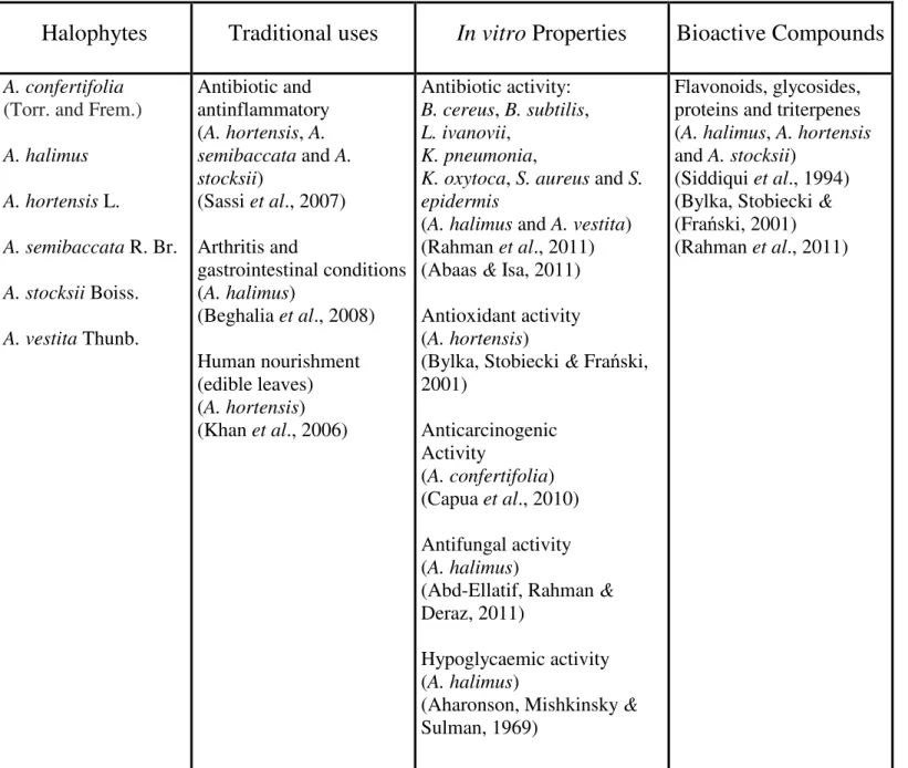
Table 2. Traditional uses, in vitro properties and bioactive compounds of medicinal salt bushes
(Atriplex spp.).
Halophytes
Traditional uses
In vitro
Properties
Bioactive Compounds
A. confertifolia(Torr. and Frem.) A. halimus A. hortensis L. A. semibaccata R. Br. A. stocksii Boiss. A. vestita Thunb. Antibiotic and antinflammatory (A. hortensis, A. semibaccata and A. stocksii) (Sassi et al., 2007) Arthritis and gastrointestinal conditions (A. halimus) (Beghalia et al., 2008) Human nourishment (edible leaves) (A. hortensis) (Khan et al., 2006) Antibiotic activity: B. cereus, B. subtilis, L. ivanovii, K. pneumonia,
K. oxytoca, S. aureus and S. epidermis
(A. halimus and A. vestita) (Rahman et al., 2011) (Abaas & Isa, 2011) Antioxidant activity (A. hortensis)
(Bylka, Stobiecki & Frański, 2001) Anticarcinogenic Activity (A. confertifolia) (Capua et al., 2010) Antifungal activity (A. halimus)
(Abd-Ellatif, Rahman & Deraz, 2011)
Hypoglycaemic activity (A. halimus)
(Aharonson, Mishkinsky & Sulman, 1969)
Flavonoids, glycosides, proteins and triterpenes (A. halimus, A. hortensis and A. stocksii)
(Siddiqui et al., 1994) (Bylka, Stobiecki & (Frański, 2001) (Rahman et al., 2011)
Table 3. Traditional uses, in vitro properties and bioactive compounds of medicinal ice plants
(Carpobrotus spp.) and spiny rushes (Juncus spp.).
Halophytes
Traditional uses
In vitro
Properties
Bioactive Compounds
C edulis C. quadrifidus (L.) L. Bolus C. mellei(L.) L. Bolus C. muirii(L.) L. Bolus Antibiotic and antinflammatory Treatment of gastrointestinal, oral, respiratory (tuberculosis) and skin conditions Human nourishment (edible fruit)(C edulis, C. quadrifidus, C. mellei and C. muirii) (Van der Watt & Pretorius, 2001)
(Springfield et al., 2003) (Springfield & Weitz, 2006)
(Buwa & Afolayan, 2009)
Antibiotic activity: B. cereus, B. subtilis, Moraxella catarrhalis, Mycobacterium aurum, M. tuberculosis, M. smegmatis, Pseudomonas aeruginosa, S. aureus, S. epidermis and Streptococcus pneumoniae (C edulis, C. quadrifidus, C. mellei and C. muirii)
(Van der Watt & Pretorius, 2001)
(Springfield et al., 2003) (Martins et al., 2005) (Springfield & Weitz, 2006) (Buwa & Afolayan, 2009)
Flavonoids, lipids, phenols, tannins, triterpenes, citric acid and malic acid (C edulis, C. quadrifidus and C. muirii)
(Van der Watt & Pretorius, 2001) (Springfield et al., 2003) (Martins et al., 2011) J. acutus J. rigidus Desf. Antinflammatory, diuretic (treatment of kidney and urinary tract conditions) and sedative (J. acutus and J. rigidus)
(Mouhajir et al., 2001) (Chen & Chen, 2004) (Khan et al., 2006) (Daoudi et al., 2008) Anti-eczematic activity (J. acutus) (Awaad, 2006) Immunostimulatory activity (J. acutus and J. rigidus) (Daoudi et al., 2008) Algicide activity (J. acutus) (Dellagreca et al., 2002) (Dellagreca et al., 2004) Dihydrodibenzoxepins, 9,10-dihydrophenanthrenes, phenanthrenes, phenolic glycosides and pyrenes (J. acutus)
(Dellagreca et al., 2002) (Dellagreca et al., 2004) (Dellagreca, Previtera & Zarrelli, 2005)
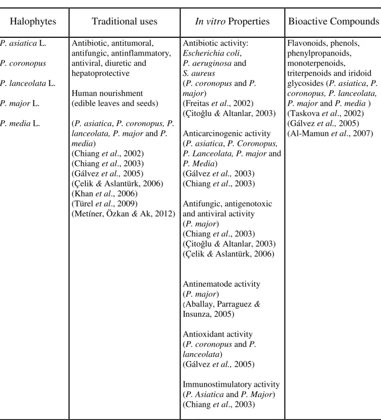
Table 4. Traditional uses, in vitro properties and bioactive compounds of medicinal plantains
(Plantago spp.).
Halophytes
Traditional uses
In vitro
Properties
Bioactive Compounds
P. asiatica L. P. coronopus P. lanceolata L. P. major L. P. media L. Antibiotic, antitumoral, antifungic, antinflammatory, antiviral, diuretic andhepatoprotective Human nourishment (edible leaves and seeds) (P. asiatica, P. coronopus, P. lanceolata, P. major and P. media)
(Chiang et al., 2002) (Chiang et al., 2003) (Gálvez et al., 2005) (Çelik & Aslantürk, 2006) (Khan et al., 2006)
(Türel et al., 2009)
(Metíner, Özkan & Ak, 2012)
Antibiotic activity: Escherichia coli, P. aeruginosa and S. aureus (P. coronopus and P. major) (Freitas et al., 2002) (Çitoğlu & Altanlar, 2003) Anticarcinogenic activity (P. asiatica, P. Coronopus, P. Lanceolata, P. major and P. Media)
(Gálvez et al., 2003) (Chiang et al., 2003) Antifungic, antigenotoxic and antiviral activity (P. major)
(Chiang et al., 2003) (Çitoğlu & Altanlar, 2003) (Çelik & Aslantürk, 2006) Antinematode activity (P. major)
(Aballay, Parraguez &
Insunza, 2005)
Flavonoids, phenols, phenylpropanoids, monoterpenoids,
triterpenoids and iridoid glycosides (P. asiatica, P. coronopus, P. lanceolata, P. major and P. media ) (Taskova et al., 2002) (Gálvez et al., 2005) (Al-Mamun et al., 2007)
1.4 Objectives
This work evaluated the potential of biomolecules of A. macrostachyum, A. halimus, C. edulis, J. acutus and P. coronopus, from the Algarve coast, to be used in aquaculture. Antibiotic, antioxidant (free radical scavenging and metal chelation) and immunostimulant properties were screened. The antibiotic properties were tested in vitro against 3 Gram-negative bacteria species: Listonella anguillarum, Vibrio fischeri (Vibrionaceae) and Photobacterium damselae piscicida (pasteurellosis). These pathogens were selected for their relevance as causative agents of morbility and mortality in fish farms. The immunostimulant properties were tested in vitro, using head-kidney phagocytes from a major aquaculture species, the gilthead seabream Sparus aurata L.
2. Materials and methods
2.1 Sample Collection
Aerial parts of A. macrostachyum, A. halimus, C. edulis, J. acutus and P. coronopus were collected on the Algarve coast (Faro Beach and Ludo, in May 2010), oven dried at 40 °C, milled and stored at - 20°C.
2.2 Preparation of the extracts
2.2.1 Organic extractsOrganic extracts were obtained by means of a sequential extraction procedure using solvents of increasing relative polarities. Aliquots (1 g) of milled samples were homogenized and sequentially extracted with hexane, ether, chloroform, methanol and water. The extracts were centrifuged (400 g, 15 min, RT), and the supernatant carefully removed. The forementioned procedure took place until a clear supernatant layer was
achieved in each individual sample. The extracts were evaporated (temp. < 40ºC), the dried extract was weighed and dissolved in dimethyl sulfoxide (DMSO), to obtain solutions in the final concentration of 500, 260, 100, 70 and 50 mg/mL.
2.2.2 Water based plant extracts
Milled samples (3 g) in Falcon tubes were homogenized and extracted with Hank's buffered salt solution (HBSS). The extracts were centrifuged (400 g, 15 min, RT), and the supernatant carefully removed. The forementioned procedure took place until a clear supernatant layer was achieved in each individual sample. The extracts were freeze-dried and dissolved in HBSS. (to obtain solutions with the final concentration of 50, 10, 5 and 1 mg/mL.
2.3 Testing of antibiotic activity
2.3.1 Pathogen collection, culture and growth curve determination
Samples of L. anguillarum, P. damselae piscicida and V. fischeri were obtained from Dr. Paulo Pedro (LAQ-University of Algarve and Dr. Teresa Baptista (Escola Superior de Turismo e Tecnologia do Mar de Peniche). The bacterial strains were inoculated on 4 mm thick culture medium (Marine Agar, Conda Laboratories) and salt enriched Tryptic Soy Agar (Himedia). In the Microbiology Laboratoy of CIMA (Center for Marine and Environmental Research), Petri dishes were innoculated to produce isolated colonies, using sterile swabs in a Biohazard laminar flow hood (Steril VBH C2 Biohazard
determine the period of exponential growth. New sub-cultures in fresh medium were done every 48 hours.
2.3.2 Antibiogram Testing
Antibiotic activity testing was made adapting the Kirby-Bauer disk diffusion method (Drew et al., 1972). The bacterial inoculum for each of the diffusion method tests was obtained from exponentially growing cultures of the selected bacteria, grown to a density of colony forming units (CFU) of approximately 1-2 x 10⁸ CFU/mL The corresponding optical density was established by previous monitoring of the same cutures both by optical density at 600nm and quantification of CFU from apropriate dilutions. Petri dishes, containing 25mL (4 mm thick) of the chosen agar medium, were inoculated using a sterile swab. Sterile paper disks (6 mm of diameter), were impregnated with 10 µL of plant extract, and dried for 15 minutes at room temperature. The disks were then put on the Petri dishes (Fig. 3) and the set was let to pre-diffuse for 15 minutes before incubation. All procedure took place in a laminar flow chamber. The Petri dishes with Listonella anguillarum and Photobacterium damselae piscicida were incubated at room temperature. Vibrio fischeri was incubated at 12 °C. After the incubation period, all Petri dishes were examined and the antibacterial activity evaluated on the basis of the presence and diameter (in millimetres) of an inhibition area surrounding the disks. Tested extracts included water based and organic fraction extracts (50 mg/mL). When low antibiotic activities were detected, disks containing higher extract concentration (70, 100, 260 and 500 mg/mL) were used. Paper disks containing or not 10 µL of HBSS, were used as negative controls. Disks impregnated with ampiciline and tetracycline (10µg of antibiotic per disk), were used as positive controls.
Fig. 3. Disposition of paper disks with halophyte extracts, antibiotics, paper and solvent controls, inside the marine agar Petri dishes. (A) A. macrostachyum, (B) A. halimus, (C) C. edulis, (D) J. acutus, (E) P. coronopus, (F) ampiciline, (G) tetracycline , (H) DMSO, (I) HBSS and (J) paper.
2.4 Testing of antioxidant activity
Antioxidant and metal-chelating activities of the water based extracts (10, 5 and 1 mg/mL) were tested in 6 replicates. Also for IC50 (half maximal inhibitory
concentration) determination 4 replicates of 1, 2, 3, 4, 5, 6, 7, 8, 9 and 10 mg/mL were tested.
After the procedure, the plate was left in the dark for 30 minutes. Absorbance was read at 517 nm (Biotek plate reader Synergy 4).
Fig. 4. Scheme of sample 96-well plate used for the antioxidant and metal chelating IC50 determination of water based halophyte extracts. A represents trial wells. B
represents colour controls.
2.4.2 Metal chelating activity for copper
Testing for metal chelating activity of Cu2+ was made according to the method described by Saiga, Tanabe & Nishimura, 2003. In a 96-well plate, 200 µL of sodium acetate buffer (50 mM, pH 6), 100 µL of copper sulphate (50 µg/mL) and 6 µL of pyrocatechol violet (40 mM in sodium acetate) were added to 30 µL of extract. In the negative and positive controls the extracts were replaced by 30 µL of, respectively, HBSS and EDTA (ethylenediaminetetraacetic acid at 1 mg/mL). For color controls, wells containing 30 µL of extract and 306 µL of sodium acetate buffer, were used. Absorbance was read at 632 nm.
2.4.3 Metal chelating activity for Iron
Testing for metal chelating activity of Fe2+ was made according to the method described by Ebrahimzadeh, Pourmorad & Bekhradnia, (2008). In a 96-well plate, 200 µL of destilled waterand 30 µL of FeCl2 (0.1 mg/mL),were added to 30 µL of extract. In the
negative and positive controls the extracts were replaced by 30 µL of, respectively, HBSS and EDTA. For colour controls, wells containing 30 µL of extract and 230 µL of destilled water were used. After 30 minutes, 12.5 µL of ferrozine were added to the set, except for the colour controls. Absorbance was read at 562 nm.
2.5 Testing of immunostimulant activity
The testing of immunostimulant activity was made using the water based extracts (10 mg/mL). Each extract was tested in triplicate using the following methods.
2.5.1 Fish collection
Gilthead seabream (Sparus aurata) with similar weights were collected from a local facility (Ramalhete Biological Station) and acclimatized to laboratory conditions for two weeks before the start of the experiment (the specimens were kindly provided by Dr. Jorge Dias, and acclimatized by Vera Rodriges and Helena Teixeira at the aquaculture laboratory in Gambelas, University of Algarve. Live specimens were slightly anaesthetized in a bucket of tank water containing 2-fenoxietanol (60 µg/L), and transported as stress-free as possible, to the local where head-kidney extraction took place. Specimens were killed by an overdose of 2-fenoxietanol before kidney extraction.
centrifuged (400g, 15 min, 15 °C). The culture medium was changed by L15 containing 0.1% FBS and the volume reduced to 1-2 mL. The cell suspension was again centrifuged (400 g, 5 min, 15 °C). The cells were re-suspended on L15 medium plus 5% FBS, pipetted to a 96-well plate (100 µL per well) and kept in the dark, at room temperature. The viable cell concentration was determined by the try-pan blue exclusion assay (Strober, 2001). After 4 hours the medium was replaced with fresh medium of equal composition and the cells verified for attachment. The cells were incubated for 24h after the extraction. The cells of different fish were kept separated or were combined, according to different authors (Tafalla & Novoa, 2000; Díaz-Rosales et al., 2007).
2.5.3 Determination of respiratory burst activity by Cytochrome c
The culture medium was removed and the cells were washed with HBSS. 50 µL of cytochrome c were added to the cells. Trial wells received 50 µL of water-based extracts. 50 µl of PMA (phorbol myristate acetate at 10 µg/ml) and PMA plus SOD (superoxide dismutase 300 U/ml), replaced the extracts respectively in the positive and negative controls. As cytochrome c exhibits photosensitivity, the whole procedure took place in under low light conditions. The absorbance was measured after 30 minutes at 550 nm, and the measurement was repeated every 30 minutes for 3 hours. A total of 9 fish were used, of which 4 had their head-kidney cells combined in 2 trials (two fish represented in each cell pool). The remaining fish had their cells treated separately in 5 different trials.
2.5.4 Determination of respiratory burst activity by Griess assays
The cell attachment medium was replaced by 100 µl of RPMI and 20 µl of extract. For negative controls, 100 µl of HBSS was used. After 30 minutes, 50 µl of each well was removed and added to 100 µl of Griess solution in a new 96-well plate. The set was let incubate for 20 minutes at room temperature and sheltered from the light. Absorbance was measured at 540 nm. A total of 9 fish were used, of witch 4 had their head-kidney cells combined in 2 trials (two fish in each cell pool). The remaining fish had their cells treated separatly in 5 different trials.
2.6 Data presentation and statistic treatment
The results are presented as mean±standard error of mean (SEM). Data was submitted to a statistical analysis, using Duncan test by the means of SPSS Statistics 17.0 software and assuming p < 0.05 (level of significance of 95%).
3. Results and Discussion
3.1 Antibiotic activity
To test the antibiotic activity of halophyte extracts, L. anguillarum, P. d. piscicida and V. fischeri were selected based upon their relevance as a source of infection in aquaculture systems, and their standardized culture guidelines. In particular V. fischeri is the most widely used aquatic test organism in toxicology studies (Kaiser, 1998; Kaplan & Greenberg, 1985; Gonzalez, Osorio & Santos, 2003; Thompson, Austin & Swings, 2006). L. anguillarum, the causative agent of haemorrhagic septicaemia, is the most common cause of economic loss among the selected pathogens, having a cosmopolitan distribution and being reported to cause infection in more then 48 warm and cold water fish species (Actis et al., 1999; Austin & Austin, 1999). P. d. piscicida is also common, particularly in Mediterranean countries were is the cause of pseudotuberculosis in mainstream aquaculture species, including gilthead seabream, seabass (Dicentrarchus labrax L.) and sole (Solea senegalensis Kaup. and Solea solea L.) (Zorrilla et al., 1999; Magariños et al., 2001, 2003; Díaz-Rosales, 2007).
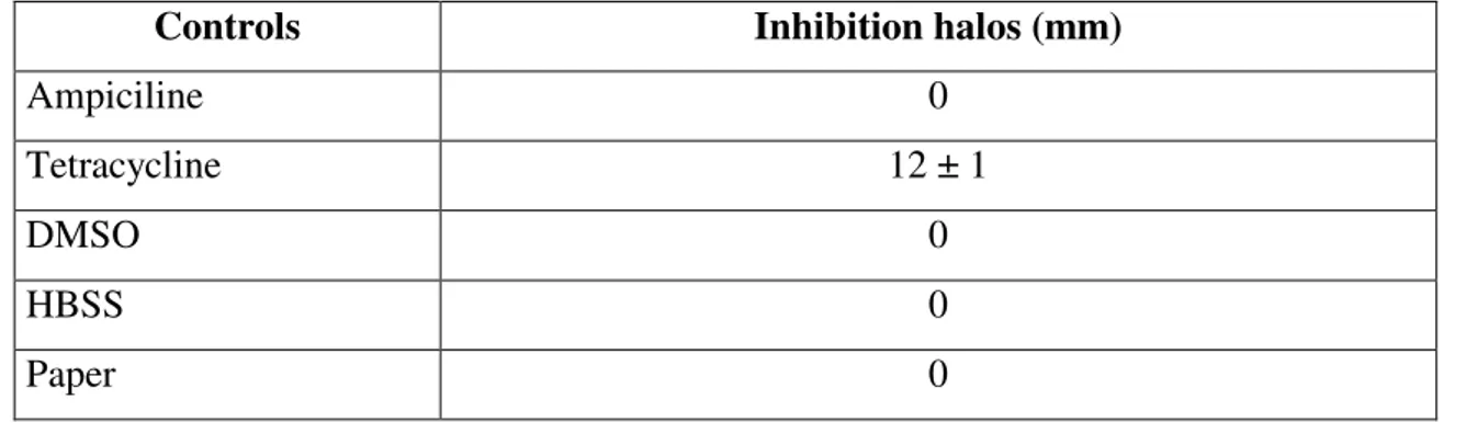
Table 5. Inhibition halos of L. anguillarum caused by antibiotics (positive controls), solvent and
paper controls (negative controls).
Controls Inhibition halos (mm)
Ampiciline 0
Tetracycline 12 ± 1
DMSO 0
HBSS 0
Paper 0
The growth of L. anguillarum was not inhibited by the water or organic halophyte extracts. Ampiciline, the solvent (DMSO and HBSS) and paper controls did not inhibit L. anguillarum. However it showed sensibility to the tetracycline impregnated paper disks, displaying a mean of 12 mm growth inhibited area surrounding the disk (Table 5. and Fig. 5).
Fig. 5. Marine agar Petri dish with L. anguillarum, control plate after incubation. A) ampiciline, B) tetracycline, C) HBSS, D) filter paper.
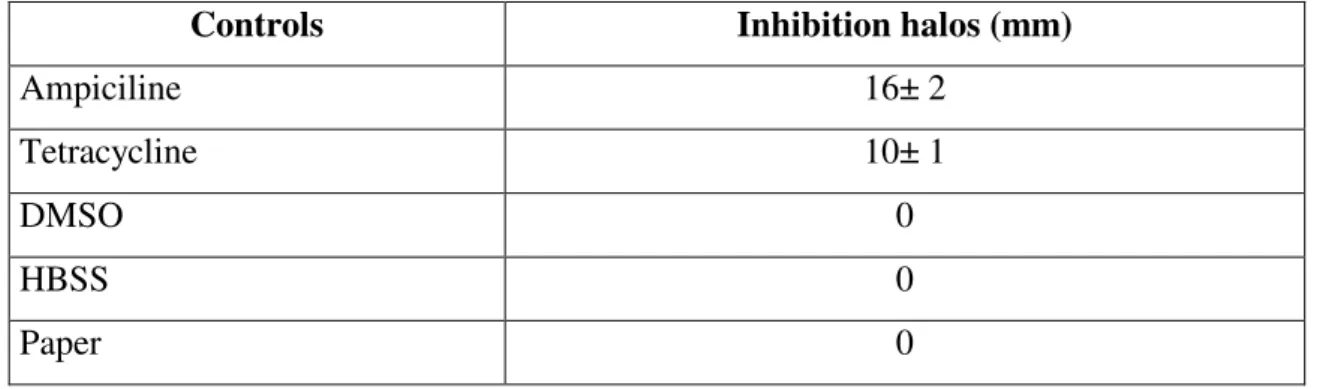
Table 6. Inhibition halos of P. d. piscicida caused by antibiotics (positive controls), solvent and
paper controls (negative controls).
Controls Inhibition halos (mm)
Ampiciline 16± 2
Tetracycline 10± 1
DMSO 0
HBSS 0
Paper 0
Of the tested halophyte extracts, P. d. piscicida displayed a 3 mm inhibiton halo surrounding the ether fraction of J. acutus. Increasing the concentration of J. acutus ether extract (50, 70, 100, 260 and 500 mg/ml were tested) did not resulted in a higher inhibition. P. d. piscicida revealed sensitivity to both antibiotic controls, causing growth inhibition halos of 16 mm (ampiciline) and 10 mm (tetracycline). P. d. piscicida growth was not inhibited by the solvent and paper controls (Table 6. and Fig. 6.).
The results suggest the presence of organic compounds in J. acutus which inhibit the growth of P. d. piscicida. Research done by several authors gives evidence about the nature of these biomolecules. Five phenolic glycosides were isolated from J. acutus: oxyresveratrol 2-O-β-D-glucopyranoside; resveratrol 3′,4′- O, O′
-di-β-D-glucopyranoside; markhamioside F; canthoside B; and caffeic acid glucorhamnoside. These compounds displayed a significant anti-immflamatory and anti-eczematic activity (Awaad, 2006). Dellagreca et al., 2002, isolated nine 9,10-dihydrophenanthrenes, three phenanthrenes and a pyrene, with algicide properties from J. acutus. These included the novel compounds: 5-(1-Ethoxy-ethyl)-2-hydroxy-7-methoxy-1,8-dimethyl-9,10-dihydrophenanthrene, 5-(1-phytoxy-ethyl)-2-hydroxy-7-methoxy-1,8-dimethyl-9,10-dihydrophenanthrene, 2,7-dihydroxy-1-methyl-5-vinylphenanthrene, 2,7-dimethoxy-1,6-dimethyl-5-vinylphenanthrene and 2,7-dihydroxy-1,6-dimethylpyrene (Dellagreca et al., 2002).
Dellagreca et al., 2004 isolated twenty-five 9,10-dihydrophenanthrenes, four phenanthrenes, a dihydrodibenzoxepin, and a pyrene, from J. acutus. Two novel phytotoxic dimeric phenanthrenoids have also been isolated from the rhizome methanolic extract of J. acutus (Dellagreca, Previtera & Zarrelli, 2005).
For A. macrostachyum, A. halimus, C. edulis and P. coronopus, the antibiotic properties described by several authors suggests that further antiotic screening and testing is needed for these halophytes. One potential explanation for absence of antibiotic activity resides on variation in phytochemical profiles in the tissues of halophytes according to location, genetics and season, among other factors. Chokoe et al., 2008 reported differences on antimicrobial testing on C. edulis extracts based on seasonal changes of phytochemicals. Organic fraction extracts (obtained by increasingly high polarity solvents: hexane, dichloromethane, ethyl acetate, acetone and methanol) from C. edulis leaf samples collected during the spring and autumn, were compared for their antibiotic activity against E. coli, Enterococcus faecalis, P. aeruginosa and S. aureus. The spring extracts were more potent against all test organisms, having lower MIC (minimum inhibitory concentration) values than the autumn extracts. When taking the total activity of each extract into account, the autumn extracts showed higher efficacy when compared with the spring samples. The compounds are indentifyed as flavonoids, tannins, malic and cytric acids. Other group displaying variation of antimicrobial molecules are the plantains. Among the compounds present in Plantago, one group of interest for therapeutic properties are the iridoid glycosides (Fig. 7).
Fig. 7. Iridoid glycosides present in Plantago spp. (A) aucubin, (B) catalpol and (C)
plantarenaloside.
These secondary metabolites function as a natural deterrent against herbivory and plant pathogens (Marak, Biere & Van Damme, 2000; Biere, Marak & Van Damme, 2004). These compounds have antibiotic, antifungal, anti-inflammatory, anti-viral and hepatoprotective activities (Davini et al., 1986, Suh et al., 1991; Shim et al., 2007). Genetic, ontogenic, phenotic, populational and seasonal variation of iridoid glycosides is described for P. lanceolata and P. major (Darrow & Bowers, 1997; Tamura, 2002; Tamura & Nishibe, 2002; Fuchs & Bowers, 2004; Zubair, 2010; Quintero & Bowers, 2012; Zubair et al., 2012). (Bowers, 1996). For P. lanceolata, based uppon phytochemical sampling in diferent seasons, targeting the concentrations of catalpol, aucubin and acteoside present in the aerial parts, it is possible to make an assessment of the most desirable period to harvest leafs for their therapeutic properties. The levels of aucubin steadily increased from spring to midfall from 2,1 to 4,8% of dry matter. The levels of acteoside increased from 3,4 to 7,1% over the same period. The data suggested
3.2 Testing of antioxidant activity
3.2.1 Antioxidant activity screening by DPPH free radical
Reactive oxygen species are continuously produced in vertebrade tissues as natural byproducts of aerobic metabolism. These reduced forms of oxygen include: hydrogen peroxide (H2O2), hydroxyl radicals (OH–), nitric oxide (NO), singlet oxigen (1O2),
superoxide anion (O2-) and the non radical peroxynitrite (ONOO-). ROS are damaging
to most cellular constituents (lipids, proteins and nucleic acids) and involved in the progression of several diseases. Oxy radical-induced damage is normally maintained at low levels due to the presence of cellular antioxidant defences. Vertebrate antioxidant defences include ROS scavenging enzymes, such as superoxide dismutase (SOD), catalase, glutathione peroxidase and glutathione reductase. Hydrophobic and hydrophilic antioxidant biocompounds such as ascorbic acid, tocopherol, carotenoids, fatty acids, and glutathione, which are synthesized or obtained from external sources, are also used to prevent ROS induced tissue damage. Oxidative stress is experienced when these endogenous antioxidant defences become overwhelmed by prooxidant forces (Babich, Palace & Stern, 1993; Krapfenbauer et al., 2003). In fish, oxidative stress has been associated with exposure to heavy metals (Rau et al., 2004; Wang, Chaung & Tung, 2004), high stock densities (Caipang et al., 2009; Aksakal et al., 2011), infectious diseases (Shoemaker, Evans & Klesius, 2000; Tanaka et al., 2002; Saurabh & Sahoo, 2008; Ali, Hashem & Al-Salahy, 2011), insecticides (Pena-Llopis, Ferrando & Pena, 2003; Parvez & Raisuddin, 2005; Monteiro et al., 2006), nanomaterials (Oberdörster, 2004; Zhu et al., 2008a, 2008b), nitrogen compounds (Yang et al., 2010) and high temperatures (Parihar & Dubey, 1995). Nutritional deficiencies also make fish prone to oxidative stress, especially in the case of non synthesized micro-nutrients such as ascorbic acid, retinol and tocopherol (Montero et al., 2001). Effects of oxidative stress in fish include a compromised immune response (Montero et al., 2001), growth inhibition and peroxidation of polyunsaturated fatty acids (Zhu et al., 2008a).
The testing of halophyte antioxidants was made using the DPPH colorimetric method. The DPPH method was choosen for the antioxidant screening based on its simplicity, reaction rate and independence of sample polarity. The DPPH radical has a maximum
absorbance of 520 nm in its oxidized form, which decreases when the radical is reduced by antioxidant compounds (Koleva et al., 2002). The antioxidant properties of the featured halophytes were compared with BHT, a commercial antioxidant (Molyneux, 2004). The antioxidant activity was calculated using IC50 which represents the
concetration of extract needed to inhibit 50% of oxidation in vitro.
Among the halophyte extracts, the lowest IC50 values (and the most antioxidant activity,
with statistically significant differences of p < 0.05 above 5 mg/mL) were displayed by P. coronopus and A. macrostachyum. The IC50 value for J. acutus was lower then C.
edulis. However at higher concentrations C. edulis exihibited more oxidant inhibition, with statistically significant differences of p < 0.05 above 5 mg/mL. For p < 0.05, J. acutus and A. halimus were significant above 10 mg/mL (Table 7. and Fig. 8.).
These results suggest differences in the amounts and/or types of compounds produced by these species. P. coronopus is a low rosette forming herb, being common in open rocky and gravel terrains. This species depends on antioxidant defenses, as well on morphological features (thick root system, numerous thin and hairy leafs, in contrast with most Plantago spp.) to grow in exposed locations. It is possible that under hydric stress, the protective role of antioxidant defenses becomes especially relevant in P. coronopus, also considering the lack of other adaptations such as leaf succulence (Golovko et al., 2012). The antioxidant activity of P. coronopus is described by several authors (Chiang et al., 2003; Gálvez et al., 2005; Çelik & Aslantürk, 2006).
The second higher antioxidant activity was detected on A. macrostachyum. A. macrostachyum is an obligate halophyte, which is linked to more antioxidant and osmoprotectant activity (Redondo-Gómez, Mateos-Naranjo & Andrades-Moreno, 2010). This species is capable of growing in interstitial soil salinities up to 1000 mM (Khan & Bilquees Gul, 2002; Redondo-Gómez et al., 2010).
salinity and sun exposure, in comparison to antioxidant defenses. Young A. halimus leafs are coated with layers of vesiculated hairs (trichomes), which function in the regulation and secretion of salt. As leafs mature, these cells collapse, forming a light-refletive waxy layer of salt crystals, which protects tissues from solar ratiation and water loss. In A. halimus stems, leafs also depend on mutual shading for photoprotection (Hassine et al., 2009). It is suggested these adaptations make A. halimus less dependent on antioxidants for photoprotection (Streb, Tel-Or & Feierabend, 1997).
The results indicate that A. macrostachyum and P. coronopus are potential sources of antioxidant compounds. However it is possible that for all the featured species, a higher antioxidant production and yield, can be achieved by means of controled growth conditions (such as exposing cultivated plants to oxidative stresses) and selective breeding ((Bowers, 1996; Pirie et al., 2013). For plantains, the development of bioactive molecule rich cultivars has taken place for species such as P. lanceolata and P. major (Tamura, 2002; Tamura & Nishibe, 2002).
Table 7. Antioxidant activity IC50 values of water based halophyte extracts.
3.2.2
Metal chelating activity for copper
Copper is a common heavy metal in the marine environment and a model prooxidant (Rau et al., 2004). In teleost fish, copper is an essential element, being a part of key proteins and in cellular respiration (Watanabe, Kiron & Satoh, 1997). Excess copper is toxic, causing oxidative stress in tissues, disrupting sodium homeostasis and causing deposits in gills and intestinal tract (Woodward et al., 1995). Copper can reach fish tissue by ingestion (in this case absorbed by the intestinal tract) or absorbed by the gill epithelium (in this case waterborne copper) (Woodward et al., 1995). In fish, the liver is the main organ for copper acummulation and homeostasis (Lorentzen, Maage & Julshamn, 1998).
Metal chelators mobilize metal compounds held in tissues by forming soluble, stable complexes that can be excreted in feces and/ or urine (Ebrahimzadeh, Pourmorad & Bekhradnia, 2008). In plants, heavy metals induce oxidative and water stresses, not very different physiologically of the effects of salinity (Poschenrieder, Gunse & Barcelo, 1989; Nedjimi & Daoud, 2009). The same metabolic pathways that allow for salt tolerance in halophytes, also make these plants more resilient towards heavy metals, when compared with glycophytes. Of particular interest are the antioxidant defences of halophytes, which make these plants pre-adapted to cope with heavy metal oxidative stress (Zhu et al., 2004). Independent research in a halophyte (Mesembryanthemum
IC50 Values (mg/mL) A. macrostachyum A. halimus C. edulis J. acutus P. coronopus 2.04 ± 0.63 5.51 ± 0.88 3.11 ± 4.4 2.56 ± 1.11 0.91 ±0.23
crystallinum L.) and a glycophyte (Lupinus luteus L.), found evidence suggesting that copper and salt stress triggered similar responses in both plant species. It is possible that copper and salt stress responses in plants overlap to some extent, functioning in common integrated mechanical and chemical signals (Thomas et al., 1998; Przymusinski, Rucinska & Gwo´zdz, 2004).
Osmoprotective compounds appear as well, to be significant for heavy metal tolerance mechanisms in halophytes. Proline, a synthesized osmoprotectant linked to ionic and osmotic stress, accumulates in halophytes in response to cadmium, copper, iron and other heavy metals. Under metal induced stress, proline acts in antioxidant defence, metal binding and signalling (Schat, Sharma & Vooijs, 1997; Shevyakova et al., 2003; Sharma & Dietz, 2006; Lefe´vre et al., 2009).
The copper chelating properties of the featured halophytes were compared with a commercial metal chelating compound, EDTA in a colorimetric test (Saiga, Tanabe & Nishimura, 2003).
The lowest IC50 value (and the higher copper chelating activity, with statistically
significant differences of p < 0.05 above 10 mg/mL) was displayed by A. macrostachyum (Table 8. and Fig. 9). A. halimus and J. acutus exihibited respectivelly the second and third lowest IC50 values, with statistically significant differences of p <
0.05 above 10 mg/mL. P. coronopus displayed the highest IC50 value, however for the
10 mg/mL concetration, it shows p < 0.05 significant differences and an chelating activity comparable to the forementioned species.
These results could be explained by the physiological adaptations and habitat of A. macrostachyum and A. halimus. A. macrostachyum is among the featured species, the most dependent and associated with marine environments, which suggests more contact with copper sources. A. macrostachyum and A. halimus resort to the uptake and accumulation of Cl− and Na+ as part of their metabolism (in the shoot internodes for A.
glycinebetaine (Lefe´vre et al., 2009). Salinity itself has been linked to the translocation of heavy metals from the roots to the aerial parts of plants (Fitzgerald et al., 2003). By comparison the least copper chelating activity was observed on C. edulis. A suggested explanation is that C. edulis normally has little contact with copper sources, and so biochemical chelators for this particular metal are less developed. The dune and rocky habitats that the sour fig inhabits are constituted by high drainage substrates. C. edulis also has a shallow root system.
Besides being potential sources of novel metal chelators, these species could be of special interest for decontamination of heavy metal poluted soils. A. halimus in particular, because of its adaptability and biomass production, has possible uses in phytoremediation research (Manousaki & Kalogerakis, 2011).
Table 8. Copper chelating activity IC50 values of water based halophyte extracts.
3.2.3 Metal chelating activity for Iron
Iron is an essential micronutrient for fish, as a constituent of proteins involved in cellular respiration and oxygen transfer. Fish obtain iron via absorption mainly by the intestinal tract but also by the gill epithelium (Peuranen et al., 1994). However excess iron is harmfull as it causes the release of ROS, which damage cells and tissue. Also excessive waterborne iron can form deposits in gills, causing respiratory distress (Dalzell & MacFarlane, 1999).
The iron chelating properties of the featured halophytes were compared with a commercial metal chelating compound, EDTA in a colorimetric assay. The water based extracts of A. macrostachyum, A. halimus, C. edulis, J. acutus and P. coronopus displayed iron chelating properties, with statistically relevant differences at concentrations of 1 mg/mL. The IC50 values for all species are bellow 1 mg/mL (Table
9. and Fig. 10.).
Iron chelators originating from halophytes are indentifyed mainly as polyphenolic compounds. Species known for having iron chelators include Anchusa azurea, Foeniculum vulgare, Limonium densiflorum, Melilotus spp., Mesembryanthemum spp., Pistacia lentiscus, Raphanus raphanistrum, Salicornia europaea, Salvia spp., Sonchus oleraceus and Tamarix gallica L. (Nehir & Sibel, 2004; Khan et al., 2006;
Halophytes IC50 Values (mg/mL)
A. macrostachyum A. halimus C. edulis J. acutus P. coronopus 8.01 ± 4,2 9,26 ± 10.37 11.09±7,4 9,89 ± 13,83 13,14 ± 20.76
Ebrahimzadeh, Pourmorad & Bekhradnia, 2008; Hanen et al., 2009; Khaznadar, Vogiatzakis & Griffiths, 2009; Ksouri et al., 2009; Medini et al., 2011).
Iron chelating compounds could be of special use to freshwater aquaculture, as many river systems originate in ground water springs containing large amounts of naturally ocurring waterborne iron, which limits aquaculture suitability in such areas (Vuori, 1995; Salami et al., 2008).
Iron chelating phytochemicals have interest for human medicine. The treatment of a variety of medical condicions, such as chronic anemia, require the use of iron chelating and antioxidant therapy. Patients with thalassemia, and other chronic anemia disorders, require regular blood transfusions for quality of life and survival. However the iron released as a result of the breakdown of transfused red blood cells is not eliminated by human beings, and is deposited in the form of hemosiderin and ferritin in the liver, spleen, endocrine organs and myocardium (Rund & Rachmilewitz, 2005; Taher, Isma'eel & Cappellini, 2006). The resulting iron acumulation causes ROS mediated tissue damage, which can lead to complications such as endocrine abnormalities, endothelial dysfunction, hypothyroidism, heart and liver failure (Olivieri et al., 1992; Taher, Isma'eel & Cappellini, 2006). The treatment of thalassemia includes both antioxidant and iron chelating therapies. This requirements could indicate potential in iron chelators present in halophytes, as research suggests that compounds such as phenols can serve a dual porpouse as antioxidants and iron chelators. Current iron chelating therapy is limited by poor oral bioavailability, short plasma half-life and severe side effects of available compounds (Kukongviriyapan et al., 2008).
Iron chelating compounds could also have pontetial for cancer therapy. Iron is an essential nutrient for cell growth, as it is necessary for nucleic acid synthesis and respiration. Due to their rapid proliferation, cancer cells have higher iron requeriments when compared with normal cells in a organism. Iron chelators targeting cancer cells
Fig. 10. Iron chelating activity of 1, 5 and 10 mg/mL water based halophyte extracts. The data
represents means of 6 trial wells in a multichamber plate. The letter