Development and characterization of an injectable bone substitute based on a hydrogel and glass-reinforced hydroxyapatite (GR-HA) granules
127
0
0
Texto
(2)
(3)
(4) DE ACORDO COM A LEGISLAÇÃO EM VIGOR, NÃO É PERMITIDA A REPRODUÇÃO DE QUALQUER PARTE DESTA TESE Universidade do Minho, ___/___/______ Assinatura: ________________________________________________.
(5) Acknowledgements Esta dissertação representa para mim o fim de uma etapa de 5 anos extremamente importante na minha vida, em que enriqueci muito o meu conhecimento intelectual e pessoal. No entanto, nunca teria chegado ao fim sem o apoio de várias pessoas que merecem todo o meu reconhecimento e gratidão. Pelo trabalho que realizei durante este ano, gostaria de agradecer todo o apoio e ensinamento aos meus orientadores, Professor Luís Rocha, Professora Cláudia Botelho e Doutora Marta Santos. Professora Cláudia, obrigada pela disponibilidade e paciência com as minhas perguntas infinitas, pela confiança no meu trabalho e pelo incentivo diário de fazer melhor. Agradeço também à Professora Ascensão Lopes pela disponibilidade constante em me esclarecer algumas dúvidas e pelas sugestões e ensinamentos para conseguir um trabalho melhor. Agradeço ao Miguel Rodrigues pela transmissão de conhecimento, pela paciência com as minhas dúvidas constantes e por incentivar sempre o meu espírito crítico. Agradeço também aos meus colegas de gabinete pelo apoio e boa disposição, ao João Coelho, Cátia Fidalgo e Vânia Rodrigues. Agradeço ainda à D. Amélia por toda a ajuda no laboratório e pela alegria que me transmitia todos os dias. Agradeço à minha amiga Sofia Alves, que sempre foi uma grande companheira, esteve sempre comigo nos momentos mais difíceis. Agradeço também ao João Póvoas, pela confiança que depositou em mim e pelo carinho, que me motivava dia após dia para continuar a lutar e a acreditar. Agradeço ainda a todos os meus amigos que não refiro aqui, mas que sabem a sua importância no caminho que percorri. Por último, tenho que agradecer à minha principal fonte de inspiração: a minha família. Obrigado pai e mãe, pelo carinho, por todo o apoio nas minhas escolhas, por confiarem sempre em mim e nas minhas capacidades, por serem um exemplo e pelos princípios que sempre me transmitiram e me ajudaram a ser mais forte e perseverante. Obrigada avó pelo carinho e palavra de conforto que sempre tiveste para mim. Obrigada mana pelas tuas gargalhadas, pelos teus abraços, pela tua perspectiva da vida, pelas tuas palavras ingénuas, mas tão genuínas, o que me ajudou sempre a continuar a lutar e não desistir do caminho que eu quero traçar. Esta tese é dedicada a ti…. iii.
(6)
(7) Abstract Bone defects caused by a disorder or injury, affect millions of patients worldwide, limiting their life quality. Thus, in the last few years, scientists have developed synthetic bone substitutes to treat those defects. Injectable bone substitutes (IBSs), which represent a minimally invasive surgical approach, are very promising systems for specific clinical applications. Therefore, this work aimed to develop a biocompatible, biodegradable and bioactive polymeric vehicle to associate with the bone substitute glass-reinforced hydroxyapatite (GR-HA) granules, allowing their injection directly to the wound site, and also improving the substitute osteoconductive properties. Firstly, three different alginate-based hydrogels were produced, one composed just by an alginate matrix cross-linked with Ca2+ ions (Alg), and other two resulted from the combination of chitosan or hyaluronic acid with that alginate matrix (Alg/Ch and Alg/HA). These vehicles, as well as the respective IBSs (GR-HA granules plus hydrogel) were fully characterized from the physical-chemical point of view. Weight change studies revealed the swelling and degradation rate of the developed materials. The rheology tests showed that the hydrogels have a non-Newtonian viscoelastic behavior, and the injectability tests revealed that low extrusion forces are required to inject the three IBSs. Thus, all the developed hydrogels can be successfully used as vehicles for the bone substitute. Furthermore, the hydrogels were also characterized from the biological point of view to evaluate their biocompatibility. The measured metabolic activity of osteoblastic cells was higher on the Alg/HA_IBS than on the other two IBSs and GR-HA granules alone. Scanning electron microscopy (SEM) analysis demonstrated different cell morphologies on the surface of each IBS. A spreader cell shape was observed on the Alg/HA_IBS surface, proving the improvement of the substitute bioactive ability by association of the hydrogel Alg/HA. This hydrogel was even subcutaneously implanted in rats revealing a slight irritating tissue response. Considering the physical-chemical and biological results, the hydrogel Alg/HA was considered as the best vehicle among the three. Additionally, significant antimicrobial properties were granted to the hydrogel Alg/HA by incorporation of Ce(III) ions, without compromising the improvement of the osteoblastic cells metabolic activity.. v.
(8) Resumo Os defeitos ósseos provocados por doença ou lesão afectam milhões de pacientes em todo o mundo, limitando a sua qualidade de vida. Assim, os cientistas têm desenvolvido, nos últimos anos, substitutos ósseos sintéticos para tratar esses defeitos. Os substitutos ósseos injectáveis (IBSs), que permitem uma abordagem de tratamento minimamente invasiva, são muito promissores para aplicações clínicas específicas. Assim, neste trabalho, pretendeu-se desenvolver um veículo polimérico, biocompatível, biodegradável e bioactivo para associar com grânulos do substituto ósseo: hidroxiapatite reforçada com vidro (GR-HA). Essa associação permite a aplicação directa dos grânulos no local lesado, bem como melhorar as propriedades osteocondutivas do substituto ósseo. Desta forma, foram desenvolvidos três hidrogéis baseados em alginato, um composto por uma matriz de alginato reticulada com iões Ca2+ (Alg), e outros dois que resultaram da combinação de quitosano ou ácido hialurónico com a matriz de alginato (Alg/Ch e Alg/HA). Estes veículos, assim como os respectivos IBSs (grânulos de GR-HA mais hidrogel), foram caracterizados do ponto de vista físico-químico. Os estudos de variação de massa revelaram a taxa de aumento de volume e de degradação dos materiais desenvolvidos. Os testes de reologia demonstraram que os hidrogéis têm um comportamento não-Newtoniano e viscoelástico. Os testes de injectabilidade revelaram baixas forças de extrusão para injectar os três IBSs. Assim, todos os materiais desenvolvidos são potenciais veículos para o substituto ósseo GR-HA. Além disso, os hidrogéis foram também caracterizados biologicamente para avaliar a sua biocompatibilidade. A actividade metabólica das células osteoblásticas registada para o Alg/HA_IBS foi superior à dos outros dois IBSs e dos grânulos de GRHA. Na superfície do Alg/HA_IBS as células apresentaram uma morfologia mais esticada do que nos outros IBSs, provando que a associação do hidrogel Alg/HA ao substituto ósseo melhora a sua bioactividade. O hidrogel Alg/HA foi implantado subcutaneamente em ratos, ocorrendo uma resposta dos tecidos ligeiramente irritante. Considerando os resultados físico-químicos e biológicos, o hidrogel Alg/HA foi considerado como o melhor veículo entre os três desenvolvidos. Adicionalmente, conferiu-se a este hidrogel propriedades antimicrobianas por incorporação de iões Ce(III), sem comprometer a melhoria da actividade metabólica dos osteoblastos. vi.
(9) Table of contents Acknowledgements ..................................................................................................... iii Abstract ........................................................................................................................ v Resumo ....................................................................................................................... vi Table of contents ........................................................................................................ vii List of abbreviations .................................................................................................... ix List of figures ............................................................................................................... xi List of tables ............................................................................................................... xv. Chapter 1 General introduction 1.1. Bone ...................................................................................................................... 3 1.1.1. Bone tissue ..................................................................................................... 3 1.1.1.1. Bone cells ................................................................................................ 6 1.1.1.2. Bone matrix ............................................................................................ 8 1.1.2. Bone physiology ............................................................................................. 8 1.1.2.1. Bone formation ....................................................................................... 8 1.1.2.2. Bone growth ......................................................................................... 10 1.1.2.3. Bone remodeling and repair .................................................................. 10 1.1.2.4. Bone disorders ...................................................................................... 13 1.2. Biomaterials ......................................................................................................... 14 1.2.1. Synthetic bone substitutes ........................................................................... 15 1.2.2. Hydrogels ..................................................................................................... 16 1.2.2.1. Alginate................................................................................................. 17 1.2.2.2. Chitosan ............................................................................................... 23 1.2.2.3. Hyaluronic acid ..................................................................................... 24 1.3. Injectable synthetic bone substitutes .................................................................. 26 1.4. Implant-associated infections............................................................................... 27 1.5. Motivation and Objectives ................................................................................... 28 1.6. References ........................................................................................................... 29. vii.
(10) Chapter 2 Synthesis and physical-chemical characterization of biodegradable alginate-based hydrogels as vehicles for glass-reinforced hydroxyapatite (GR-HA) granules Abstract ...................................................................................................................... 37 2.1. Introduction ......................................................................................................... 37 2.2. Materials and methods ........................................................................................ 40 2.3. Results and discussion .......................................................................................... 44 2.4. Conclusions .......................................................................................................... 56 2.5. References .......................................................................................................... 57. Chapter 3 Biological evaluation of biocompatible alginate-based hydrogels as vehicles for glassreinforced hydroxyapatite (GR-HA) granules Abstract ...................................................................................................................... 65 3.1. Introduction ......................................................................................................... 65 3.2. Materials and methods ........................................................................................ 68 3.3. Results and discussion .......................................................................................... 73 3.4. Conclusions .......................................................................................................... 83 3.5. References ........................................................................................................... 84. Chapter 4 Implantation of one developed injectable bone substitute 4.1. Surgical procedure .............................................................................................. 93 4.2. Samples preparation for histological analysis ....................................................... 95. Chapter 5 General discussion, conclusions and future work 5.1. General discussion and conclusions...................................................................... 99 5.2. Future work ....................................................................................................... 104 5.3. References ........................................................................................................ 106. viii.
(11) List of Abbreviations Alg. Hydrogel of alginate. Alg/Ch. Hydrogel of alginate and chitosan. Alg/Ch_IBS. Injectable bone substitute with the hydrogel of alginate and chitosan. Alg/HA. Hydrogel of alginate and hyaluronic acid. Alg/HA_IBS. Injectable bone substitute with the hydrogel of alginate and hyaluronic. acid Alg_IBS. Injectable bone substitute with the hydrogel of alginate. ASTM. American Society for Testing and Materials. BCP. Biphasic Calcium Phosphate. BMP. Bone Morphogenetic Protein. BMP’s. Morphogenetic Proteins. CFU. Colony-Forming Units. CMC. Carboxymethyl Cellulose. DBM. Demineralized Bone Matrix. DD. Deacetylation Degree. ECM. Extracellular Matrix. FTIR-ATR. Fourier Transform Infrared-Attenuated Total Reflectance. G. α-L-Guluronic Acid. GR-HA. Glass-Reinforced Hydroxyapatite. HA. Hyaluronic Acid. HAP. Hydroxyapatite. HCl. Hydrochloride. HMDS. Hexamethyldisilazanes. IBSs. Injectable Bone Substitute. ISO. International Organization for Standardization. KHP. Potassium Hydrogen Phthalate. M. β-D-Mannuronic Acid. MTT. 3-[4,5-Dimethyl-Thiazol-2-yl]-2,5-Diphenyltetrazolium. PBS. Phosphate Buffered Saline. PEG. Poly(Ethylene Glycol) ix.
(12) PEO. Poly(Ethylene Oxide). PNIPAAm. Poly(N-isopropylacrylamide). PPO. Poly(Propylene oxide). RGD. Arginine-Glycine-Aspartic Acid. SEM. Scanning Electron Microscopy. TCP. Tricalcium Phosphate. USA. United States of America. x.
(13) List of Figures Chapter 1 General introduction Figure 1.1: The four bone categories ........................................................................ 3 Figure 1.2: Structure of the long bones .................................................................... 3 Figure 1.3: Structure of a flat bone ........................................................................... 4 Figure 1.4: Structure of the compact bone ............................................................... 5 Figure 1.5: Structure of the cancellous bone ............................................................ 5 Figure 1.6: Photomicrograph of an osteocyte in a lacuna ......................................... 7 Figure 1.7: Transmission electron micrograph of a human osteoclast. Black arrows: ruffled border; N: nucleus ........................................................................................ 7 Figure 1.8: Phases of the bone repair process ........................................................ 12 Figure 1.9: Different bone disorders ....................................................................... 13 Figure 1.10: (a): Chemical structure of alginate monomers; (b): Example of a monomers sequence of alginate............................................................................. 18 Figure 1.11: Accommodation of the calcium ions, between two alginate chains, in the different glycosidic linkages: (a) GG/GG interaction; (b) MG/MG interaction; (c) GG/MG interaction ................................................................................................. 18 Figure 1.12: The egg-box model of cross-linking: (a): binding of the divalent cations in the G monomers of two polymeric chains; (b): formation of junction zones between polymeric chains ...................................................................................... 19 Figure 1.13: Chemical structure of: A. chitin; B. chitosan ........................................ 23 Figure 1.14: Chemical structure of the two monosaccharides of hyaluronic acid .... 25 Figure 1.15: The main pathogenic species among orthopedic implant associated infections ............................................................................................................... 27. Chapter 2 Synthesis and physical-chemical characterization of biodegradable alginate based hydrogels as vehicles for glass-reinforced hydroxyapatite (GR-HA) granules Figure 2.1: Syringe with the injectable system on the texture analyzer .................. 43. xi.
(14) Figure 2.2: Macroscopic appearance of the three developed hydrogels: A-Alg hydrogel; B- Alg/Ch hydrogel; C- Alg/HA hydrogel .................................................. 44 Figure 2.3: Macroscopic appearance of the Alg_IBS ............................................... 44 Figure 2.4: SEM images of the three hydrogels: A-Alg; B-Alg/Ch; C-Alg/HA ............ 45 Figure 2.5: SEM images of the three injectable systems: A1,A2-Alg_IBS; B1,B2Alg/Ch_IBS; C1,C2-Alg/HA_IBS................................................................................ 46 Figure 2.6: FTIR-ATR spectra of the three developed hydrogels and the polymeric solutions used to produce them ............................................................................. 47 Figure 2.7: FTIR-ATR spectra of the sodium alginate powder autoclaved and nonautoclaved.............................................................................................................. 48 Figure 2.8: Weight change of the three hydrogels at 37 oC: A-PBS (pH 7.4); B- KHP (pH 4) ..................................................................................................................... 49 Figure 2.9: Degradation of the three hydrogels at 37 oC and 1 Hz orbital agitation: APBS (pH 7.4); B-KHP (pH 4) ..................................................................................... 51 Figure 2.10: Rheological characterization of the three developed hydrogels: Aviscosity frequency dependence; B-storage modulus (G’) and loss modulus (G’’) frequency dependence ........................................................................................... 52 Figure 2.11: Injectability curves of the three IBSs,at an extrusion velocity of 1 mm/s ................................................................................................................... 55. Chapter 3 Biological evaluation of biocompatible alginate based hydrogels as vehicles for glassreinforced hydroxyapatite (GR-HA) granules Figure 3.1: Subcutaneous implantation of the hydrogel Alg/HA: A- the dorsal incisions made on the dorsal area of the Sprague Dawley® rats; B-incisions suture after implantation .................................................................................................. 71 Figure 3.2: Metabolic activity of the MG63 cells seeded on the three developed IBSs, after 24, 48 and 72 hours of incubation .......................................................... 74 Figure 3.3: SEM appearance of the MG63 cells adhered on the three developed IBSs after 24 hours of incubation: A1-Alg_IBS non-cell seeded; A2-Alg_IBS cell seeded; B1-Alg/Ch_IBS non-cell seeded; B2-Alg/Ch_IBS cell seeded; C1-Alg/HA_IBS non-cellseeded; C2-Alg/HA_IBS cell seeded ........................................................................ 76 xii.
(15) Figure 3.4: Interaction of the hydrogels and IBSs with sheep blood: A-hydrogel Alg; B-hydrogel Alg/Ch; C-hydrogel Alg/HA; D-Alg/HA_IBS ............................................ 77 Figure 3.5: Histological image (magnification x 400) of a subcutaneous tissue sample, after 2 weeks of the hydrogel Alg/HA implantation ................................... 78 Figure 3.6: Antimicrobial activity of the hydrogels Alg/HA, Alg/HA1 and Alg/HA2 after 24 hours of incubation ................................................................................... 81 Figure 3.7: Metabolic activity of the MG63 cells seeded on Alg/HA2_IBS and Alg/HA_IBS, after 24, 48 and 72 hours of incubation. ............................................. 82. Chapter 4 Implantation of one developed injectable bone substitute Figure 4.1: Exposed femur before the performance of the bone defects ................ 94 Figure 4.2: Performance of the bone defects.......................................................... 94 Figure 4.3: Bone defects before the biomaterials implantation. ............................. 94 Figure 4.4: Bone defects filled with the biomaterials: A-GR-HA granules; BAlg/HA_IBS ............................................................................................................. 94 Figure 4.5: A sample of a bone defect filled with the Alg/HA_IBS embedded in resin ................................................................................................................... 95. xiii.
(16)
(17) List of Tables Chapter 1 General introduction Table 1.1: Examples of injectable bone substitutes ................................................ 26. Chapter 2 Synthesis and physical-chemical characterization of biodegradable alginate based hydrogels as vehicles for glass-reinforced hydroxyapatite (GR-HA) granules Table 2.1: Proportions of GR-HA and hydrogel for each IBS: Alg_IBS: substitute with hydrogel Alg; Alg/Ch_IBS: substitute with hydrogel Alg/Ch; Alg/HA_IBS: substitute with hydrogel Alg/HA ............................................................................................. 41 Table 2.2: Peaks intensity reduction in hydrogels, when compared to sodium alginate solution ..................................................................................................... 48. Chapter 3 Biological evaluation of biocompatible alginate based hydrogels as vehicles for glassreinforced hydroxyapatite (GR-HA) granules Table 3.1: Proportions of GR-HA and hydrogel for each IBS: Alg_IBS: substitute with hydrogel Alg; Alg/Ch_IBS: substitute with hydrogel Alg/Ch; Alg/HA_IBS: substitute with hydrogel Alg/HA; A/HA1_IBS: substitute with hydrogel Alg/HA1; A/HA2_IBS: substitute with hydrogel Alg/HA2 ........................................................................... 69 Table 3.2: Semi-quantitative scoring of the biological response to the implanted material, according to ISO 10993-6 ........................................................................ 78. xv.
(18)
(19) Chapter 1 General introduction.
(20)
(21) Chapter 1. 1.1. Bone 1.1.1. Bone tissue The skeletal system plays several functions in the body, it provides physical support, vital internal organs and structures protection, body movement, blood cells production and mineral storage, thus having an important action in the ionic gradient balance and in the organic homeostasis regulation.[1-3] This system is composed of bones, cartilages and ligaments, which together form a strong and flexible framework that confers structure to the body.[2] Bone is the main calcified tissue present in the skeletal system of vertebrates, it is a connective tissue formed by the process named osteogenesis.[4] There are four general categories of bones, according to their shape and function, short bones, long bones, flat bones, and irregular bones (figure 1.1). Long bones have four main components, the diaphysis, the epiphysis, the metaphyses and the epiphyseal plate (figure 1.2). The diaphysis is mainly composed of compact bone and this component encloses a space named the medullary cavity, where there is bone marrow. The epiphysis (above the epiphyseal plate) and the metaphyses (below the. Figure 1.2: Structure of the long bones.. 3. [adapted from [1]]. Figure 1.1: The four bone [adapted from [1]] categories..
(22) Chapter 1 epiphyseal plate) are mainly composed of cancellous bone, but the outer surface is a layer of compact bone. Between the epiphysis and diaphysis there is the epiphyseal, or growth, plate, which is hyaline cartilage.[1-3] Flat bones, normally, have no diaphyses or epiphyses, they consist in a cancellous bone layer between two compact bone layers (figure 1.3). Short and irregular bones are not elongated and they have a compact bone surface surrounding a cancellous bone center.[1, 2] The outer surface of the compact bone is covered by a connective tissue membrane, called the periosteum (figure 1.2). This Figure 1.3: Structure of a flat bone.. [adapted from [1]]. membrane is composed by an outer collagen fibrous layer, which contains blood vessels and nerves, and an inner single layer of bone cells, namely osteoblasts, osteoclasts, and osteochondral progenitor cells. There is also another connective tissue membrane just composed by a thin single layer of bone cells (including osteoblasts, osteoclasts, and osteochondral progenitor cells), called endosteum (figure 1.2). This membrane covers the internal surfaces of the bone cavities, like the medullary cavity in the diaphysis and the cavities in cancellous and compact bone.[1, 2] The bone tissue consists of extracellular bone matrix with bone cells, but according to its macroscopic features, it can be distinguished in two different types: the compact bone, which represents 80% of bone tissue, and the cancellous bone, which represents 20% of bone tissue.[3, 5] The compact bone is mostly comprised of bone matrix, but with few small cavities. On the other hand, the cancellous bone tissue is mostly composed by small cavities surrounded by bone matrix.[1, 2] The compact bone tissue has blood vessels and the bone lamellae are mainly oriented around them. The vessels parallel to the long axis of the bone are in central (or haversian) canals, which have blood vessels, nerves and loose connective tissue, and are lined with endosteum. The central canals are surrounded by circular layers of bone matrix, named concentric lamellae, which have osteocytes between them (figure 1.4). The system composed by a central canal, the concentric lamellae and osteocytes, is called of osteon or haversian system (figure 1.4). Moreover, in the outer surface of. 4.
(23) Chapter 1 the compact bone there are flat plates extended around the bone, which are named of circumferential lamellae, and between the osteons, exist also the interstitial lamellae (older bone in the bone remodeling process).[1-3]. Figure 1.4: Structure of the compact bone.. [adapted from [1]]. The cancellous bone (or spongy bone) tissue consists in a network composed by slender interconnecting rods or plates of bone called trabeculae (figure 1.5).[1-3] In the trabeculae there are pores, that are filled with bone marrow and blood vessels. The majority of trabeculae are thin, varying between 50-400 μm, and are composed by some lamellae, with osteocytes between them (figure 1.5). Besides that, the surface of the trabeculae is coated with a single layer that mostly contains osteoblasts, and few osteoclasts (figure 1.5).[1, 2] It should be referred, that the trabeculae are not randomly arranged, they are oriented along the bone’s lines of stress. So, if for some reason, the direction of weight-bearing stress is changed, the trabeculae orientation changes to realign with the new lines of stress.[1, 2]. 5. Figure 1.5: Structure of the [adapted from [1]] cancellous bone..
(24) Chapter 1 1.1.1.1. Bone cells In bone tissue different cell types can be distinguished according to its origin and function, standing out the osteoblasts, osteoclasts and osteocytes. a) Osteochondral progenitor cells or osteogenic cells The connective tissue is embryologically developed from mesenchymal cells and some of them become stem cells. This cell type is characterized for being able of giving rise to more differentiated cells types.[1, 2] In this case, osteogenic cells are stem cells that can become to osteoblasts (bone cells) or chondroblasts (cartilage cells). These bony cells lie, for instance, in the inner layer of the periosteum, and in the endosteum, and, through their mitosis and differentiation, they are the only source of new osteoblasts, since these ones are nonmitotic cells.[1, 2, 6] The mitosis and differentiation of the osteogenic cells are accelerated by cases of stress and fractures, which consequently cause a rapid osteoblasts number increase.[2] b) Osteoblasts Osteoblasts are responsible for bone formation, by a process named ossification, or osteogenesis, they produce the organic material of the bony matrix and they also participate in the mineralization of the bone tissue.[1, 2] These cells have an extensive endoplasmic reticulum and numerous ribosomes, they are arranged in rows in the endosteum and inner layer of periosteum, and they are connected by gap junctions throughout the cell processes. Osteoblasts synthesize collagen and proteoglycans, which are incorporated into vesicles by the Golgi apparatus and then released to the extracellular medium by exocytosis. Besides that, inside these cells there are other vesicles which incorporate calcium ions (Ca2+), phosphate ions (PO43-), and various enzymes (for example, the alkaline phosphatase). These components are also released from the intracellular medium to the extracellular medium by exocytosis to form hydroxyapatite crystals, occurring by this way the mineralization of the bony matrix.[1, 2, 6]. 6.
(25) Chapter 1 c) Osteocytes An osteocyte is a mature bone cell, more exactly, it is a terminally differentiated osteoblast that has became surrounded by the bone matrix that it has produced. In spite of becoming less active than the most osteoblasts, they function within networks and are able to produce components which are needed to maintain the bone structure and metabolism.[2, 5, 6] By this way, osteocytes do not have a significant action in formation or resorption bone event. However, when they detect strain in a bone, they pass the information to osteoblasts at the surface, that will provide bone formation where it is needed and inform osteoclasts to reabsorb bone in another place.[1, 2, 6] The osteocyte cells bodies are positioned in tiny cavities in the bony matrix named lacunae, and its extensive processes are in slender channels called canaliculi (figure 1.6). These channels allow the contact between the processes of neighboring osteocytes, which are connected by gap junctions. Through these. gap. exchange chemical. junctions nutrients,. signals,. osteoblasts gases. and. and. transport. wastes to the closest blood vessel for. Figure 1.6: Photomicrograph of an osteocyte in a lacuna. [adapted from [3]]. disposal.[7, 8] d) Osteoclasts Osteoclasts are large and multinucleated cells (3 or 4 nuclei, and sometimes up to 50), responsible for bone resorption, and most of them are derived from. bone. marrow. monocyte-macrophage. precursor cells.[2, 3, 9] The plasma membrane of osteoclasts forms many deep infoldings in the contact zone with the bone matrix, called a ruffled border (figure 1.7).. [3, 6, 9]. Across this border hydrogen ions are expelled to the. 7. Figure 1.7: Transmission electron micrograph of a human osteoclast. Black arrows: ruffled border; N: [adapted from [9]] nucleus..
(26) Chapter 1 extracellular medium, causing an environment acidification, giving rise to the bone matrix decalcification.[8,. 9]. Moreover, the protein components of the matrix are. digested by enzymes also released by the osteoclasts. After the breakdown of the matrix components, some of the resultant products of it are then reabsorbed by these cells through the endocytosis process.[1, 6] Osteoclasts action is improved when they contact directly with mineralized bone matrix. This event is enhanced by the osteoblasts, once they produce enzymes which breakdown the unmineralized organic matrix layer that usually covers the bone, enabling the direct contact of osteoclasts with the mineralized bone part.[1, 6] 1.1.1.2. Bone matrix The bone matrix is about 35% organic and 65% inorganic matter, by weight. The organic material mainly consists in 90% of type I collagen fibers and various proteincarbohydrate complexes such as glycosaminoglycans, proteoglycans, glycoproteins and growth factors. The inorganic material is approximately 85% of hydroxyapatite (HAP), a crystallized calcium phosphate salt [Ca10(PO4)6(OH)2], 10% of calcium carbonate (CaCO3), and smaller amounts of magnesium, sodium, potassium, fluoride, sulfate, carbonate, and hydroxide ions.[1, 2, 5] The bone functional characteristics are defined by the collagen and the mineral components. The mineral components give the matrix compression strength, whereas the collagen fibers determine the elasticity and toughness of the tissue. Thus, if the mineral components are removed from the bone, it becomes extremely flexible, but, if the collagen does not exist, the bone becomes overly brittle. So, the bone physical features are ensured by the complementary relation between the organic and mineral part.[1, 2, 5]. 1.1.2. Bone physiology 1.1.2.1. Bone formation The process by which occurs the bone formation is named of ossification or osteogenesis. During fetal development, there are two types of ossification, the intramembranous, in which the process occurs in connective tissue, and the endochondral, in which the process occurs in cartilage.[1, 2, 6]. 8.
(27) Chapter 1 • Intramembranous ossification: In this process are produced, for instance, the flat bones of the skull and most of the clavicle. Around the fifth week of evolution, the embryonic mesenchyme forms a membrane of connective tissue with collagen fibers, and about the eighth week, some of the mesenchymal cells in the membrane become osteogenic cells, which are then differentiated in osteoblasts.[1, 2, 6] After this, osteoblasts start to produce an organic matrix, and the formation of thin trabeculae occurs. Meanwhile, the trabeculae become larger and longer, because osteoblasts continue to produce more and more bone, calcium phosphate is deposited in the matrix and some osteoblasts stay trapped in the lacunae, becoming osteocytes. Then the formation of cancellous bone occurs, by the junction of the trabeculae. The trabeculae at the surface of the developing bone continue to calcify in order to fill the empty spaces and produce compact bone. Besides that, the cells at the new bone tissue surface specialize to form the periosteum.[1, 2, 6] • Endochondral ossification: The endochondral formation occurs from the cartilage and begins about the eighth week of fetal development. This process is responsible for the production of, for example, the vertebrae, pelvic bones and bones of the limbs.[1, 2, 6] In this process, mesenchymal cells are differentiated into chondroblasts, which produce a hyaline cartilage structure with a similar shape to the future bone. Chondroblasts become chondrocytes as they stay surrounded by cartilage matrix produced by them. The hyaline cartilage model is wrapped by perichondrium (fibrous connective tissue membrane), and some of its cells become osteoblasts. These bone cells start to produce compact bone on the cartilaginous structure surface, creating a bone collar. At this time, the surrounding fibrous membrane is considered as periosteum. Besides that, by this time, the chondrocytes inside the cartilage structure hypertrophy, and the cartilage matrix is calcified by deposition of calcium carbonate. In this calcified medium, the death of chondrocytes eventually occurs, appearing lacunae in the matrix.[1, 2, 6] Then, the blood vessels and osteoblasts from periosteum invade the calcified cartilage, forming a primary ossification center. Osteoblasts form bone on the calcified cartilage surface, producing cancellous bone. After this, osteoclasts reabsorb bone. 9.
(28) Chapter 1 existent in the central part of the diaphysis, forming the medullary cavity. Besides the primary center, there are also the secondary ossification centers located in the epiphyses. The process of bone formation in these centers is the same of the primary, but, in this case, the medullary cavity formation does not exist.[1, 3] 1.1.2.2. Bone growth Mature bone growth occurs by the appositional mechanism, which consists in the formation of new bone on the surface of older bone or cartilage.[1, 2] In the appositional growth, osteoblasts produce and deposit new bone in layers parallel to the surface of the existent bone. During the deposition of new bone tissue at the outer bone surface, osteoclasts remove bone from the inner surface, increasing the marrow cavity size. These two processes, the bone formation and removal, function in a balanced way between them. If one of them is intensified relatively to the other, several bone abnormalities occur.[1, 2] The longitudinal and radial bone growth happens in the childhood and adolescence. The width of long bones, as the size or thickness of other bones, increases through the appositional method. The length of long bones increases through the growth at the epiphyseal plate by new cartilage formation, and the appositional bone growth on the surface of the new cartilage.[1-3] 1.1.2.3. Bone remodeling and repair • Bone remodeling The bone remodeling process is responsible for replace old bone for new bone to maintain the bone strength and the mineral homeostasis. The remodeling starts before the birth and is maintained until the death to prevent accumulation of bone microdamage. The system responsible for this phenomenon is composed by osteoclasts and osteoblasts, which reabsorb old bone and form new bone, respectively. In women the remodeling process increases in perimenopausal and early postmenopausal period, but with further aging it is slowed besides continuing at a higher rate than in premenopausal period. In aging men, the remodeling process is thought to exhibit a mild increase.[1-3] The remodeling is present in bone growth, bone repair, changes in bone shape, bone adjustment to stress, and calcium ion regulation. The remodeling points can be. 10.
(29) Chapter 1 randomly developed, but they also exist in specific sites which need to be repaired. The process is composed by four sequential phases, activation, resorption, reversal, and formation: [1, 3] 1) Activation: this phase mainly consists in recruitment and activation of mononuclear monocyte-macrophage osteoclast precursors from the circulation, which will give raise to the osteoclasts;[1, 3] 2) Resorption: this lasts about two to four weeks in the process, and it consists in the resorption of old bone by the osteoclasts. These cells digest the organic matrix forming resorption pits, named as Howship’s lacunae, on the cancellous bone surface and in the Haversian canals of the compact bone;[1, 3] 3) Reversal: during this phase bone resorption transits to bone formation. The chemical signals between the end of bone resorption and the beginning of bone formation are as yet unknown, but some signal candidates has already been proposed, for instance, bone morphogenetic proteins and bone matrix-derived factors. Besides that, it has also been proposed that this phase can be mediated by the strain gradient in the lacunae. Thus, osteoclasts are activated when there is a reduced strain, and osteoblasts are activated when there is an increased strain;[1, 3] 4) Formation: the formation phase lasts about four to six months, and it consists in the production of new organic matrix by the osteoblasts to substitute the old bone that have been removed.[1, 3] • Bone repair The bone tissue has the ability to repair after damage to it. The bone repair process can be described in four main steps: 1) Hematoma formation (figure 1.8-1): in case of a bone fracture a hematoma (localized blood mass, released from blood vessels, limited in an organ or space) is formed, caused by the damage of the bone blood vessels and the periosteum. The damage of blood vessels in the central canals causes inappropriate blood sustainability of osteocytes, which induces the death of the bone tissue next to the fracture local. Furthermore, the tissues around the bone usually suffer inflammation and swelling.[1, 6] 2) Callus formation (figure 1.8-2): in this phase at the fracture local a callus is formed, which is a tissue mass that connects the bone broken ends. The internal callus,. 11.
(30) Chapter 1 exists between the bone ends and in the medullary cavity (in case of a long bone. Some days after the fracture macrophages eliminate cell debris, osteoclasts reabsorb dead bone tissue, and fibroblasts (constituent cells of connective tissue) produce a collagen fibrous network. Besides that, the osteoblasts and chondroblasts produce woven bone and cartilage respectively. The external callus is a collar around the fracture site. This collar is formed by the osteoblasts and chondroblasts, which produce woven bone and cartilage respectively. The external callus allows the stabilization of the bone broken ends.[1, 6] 3) Callus ossification (figure 1.8-3): in this phase the cartilage present in the external callus is converted in woven, cancellous bone by endochondral ossification. In the internal callus, the cartilage and fibers are also substituted by woven, cancellous bone. This callus ossification becomes the broken bone even more stable.[1, 6] 4) Remodeling of bone (figure 1.8-4): in the last phase of the process the woven bone in the internal callus and the death bone next to the fracture local are substituted by compact bone. In this new bone, the osteons from the broken ends are extended across the fracture zone, in order to link the two parts. This repair stage is long and can last more than a year.[1, 6]. Figure 1.8: Phases of the bone repair process.. 12. [adapted from [1]].
(31) Chapter 1 1.1.2.4. Bone disorders The natural bone tissue physiolology can be influenced by several types of bone disorders, as presented in figure 1.9.[1, 2, 10] These disorders can interfere with the bone growth process giving rise to bones with abnormal dimensions or very brittle, being susceptible to fracture easily. Other bone disorders can interfere with the bone remodeling process, namely, causing a bone resorption rate higher than its formation rate. This difference may cause small or large bone defects, or even its fracture.[1, 2, 10, 11]. Moreover, bone defects or fractures can occur due to injuries caused by external factors, such as falls, in which bone is exposed to an excessive stress.[1, 2]. Growth and Development Disorders. •Giantism: abnormal growth caused by the excessive cartilage and bone formation at the epiphyseal plates of long bones. •Dwarfism: is the opposite of giantism, in this case the pacient is abnormally short. •Osteogenesis imperfect: genetic disorders, which cause brittle bones more susceptible to fracture, due to a defect in collagen deposition .. Bacterial Infections. •Osteomyelitis: inflammation of bone tissue and bone marrow, caused by a bacterial infection.. Tumors. •Osteoma: benign bone tumor, that mainly appears in the flat bones of the skull. •Osteochondroma: benign bone and cartilage tumor that usually forms bone spurs at the long bones ends. •Osteosarcoma: the most common and deadly cancer. It usually occurs in the femur, tibia and males humerus. •Chondrosarcoma: slow-growing hyaline cartilage cancer. It's most common in middle age.. Decalcification. •Osteomalacia: bone becomes soft and weakened, and more susceptible to fracture, caused by a deficiency of calcium in bones. •Osteoporosis: bone mass loss that occurs when the rate of bone resorption exceeds the rate of bone formation. It is provoked by the absence of physical exercise or the estrogen lack, after menopausa. This disorder gives rise to an increased brittleness and susceptibility to fractures.. Paget disease. •Rapid and disorderly bone remodeling and weak and deformed bones. It is caused by an excessive osteoclasts proliferation and consequent excessive bone resorption, what osteoblasts try to compensate by depositing more bone.. Figure 1.9: Different bone disorders.. 13. [1,2,10].
(32) Chapter 1. 1.2. Biomaterials The development of biomaterials has presented a big impact on the treatment of injuries and diseases. Biomaterials may be of synthetic or natural origin. Briefly, a biomaterial can be defined as a material used in contact with biological systems to replace a part or a function of the body, without harming the living organism and its components.[12-15] These materials must meet several criteria, such as biocompatibility, which is defined as “the ability of a material to perform with an appropriate host response in a specific application”.[12, 13, 16] Thus, a biomaterial must simultaneously be non-toxic and satisfy the requirement of functionality for what it was designed.[16] Biomaterials can be classified according to two different parameters, bulk and surface properties. The characterization based on bulk properties is based on the atomic composition and the respective inter-atomic bonding, according to these biomaterials can be metals, ceramics, polymers and composites. The characterization based on surface properties based describes the interaction type that happens at the material interface with the biological environment: Bioinert: materials that retain their physical and chemical properties when implanted, presenting a minimal or even none interaction with the surrounding tissues. Examples: titanium and alumina.[15, 17, 18] Bioactive: materials that chemically bond and interact with the surrounding tissues at the interface, presenting the ability to initiate a biological response, namely cell adhesion, proliferation, or even in some cases differentiation of some progenitor cells. Examples: synthetic HAP.[13, 15, 18] Biotolerated: materials that are moderately accepted by the host organism when implanted, being normally involved by a fibrous capsule. Example: stainless steel.[18] Bioresorbable: materials that, after implantation, are degraded over time and simultaneously and gradually replaced by regenerating endogenous tissue. The degradation resultant products are absorbed and released through metabolic processes. Example: tricalcium phosphate (TCP) and hyaluronic acid (HA).[15, 17, 18]. 14.
(33) Chapter 1. 1.2.1. Synthetic bone substitutes In order to treat bone defects, different approaches can be used: grafts either from the patient (autograft), from a donor (allograft) or from another animal specie (xenografts); demineralized bone matrix (DBM) extracted from the allografts; or synthetic bone substitutes that have been developed in the last few decades.[19-24] Three essential elements are convenient for an ideal bone regeneration, osteogenesis, osteoinduction, and osteoconduction, resulting in an osteointegration of the graft or substitute in the host bone.[21,. 22]. Thus, autografts are the most used. option, because they present the three essencial elements and no immune response after implantation. However, their availability is limited and they can cause chronic pain.[19-22] Allografts and xenografts present only osteoinductive and osteoconductive properties, and have risks of bacterial contamination, viral transmission, and immunogenicity.[19-21, 23] Synthetic bone substitutes, such as HAP and bioglasses, are bioactive and bioresorbable biomaterials that just present osteoconductive properties. However, they eliminate the risk of disease transmission, their availability is unlimited and possible to produce in different forms and porosity levels.[19-22] Moreover, these synthetic substitutes can be combined with biologic agents, which grant them osteoinductive and osteogenic properties.[19, 21, 23] Synthetic substitutes options: • Ceramics HAP and TCP The most used ceramics are synthetic HAP and β-crystalline form of TCP (β-TCP, Ca3(PO4)2). These calcium-phosphate-based ceramics allow bone cells attachment, proliferation and migration. β-TCP and HAP present chemical similarity to the bone mineralized phase, that provides their osteoconductive potential and good biocompatibility. Although, TCP is resorbed more quickly by the osteoclasts and mechanically less stable than HAP.[20-23] Bioactive glasses The main components of the bioactive glasses are sodium oxide (Na2O), calcium oxide (CaO), phosphorus pentoxide (P2O5) and silicon dioxide (SiO2). However, there are different bioglasses depending on the percentage of each one of those 15.
(34) Chapter 1 components in the glass. When bioglasses are exposed to physiologic aqueous solutions, a mechanically strong bond between the glass and the bone is established due to the synthesis of apatite crystals similar to that of bone, promoting the osteointegration. Thus, these materials are thought to be not only osteoconductive, but also osteoinductive. Moreover, these materials present greater mechanical strength when compared to calcium phosphate preparations.[21, 22, 25] Therefore, in order to improve the biological and mechanical properties of HAP, bioactive glasses have been associated to it, giving rise to a material named glassreinforced hydroxyapatite (GR-HA).[26, 27] • Hybrid materials These materials can be obtained from the combination of a ceramic matrix, which supply a structural underlay, with osteogenic cells (which can be obtained from bone marrow) and/or osteoinductive factors (namely morphogenetic proteins (BMP’s)). These elements confer to the substitute osteogenic and osteoinductive properties, providing a better and quicker bone repair process.[20, 21, 23]. 1.2.2. Hydrogels Hydrogels are cross-linked three-dimensional polymeric structures, which swell in aqueous solutions.[12, 16, 28] These structures can be produced with natural polymers (such as alginate, collagen, agar and chitosan) or synthetic polymers (such as poly(ethylene glycol) (PEG), poly(N-isopropylacrylamide) (PNIPAAm), poly(ethylene oxide) (PEO) and poly(propylene oxide) (PPO)). [16, 28-33] These cross-linked structures can contain covalent bonds between monomers, physical cross-links of entanglements, hydrogen bonds or van der Waals interactions between chains or even ionic interactions. Besides that, they can be homopolymeric hydrogels: when the network is composed by just one hydrophilic monomer type; copolymeric/multipolymeric hydrogels: when are composed by two/or more distinct monomers, being at least one of them hydrophilic to ensure the swelling property; interpenetrating polymeric hydrogels: when are composed by two intermeshed networks.[12, 28] In recent years, hydrogels have been studied for several biomedical and pharmaceutical applications. Due to their properties they can be potentially used as. 16.
(35) Chapter 1 drug delivery injectable systems and as injectable scaffolds in tissue engineering. They present an inherent ability to mimic the ECM structural and compositional properties, allowing cell adhesion and proliferation and promoting the diffusion of hydrophilic nutrients and cell metabolites.[12, 16, 28, 29, 34] 1.2.2.1. Alginate “Alginates” is the designation given to a natural family of biodegradable, biocompatible, hydrophilic (in normal physiological conditions) and non-toxic polysaccharides extracted from some marine algae and some microorganisms. [28, 35-38] This family includes alginic acid and its salts, such as sodium alginate. The alginic acid was discovered by the British chemist Stanford in 1880, and it exists in all species of the brown seaweed (Phaeophycae), constituting up to 40% of the dry matter, and in a few species of red algae (Corallinacae). [36, 39-41] • Chemical structure Alginates are linear block co-polymers composed of two different monomers, βD-mannuronic acid (M) and α-L-guluronic acid (G), which are linked by (1-4) glycosidic bonds (figure 1.10-a). These two components are associated in a certain order, in a block-structure pattern, which can be homopolymeric, if the block is only constituted by just one monomer type (M or G) or heteropolymeric, if the block is simultaneously constituted by the two monomers alternated (figure 1.10-b). The blocks composition and their sequence in a certain alginate depends of the source of which it was extracted, however, some investigations have revealed that the most common structure comprises homopolymeric M blocks and homopolymeric G blocks interspersed by heteropolymeric MG blocks.[16, 39-41]. 17.
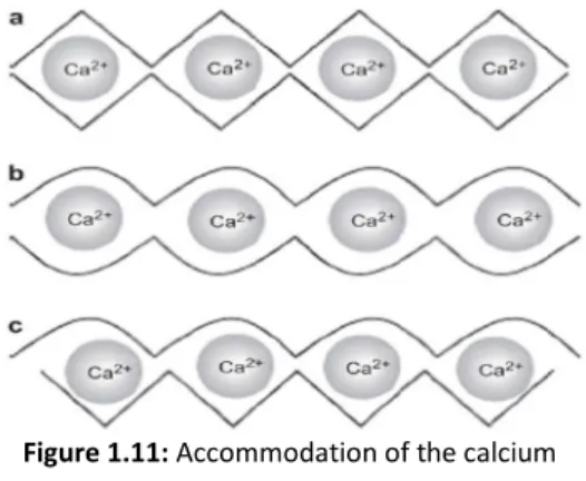
(36) Chapter 1. Figure 1.10: (a): Chemical structure of alginate monomers; (b): Example of a [adapted from [36]] monomers sequence of alginate.. The two monomers present distinct conformations (M presents 4C1 chair conformation and G presents 1C4 boat conformation) to allow the bulky carboxyl group being in the energetically best equatorial position.[36,. 39]. Thus, due to the different. monomers conformations, the polysaccharide one will be influenced by the orientation of the glycosidic bonds, that depends of the monomers sequence in the polymer. Between two M monomers exist a diequatorial glycosidic linkage, between two G monomers exist a diaxial, in a MG sequence exist an equatorial-axial and in a GM sequence exist an axial-equatorial. So, a region composed of a M sequence (M-MM-M) reveals a flat ribbon structure, whilst a region composed of a G sequence (G-GG-G) reveals a buckle structure (figure 1.10-b), and besides that, it was reported that the stiffness of the chain blocks decreases in the order GG>MM>MG.[36, 39] • Alginate hydrogels The main property of alginate that potentiates its use in different areas, it is its ability to bind some divalent cations, such as Ca2+, Ba2+, Sr2+, Cu2+ and Pb2+ in the carboxylic groups providing the gelation of the alginate solution.[36, 39, 41, 42] Both of the constituent monomers of an alginate salt possess a carboxylic moiety, which at a pH value above its pKa (about about 3.38 and 3.65 for M and G residues,. 18. Figure 1.11: Accommodation of the calcium ions, between two alginate chains, in the different glycosidic linkages: (a) GG/GG interaction; (b) MG/MG interaction; (c) GG/MG [adapted from [42]] interaction..
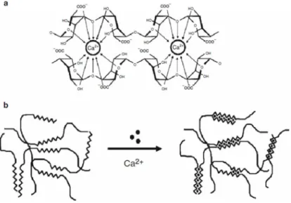
(37) Chapter 1 respectively), are negatively charged, transforming the material in a polyanion, named alginate.[35, 36, 43, 44] Thus, in these conditions, the ions responsible for the gelation can bind to the carboxylic groups, happening the cross-linking of the polymeric chains. This reaction is very rapid and irreversible.[36, 41] The two constituent monomers of alginate reveal just one difference between them, their conformation, whic results in a selective ion-binding phenomenon. It has been reported that the divalent ions have a higher tendency to bind to guluronate blocks of the polymer chains. This fact is owed to the geometrical requirements of the cavity originated by the diaxial linkage between the G monomers, that allows a higher degree of accommodation of the divalent ions than the other glycosidic linkages types (figure 1.11). Each cross-linking ion interacts with two consecutive G monomers of a polymeric chain and with two consecutive G monomers of an adjacent polymeric chain. This phenomenon establish junctions between the chains, what is named of the egg-box model of crosslinking (figure 1.12), forming a gel structure.[16, 36, 39-42]. Figure 1.12: The egg-box model of cross-linking: (a): binding of the divalent cations in the G monomers of two polymeric chains; (b): formation of [adapted from [36]] junction zones between polymeric chains.. Therefore, the properties of the gel are dependent of the ratio between M and G monomers (M:G ratio). If the proportion of the G monomer is higher, it will be obtained a strong brittle gel. On the other hand, if the proportion of the M monomer is higher, the formed gel will be weaker, but more flexible, because there are less junction zones between the polymer chains.[16, 36, 40]. 19.
(38) Chapter 1 Besides that, it is possible to produce a hydrogel of alginate with another associated polymer, in order to better some alginate properties or even to obtain, in the gel, some inexistent features in the alginate. As alginate is a polyelectrolyte, more specifically a polyanion, it can be ionically associated with a polycation existent in the same solution through hydrogen bonding or electrostatic interactions, forming a polyelectrolyte complex. Although, to achieve this complex formation it is necessary to control the solution pH to ensure that the two polymers are on the charged state. So, the solution pH has to be above the pKa of the polyanionic polymer, and below the pKa of the polycationic polymer to obtain two polyelectrolytes oppositely charged.[35, 40, 42, 45]. • Physical-chemical Properties Molecular-weight and Viscosity Alginates are considered polydisperse relatively to the molecular weight. This fact can be justified by two aspects: alginate is not gene-encoded, their production is enzymatically. controlled;. the. extraction. process. provokes. a. significant. depolymerisation of the polymer chains. Thus, due to the polydispersity in the polymerization degree of each sample, the molecular weight of an alginate is an average of the different existent molecular weights.[36, 41] The viscosity observed in alginate solutions depends mostly of the temperature, alginate molecular weight, solution concentration and pH value. With an alginate molecular weight increase it is verified a viscosity increase of the solution, to an increase in the concentration happens the same.[36, 40, 41] When an alginate solution is exposed to a temperature increase its viscosity decreases, what is owed to a depolymerization of the polymeric chains.[36,. 41]. With a pH decrease, the viscosity. slightly increases and it is maximum when the pH value of the solution is below the pKa of the alginate, because the carboxylic groups along the chains become protonated forming hydrogen bonds.[40]. 20.
(39) Chapter 1 Rheological properties About the rheological properties of alginates hydrogels, several studies have proved that these materials present a non-Newtonian behavior, which means that the shear rate and the shear stress are not directly proportional, so the viscosity depends of the shear rate (apparent viscosity, η).[46-49] More exactly, they present a shear thinning behavior, the viscosity decreases when the shear rate increases. Besides that, they are classified as presenting a viscoelastic behavior. A viscoelastic fluid simultaneously presents typical properties of a fluid (viscous) and a solid (elastic). The elasticity of a viscoelastic fluid, which is subjected to a not hydrostatic stress state, is revealed by its capacity of partially invert the deformation process after the removal of the stress, resembling to the elastic response of a solid.[36, 41, 46-49] • Biocompatibility In vitro and in vivo alginate biocompatibility has been widely studied. Although, it is considered as a biocompatible material, some researchers still disagree regarding the impact of the material in the organism. It has already been reported that alginates with high content of M monomer were immunogenic and induce cytokine production about 10 times more than alginates with high G monomer content. On the other hand, in some other studies alginate implants with a high M percentage were used, and it was observed an insignificant or even none immunoresponse.[40, 50, 51] • Biodegradation Alginate, like the other biodegradable polymers, when exposed to the body fluids and tissues can be degraded by chemical oxidation. When a material is implanted into the body, happens a normal and minimal inflammatory response to the foreign material. In this response inflammatory cells, mainly leukocytes and macrophages, produce highly reactive oxygen species (for example, superoxide O2– and hydrogen peroxide H2O2), which can cause the cleavage of the polymeric chains through their oxidative effect.[52] Besides that, alginate can also be degraded by non-enzymatic hydrolysis, which consists in the cleavage of the chemical bonds in the polymer structure by the attack of water, resulting in the formation of oligomers and monomers. In addition, the hydrolysis process can also be catalyzed by the enzyme alginase, which catalyzes the. 21.
(40) Chapter 1 cleavage of the (1-4)-glycosidic bond by a β-elimination reaction, breaking down the polymeric chains.. [36, 39, 40, 52]. Nevertheless, this enzyme does not exist in mammals.. However, alginate hydrogels that were ionically crosslinked can be dissolved in vivo through the cross-linking ions release into the surrounding media. This phenomenon is owed to an exchange process with monovalent cations present in the media, mainly Na+ ions. This happens when alginate gels are exposed for some time (several hours) to a physiological concentration of Na+. By this way, for medical applications, the average molecular weight of alginate must be a concern, it must be as low as possible, in order to respect the renal clearance threshold to ensure a complete removal of the substance from the organism.[36, 39, 40, 53] • Biomedical Applications Due to the biocompatibility, non-toxicity, biodegradability and relatively low cost of alginates, they have been studied to be used in several biomedical applications.[39, 40, 42, 54]. In the case of bone tissue engineering, to promote bone regeneration, alginate hydrogels have shown potential to be used as a structure to encapsulate and release osteoinductive factors and bone forming cells inside the body.[29, 40, 53] Furthermore, alginate hydrogels have also been used as a vehicle of inorganic materials, such as HAP, allowing its injectability.[34, 50, 55, 56] Thus, the synthetic bone substitutes can be introduced into the bone defect in a minimally invasive way and allow to fill the whole bone defect, even the most irregular one.[34, 40, 56] However, an alginate matrix can reveal some disadvantages in biomedical applications, namely, a reduced cell adhesion and unstable and weak mechanical properties.[37, 40, 42, 53, 54] Although, to improve the low cell adhesion, alginate hydrogels have the advantage of being easily chemically modified with adhesion ligands (such as Arginine-Glycine-Aspartic acid residues (RGD)), and associated with another polyelectrolyte that presents a better cell adhesion, such as chitosan.[16, 29, 37, 40, 54, 57] Similarly, the mechanical properties can also be improved through the association of alginate with another polymer that presents better properties.[37, 40, 54, 57]. 22.
(41) Chapter 1 1.2.2.2. Chitosan Chitosan is a natural and hydrophilic copolymer, and it is composed by two monomeric units, D-glucosamine and N-acetyl-D-glucosamine linked by β(1–4)glycosidic bond, as presented in figure 1.13-B.[16, 58-60] This linear polysaccharide has been widely studied for medical applications, due to many properties that it presents, such as, biocompatibility, biodegradability, non-toxicity, antimicrobial activity, noncarcinogenicity, notable affinity to proteins, promotion of cell adhesion, as well as proliferation and differentiation.[53, 58-61] The cell adhesion is mainly promoted by the fact of chitosan having a positive charged surface, which allows this phenomenon to occur on the surface of the implanted biomedical material, due to the negatively charged cell surface.[57, 61] This polymer is obtained from the chitin (figure 1.13-A), which is extracted from exoskeleton of crustaceans, like crabs and shrimps, and even from cell walls of several fungi.[16,. 35, 59]. Chitosan results from the alkaline deacetylation of the chitin, which. means, the removal of some acetyl groups. Thus, the ratio between the two monosaccharides depends of the deacetylation degree (DD), what determines the percentage of amino groups in the polymeric chain. [16, 59]. D-glucosamine. N-acetyl-D-glucosamine. Figure 1.13: Chemical structure of: A. chitin; B. chitosan.. [adpted from [61]]. At neutral or basic pH conditions, chitosan contains free amino groups, being insoluble in water. But in weekly acidic solutions, with a pH lower than its pKa (which varies between 6.2 and 7), the amino groups of the D-glucosamine monomers along the molecular chains are protonated, making chitosan soluble in water. In these conditions, chitosan is in the polycationic form and it can ionically complex with. 23.
(42) Chapter 1 polyanions, for instance, the alginate, resulting in a polyelectrolyte complex formation.[35, 45, 53, 61] Chitosan insolubility at neutral pH is a limitation in some medical applications, because most biological applications for chemical substances need the material to be processible and functional at that pH value. The chitosan solubility is mainly influenced by its molecular weight and degree of deacetylation. Consequently, some techniques have been developed to lower the molecular weight of chitosan. This phenomenon is achieved by the hydrolysis of the polymeric chains, in order to produce chitosan salts which are soluble in water. To achieve the depolymerization of the chains there are chemical, physical and enzymatic techniques, for instance, the chemical process can be done by acidic hydrolysis promoted by HCl, resulting in the salt chitosan HCl.[59, 61] In vivo, chitosan, like alginate, can be degraded by chemical and enzymatic oxidation, non-enzymatic hydrolysis and enzyme-catalyzed hydrolysis. This last process can be oriented by different proteases, such as, lysozyme, papain, chitosanase and pepsin. The lysozyme, which is present in all mammalian tissues, seems to be the main protease involved in chitosan degradation by catalyzing the acetylated residues hydrolysis. Thus, the degradation rate of the polymer is inversely dependent of the DD, for a higher DD it is observed a lower rate of enzymatic degradation.[53,. 61, 62]. The. products of its biodegradation are non-toxic oligosaccharides of variable length, that can be incorporated to glycosaminoglycans and glycoproteins, to metabolic pathways or be excreted by the organism.[52, 53, 61] 1.2.2.3. Hyaluronic Acid (HA) HA is a natural, hydrophilic and non-sulfated glycosaminoglycan. This polymer is a linear polysaccharide, in which, the repeating unity is a disaccharide composed by two monomers, D-glucuronic acid and N-acetyl-D-glucosamine, linked through alternating β(1-3) and β(1-4)-glycosidic bonds, figure 1.14.[63-66] HA has been very investigated to be used as a biomaterial in various medical applications, due to its biocompatibility, biodegradability, and nonimmunogenicity.[60, 64, 65]. 24.
(43) Chapter 1. N-acetyl-D-glucosamine. D-glucuronic acid. Figure 1.14: Chemical structure of the two monosaccharides of [adpted from [68]] hyaluronic acid.. HA is the main component existent in the ECM of living tissues, namely in the connective, epithelial and neural.[60,. 64, 65, 67]. This polymer, due to its structural and. biological properties, has the ability to mediate the cell signaling and behavior, and the matrix organization. HA is able to interact with some cell surface receptors, being involved in the tissue hydrodynamics, cell migration and proliferation.[60, 64, 65] This natural polymer HA is water-soluble and it forms highly viscous solutions with specific viscoelastic properties.[53] At a pH value above its pKa of approximately 3, HA is in the polyanionic form due to the deprotonation of the carboxylic groups existent in the D-glucuronic acid monomers, being usually called as hyaluronate or hyaluronan.[60, 66] When the polymer is in this form it can also establish, as alginate and chitosan, ionic interactions with oppositely charged polyelectrolytes.[45, 60] In the case of bone, HA has been discovered, in high concentrations, in the early fracture callus, in the osteoprogenitor cells cytoplasm, and in lacunae, in the growth plate, surrounding chondrocytes.[63] Besides that, it was shown that high molecular weight HA can inhibit osteoclasts differentiation and can be involved in the migration of mesenchymal stem cells. Therefore, HA is a material with a big potential to be used in bone tissue engineering to treat bone defects or fractures, stimulating the bone repair process.[67] In vivo conditions, the enzyme-catalyzed hydrolysis of HA is oriented by hyaluronidases. These enzymes catalyze the hydrolysis of the β-N-acetyl-Dglucosaminidic linkages in the polymer chains, resulting in the formation of mono and disaccharides, which then are converted into ammonia, carbon dioxide and water via the Krebs cycle. Moreover, HA can also be degraded by chemical and enzymatic oxidation and non-enzymatic hydrolysis. [52, 53, 68, 69]. 25.
(44) Chapter 1. 1.3. Injectable synthetic bone substitutes In order to improve the clinical potential of the synthetic bone substitutes, scientists have developed injectable bone substitutes systems able to mould to the shape of the bone defect.[24,. 46, 50, 70]. This minimally invasive surgical approach can. decrease the surgery time, reduce the damages caused by large muscle retraction, reduce the size of scars and the post-operative pain, which provides a more rapid and economic recovery of the patient.[70,. 71]. Additionally, these systems are even more. beneficial in clinical situations that present difficulties in the access to the bone defect, namely in the augmentation of osteoporotic fractures, in some applications in the spine and in the treatment of maxillofacial defects.[70, 71] Therefore, to produce the injectable systems, the association of synthetic bone substitutes with hydrogels has been proposed, and several products based on this concept are already available in the market. Some examples are presented in table 1.[24, 72] Table 1: Examples of injectable bone substitutes. Product. .[adapted from [24,72]]. .. Composition. Company. MBCP Gels ®. Biphasic calcium phosphate (BCP) granules (60% HAP, 40% β-TCP; 0.08-0.2mm) and 2% HPMC.. Biomatlante (France). Pepgen P-15®. HAP (0.25-0.42mm), P-15 peptide and aqueous sodium hyaluronate solution.. Dentsply (USA). HAP (20-30%) and collagen.. DePuy Spine (USA). β-TCP (80%) and type 1 collagen (20%).. Integra LifeSciences (USA). β-TCP granules (0.125-0.71mm; 94%) and recombinant sodium hyaluronate(6%).. Mathys Ltd (Switzerland). BCP (85% HA, 15% β-TCP) and bovine collagen.. Medtronic (USA). Bioglass and synthetic binder.. NovaBone (USA). β-TCP granules and carboxymethyl cellulose (CMC).. Stryker Biotech (USA). β-TCP granules and polymer.. Therics (USA). BCP granules (65% HA, 35% β-TCP; 0.5-1.0 mm), bovine collagen, and bone marrow aspirate.. Zimmer (USA). ®. Healos Fx ™. Integra Mozaik. Ceros® TCP Putty/ cyclOS® Putty Mastergraft® NovaBone® Calstrux™ Therigraft™. Collagraft. 26.
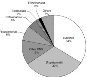
(45) Chapter 1. 1.4. Implant-associated infections The risk of infection is present in every surgical process, namely when a biomaterial is implanted into the body, such as a synthetic bone substitute in orthopaedic and maxillofacial surgery. The occurrence of infection can negatively affect the regenerative ability to the implanted material, increasing the patient recovery time and associated costs.[73,. 74]. In orthopedic surgery the main bacteria. responsible for infections are: Staphylococcus aureus, Staphylococcus epidermidis and Pseudomonas aeruginosa, as presented in figure 1.15.[73, 75] The decreased hygienic. infections. incidence. has. by. antibiotics. and. using. protocols.. However,. these. protocols are not efficient enough and the recurrent using of antibiotics can lead to antibiotic-resistant bacteria, not being affected by their action.[73, 76] Thus, ideally, a bone substitute should not only perform its supposed regeneration function, but Figure 1.15: The main pathogenic species among. also have the ability to prevent infection. orthopedic implant associated infections.. [adapted from[73]]. by microorganisms colonization. Therefore, alternative methods to avoid the infection phenomenon have been tried, such as, the incorporation in the biomaterials of effective ions against bacteria growth. The most studied and used ion is Ag+, which has already revealed an efficient antimicrobial activity against several microorganisms. However, when this ion is used in high concentrations it can be toxic to mammals.[74] More recently, it has been investigated the antimicrobial ability of some lanthanides, such as Ce(III). This ion has revealed an efficient bacteriostatic and bactericidal ability against several microorganisms. Moreover, it has been reported as non-toxic to mammals even at high concentrations.[77, 78]. 27.
(46) Chapter 1. 1.5. Motivation and objectives A few millions of patients worldwide, per year, need a bone graft or synthetic bone substitute to repair a bone defect resulting from an injury or a disorder. For instance, in USA, more than 500.000 bone grafting procedures are happening to treat bone defects, annually. The associated costs from the provided medical care by this country, in orthopaedic defects, are estimated to be around $849 billion/year. In last few years, several synthetic injectable bone substitutes have been developed due to the advantages already discussed regarding to its easy application in certain clinical conditions. Moreover, one of the most attractive features of injectable bone substitutes, it is the possibility to grant additional properties to the bone substitute through the associated hydrogel. The hydrogel chemical structure and morphology can improve the cell adhesion on the substitute; it can have incorporated osteogenic cells, osteoinductive factors or even therapeutic agents. So, the hydrogel association to the substitute can promote a better integration of the implant with the host tissue, which can improve the overall treatment outcome. Therefore, the main objective of this master dissertation was to develop a hydrogel to associate with GR-HA granules, allowing their easy injection and application in a bone defect. The developed injectable system is intended to be used in orthopaedic and maxillofacial bone defects. Furthermore, the hydrogel should be biocompatible and biodegradable, being the desired degradation time of about 2 or 3 days. The vehicle should also enhance the osteoconductive properties of the bone substitute, improving the bone regeneration process. As an additional property, the vehicle should also present an antimicrobial activity against the main microorganisms involved in bone infections. The developed material should be properly characterized in a physical-chemical and biological (in vitro and in vivo) point of view.. 28.
Imagem
![Figure 1.2: Structure of the long bones. [adapted from [1]]](https://thumb-eu.123doks.com/thumbv2/123dok_br/17802857.840952/21.892.146.761.839.1100/figure-structure-long-bones-adapted.webp)
![Figure 1.5: Structure of the cancellous bone. [adapted from [1]]](https://thumb-eu.123doks.com/thumbv2/123dok_br/17802857.840952/23.892.281.617.248.506/figure-structure-cancellous-bone-adapted.webp)
![Figure 1.8: Phases of the bone repair process. [adapted from [1]]](https://thumb-eu.123doks.com/thumbv2/123dok_br/17802857.840952/30.892.113.745.701.936/figure-phases-bone-repair-process-adapted.webp)
![Figure 1.9: Different bone disorders. [1,2,10]](https://thumb-eu.123doks.com/thumbv2/123dok_br/17802857.840952/31.892.140.748.468.1039/figure-different-bone-disorders.webp)
+7
![Figure 1.13: Chemical structure of: A. chitin; B. chitosan. [adpted from [61]]](https://thumb-eu.123doks.com/thumbv2/123dok_br/17802857.840952/41.892.285.631.678.911/figure-chemical-structure-chitin-b-chitosan-adpted.webp)
![Figure 1.14: Chemical structure of the two monosaccharides of hyaluronic acid. [adpted from [68]]](https://thumb-eu.123doks.com/thumbv2/123dok_br/17802857.840952/43.892.313.599.117.242/figure-chemical-structure-monosaccharides-hyaluronic-acid-adpted.webp)
Documentos relacionados