Magda Sofia Catroga Ferreira
Licenciada em Bioquímica
Antimicrobial Effect of Polymeric
Biomaterials for Bone Infection Treatment
Dissertação para obtenção do Grau de Mestre em Bioquímica
Orientador: Ana Bettencourt, Professora Doutora, Faculdade
de Farmácia, Universidade de Lisboa
Co-orientador: Luísa Jordão, Professora Doutora, Instituto
Nacional de Saúde Dr. Ricardo Jorge
Júri:
Presidente: Prof. Doutor Carlos Alberto Gomes Salgueiro Arguente(s): Prof. Doutora Lídia Maria Diogo Gonçalves
Vogal(ais): Prof. Doutora Ana Francisca de Campos Simão Bettencourt
iii
Copyright
“Antimicrobial Effect of Polymeric Biomaterials for Bone Infection Treatment”
Copyright © Magda Sofia Catroga Ferreira, Faculdade de Ciências e Tecnologia, Universidade Nova de Lisboa
v
Acknowledgements
Firstly, I would like to thank my supervisors. To my main supervisor, Professor Ana Bettencourt from Faculdade de Farmácia da Universidade de Lisboa, and to my co-supervisor, Dra. Luísa Jordão from Instituto Nacional de Saúde Dr. Ricardo Jorge. For accepting me in this project, for all the support, help, advices, teaching, for guiding me in the best possible way during all stages of this work and for making me feel comfortable along this year. Without them the completion of this work would not have been possible.
Also, a special acknowledgment to Professor Lídia Gonçalves for all the support, teaching, advice and time given to me even without being her responsibility. To Inês Ferreira for all the support and teaching at the beginning of my work. To Professor António Almeida for accepting me in his research group and to all master and PhD students from NanoBB (Lab112) and Chem Tox research group for the companionship.
To the institutions who received me to develop my master’s dissertation and for all opportunities
presented, Faculdade de Farmácia da Universidade de Lisboa and Instituto Nacional de Saúde Dr. Ricardo Jorge.
To Faculdade de Ciências e Tecnologia da Universidade Nova de Lisboa, for an academic education of excellence.
To my mother, father, sister and maternal grandparents for all support, emotional and financial, affection and patience not only this year but throughout my academic formation.
Last but not least, to João Morgado for being always present, for all the help mainly with writhing in English and for unconditional love.
Funding:
vii
Abstract
Bone infection, mainly caused by Staphylococcus aureus, is a public health concern. Treatment is
challenging due to multi-resistant strains, and S. aureus ability to adhere and form biofilm on bone and implant surfaces, as well as to invade and persist in osteoblast cells.
The present work consisted in the preparation and evaluation of novel acrylic polymeric systems that provide local and controlled antibiotic delivery for the treatment of bone infection, namely levofloxacin-loaded acrylic bone cement (BC), and vancomycin or daptomycin-levofloxacin-loaded acrylic microparticles (MP).
Properties of both delivery systems with high impact on clinical performance were tested. Namely, contact angle and surface energy were determined in BC matrices and encapsulation efficiency in MP formulations. Release studies of levofloxacin-loaded BC matrices were also conducted. Also, the anti-biofilm activity of these systems was evaluated against S. aureus strains. Furthermore, BC and MP formulations were tested concerning the antibacterial intracellular activity using a human osteoblast infection model.
Overall, both BC formulations’ surface characteristics and MP encapsulation efficiency were in
agreement with previously published data. The release studies of levofloxacin from BC matrices showed that the drug release is size- and incubation medium-dependent. All BC matrices loaded with levofloxacin concentrations of 1.5 % or higher exhibited anti-biofilm activity against all S. aureus tested
strains. For BC matrices and Vancomycin-loaded MP, a decrease of viable intracellular bacteria was observed. For Daptomycin-loaded MP, no viable intracellular bacteria were detected.
In conclusion, this work has shown that the BC formulations with drug concentration of 1.5 % or 2.5 % and daptomycin-loaded MP show potential to be used in the context of bone infection treatment.
Keywords: Bone infection; bone cement; microparticles; levofloxacin; Staphylococcus aureus; intracellular
ix
Resumo
A infeção óssea, causada principalmente por Staphylococcus aureus, é um grave problema de saúde
pública. O seu tratamento é difícil devido a estirpes multi-resistentes, à sua habilidade de aderir e formar biofilmes em osso e implantes, bem como à sua capacidade de invadir e persistir em osteoblastos.
Este trabalho consiste na preparação e avaliação de novos sistemas poliméricos acrílicos, que promovem a libertação local e controlada de antibióticos para o tratamento de infeções ósseas, nomeadamente cimento ósseo (BC) carregado com levofloxacina, bem como vancomicina ou daptomicina encapsulada em micropartículas (MP).
Foram testadas propriedades com elevado impacto no desempenho clínico de ambos os sistemas, nomeadamente ângulos de contacto e energia de superfície das matrizes de BC, assim como a eficiência de encapsulação das formulações de MP. Foram também realizados estudos in vitro de
libertação de levofloxacina das matrizes de BC e da sua atividade anti-biofilme contra estirpes de S. aureus. Usando um modelo de infeção de osteoblastos humanos, ambos os sistemas foram avaliados
quanto à sua atividade antibacteriana intracelular.
Tanto as características de superfície das matrizes de BC como a eficiência de encapsulação de MP estão em concordância com estudos realizados anteriormente. Os estudos de libertação de levofloxacina a partir de diferentes formulações de BC revelaram que esta é dependente do tamanho das placas e do meio de incubação. As formulações de BC com concentração de fármaco igual a 1.5 e 2.5 % demonstraram atividade anti-biofilme. Todas as formulações testadas reduziram o número de bactérias intracelulares viáveis, sendo mais eficazes as MP com daptomicina.
Este trabalho conclui que as formulações de BC com concentração de fármaco igual a 1.5 e 2.5 % e as MP carregadas com daptomicina demonstram potencial para utilização no tratamento de infeções ósseas.
xi
Index
Acknowledgements ... v
Abstract……… ... vii
Resumo…….... ... ix
Index…….…… ... xi
Index of figures ... xv
Index of tables. ... xvii
List of abbreviations ... xix
Objective and thesis structure ...1
Chapter 1. Introduction ...3
1.1. Bone infection ...3
1.1.1. Aetiology ...5
1.1.2. Bone infection caused by S. aureus ...6
1.1.3. Bone infections caused by S. aureus biofilms ...7
1.1.4. Intracellular S. aureus infection in osteoblasts ...9
Conventional antibiotic treatment of bone infection ... 11
1.2.1. Fluoroquinolones: Levofloxacin ... 13
1.2.2. Glycopeptides: Vancomycin ... 15
1.2.3. Lipopeptides: Daptomycin ... 16
Local drug delivery systems as new therapeutic strategies ... 20
1.3.1. Antibiotic-loaded acrylic bone cement (ALABC) ... 21
1.3.2. Antibiotic-loaded polymeric microparticles ... 23
Compliance of novel local drug delivery systems ... 25
1.4.1. Surface studies ... 25
1.4.2. In vitro drug release studies ... 25
1.4.3. Microbiological assays ... 26
1.4.4. Antibacterial intracellular activity assays ... 26
Chapter 2. Materials and methods ... 27
Preparation of biomaterials... 27
2.1.1. Acrylic bone cement ... 27
2.1.2. Polymeric microparticles ... 28
Characterization of biomaterials ... 29
2.2.1. Acrylic bone cement ... 29
2.2.1.1. Contact angle and surface energy determination ... 29
2.2.1.2. In vitro release studies ... 29
2.2.1.2.1. Levofloxacin release assays in PBS ... 29
2.2.1.2.2. Levofloxacin release assays in culture media ... 31
2.2.1.2.3. Release of lactose ... 31
2.2.2. Polymeric microplarticles ... 32
xii
Microbiological assays ... 33
2.3.1. Bacterial strains and cell line ... 33
2.3.2. Biofilm assembly ... 34
2.3.3. Bacteria susceptibility to levofloxacin ... 34
2.3.3.1. Standard levofloxacin ... 34
2.3.3.2. Levofloxacin released from BC matrices ... 35
2.3.3.3. Levofloxacin-loaded BC matrices ... 35
2.3.4. Biofilm assembly by scanning electron microscopy (SEM) ... 35
Antibacterial intracellular activity ... 36
2.4.1. Survival assay ... 36
2.4.1.1. Acrylic bone cement ... 36
2.4.1.2. Polymeric microparticles ... 36
2.4.2. Intracellular distribution of S. aureus ... 37
2.4.3. Statistical analysis ... 37
Chapter 3. Results and discussion ... 39
Characterization of biomaterials ... 39
3.1.1. Acrylic bone cement ... 39
3.1.1.1. Contact angle and surface energy determination ... 39
3.1.1.2. In vitro release studies ... 40
3.1.1.2.1. Levofloxacin release assays in PBS ... 41
3.1.1.2.2. Levofloxacin release assays in culture media ... 42
3.1.1.2.3. Release of lactose ... 45
3.1.2. Polymeric microparticles ... 46
3.1.2.1. Preparation and characterization ... 46
Microbiological assays ... 47
3.2.1. Biofilm assembly ... 47
3.2.2. Bacteria susceptibility to levofloxacin ... 48
3.2.2.1. Standard levofloxacin ... 48
3.2.2.2. Levofloxacin released from BC matrices ... 50
3.2.2.3. Levofloxacin-loaded BC matrices ... 51
3.2.2.4. Biofilm assembly by scanning electron microscopy (SEM) ... 51
Antibacterial intracellular activity ... 52
3.3.1. Intracellular distribution of S. aureus ... 52
3.3.2. Acrylic bone cement ... 53
3.3.3. Polymeric microparticles ... 55
Chapter 4. Conclusion and future work ... 57
Chapter 5. Bibliography ... 61
Chapter 6. Annex ... 71
A – Contact angle and surface energy determination ... 71
xv
Index of figures
Figure 1.1 – Sequential phases of the formation of biofilms on a biomaterial surface. ...8
Figure 1.2 – Schematic diagram of current model of the S. aureus invasion mechanism. ... 10
Figure 1.3 – Structure of levofloxacin. ... 13
Figure 1.4 – Mechanism of action of levofloxacin. ... 14
Figure 1.5 – Structure of vancomycin. ... 15
Figure 1.6 – Mechanism of action of vancomycin... 16
Figure 1.7 – Structure of daptomycin. ... 17
Figure 1.8 – Mechanism of action of daptomycin. ... 19
Figure 1.9 – Structure of PMMA. ... 22
Figure 1.10 – Artificial hip replacement – cemented prosthesis. ... 22
Figure 1.11 – Schematic representation of the techniques commonly used in the preparation of PMMA particulate drug carriers... 24
Figure 2.1 – A) Aluminium moulds used for preparing the BC plates; B) Example of a BC matrix. ... 28
Figure 2.2 – Bone cement matrices. ... 30
Figure 2.3 – Example of calibration curve of levofloxacin in water, by HPLC measurement. ... 30
Figure 2.4 – Example of calibration curve of levofloxacin, by fluorescence measurement. ... 31
Figure 2.5 – Representation of the oxidation reaction of a general reducing sugar, in the presence of 3,5-Dinitrosalicilic. ... 32
Figure 2.6 – Example of calibration curve of Lactose, by DNS method... 32
Figure 2.7 – Examples of calibration curves for determination of encapsulation efficiency of antibiotics. ... 33
Figure 3.1 – A liquid droplet on the biomaterial surface. ... 39
Figure 3.2 – Results obtained for the surface energy of the BC matrices. (A) Total (γ); (B) Dispersive (γd); (C) Polar (γp). ... 40
Figure 3.3 – Kinetic of biofilm assembly for S. aureus strains... 48
Figure 3.4 – SEM images of biofilm assembly. ... 52
Figure 3.5 – Intracellular distribution of S. aureus. ... 53
Figure 3.6 – In vitro survival assay of intracellular MSSA. ... 54
Figure 3.7 –In vitro survival assay of intracellular MRSA strains... 54
Figure 3.8 –In vitro survival assay of intracellular MSSA. ... 56
Figure 6.1 – In vitro accumulative release profiles of levofloxacin. ... 71
Figure 6.2 –In vitro release of levofloxacin. ... 72
xvii
Index of tables
Table 1.1 – Summary of causative organisms. ...5
Table 1.2 – Bone penetration of antibiotics. ... 12
Table 1.3 – Local drug delivery systems. ... 20
Table 1.4 – Components of bone cement. ... 22
Table 2.1 – Composition of the commercial acrylic bone cement DePuy CMW1®. ... 27
Table 2.2 – Composition of different BC formulations. ... 27
Table 3.1 – Results obtained for advancing contact angle surface in water and in 1,2-propanodiol. ... 40
Table 3.2 –In vitro results of levofloxacin cumulative release (µg/mm2) in PBS. ... 41
Table 3.3 –In vitro results of levofloxacin cumulative release (µg/mL) in PBS. ... 42
Table 3.4 –In vitro results of levofloxacin cumulative release (µg/mm2) after 24 h of incubation in RPMI or MH broth obtained by fluorescence technique. ... 43
Table 3.5 –In vitro results of levofloxacin cumulative release (µg/mL) after 24 h of incubation in RPMI or MH broth obtained by fluorescence technique. ... 44
Table 3.6 –In vitro results of levofloxacin cumulative release (µg/mm2). ... 44
Table 3.7 –In vitro results of levofloxacin cumulative release (µg/mL). ... 45
Table 3.8 –In vitro results of lactose cumulative release (µg/mm2) after 24 h of incubation in PBS or MH broth obtained by DNS method. ... 46
Table 3.9 – Encapsulation efficiency (EE) and drug loading (DL) for PMMA-EUD microparticles formulations. ... 47
Table 3.10 – Results obtained for S. aureus strains susceptibility to free levofloxacin. ... 49
Table 3.11 –S. aureus strains susceptibility to levofloxacin released from BC matrices. ... 50
Table 6.1 – Results obtained for determination of contact angle and surface energy of the BC matrices. ... 71
Table 6.2 – Composition of MH broth. ... 73
xix
List of abbreviations
Ala– Alanine
ALABC– Antibiotic-loaded Acrylic Bone Cement ATCC - American Type Culture Collection ATP– Adenosine triphosphate
a.u.– Absorvance units BC– Bone Cement
Bis-GMA/TEGDMA –2,2’-bis-[4-(methacryloxypropoxy)-phenyl]-propane/Tri(ethyleneglycol) dimethacrylate
BPO– Benzoyl peroxide CFU– Colony Forming Unit
CLSI – Clinical & Laboratory Standards Institute CPC – Calcium Phosphate Cement
CRP– C-reactive Protein CT– Computed Tomography Dapto– Daptomycin
DBM– Demineralized Bone Matrix DCM– Dichloromethane
DL– Drug Loading
DMPT– N, N-Dimethyl para-toluidine DMpt– Dimethyl para-toluidine DNA– Deoxyribonucleic Acid DNS– Dinitrosalicylic Acid
Eap– Extracellular Adhesion Protein ECM– Extracellular Polymeric Matrix EE – Encapsulation efficiency
ESR - Erythrocyte Sedimentation Rate EUD – Eudragit RL 100
FDG-PET – 18-fluoro-D-deoxyglucose Positron Emission Tomography FnBPA– Fibronectin Binding Protein A
FnBPB– Fibronectin Binding Protein B GlcNAc–N-acetylglucosamine Acid HA– Hydroxyapatite
HAI – Healthcare Associated Disease
HPLC– High Performance Liquid Chromatography Lac– Lactose
Lev – Levofloxacin
xx MIC90– Minimum Inhibitory Concentration necessary to inhibit bacteria growth in 90 %
MH –Mϋeller-Hinton MMA– Methyl methacrylate MOI– Multiplicity of Infection MP– Microparticles
MRI – Magnetic Resonance Image
MRSA– Methicillin-resistance Staphylococcus aureus
MSCRAMM– Microbial Surface Components Recognizing Adhesive Matrix Molecules MSSA - Methicillin-susceptible Staphylococcus aureus
MurNAc–N-acetylmuramic Acid
Nano-HA-PHBV/PEG-GM – Nano-hydroxyapatite/poly (3-hydroxybutyrate-hydroxyvalerate)-polymethyl lene glycol gentamicin drug delivery system
OD – Optical density
PBS– Phosphate Buffered Saline Pls– Plasmin-sensitive Protein PMMA – Poly(methyl methacrylate) PVA– Poly(vinyl alcohol)
RPMI– Roswell Park Memorial Institute medium
Sccmec I – Staphylococcal Chromosomal Cassette type I SCV – Small Colony Variants
SEM– Scanning Electron Microscopy SD – Standard Deviation
TRAIL– TNF-Related Apoptosis-inducing Ligand UDP– Uridine Diphosphate
UV– Ultraviolet Vanco– Vancomycin
1
Objective and thesis structure
The present thesis’ main objective is the evaluation of antibacterial activity of antibiotic-loaded polymeric biomaterial systems, namely bone cement and microparticles, against the main pathogenic organism causing bone infections (Staphylococcus aureus).
Specific aims were:
Production of polymeric systems with incorporation of respective drugs, levofloxacin in bone cement formulations and daptomycin or vancomycin in microparticles;
Brief characterization of drug delivery systems;
Release studies of drug and lactose (release modulator) from bone cement matrices for method optimization;
Assessment of antibacterial activity, namely anti-biofilm efficacy of bone cement matrices;
Evaluation of the antibacterial intracellular activity of bone cement formulations and microparticles.
This work is organized in four chapters and annexes including: Introduction, Materials and Methods, Results and Discussion, and Conclusion and Future Work.
Chapter 1 – Introduction
In Introduction, a brief clinical context of the bacterial bone infection is presented. It contains a sum of microorganisms that are usually responsible for bone infections and a description of the most common pathogen causing this disease, particularly the mechanisms and the problems with its biofilm assembly ability and its capacity to invade and persist within osteoblast cells. The challenge of the conventional antibiotic treatment and the characteristics of antibiotics used in this study are also detailed. Novel local drug delivery systems as a new therapeutic strategy including a list of some systems that have been developed, and a description of antibiotic-loaded acrylic bone cement and antibiotic-loaded polymeric microparticles. Finally, the assays that should be performed to characterize and evaluate the antibacterial activity of novel systems for applications in the treatment of bone infection. As such, for bone cement systems, surface and release studies and microbiological assays were performed and, for polymeric microparticles, encapsulation efficiency was determined. Also, for both systems, intracellular assays were conducted.
Chapter 2 – Materials and Methods
2 both antibiotic-loaded polymeric systems. The reagents, equipment and experimental protocols used in this work are detailed in this chapter.
Chapter 3 – Results and Discussion
In this chapter, all results obtained from the experiments performed throughout this study and subsequent discussion are presented. It concerns the production of bone cement and microparticles formulations, their characterization, drug and lactose release studies, as well as the effects of bone cements matrices on microbiological activity and the antibacterial intracellular performance of all formulations.
Chapter 4 – Conclusion and Future Work
3
Chapter 1. Introduction
1.1.
Bone infection
Infection is defined as a homeostatic imbalance between the host tissue and the presence of microorganisms [1]. Infections occur when pathogens (bacteria or fungi) successfully invade the host and begin to reproduce [2]. A major concern in terms of medical care is the infection involving bone, due to this being a significant cause of morbidity and mortality. Furthermore, they can result in prolonged hospital stays, long courses of systemic antibiotics and frequently will require a new surgical intervention [3].
Bone infection is associated with a variety of occurrences, mainly from complications following surgery and device implantation, leading to orthopedic implant failure and resulting in diseases such as osteomyelitis, and septic arthritis. Therefore, this infection can be acquired after bone surgery, or joint replacement, or even in consequence of a trauma. Consequently, people that suffer from immunosuppressive disorders face a higher risk of infection. Furthermore, the evidence of bacterial resistance is on the rise, and complications associated with infections are therefore expected to increase in the general population [1], [4]–[6]. This type of infection leads to necrosis and destruction of the bone. This pathology can affect all ages and involve any bone, and it can be limited to a single proportion of the bone or can involve several regions, such as marrow, cortex, periosteum, and the surrounding soft tissue, becoming a chronic disease and causing persistent morbidity [4], [7], [8].
Nowadays, with the emergence of modern standards in the control of sterility within the operating room environment and adequate protocols of peri-operative antibiotic prophylaxis, there is a decrease of the incidence of infections associated with orthopedic implants. Nevertheless, this infection still represents one of the most serious and devastating complications which may involve prosthetic devices [9]. Surgical implant procedures have become valid and extremely common procedures to restore the function of affected joints, fractured bone segments and impaired limbs. The enormous population of patients with orthopedic implants estimates only about 0.5-5 % of risk of infection. However, it has to be considered very relevant due to its serious consequences [9]. Thus, exposure to invasive medical devices is one of the most important risk factors. These devices predispose to infection by damaging or invading epithelial or mucosal barriers and by supporting growth of microorganisms, thus functioning as a reservoir [10].
The diagnostic and therapeutic of bacterial bone infections are a challenge to all clinicians [11]. The clinical diagnostic is made by different and complementary approaches, as laboratory tests, microbiological analysis of specimens and imaging modalities [3], [12]. Normally, main clinical symptoms are persisting local pain, erythema, oedema, warmth, tenderness, necrosis of wound edges, large post-operative haematoma and abrupt onset of high fever [12].
4 analysis consists of the culture of pathogens. These assays, although helpful in the diagnostic of bone infection, entail several disadvantages that influence the results of the test. One such example is the administration of antibiotics before collection of samples, or the improper management of specimens that may alter the results and the growth of microbes [12]. For imaging modalities, such as radiographs, it is only possible to collect evidence of infection up to 10-14 days after onset, although there may be some observable soft-tissue changes before. Moreover, the presence of implants makes the interpretation of radiographs difficult, decreasing the sensitivity and specificity of diagnostic active infection. Several imaging techniques besides radiography are also required for diagnostic of bone infection, mainly when the site of infection is unknown, such as 18-fluoro-D-deoxyglucose positron emission tomography (FDG-PET) or magnetic resonance image (MRI). Nevertheless, these techniques also entail a number of factors that influence the results, such as post-operative scars or artefacts to residual abrasions of metallic implants, in the case of the MRI.
When it is known that it is bone infection, computed tomography (CT) is routinely used for pre-operative planning. This method demonstrates better the details of the cortical bone, cortical erosion or destruction, peri-osteal and endoesteal reaction, as well as the presence of sequestrum and intraosseous fistulas. This happens due to optical imaging of the bone structure, regarding mineralisation and extension of disease. However, in presence of metallic implants, the CT loses quality. It is also possible to carry out directly a bone biopsy. Still, many other techniques of diagnostic in bone infection can be applied, all of which with great difficulty in interpreting the results, which makes diagnostic a challenge in clinic context [3], [12].
When an infection is diagnosed, a number of therapeutic approaches may be adopted. The selection of the proper approach depends on host factors, such as age, baseline mobility, comorbidities, etc., virulence and antimicrobial susceptibility of the infecting organism, duration of infection, prosthesis factors, such as stability of implant, loss of bone stock and soft tissue because of previous surgeries or infections, and patient expectation [5]. The treatment of bone infections involves operative debridement and chemotherapy with antimicrobial agents. However, both the simple debridement procedures with retention of prosthesis and antibiotic therapy treatments are not always effective on infections that have already established. Often, prosthesis removal and replacement, when not even joint fusion, it is the only solution to definitively eradicate severe infections [8], [9]. Another inherent problem is that the therapy with antibiotic takes long periods of time. By intravenous route, the antibiotics are prescribed for three weeks, followed by more three weeks of oral antibiotics. Furthermore, to achieve effective therapeutic drug concentration in the bone, at the precise site of infection, there is a need of high parenteral dose of antibiotic. These facts can lead to systemic toxicity of the antibiotic. [8], [13].
5 are the most relevant etiologic agents involved, the pathogenetic mechanisms leading to microbial adhesion, colonization of implant surfaces, and evasion of the host defences, the most important virulence factors, the nature and properties of microbial biofilms and the progressive alarming appearance of antibiotic resistant strains [9].
1.1.1. Aetiology
Bone infection is usually caused by bacteria but sometimes can also be caused by a fungus. The microorganisms that normally are responsible for bone infection are staphylococci, streptococci, Gram-negative bacilli enterococci and anaerobes. In Table 1.1 it is listed a series of organisms present in bone and joint infections [15]. The most common pathogens that cause these infections, mainly implant-associated infections, are Staphylococcus aureus and Staphylococcus epidermidis. S. aureus is generally responsible for metal-biomaterial, bone-joint, and soft-tissue infections. Whereas that, S. epidermidis is better known for causing polymer-associated implant infections [6], [13], [16], [17].
Table 1.1 – Summary of causative organisms.
Disease Causative organisms
Osteomyelitis Staphylococcus aureus (90 % adults, >50 % children); Staphylococcus epidermidis;
Streptococcus groups A, B, C and G;
Gram-negative bacilli (e.g. Escherichia coli, salmonella);
Polymicrobial (5 %);
Brucella Spp; Fungi
Haemophilus influenzae;
Kingella Kingae;
Bartonella Henselae;
Septic arthritis Staphylococcus aureus;
β-Haemolytic Streptococci;
Streptococcus pneumoniae; Prosthetic joint infection Early (<3 months after surgery):
Staphylococcus aureus, Staphylococcus epidermidis; β
-Haemolytic Streptococci and enterococci;
Delayed (3-24 months after surgery): coagulase-negative
staphylococci, Propionobacterium acnes;
Late (>24 months after surgery): Staphylococcus aureus, Gram-negative bacilli.
6 1.1.2. Bone infection caused by S. aureus
S. aureus is considered to be a major virulent pathogen, capable of colonizing and infect hospitalized patients with decreased immunity, as well as health immuno-competent people in the community [17]. Therefore, this organism has a more relevant approach in this work.
S. aureus is a gram-positive facultative anaerobic bacterium and it is found naturally on the skin and in
the nasopharynx of the human body. It is also found within environment hospital, mainly resistant S. aureus strains. Members of the staphylococci genus, as S. aureus, are catalase-positive and its cell wall
is composed of peptidoglycan and teichoic acids, attached to which are adhesins and exotoxins [17].
This bacterium is an important human pathogen that causes several diseases, such as skin infections, scalded-skin syndrome, toxic shock syndrome, endocarditis, septic arthritis, and bone infections. This happens because, although the skin and mucous membranes are excellent barriers against local tissue invasion by S. aureus, after they are breached due to trauma or surgery, this bacterium can enter the underlying tissue, reaching the lymphatic channels or blood vessels as well. Once it penetrates the subcutaneous tissues and reaches the lymphatic channels or blood, it can infect almost any organ and can cause septicemia, but it mostly affects bone tissue and cardiac valves. It is responsible for about 80 % of all cases of human bone infections. Infections caused by S. aureus are a worldwide public health
concern. The difficulty in treatment of clinical cases and the wide variety of staphylococcal disease, reflects the evolutionary versatility of this pathogen and the major challenges faced by health care providers [8], [17]–[20]. The treatment of bone infection is challenging, because the approaches are scarce, expensive and infections caused by S. aureus are subject to recurrence months and even years after apparently successful therapy [8].
How S. aureus targets bone may relate to its ability to adhere to bone. This capacity of this organism is directly related to its expression of the receptors (adhesins) for components of bone matrix such as fibronectin, collagen, and bone sialoprotein [6], [14]. Therefore, this bacterium is capable of forming an extracellular matrix, designated biofilm that adheres to bone. Bacteria in biofilm may cause persistent infection due to increased resistance to antibiotics [7], [21]. Additionally, S. aureus expresses toxins and exoenzymes that may defend staphylococci against immune cells and destroy host tissue, which allows the pathogens to enter in deep tissue structures [14].
Therefore, this bacterium has the capacity to persist within bone cells, as osteoblasts, facilitating the development of chronic infections, as it has been demonstrated in vitro and in vivo. This fact leads to an urgent need for development of novel therapeutic approaches in bone infection caused by this pathogen [7], [21]. Furthermore, this bacterium also expresses cytotoxic and pro-inflammatory virulence factors. [14].
The existence of highly virulent methicillin-resistance S. aureus (MRSA) is also an important and
7 penicillin, and amoxicillin) including cephalosporins, streptomycin, tetracycline, and sulphonamide. There are also reports that, upon exposure to vancomycin and other glycopeptide antibiotics, certain MRSA strains become less susceptible to these antibiotics[17], [20], [22].
1.1.3. Bone infections caused by S. aureus biofilms
The surface of implanted biomaterials may suffer attachment of a biological active layer. This includes an extracellular matrix, proteins, host cells (fibroblasts, osteoblasts, endothelial cells) and bacteria. The extracellular matrix consists in a complex mixture of macromolecules, such as fibronectin, fibrinogen, albumin, vitronectin, and collagen. This matrix serves as a substrate for the host cells as well as for bacteria, such as S. aureus [17].
S. aureus expresses many surface adhesins that promote attachment to plasma and to extracellular matrix proteins of host cells or those adsorbed onto metal or polymer surfaces, and possess a variety of adhesion mechanisms. These mechanisms include microbial surface components recognizing adhesive matrix molecules (MSCRAMMS-fibronectin binding proteins, clumping factor binding proteins, collagen binding protein), which facilitate its adhesion to biomaterials and to the extracellular matrix proteins deposited on the biomaterial surface. When S. aureus attaches to a surface, host cells are
unable to dislodge it [17].
Nevertheless, many staphylococcus strains have the capacity to produce its own extracellular matrix, resulting in biofilms [17]. Biofilms are communities of microorganisms that are attached to a solid surface, such as a biomaterial used in implants, and encased in extracellular polymeric matrix (ECM). This ECM is constituted by polysaccharides, proteins and extracellular DNA, that provides structural stability and protection to bacteria within the biofilm [23]–[25].
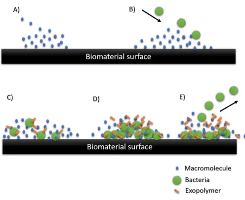
Biofilm formation on biomaterial surface occurs in four main phases (Figure 1.1). The first is the reversible adhesion of planktonic bacteria (freely moving) on a biomaterial surface, which may be covered by a layer of proteins or other host cells. In this phase, the bacteria are still susceptible to antibiotics, followed by irreversible binding to the surface and multiplication of the bacteria. This happens in a few hours, forming microcolonies and beginning to produce their own ECM around these microcolonies. In the next phase, biofilm grows in thickness to form a mature biofilm. When in vitro
8 Consequently, biofilm-growing bacteria cause chronic infections. These are infections that persist despite antibiotic therapy and innate and adaptive immune and inflammatory responses of the host [27]. It is important to note that cells within a biofilm become phenotypically different from their planktonic analogs. Bacteria in biofilms have reduced mobility, distinct gene transcription patterns, and exhibit a spectrum of metabolic activity with cells at the biofilm periphery growing more rapidly than the nutrient-deprived cells in the biofilm’s inner layers. This fact leads to significant consequences of diagnostic and
therapy of disease [27], [28].
Traditional sampling techniques may not be sufficient to culture biofilm-growing bacteria sticking to a surface, because the biofilm is subject to an ultrasonic pre-treatment for bacteria release. Thus, these techniques reveal only the properties of planktonic bacteria, for example, the results of antibiotic susceptibility testing do not reflect the increased resistance of the bacteria living in biofilms. The minimum inhibitory concentration (MIC) and minimum bactericidal concentration (MBC) of antibiotics to biofilm bacteria may be up to 100-1000 fold higher compared with free bacteria [27].It is known that bacterial biofilms develop mechanisms that provide resistance to antibiotics, disinfectant chemicals and to phagocytosis and other components of the innate and adaptive inflammatory defense system of the host [17], [23], [25]. These mechanisms consist in physical or chemical diffusion barriers to antimicrobial penetration into biofilm, in biofilm slow growth due to nutrient limitation, in activation of general stress response and the emergence of a biofilm-specific phenotype [23].
Figure 1.1– Sequential phases of the formation of biofilms on a biomaterial surface.
9 1.1.4. Intracellular S. aureus infection in osteoblasts
Cells such as neutrophils and macrophages, designated by “professional phagocytic cells”, are designed to engulf microorganisms and their subsequent death. Nevertheless, some pathogens have the ability to escape these phagocytic cells by their internalization in other cells, that may be called “non
-professional phagocytic cells”, which possess mechanisms that permit endocytic uptake of
microorganisms [20].
S. aureus is not considered a significant intracellular pathogen when compared with some
microorganisms, such as Listeria and Shigella. This bacterium has been regarded as non-invasive
extracellular pathogen that damages host cells after adhering to the extracellular matrix [8]. However, this organism has demonstrated to have the capacity to invade and persist within eukaryotic cells for long periods of time. Several studies have been undertaken in this scope in order to understand their intracellular mechanism and persistence. S. aureus internalization and intracellular persistence have been performed in endothelial cells, epithelial cells, fibroblasts, osteoblasts and keratinocytes. However, recent studies have been documenting bacterial survival also within professional phagocytes [8], [19]–
[21]. Intracellular invasion and persistence of S. aureus provides an ideal strategy to escape from
immunological defence mechanisms as well as promote protection against several classes of antibiotics, due to its limited exposure to antibiotic treatment, namely extracellular bactericides. This promotes recrudescent infection, which could explain the persistence of disease [20], [21], [29]. Thus, the internalization of S. aureus into osteoblast cells has led to an increase of concern in terms of public health care, because of its likely interconnection with persistent infections and chronic disease, which leads to a consequent increase in morbidity and mortality. This happens because host cells, in this case osteoblast cells, are responsible for the continual process of bone remodelling, together with another type of cells, the osteoclasts. Osteoclasts are responsible for driving the resorption of bone by acidification and release of lysosomal enzymes. Osteoclasts derive from myeloid precursors, whereas osteoblasts derive from mesenchymal bone marrow precursors and produce components of bone, mainly type I collagen. These cells also catalyse the calcification process and produce soluble factors that serve to modulate the activity and formation of osteoclasts. This way, osteoblasts are an important and essential component of bone, due to their purpose as a principal director of osteoclast function, and thus, control net bone formation or resorption [29].
However, the evidence that S. aureus might be a facultative intracellular pathogen is particularly demonstrated in certain subpopulations of this organism. These subpopulations, called small colony variants (SCVs), are phenotypically very different from the parent strain and naturally occurring mainly during the course of antibiotic therapy. They have a slow growth that lead to small colonies, normally these being one-tenth the size of “normal” S. aureus. SCVs show altered drug-resistance profiles and
10 integral part of the S. aureus life cycle, although it is already known for several years that SCVs cause subacute antibiotic-resistant infections [20], [30].
Nevertheless, evidence to support the hypothesis that intracellular S. aureus promotes the persistent
infection in humans or animals are few, because the current techniques aren’t viable to prove this
hypothesis. Furthermore, another recognized problem with human studies of S. aureus infection is the
time point and duration of infection, that frequently are unknown and patients often present an acute disease [20].
In bacterial bone infections, bacteria can destroy bone by several possible mechanisms, including the production of certain compounds, such as acids or proteases, or by indirectly stimulating osteoclastogenesis. Furthermore, it has been reported that S. aureus surface-associated proteins are potent stimulators of bone resorption and that stimulation of osteoclast formation due to these proteins plays a role in bone destruction. Other studies have been demonstrating that exposure of mouse and human osteoblasts to S. aureus induces the expression of tumor necrosis factor-related apoptosis-inducing ligand (TRAIL) by these cells. This fact suggests an additional mechanism whereby S. aureus
can mediate bone destruction via induction of apoptosis in bone-forming osteoblasts. However, the most important mechanisms in the pathophysiology of bacterium-induced bone resorption are not yet fully known [29].
Studies with osteoblast cell lines have been demonstrating that actin microfilaments, microtubules, and receptor-mediated endocytosis are required in the internalization of S. aureus into osteoblasts. Furthermore, other studies have revealed that the mechanism of S. aureus host cell invasion is mediated via fibronectin bridging between host α5β1 integrins and staphylococcal surface proteins, FnBPA and
FnBPB (Figure 1.2) [22], [29].
Moreover, there are other proteins known to be required for intracellular invasion. The Eap is a multifunctional protein, consisting of 4 to 6 tandem-repeat domains. Two tandem-repeat domains have been identified as minimal structural requirements for Eap-mediated host cell invasion. Eap may also Figure 1.2 – Schematic diagram of current model of the S. aureus invasion mechanism.
Host cell invasion mediated via fibronectin bridging between host α5β1 integrins and bacteria surface proteins,
11 bind fibronectin, which promotes its capacity to partially compensate for the loss of FnBP functions. Another one is an additional surface protein, Pls (plasmin-sensitive protein). Pls is a protein of MRSA located on staphylococcal chromosomal cassette type I (Sccmec I), which down-modulates invasiveness of these strains and acts by steric hindrance, rather than other mechanisms for down-modulation of host cell invasion [22].
The majority of intracellular S. aureus is contained within vesicles in osteoblasts, although some bacteria
appear free in the cytoplasm. It should be noted that, following lysis or trypsinization of the human osteoblasts, the bacteria are released while viable, which promotes the invasion onto other osteoblasts. Thus, S. aureus is sequestered from the host immune system within osteoblast cells, acting as a reservoir of bacteria, which may explain why chronic bacterial infections of bone are associated with multiple recurrences, even when in the presence of a proper humoral response [29].
Therefore, host cells have developed powerful mechanisms to destroy invading pathogens. These mechanisms generally consist of generation of reactive oxygen species, modulation of essential cations and nutrients, and degradation by proteolytic enzymes. However, some intracellular pathogens, such as S. aureus, have also developed sophisticated mechanisms to survive and persist within this
intracellular environment. One of the mechanisms employed by bacteria to escape host cells defence is to avoid lysosomal killing. This way, the invading pathogen can go into the cytoplasm [22].
Destruction of the phagosomal compartment of non-professional phagocytes cells is a prerequisite for induction of host cell death. Phagosomal escape of S. aureus has been described as pore-forming toxins as well as phospholipases for phagolysosomal membrane destruction [22].
Conventional antibiotic treatment of bone infection
The conventional treatment for bone infection, is usually the antibiotic treatment. Antibiotics are substances that either stop bacteria from growing, designated by bacteriostatic agents, or kill them, depending on their capacity to block critical bacteria cellular processes, these being referred to as bactericidal agents [2], [31].
The introduction of antibiotics in the medical field was one of the most important interventions to reduce the onslaught of many diseases. As a result, antibiotics are the economic powerhouses of our society [2].
12 Generally, antibiotics can be categorized according to their principal action mechanisms. The main action mechanisms performed by antibiotics for bone infection treatment are: 1 - β-lactams and glycopeptides, agents that interfere with cell wall synthesis; 2 - macrolides, aminoglycosides, tetracyclines, and oxazolidinones which are agents that inhibit protein synthesis; 3 - fluoroquinolones and rifampin are compounds that interfere with nucleic acid synthesis; 4 - sulfamides and folic acid analogues inhibit a metabolic pathway; and 5 - compounds that disrupt the bacterial membrane structure, which include polymyxins and daptomycin [2].
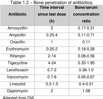
The therapeutic success in bone infections is also determined by the rate and extent of antibiotic penetration in bone tissues. An important factor to take into account is the choice of the appropriate antibiotic treatment [34]. This way, the bone penetration of several antibiotics has been studied. However, methodologies have not been standardized, which makes difficult the interpretation of results, and a consequent variation of these [35]. Penetration of an antibiotic into infected bone tissue depends on its pharmacological characteristics, the degree of vascularization, good conditions of soft tissues, and the presence of foreign bodies. Some of the antibiotics used in this pathology are described in Table 1.2, as well as their bone/serum concentration ratio and respective time interval since last dose (h) [34].
Table 1.2 – Bone penetration of antibiotics.
Antibiotic
Time interval
since last dose
(h)
Bone/serum
concentration
ratio
Amoxycillin 2 0.17-0.31
Ampicillin 0.25-4 0.11-0.71
Oxacillin 1 0.11
Erythromycin 0.25-2 0.18-0.28
Rifampin 2-14 0.08-0.56
Tigecycline 4-24 0.35-1.95
Levofloxacin 0.7-2 0.36-1.0
Vancomycin 0.7-6 0.05-0.67
Linezolid 0.5-1.5 0.4-0.51
Daptomycin 2 1.08
Adapted from [34].
Furthermore, many other problems have been revealed by antibiotic treatment of bacterial infections. When there is clinical treatment failure of bacterial infectious disease, it is usually associated with low bioavailability of antibiotics and their side effects, tissue and cellular barriers, biofilm-related infections and the emergence of resistant bacteria [2]. MRSA strains, resistant to methicillin, are normally synonymous of multidrug-resistant S. aureus, because many of these strains are also resistant to many other commonly used antibiotics. In Europe, it was reported that approximately 20 % of S. aureus
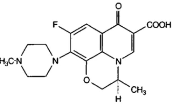
13 1.2.1. Fluoroquinolones: Levofloxacin
Fluoroquinolones have proved to be potent antibacterial agents, demonstrating a broader spectrum of antibacterial activity, a great efficacy against resistant organisms, and a better safety profile than other antimicrobial agents, including the older quinolones [37], [38]. Fluoroquinolones have several characteristics that have led to their increased use in bone infections. They have a rapid bactericidal effect against most susceptible organisms, and show one of the highest median extents of bone penetration of all antibiotic classes, partly due to quinolone binding to calcium in the bone [39], [40]. Levofloxacin is being described as the quinolone with the higher values in this group. Levofloxacin belongs to the fluoroquinolone class, widely used in treatment of certain bacterial infections (Figure 1.3). This drug has an oral broad spectrum of activity and excellent tissue penetration, above the minimum inhibitory concentration (MIC) for susceptible pathogens generally associated to bone and joint infections. The MIC of levofloxacin, as described in literature, for MSSA and MRSA is 0.25 – 0.5 µg/mL and 0.5 -16.0 µg/mL, respectively [41]. Moreover, it has availability in both oral and intravenous formulations, with lower toxicity. It is active against Gram positive and Gram negative bacteria, as well as other pathogens such as Mycoplasma, Chlamydia, Legionella and Mycobacteria spp. Futhermore, it
is being referred as the fluoroquinolone with the greater in vitro and in vivo anti-staphylococcal activity
against both intracellular and extracellular pathogens. Levofloxacin is the L-isomer of the racemic drug substance ofloxacin, a quinolone antibacterial agent. In chemical terms, levofloxacin is a chiral fluorinated carboxyquinolone, the pure enantiomer of ofloxacin. However, levofloxacin is more active against bacterial pathogens than its enantiomer. [36], [40], [42].
After oral administration of 50 to 200 mg of levofloxacin in healthy volunteers, the mean maximum plasma concentrations range from 0.57 to 2.04 mg/L achieved within 0.8 to 2.4 h. These values are linearly related to dose. Levofloxacin has an oral bioavailability approaching 100 %. Furthermore, with this administration method, levofloxacin penetrates rapidly and efficiently throughout the body, achieving concentrations in tissues or body fluids which are generally higher than those in plasma. This antibiotic is approximately 24 to 38 % bound to serum plasma proteins, mainly to albumin. The plasma elimination
14 half-life is 4 to 7 h, further within 24 h of an administrated oral dose, about 80 to 85 % of the drug is excreted unchanged in the urine, though glomerular filtration and tubular secretion [36], [43].
Like other fluoroquinolones, levofloxacin exerts its antibacterial effects through inhibition of deoxyribonucleic acid (DNA) gyrase, a type II topoisomerase (Figure 1.4). DNA topoisomerases are a class of enzymes that alter the topology of DNA by catalysing reactions called supercoiling, relaxing, knotting and catenating. Mainly, it controls the supercoiling of DNA. DNA gyrase has two subunits A and B. The subunits A, encoded by the gyrA gene, cause strand breaks on a bacterial chromosome and then reseal the chromosome after supercoiling. The B subunits, encoded by the gyrB gene, are ATP hydrolysis-dependent and introduce negative supercoils into the DNA double strand after the initial strands are resealed by subunits A. So, the principle bactericidal activity of levofloxacin results from the inhibition of the A subunits of DNA gyrase following supercoiling, by stabilizing the DNA-DNA gyrase complex. This stabilized complex blocks movement of the replication fork, causing formerly reversible DNA-DNA gyrase complexes to became irreversible, leading to inhibition of bacterial DNA replication and transcription [36], [43], [44].
1)
2)
3)
Figure 1.4 – Mechanism of action of levofloxacin.
1) DNA double strain with the enzyme DNA gyrase, DNA-DNA gyrase complex; 2) Introduction of the antibiotic levofloxacin which binds to the A subunits of the enzyme. Levofloxacin stabilizes the DNA-DNA gyrase complex, resulting in an irreversible complex; broken strands cannot be released leading to inhibition of DNA replication; 3)
Broken strands are released resulting in cell death. A – Subunits A of DNA gyrase; B –Subunits B of DNA gyrase.
15 1.2.2. Glycopeptides: Vancomycin
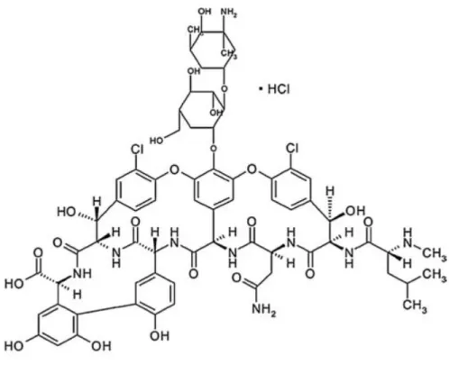
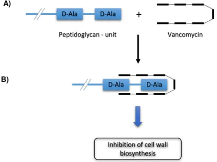
Vancomycin is a glycopeptide antibiotic used clinically to treat serious Gram-positive bacterial infections that are resistance to other antibiotics, such as β-lactams (Figure 1.5). Vancomycin is a first-line treatment for many bone and joint infections caused by typical organisms, including methicillin-resistant
S. aureus (MRSA). However, S. aureus reduced susceptibility to vancomycin may be partially due to its
ability to produce biofilms, which may facilitate resistance by promoting horizontal gene transfer. The frequency of resistance to the glycopeptide antibiotics has increased significantly, which represents a serious threat to public health. Furthermore, vancomycin has poor bone penetration [33], [45].
Vancomycin mechanism of action consists in blocking steps in the biosynthesis of the peptidoglycan layer of bacterial cell walls (Figure 1.6). Bacterial cells are surrounded by layers of peptidoglycan that provide the mechanical support necessary to prevent osmotic pressure oscillations, as well as cell lysis. Peptidoglycan is a rigid polymer of alternating units of N-acetylglucosamine (GlcNAc) and N
-acetylmuramic acid (MurNAc) connected by peptides cross-links. Peptidoglycan biosynthesis takes place in three stages. The first two stages lead to the production of lipid II, where the first step involves synthesis of the wall precursors in the cytoplasm, and the second consists in formation of the wall subunit on a mobile lipid in the membrane followed by its transfer to the outer surface of the membrane. The final stage involves glycan polymerization and cross-linking by transglycosylation and transpeptidation. Cell wall active antibiotics, such as vancomycin, inhibit the final step of peptidoglycan biosynthesis, by binding to the substrates, mainly of transglycosylases reaction. The enzymes involved in this step are
16 extracellular and are thus accessible to vancomycin, that needs not penetrate to the cytoplasm to exert its antibiotic effects. Vancomycin interacts with terminal acyl-D-Ala-D-Ala residues in peptidoglycan
precursors. It involves blocking the utilization of a substrate rather than acting directly on a biosynthetic enzyme. The heptapeptide backbone of the drug assumes a rigid conformation and forms a carboxylate bonding pocket that binds acyl-D-Ala-D-Ala residues via five hydrogen bonds and hydrophobic
interactions. The result is a shielding of precursor substrates from the enzymes that catalyze transglycosylation and transpeptidation. Inhibition of this reaction leads to the accumulation of lipid intermediates in the biosynthetic pathway and of UDP-MurNAc-pentapeptide in the cytoplasm [45]–[47].
1.2.3. Lipopeptides: Daptomycin
Daptomycin is a novel cyclic lipopeptide antimicrobial agent that was developed as an alternative therapy in bone infections treatment after vancomycin failure. The drug has excellent bactericidal activity against most Gram-positive organisms, including multiple antibiotic-resistant and -susceptible strains, such as methicillin-resistant strains, beta-hemolytic groups A, B, C, and G streptococci and enterococci, and ampicillin- and vancomycin-resistant strains (Figure 1.7) [16], [48]–[51]. Its bactericidal activity is concentration dependent and very fast. Furthermore, it also retains this advantage in biofilms. Several studies have been demonstrating that daptomycin penetrates rapidly into biofilms. Due to its unique mechanism of action on a cell membrane, daptomycin retains antibacterial activity against both stationary-phase cultures of staphylococci within the biofilm and bacteria in the multiplication phase [51], [52]. Moreover, daptomycin penetrates bone effectively [51].
Figure 1.6 – Mechanism of action of vancomycin.
A) Peptidoglycan unit and antibiotic vancomycin; B) Interaction between vancomycin and acyl-D-Ala-D-Ala residues
of peptidoglycan unit – inhibition of cell wall biosynthesis. Adapted from [45]–[47].
A)
17 With a half-life of 8 h allowing for once daily dosing results in linear pharmacokinetics at doses up to 12 mg/Kg, with minimal drug accumulation. Daptomycin distributes primarily in the plasma, with penetrations to vascularized tissues. This antibiotic is highly protein-bound (92 %) [33], [50]. Its excretion occurs first via the kidneys. Approximately 80 % of the total dose, of which two-thirds is intact drug, is recovered in the urine. The daptomycin single mechanism of action and its lack of metabolization by cytochrome P450 or other hepatic enzymes results in an absence of drug interactions between itself [33].
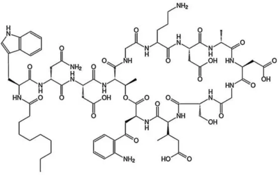
Figure 1.7 – Structure of daptomycin.
Daptomycin has an unique structure, which consists of a 13-member amino acid cyclic lipopeptide with a decanoyl side-chain. This structure confers a novel mechanism of action. The mechanism involves the calcium-dependent insertion of the lipophilic daptomycin tail into the bacterial cytoplasmatic membrane, causing rapid membrane depolarization and a potassium ion efflux. This is followed by arrest of DNA, RNA and protein synthesis resulting in bacteria cell death (Figure 1.8) [49], [53]. Calcium binding between two of the aspartate residues on daptomycin decreases its net negative charge and increases the area of its hydrophobic surface, improving the interaction with the membrane [53]. In addition, recently, studies have been demonstrated that calcium, as Ca2+ ions, is needed to generate two
structural transitions in daptomycin [54]. Calcium binds to daptomycin in solution and there is an aggregation of drug, resulting in more tightly defined family of structures of the apo-form of this drug. This is consistent with the suggestion that Ca2+ is needed to lock the molecule into an active
18 observed requires the presence of both Ca2+ and lipids with negatively charged headgroups (e.g.,
phosphatidyl glycerol). The effect of daptomycin on these lipid bilayers in the presence of Ca2+ is to
19 Figure 1.8 – Mechanism of action of daptomycin.
A) Daptomycin molecule without calcium adopts a structure which is reasonably well defined but not highly amphiphilic; B) In presence of calcium ions, the lipopeptide oligomerizes and most likely arranges itself into a micelle. This process is accompanied by change of conformation; C) Once daptomycin comes into close proximity with the bacterial membrane, the multimer dissociates, and drug inserts into the bilayer and the second structural transition is formed; D) Daptomycin causes the rupture of the bacterial membrane; E) Insertion of daptomycin into the membrane is accompained by the induction of positive membrane curvature and oligomerization in the membrane may occur; F) Bacterial cells are killed by membrane perforation (assessed as depolarization), and potassium ions efflux, or some other membrane associated event. Adptaded from [54].
A)
B)
C)
D)
F)
Dissociation and membrane insertion; Change in conformation
(2nd structural
transition) Oligomerization
and change in conformation
Membrane curvature
Leakage, potassium ion efflux and cell
death Apo-form
20
Local drug delivery systems as new therapeutic strategies
Due to problems of the conventional treatment of bone infection, new approaches are required. Drug delivery systems arise as new therapeutic strategies for treatment of bone infections, which achieve elevated antibiotic concentration at the site of infection without exceeding the systemic toxicity [13]. Furthermore, drug delivery systems can provide drugs more effectively and conveniently than those conventionally used, increase patient compliance, extend the antibiotic life cycle, provide product differentiation and reduce healthcare costs [55]. These controlled drug delivery systems usually include particulate carriers composed primarily of lipids and/ or polymers, and their associated therapeutics [56].These systems may be divided into biodegradable and biodegradable carriers. The non-biodegradable delivery systems have been approved for use in treatment of osteomyelitis in Europe, an example are the polymethylmethacrylate (PMMA) beads containing gentamicin. However, this product although effective, suffers from the major drawback of requiring subsequent removal of the beads at completion of drug release. Furthermore, in recent years biodegradable systems have also been developed and evaluated for local delivery of antibiotics in the treatment of bone infections [13]. Examples of these systems are shown in Table 1.3.
Table 1.3 – Local drug delivery systems.
Carriers used for local delivery and antibiotics released.
Carrier system Antibiotic released
Non-biodegradable
Acrylic bone cement
Oxacillin [57] Cefazolin [57]
Gentamycin [58]–[60] Fucidin [58]
Cephalosporin [61]
Cement of BIS-GMA/TEGDMA resin Plaster of Paris pellets/beads
Cephalexin [62] Teicoplainin [63] Gentamicin [64]–[66] Fucidin [64]
Polymethylmethacrylate (PMMA) beads Gentamicin [59], [60], [67]Tazocin [71] –[70] Vancomycin [72]
Polymethylmethacrylate (PMMA) cement
Vancomycin [73]
Vancomycin and Tobramycin [74] Minocycline [75]
Daptomycin [76] Gentamicin [77], [78] Biodegradable
Collagen-gentamicin sponge Gentamicin [59], [70], [79]–[81]
Hydroxyapatite blocks
Vancomycin [82], [83] Gentamicin [84], [85] Arbekacin [86]
Hydroxyapatite cement Vancomycin [87]
Nano-HA-PHBV/PEG-GM microsphere Gentamicin [88]
Bone cement Ciprofloxacin [89]
Hydroxyapatite-β-tricalcium phosphate
composite Gentamicin [90]
Β-tricalcium phosphate-chitosan scaffold Gentamicin [91]
21 Vancomycin [93]
Apatite-wollastonite glass ceramic blocks Cefmetazole [94] Isepamicin sulfate [94] Bioglass reinforced plaster of Paris,
hydroxyapatite and sodium alginate Cephazoline [95]
Polylactide and/or polyglycolide implants
Gentamicin [96]–[100] Ciprofloxacin [101], [102] Vancomycin [103]–[106] Tobramycin [107], [108] Sodium fusidate [109]
Poly(acrylic acid) and gelatin crosslinked Gentamycin or Vancomycin [110] Polyanhydride and polylactide blend Ofloxacin [111]
Polycaprolactone Tobramycin [112]
Polyanhydride implant (Septacin) Gentamicin [113] Injectable gelling polymer Gentamicin [114] Fibrin clots/ sealant Arbekacin [115] Tobramycin [116] Ciprofloxacin [117] Fibrin gel (vanco-AB-FG) with bone
marrow-derived mesenchymal stem cells (BMMSCs) Vancomycin [118]
Dilactate polymers
Teicoplanin [119] Tobramycin [120] Sulperazone [121] Fleroxacin [122] Ciprofloxacin [123] Pefloxacin [123]
Bone xenograft Gentamicin [124]
Bone graft/ demineralized bone matrix Tobramycin [125]
Β-tricalcium phosphate Gentamicin [126] Vancomycin [126]
Calcium sulphate Tobramycin [127] Daptomycin [128] Calcium sulphate with demineralized bone
matrix (DBM) Vancomycin [129]
Calcium phosphate cement (CPC)/ injectable CPC
Gentamicin [130], [131] Teicoplanin [132]
Biomedical polyurethanes Flucloxacillin [133] Ciprofloxacin [133] Fosfomycin [133] Gentamicin [133]
Fibres Tetracycline [134], [135]
Cross-linked hyaluronic acid (HA) gel Gentamicin [136]
Monoolein-water gels Gentamicin [137]
1.3.1. Antibiotic-loaded acrylic bone cement (ALABC)
22 liquid (monomer) and a solid phase (powder). The solid phase is characterized by the polymer (PMMA), the catalyst of the polymerization reaction and by the ratio-opacifier,
while the liquid phase is characterized by the monomer (MMA), by the reaction accelerator and by the stabilizer (Table 1.4). The solid and the liquid phase components are usually in a 2:1 ratio [138], [141]. The two components are mixed, the liquid monomer polymerizes around the pre-polymerized powder particles to form hardened PMMA. This is an exothermic reaction, where the cement heats up, initiated by the decomposition of a catalyst (e.g. benzoyl peroxide) producing free radicals that set off additional polymerization of the MMA. The exothermic reaction reaches temperatures of around 82-86 °C in the body [140],
[141]. Thus, the exposure of bone to these high temperatures have been mentioned as a cause of necrosis and tissue damage, resulting in failure of the prosthetic fixation [142].
Table 1.4 – Components of bone cement.
Powder Liquid
Poly (methyl methacrylate) (PMMA) – Polymer Benzoyl peroxide (BPO) – Initiator
Barium sulphate (BaSO4)/ Zirconia (ZrO2) –
Radio-opacifier
Methyl methacrylate (MMA) – Monomer
N, N-Dimethyl para-toluidine (DMPT)/dimethyl para-toluidine (DMpt) – Accelerator
Hydroquinone – Stabilizer
Adapted from [141]
Bone cement mixed with active agents, such as antibiotic, releases the agents slowly, thus serving as a vector for specific controlled in situ therapy [143]. PMMA bone cement with antibiotics incorporated
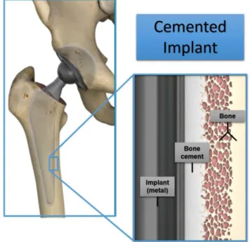
Figure 1.10 – Artificial hip replacement – cemented prosthesis.
Adapted from [178].
Figure 1.9 – Structure of PMMA.
23 reduces the infection rates in orthopedic surgery. These drug delivery systems are already used for primary and revision surgery, while antibiotic-loaded PMMA beads are part of a more complete treatment of infection and supplement other interventions (mostly surgical). Antibiotic-loaded PMMA bone cement is normally used in multistage revision of infected implants, where they have not only an antibacterial effect, but also prevent contraction of ligaments, and scar tissue from growing into the joint space. Several studies indicate that antibiotic released from PMMA bone cements is a surface phenomenon, but the mechanism by which these drugs are released is still debated. Moreover, studies reported that S. aureus biofilm formation was reduced on different gentamicin-loaded bone cements, when compared to unloaded bone cements only during a short period, which depends on the initial drug release of the bone cement [140].
As described by Arora et al., several kinds of additives started to be added to BC, besides antibiotics, with different aims, while still maintaining structural and mechanical integrity [144]. An example of this is the addition of release modulators, as a means to increase the drug release rate from BC matrices. In the case of PMMA BC the rate of antibiotic release is low, since in vitro and in vivo studies have
demonstrated that, in this type of cement, the initial liberation of the drug is a surface phenomenon, due to the BC matrix being impermeable to drugs. The antibiotics must be released through an interconnecting series of voids and cracks in the cement. Therefore, an increase of superficial porosity of the cement is required to increase the efficiency of drug release from bone cement matrices, since the sustained liberation of antibiotics is largely affected by the penetration of fluids into the polymer matrix. So, the inclusion of water soluble compounds (release modulators) promotes the increase of porosity of the bone cement and consequently the drug release [145]. Previous data published by Matos and colleagues using levofloxacin-loaded PMMA bone cement describes the study of addition of lactose as enhancer of drug release. In this study, 10 % of this release modulator was added to a novel levofloxacin-loaded bone cement matrix. The results (during a 7-week period) demonstrated an improve of the amount of levofloxacin released from lactose-loaded BC matrices of 3.5-fold higher than from plain BC matrices [40].
1.3.2. Antibiotic-loaded polymeric microparticles
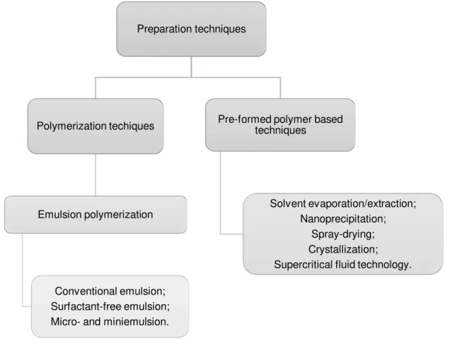
Polymeric nanoparticles and microparticles, made of natural and synthetic polymers, have revolutionized the administration of medicines, due to the important impact in the treatment and management of several conditions with high social and economic effect, such as cancer, respiratory and metabolic diseases, infections and tissue regeneration. These shown several advantages as drug carriers, such as high stability both in vitro and in vivo, multifunctionality and good biocompatibility. In
this way, the interest of these systems has increased in the medical field [2], [146], [147].