1. Neuropsychiatry Department, Sohag University, Egypt 2. Rheumatology and Rehabilitation Department, Mansoura University, Egypt
3. Radiodiagnosis Department, Bani Sweef University, Egypt 4. Radiodiagnosis Department, Zagazig University, Egypt
Psychiatric disorders and MRI brain findings
in patients with systemic lupus erythematosus
and Behcet’s disease: A cross sectional study
ACTA REUMATOL PORT. 2013;38:252-260
AbstrAct
Introduction: Neuropsychiatric systemic lupus ery -thematosus (NPSLE) shows some similarities to neu-roBehçet’s disease (NBD).
Aim of the work: to investigate and compare the psy-chiatric manifestations in a cohort of patients of syste-mic lupus erythematosus (SLE ) versus Behcet’s Disea-se (BD). Also, a comparison of MRI brain findings in SLE patients and BD patients presented with psychia-tric disorders was done. Finally, we correlate these ma-nifestations with disease activity indices of the patients. Patient and Method: The study included 50 patients of SLE, 34 patients of Behcet’s disease (BD) and 44 healthy volunteers as a control group. All patients were subjected to psychiatric interview to diagnose any psy-chiatric disorders clinically. MRI brain was done for SLE patients and BD patients presented with psychiatric di-sorders. Overall clinical assessment and disease activi-ty of SLE and BD were evaluated.
Results: Psychiatric disorders were detected in 28 (56%) of SLE patients which were significantly more prevalent than psychiatric disorders that were detected in 9 (26.47%) of BD patients. Psychiatric disorders in healthy volunteers were significantly less prevalent than either SLE patients or BD patients. MRI brain of SLE pa-tients presented with psychiatric disorders commonly showed cerebral white matter abnormalities while in BD patients presented with psychiatric disorders com-monly showed brain stem lesions.
Conclusion: (1) High prevalence of psychiatric
disor-Taher Abdelraheem1, Hisham M. Habib2, Ashraf A Eissa3, Nesreen M. Radwan4
ders in SLE and BD with a higher significant prevalen-ce in SLE. (2) Evident MRI brain findings in SLE and BD patients presented with psychiatric disorders. Keywords: Psychiatric disorders; Systemic Lupus Ery -thematosus; Behcet’s disease; MRI brain.
IntroductIon
Neuropsychiatric systemic lupus erythematosus (NPSLE) shows some similarities to neuro Behçe ts di-sease (NBD) and both conditions have some analogous clinical features and they are both pathologically asso-ciated cerebral vasculopathy1.
Systemic lupus erythematosus (SLE) is a systemic autoimmune disease that can affect any part of the body. As occurs in other autoimmune diseases, the immune system attacks the body cells and tissue, resulting in inflammation and tissue damage2. In the course of SLE,
a variety of neuropsychiatric disturbances is reported, with prevalence rates ranging from 17% to 75%3.
Psy-chiatric manifestations of SLE include depression, an-xiety, cognitive deficits, psychosis and mania4,5.
Immu-nologic and cerebral imagery research suggests that psychiatric disorders are related to vasculitis and non--inflammatory vasculopathy of the small cerebral blood vessels6. A common MRI brain finding in patients with
SLE is cerebral white matter abnormality1.
Behçet’s disease (BD) is a multi-system inflammato-ry disorder dominated clinically by recurrent oral and genital ulceration, uveitis, and erythema nodosum. The disease tends to wax and wane. BD is a vasculitis, af-fecting vessels of different types, sizes, and localiza-tions7. Although the cause of BD is unknown,
autoim-mune, infectious, and genetic causes have been sus-pected. Environmental factors, such as microbial
in-fections, are also suspected to be factors that contribute to the development of BD8. Central nervous system
(CNS) involvement can be either parenchymal or non-parenchymal. Parenchymal involvement primarily af-fects the brainstem, spinal cord, and cerebral hemis-pheres. Nonparenchymal involvement includes intra-cranial hypertension, aseptic meningitis, intra-cranial neu-ropathy, and cerebrovascular disorders such as dural sinus thrombosis, arterial dissection, occlusion, and aneurysm9. The prevalence of psychological symptoms
was remarkable in the patients with BD. The psycho-logical symptoms in BD could be aggravated by the ill-ness itself or by the immunosuppressive drugs used during the treatment course10. Psychiatric reactions to
BD may include anxiety, insomnia, manic/depressive episodes and psychosis11. The characteristic MRI brain
lesion in parenchymal involvement of BD is an upper brainstem lesion that extends into the thalamus and basal ganglia on one side12.
AIm of the work
The purpose of this study is to investigate and compare the psychiatric manifestations in a cohort of patients with SLE versus BD and to correlate these manifesta-tions with disease activity indexes of the patients. Li-kewise, a comparison of MRI brain findings in SLE pa-tients and BD papa-tients presented with psychiatric di-sorders has been carefully analyzed.
PAtIents And methods PAtIents And controls
Fifty patients of SLE and 34 patients of BD and 44 con-trol subjects with similar demographic characteristics were included in the study. All SLE patients had fulfil-led American College of Rheumatology criteria for the classification of systemic lupus erythematosus13,
whi-le BD patients had fulfilwhi-led the International Study Group for BD diagnostic criteria14. The study was
approved by the hospital ethical committee and a writ-ten informed consent was taken from all patients who agreed to contribute.
PsychIAtrIc Assessment
All patients and control group were subjected to inpa-tient based structured psychiatric interview and neu-ropsychological evaluation to diagnose clinically any
psychiatric disorders. All subjects included in the stu-dy were asked to perform a series of neuropsychologi-cal test done by expert psychologist to detect depres-sion, anxiety, cognitive impairment, mania or psycho-sis.
1. H
AMILTON DEPRESSION INVENTORY TO DETECT DEPRESSION15It is a 23-item self report inventory that assesses de-pressive symptomatology for the previous 2weeks. The administration time is 10-15 minutes. Scoring varies by item, with a total range of 0-73 and a score of 19 has been suggested as a cut off score when screening for depression.
2. S
TATE-T
RAIT ANXIETY SCALE TO DETECT ANXIETY16It is a self-report and one of the widely used scales in a variety of research studies. The administration time is 20 minutes. It is a two 20-item scale and each item is scored on a 4-point scale and scores ≥ 20 indicate an-xiety.
3. M
INIM
ENTALS
TATEE
XAMINATION TO DETECT COGNITIVE DYSFUNCTION17The most common neurocognitive screening tool used to detect cognitive losses through a score (maximum score = 30) of the five areas of cognition, orientation, registration, attention and calculation, as well as recall and language. Scores < 24 suggest the presence of de-cline18.
4. M
OOD DISORDER QUESTIONNAIRE TO DETECT MANIA19It is a self-report questionnaire designed to screen for mania. The administration time is 5-10 minutes. It is a brief 13-item questionnaire in a yes/no format. The screen is considered positive when 7 or more symp-toms have occurred.
5. B
RIEFP
SYCHIATRICR
ATINGS
CALE TO DETECT PSYCHOSIS(BPRS)
20BPRS remains one of the most widely used clinician--admi nistered tools designed to assess overall psycho-pathology in patients with a major psychiatric disorder, particularly psychosis. It is administered in a semi structured manner and it takes about 30 minutes to complete. It comprehends 24 items that can be scored from 1 (not present) to 7 (very severe). The total BPRS--E score is the sum of the scores for each of the 24 items.
brAIn mAgnetIc resonAnce ImAgIng (mrI)
All SLE and BD patients presented with psychiatric di-sorders underwent brain MRI including T1- weighted images, T2- weighted images and fluid-attenuated in-version-recovery images (FLAIR) images. The MRI was performed using a 1.5 T MRI system (GE SIGMA Advantage version 4-8). The conventional spin echo pulse sequence with a TE of 20 ms and a TR of 400 ms was used to obtain the T1-weighted NR images. The fast spin echo pulse sequence with a TE of 90 ms and a TR of 2,500 ms was used to obtain the T2-weighted MR images. Contrast medium was administered to all SLE and BD patients presented with psychiatric disor-ders and this was followed by obtaining the axial/sa-gittal/coronalT1-weighted images. MRI were studied for: an abnormal T2-weighted image, infarct-like le-sions (moderate to large, roughly wedge-shaped areas of abnormal high signal on the T2-weighted images and/or encephalomalacia involving the gray and whi-te matwhi-ter), parenchymal hemorrhage, and loss of brain volume or abnormal intracranial enhancement. The location of the brain lesion was addressed. Finally, the abnormal MRI findings were classified into two main categories: parenchymal (mainly located in upper brainstem thalamus and basal ganglia) and non-pa-renchymal (mainly venous sinus thrombosis).
dIseAse ActIvIty meAsurement
All the patients were subjected to disease activity mea-surement in the following manner:
*Overall clinical assessment and disease activity index of SLE patients: was done using the BILAG (British Is-les Lupus Assessment Group) disease activity index21.
It distinguishes activity in 8 organs or systems name-ly general, mucocutaneous, Central Nervous System, musculoskeletal, Cardio-Vascular System/respiratory, vasculitis, renal and haematological systems. It provi-des an accurate means of grading disease activity from the “most active” to “no evidence of disease activity currently”. Patients are classified to 5 grades:
• Grade A= “Active” the most active disease state re-quiring major immunosuppressive drug. A= 9 points.
• Grade B= “Beware” patients known to have active disease but is already on immunosuppressive the-rapy. B= 3 points.
• Grade C= “Contentment”, patients has relatively mild disease controlled by little specific therapy if
any. C= 1 point.
• Grade D= “Discount”, there is no activity in this sys-tem now. D= 0 point.
• Grade E= no “Evidence”, of activity in this system now or previously. E= 0 point.
A global score can finally be calculated in the pa-tients collecting the 8 system/organ score together. * Iranian Behecets Disease Dynamic Activity Measure (IBDDAM)22: patient is evaluated on several weeks or
months. Each attack is measured separately and given an index. The obtained indexes are added together and the total is divided by the number of months of the evaluated period. The final result is the mean disease activity index. One point is given for: oral ulcers (2 for more than 5 ulcers at a time), genital ulcers (one for each ulcer), pseudofolliculitis (2 for more than 10), erythema nodosum (2 if more than 5), arthralgia, su-perfacial phlebitis and pathergy. Two points are given for phlebitis (each vessel), epididymitis, and mo-noarthritis. Three points for polyarthritis, mild CNS, and intestinal manifestations. Six points are given for large vessel thrombosis, moderate to sever intestinal lesions. Each inflammatory sign of the eye is given 1 to 4 points. If an attack does not heal in 1 month, the same point is given for each additional month.
stAtIstIcAl AnAlysIs
Comparison of the studied groups was done using the students T test. The Chi-squared test was used to com-pare categorical variables. Correlations between groups were evaluated using the Spearman test. Non-para-metric Mann–Whitney U test was used for the com-parison of variables between tested groups with and without psychiatric disorders. Pearson’s correlation analysis was used to investigate relation between SLE duration and psychiatric disorder. A probability value (P-value) less than 0.05 was considered significant. Data were collected and tabulated using Microsoft ex-cel version 7 (Microsoft Cooperation. NY, USA) and analyzed using SPSS for windows (Statistical Package for the Social Science, version II, SPSS, Inc, Chicago, IL, USA).
results
Table I demonstrates the Demographic and clinical characteristics of SLE and BD patients as well as
con-trols. The study comprised 50 SLE patients; 44 fema-le and 6 mafema-les with mean age of 29.5 (range: 18-42) years, and 34 BD patients with a mean age of 39.5 (ran-ge: 22-55) 18 female and 16 males. Disease duration of SLE patients was 7.5±2.3 with range of 6 months to 11 years. Disease duration of BD patients was 8.4±3.3
with range of 1 year to 13.5 years.
Corticosteroids medication in patients: approxima-te cumulative dose of corticosapproxima-teroids and current dai-ly dose of corticosteroids, for each patient were calcu-lated. Cumulative corticosteroid dose in SLE patients was in range 0–98g, mean 18.55 ± 11.2g and current dose 0–45 mg, mean 12.4 ± 5.19mg. Cumulative cor-ticosteroid dose in BD was in range 0–22g, mean9.12 ± 3 g and current dose 0–30 mg, mean 7.5 ± 2.5 mg. Table II specifies the steroid dosage in SLE and BD pa-tients in detailed manner. No significant difference in SLE and BD patients in steroid dosage in different ca-tegories (p <0.05).
Overall clinical assessment and disease activity in-dex of SLE patients were done using the BILAG: • Group (1): 12 patients with 0-3 points. • Group (2): 10 patients with 4-6 points. • Group (3): 12 patients with 7-12 points. • Group (4): 7 patients with 13-15 points.
tAble I. demogrAPhIc And clInIcAl chArActerIstIcs of sle, bd PAtIents And controls
SLE (No=50) BD (No=34) Controls (No=44)
Clinical characteristic No (%) No (%) No (%) P value
Gender: Male 6 (12%) 16 (47%) 20 (45.5%) 0.8 Female 44 (88%) 18 (53%) 24 (54.5%) Age in years: Mean ±SD 29.5±11.5 39.5±9.5 36.5±9 0.3 Range (18-42) (22-55) (16-49)
Disease duration in years:
Mean ±SD 7.5±2.3 8.4±3.3 0.1 Range (0.5-11) (1-13.5) Skin involvement, No (%) 25 (50%) 17 (50%) 0.5 Renal involvement, No (%) 12 (24%) 1 (3%) 0.05 Neurological involvement, No (%) 9 (18%) 5 (14.7%) 0.2 Gastrointestinal involvement, No (%) 8 (16%) 4 (11.7%) 0.5 Oral ulceration, No (%) 31 (62%) 34 (100%) 0.05 Genital ulceration, No (%) 0 (0%) 22 (64.7%) 0.001 Ocular involvement, No (%) 5 (10%) 25 (73.5%) 0.001 Vascular events, No (%) 3 (6%) 17 (50%) 0.01 Lung involvement, No (%) 18 (36%) 6 (17.6%) 0.05
Cumulative corticosteroid dose (gm)
Mean ±SD 18.55 ± 11.2 9.12 ± 3 0.03
Range 0–98 0–22
Current corticosteroid dose (mg)
Mean ±SD 12.4 ± 5.19 7.5 ± 2.5 0.01
Range 0-45 0-30
tAble II. steroId dosAge In sle And bd PAtIents
SLE patients BD patients Steroid (n = 50) (n = 34) P dosage (mg) No (%) No (%) value 0 8 (16%) 5 (14.7%) 0.1 5-15 15 (30%) 11 (32.3%) 0.08 15-45 27 (54%) 18 (53%) 0.06 > 45 0 (0%) 0 (0%)
• Group (5): 9 patients > 15 points.
Iranian Behecets Disease Dynamic Activity Measure (IBDDAM): patients were evaluated month ly for 12 months without appointment failure for those 24 patients: • Group (1): 6 patients < 1 point.
• Group (2): 7 patients with 1-3 points. • Group (3): 8 patients with 4-6 points. • Group (4): 5 patients with 7-12 points. • Group (5): 8 patients > 12 points.
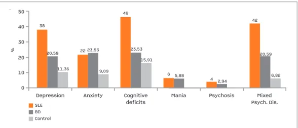
clInIcAl And PsychometrIc Assessment of PsychIAtrIc dIsorders (fIgure 1)
Psychiatric disorders were detected in 28 (56%) pa-tients with SLE which were statistically significant compared to 9 (26.47%) patients with BD. Psychiatric disorders in each of SLE or BD patients were signifi-cantly more prevalent than that of control group (6 persons = 13.63%).
As shown in Figure 1 depression was more preva-lent in SLE patients (19 patients =38%) compared to BD patient (7 patients =20.59%) in a statistically sig-nificant manner. In either SLE patients or BD patients the prevalence of depression is significantly higher than the control group (5 persons = 11.36%) (p<0.05). No significant difference in the prevalence of an-xiety in SLE and BD patients (11 patients = 22% vs 8 patients =23.53% respectively). Also, in SLE and BD patients the prevalence of anxiety was significantly hi -gher than control group (4 persons = 9.09%) (p>0.05). Cognitive deficits were significantly high in SLE pa-tients (23 papa-tients = 46%) compared to BD papa-tients (8 patients = 23.53%) and the frequency of cognitive
de-ficits from either patients were notably higher than the control group (7 persons = 15.91%) (p <0.05).
Three SLE patients (6%) had mania compared to 2 BD patients (5.88%) with no statistical significant dif-ference (p >0.05). No reported cases in control group had mania.
For psychosis, 2 SLE patients (4%) and 1 (2.94%) BD patients were affected, with no statistical signifi-cant difference (p >0.05). Mixed psychiatric disorders were detected in a statistically significant higher per-centage in SLE patients compared to BD patients (21 =42% vs 8 = 20.59%) (p <0.05). Likewise, mixed psychiatric disorders were statistically more prevalent in SLE and BD patients compared to control group (3 persons = 6.82%) ( >0.05).
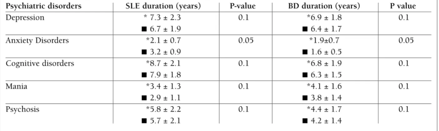
Table III explains the effect of disease duration on psychiatric disorders of both SLE and BD patients. It shows that shorter duration of illness in SLE and BD patients is statistically correlated with anxiety disor-ders if compared with those without anxiety disorder. No significant correlation with depression, cognitive impairment, mania or psychosis.
For MRI of brain (Table IV), there was a statistical-ly significant (p=0.05) frequency of abnormalities in BD patients presented with psychiatric manifestations (7/9 patients = 77.78%) than that in SLE patients (18/28 patients = 64.29%). Abnormal T2-weighted images were more common (p = 0.1) in BD patients (5/7 = 71.43%) than SLE patients presented with psy-chiatric manifestations (4/18 = 22.22%). MRI brain showed no abnormal findings in 10 SLE patients (35.71%) out of 28 and 2 BD patients (22.22%) out of
0DepressionAnxietyCognitivedeficitsManiaPsychosisMixedPsych. Dis.1040203050%3811,36BDControlSLE20,59229,0923,534623,536 5,884 2,9415,914220,596,82
0
Depression Anxiety Cognitive deficits
Mania Psychosis Mixed Psych. Dis. 10 40 20 30 50 % 38 11,36 BD Control SLE 20,59 22 9,09 23,53 46 23,53 6 5,88 4 2,94 15,91 42 20,59 6,82
fIgure 1shows the prevalence of different psychiatric disorders in Systemic Lupus Erythematosus (SLE), Behcet’s Disease (BD) and control group
9 despite presentation with psychiatric manifestations with statistically insignificant difference in both groups (p= 0.06).
For location of the lesion in MRI (Table V), Cerebral
white matter lesions on MRI were more frequently en-countered in a significant ratio (p= 0.01) in SLE pa-tients presented with psychiatric disorders (10 papa-tients = 55.56%) than those of BD (1 = 14.29%) whereas the
tAble III. effect of Illness durAtIon In sle And bd PAtIents on PsychIAtrIc dIsorders
Psychiatric disorders SLE duration (years) P-value BD duration (years) P value
Depression * 7.3 ± 2.3 0.1 *6.9 ± 1.8 0.1 ■ 6.7 ± 1.9 ■ 6.4 ± 1.7 Anxiety Disorders *2.1 ± 0.7 0.05 *1.9±0.7 0.05 ■ 3.2 ± 0.9 ■ 1.6 ± 0.5 Cognitive disorders *8.7 ± 2.1 0.1 *6.8 ± 1.9 0.1 ■ 7.9 ± 1.8 ■ 6.3 ± 1.5 Mania *3.4 ± 1.3 0.1 *4.1 ± 1.6 0.1 ■ 2.9 ± 1.1 ■ 3.8 ± 1.4 Psychosis *5.8 ± 2.2 0.1 *4.4 ± 1.7 0.1 ■ 5.7 ± 2.1 ■ 4.2 ± 1.4
* Present active disorder ■ Absent active disorder
tAble Iv. comPArIson of the brAIn mrI fIndIngs In sle PAtIents And bd PAtIents Presented wIth PsychIAtrIc mAnIfestAtIons (AccordIng to tyPe of AbnormAlIty)
SLE patients with Psychiatric BD Patients with Psychiatric
Brain MRI findings Manifestations (no.28) Manifestations (no.9) P
I. MRI Abnormalities 18/28 (64.29%) 7/9 (77.78%) 0.05
1. Abnormal T2-weighted image 4/18 (22.22%) 5/7 (71.43%) 0.01 2. Infarct like lesion 3/18 (16.67%) 1/7 (14.29%) 0.1 3. Parenchymal hemorrhage 4/18 (22.22%) 0/7 (0%) 0.01
4. Volume loss 5/18 (27.78%) 2/7 (28.57%) 0.1
5. Abnormal intracranial enhancement 3/18 (16.67%) 1/7 (14.29%) 0.1 6. Mixed abnormalities 1/18 (5.56%) 2/7 (28.57%) 0.05
II. Negative 10/28 (35.71%) 2/9 (22.22%) 0.06
tAble v. comPArIson of the brAIn mrI fIndIngs In sle PAtIents And bd PAtIents Presented wIth PsychIAtrIc mAnIfestAtIons (AccordIng to the locAtIon of lesIon)
SLE Patients with Psychiatric BD Patients with Psychiatric
Brain MRI findings Manifestations (no.28) Manifestations (no.9) P
Location of the lesion 18 (64.29%) 7 (77.78%)
1. Cerebral white matter 10/18 (55.56%) 1/7 (14.29%) 0.01
2. Basal ganglia 4/18 (22.22%) 2/7 (28.57%) 0.1
3. Thalamus 2/18 (11.11%) 1/7 (14.29%) 0.3
4. Brain stem 4/18 (22.22%) 4/7 (57.14%) 0.01
fIgure 3. (A,B) axial T2WI (C) axial FLAIR: 45 years old female with SLE show bilateral diffuse periventriclular ischemic high sig-nal (arrow) and infarct like lesions (filled arrows).No signifcant atrophy, no haemorage, no enhancement in T1 study (not shown) .
frequency of brain stem lesion tended to be statistical-ly higher (p= 0.01) in BD patients presented with psy-chiatric disorders (4 patients = 57.14%) than those of SLE (4 patients = 22.22%). In BD we noticed that 6 (85.5%) patients had parenchymal lesion, while 1
(14.5%) patient had non- parenchymal lesion, with a significant difference (p = 0.05) (data not shown in the table). Figures 2 and 3 showed MRI findings in a pa-tient with SLE with brain involvement and a papa-tient with neuro-Behcet’s respectively.
fIgure 2. (A) axial T2WI (B) axial FLAIR: 50 years old male patient with BD, show bifrontal atrophic changes with loss of volume (arrow) as well bilateral advanced periventriclular ischemic high signal (filled arrow), no area of infarction, no hamorrage, no enhancement in T1 study (not shown)
A b
Correlation of psychiatric disorders with disease ac-tivity in both diseases yields a significant accumula-tion of patients with major psychiatric disorders like psychosis and mania in groups with more disease fla-res. Five SLE patients with mania and psychosis were of groups (4) and (5) of BILAG disease activity and 3 BD patients with mania and psychosis were of groups (4) and (5) of IBDDAM (p< 0.005). On the other hand, the groups with less disease activity in both SLE and BD such as groups (1), (2) and (3) BILAG of SLE and IBDDAM of BD showed significant accumulation of anxiety, depression and cognitive disorders (28 SLE patients and 9 BD patients) ( p < 0.001).
dIscussIon
In our study, we addressed the different psychiatric di-sorders in both SLE and BD in a comparative way. Our results were comparable with other studies. We revea-led that the prevalence of psychiatric disorders in SLE (56%) was higher than in BD (26.47%). Those figures were comparable with that of other studies. Kaplan and Sadock’s mentioned that 50 percent of patients with SLE show neuropsychiatric manifestation23.
Ka-kalamani et al reported that neuropsychiatric involve-ment occurs in 10-20% of cases with BD24. We
revea-led higher frequency of depression in SLE patients (38%) than BD patients (20.59%) in a statistically signi ficant manner, which is consistent with the results of Cho et al1who reported that a common feature in
SLE patients was depression (32%) which was un-common in BD patients. However, depression was more prevalent in SLE and BD than the general popu-lation (17%)25. Cohen et al4 stated that depression is
the second most common psychiatric disorder in SLE, with a prevalence of almost 50%. Moreover, we re-ported that prevalence of cognitive dysfunction in SLE patients (46%) was higher than BD patients (23.53%). This was in agreement with the results of Denburg and Ainiala26,27but it was contradictory with the results of
Cho1. He reported that cognitive dysfunction in BD
patients (44.4%) was more frequent than in SLE pa-tients (36.4%). A possible plausible explanation of this discrepancy was different cultures and tools of psy-chometric assessment in addition to the intermingling features of cognitive deficits that are considered a com-mon feature of depression which is prevalent in SLE. In our study no statistical significant difference bet-ween the prevalence of anxiety disorder in SLE patients
(22%) and BD patients (23.53%) was noted but in each disease, anxiety disorder is more prevalent than the general population (17.7%). We reported no signifi-cant difference of mania frequency in SLE patients (6%) and BD patients (5.88%). Berlit27reported that
mania is commonly occurs in SLE and also related to dose dependent corticosteroid therapy. Also, we noti-ced no significant difference in the prevalence of psy-chosis in SLE patients (4%) and BD patients (2.94%), which is similar to the results of other publications29,30.
For the effect of disease duration on psychiatric disor-ders of both SLE and BD patients, we found that the duration of SLE and BD in patients with anxiety di-sorders was short if compared with those without an-xiety disorders. This is consistent with the results of MeCracken et al who relate this finding to the deve-lopment of coping strategies with better social func-tioning31.
MRI is a useful tool for detecting CNS lesions in pa-tients suffering with SLE and Behçets disease32. The
present study revealed that MRI brain of BD patients presented with psychiatric disorders frequently sho-wed abnormal T2-images (71.43%) than those of SLE patients (22.22%). In the current work we addressed that cerebral white matter involvement in brain MRI of SLE patients presented with psychiatric disorders (55.56%) were more frequent than those of BD pa-tients (14.29%), while brain stem lesions were more encountered in BD patients presented with psychia-tric disorders (57.14%) than those of SLE patients (22.22%). This findings is comparable with the results of Byung-Sik Cho, et al1who found that cerebral
whi-te matwhi-ter abnormalities are more common in NPSLE (48%) than NBD (22.29%) whereas brain stem lesions on MRI are more common in NBD (55.6%) than NPSLE (20.2%).
Furthermore, we have tried to correlate disease activity scoring in both SLE and BD and psychiatric disorders taking severity in mind. Accordingly, we have divided psychiatric disorders into both divisions; na-mely anxiety, depression and cognitive disorders in one “pole” and the “major psychiatric disorders” like mania and psychosis in another “pole”. Our results found a positive correlation between high disease activity scores in both diseases with major psychiatric disorders like mania and psychosis. Explanation may be linked to the more chance of cerebral vasculitis in higher disease activity score patients with higher pro-pensity to have more aggressive psychiatric disorders. To our knowledge, no reports correlating disease
acti-vity measures in SLE and BD with psychiatric illness in both diseases. In this respect, a wide scale study with more stress on a cohort of higher disease activity sco-re patients in both diseases may appear intesco-resting.
Finally, a wider scale study may be recommended to address the effect of chronic pain, the impact of func-tional disability and socioeconomic status as well as the effect of medications -mainly steroid- in the psy-chiatric make up of SLE and BD patients.
We therefore conclude that there is a high prevalence of psychiatric disorders in both SLE and BD with a sig-nificant increase of prevalence in SLE. There is accumu-lation of patients with major psychiatric disorders like psychosis and mania in groups with more disease flares in both diseases. MRI is a useful tool for detecting CNS lesions in patients suffering with SLE and BD at the level of type of abnormality and location of the lesion.
corresPondence to
Hisham Mohamed Habib
Kigdoom of Saudia Arabia, Al Ahsa 31982, Al Ahsa Hospital, P.O. Box 3230
E-mail: hesham_habib@yahoo.com
references
1. Cho B, Kim HYO, Oh SJ, Koh HJ, Yoon CH, Jung SL, et al. Comparison of the clinical manifestations, Brain MRI and prog-nosis between NeuroBehcet’s Disease and neuropsychiatric lu-pus.Korean J Intern Med, 2007; 22(2) 77-86.
2. James, William; Berger, Timothy; Elston, Dirk. Andrews’ Di-seases of the Skin: Clinical Dermatology. (10th ed.), 2005; Saunders. ISBNo-7216-2921-0.
3. Colasanti T, Delunardo F, Margutti P, Vacirca D, Piro E, Siracu-sano A, et al. Autoantibodies involved in neuropsychiatric ma-nifestations associated with systemic lupus erythematosus. J Neuroimmunol. 2009; 212:3–9.
4. Cohen W, Roberts WN and Levenson JL. Psychiatric aspects of SLE. In: Lahita R ed. Systemic Lupus Erythematosis. 4th ed. San Deigo. CA: Academic press. 2004; 785-825.
5. Hanly JG. Neuropsychiatric Lupus: Rheum Dis Clin North Am., 2005; 31 (2): 273-298.
6. Ampélas JF, Wattiaux MJ, Van Amerongen AP. [Psychiatric ma-nifestations of lupus erythematosus systemic and Sjogren’s syn-drome].Encephale; 2001; 27(6):588-99.
7. Al-Otaibi LM, Porter SR, and Poate TW. Behect’s disease: a re-view. Journal of Dental Research, 2005; 8, 3: 209-222. 8. Shahien R and Bowirrat A. Neuro-Behçet’s disease: A report of
sixteen patients. Neuropsychiatr Dis 2010; 6: 219– 225. 9. Farah S, Al-Shubaili A, Montaser A. Behçet’s syndrome. A
re-port of 41 patients with emphasis on neurological manifesta-tions. J Neurol Neurosurg Psychiatry. 1998; 64:382–384. 10. Bagheri F, Mani A, Tadayyoni A, Firozi F, Nazarinia MA. The
pre-valence of psychiatric symptoms in the patients with Behcet’s di-sease in Shiraz, Southwest of Iran. JMOOD. 2013; 3(1): 28-32. 11. Kontogiannis V and Powell R. Behcet’s disease. Postgrad Med J
2000; 76 (900): 629-637.
12. Al-Araji A, Kidd PD. Neuro-Behçet’s disease: epidemiology,
cli-nical characteristics, and management Lancet Neurol 2009; 8: 192–204.
13. Tan EM, Cohen AS, Fries JF, Masi AT, McShane DJ, Rothfield NF, et al. The 1982 revised criteria for the classification of systemic lupus erythematosus. Arthritis Rheum 1982; 25: 1271-1277. 14. O’Neill TW, Rigby AS, Silman AJ, Barnes C. Validation of the
International Study Group criteria for Behçet’s disease. Br J Rheumatol. 1994;33:115–117.
15. Dozois DJ. The psychometric characterstics of the Hamilton Depression Inventory. J Pres Assess2003; 80 (1): 31- 40. 16. Kennedy BL, Schwab JJ, Morris RL, Beldia G. Assessment of
state and triat anxiety in subjects with anxiety and depressive disorders. Psychiatry Q 2001; 72 (3): 263-76.
17. Folstein MF, Folstein SE, and Mc Hugh PR. “Mini-mental state”. A practical method for grading the cognitive state of patients for the clinicians. Journal of psychiatric research, 1975; 12 (3): 189-198. 18. Conde SA, Fernandes N, SantosFR, Chouab A, Mota MM, Bas-tos MG. Cognitive decline, depression and quality of life in pa-tients at different stages of chronic renal disease. J.Bras.Nefrol, 2010; 32 (3) :242-248.
19. Hirschfeld RM, Williams JB, Spitzer RL, Calabrese JR, Flynn L, Keck PE Jr,.et al. Development and validation of a screening instrument for bipolar spectrum disorder: the Mood Disorder Questionnaire. Am J Psychiatry, 2000; 157 (11): 1873-1875. 20. Silverstein ML, MavrolefterosG and Close D. BPRS syndrome
scales during the course of episode of psychiatric illness. J Clin Psychol; 1997; 53(5): 455-458.
21. Hay EM, Bacon PA, Gordon C Isenberg DA, Maddison P, Snaith ML, et al. The BILAG index: a reliable and valid instrument for measuring clinical disease activity in systemic lupus erythe-matosus. QJ Med 1993; 86: 447-458.
22. Shahram F, Khabbazi A, Nadji A, Ziaie N, Banihashemi AT, Da-vatchi F. Comparison of existing disease activity indices in the follow-up of patients with Behçet’s disease. Mod Rheumatol. 2009;19(5):536-41. Epub 2009.
23. Sadock K, James B and Alcott V. Kaplan and Sadek’s Synopsis of Psychiatry. Behavioral Sciences/ Clinical Psychiatry, 10th edi-tion. Psychiatric history and mental status examination, 2007; 7.1; p 227-243. Lippincott Williams and Wilkins.
24. Kakalamani VG, Vaiopoulos Gand Kaklamanis PG. Behcet’s Di-sease. Semin Arthritis Rheum, 1998; 27(4): 197-217. 25. Antoijevic IA. Depressive disorders. Is it time to endorse different
pathphysiologies? Psychneuroendocrinology , 2006; 31: 115. 26. Denburg SD, Carbotte RM, and Denburg JA. Corticosteroids and
neuropsychological functioning in patients with systemic lupus erythematosus. Arthritis Rheum.1994; 37 (9): 1311-1320. 27. Ainiala H, Loukkola J, Peltola J, Korpela M, Hietaharju A. The
prevalence of neuropsychiatric syndromes in systemic lupus erythematosus Neurology, 2001; 57 (3): 496- 500.
28. Berlit P. Nuropsychiatric diseases in collagen vascular diseases and vasculitis. J Neurol, 2007; 254 Suppl 12: 887-889. 29. Alevizos B, Anagnostara C and Christodoulou GN. Resistant
bipolar disorder precipitated by Behcet’s Syndrome. Bipolar Di-sord, 2004; 6(3).260-263.
30. Nkam J and Cottereau MJ. [Acute psychosis and Behcet’s di-sease: a case report] Encephale, 2006; 32 (3): 385-388. 31. MeCracken LM, Semenchuk EM, Goetsh VL. Cross-sectional
and longitudinal analysis in systemic lupus erythematosus. Be-hav Med. 1995;179-187.
32. Graham JW, Jan W. MRI and the brain in systemic lupus eryt-hematosus. Lupus, 2003; 12:891-896.