1. Department of Physical and Rehabilitation, Adnan Menderes University Hospital
have a pathogenic role2-9. The onset of the symptoms
usually present with acute edema and pain in the limbs, followed by thickening of the dermis or subcutaneous fascia and hyperpigmentation. Skin tightness may pre-sent with woody induration, “peau d’orange” appear-ance, and groove sign. Laboratory analysis reveals in-creased eosinophilia, hypergammaglobulinemia and el-evated acute phase reactants in peripheral blood sam-ples. The definitive diagnosis is based on the presence of inflammatory cell infiltration in the fibrotic fascia, as assessed by a full-thickness skin/fascia/muscle biopsy. Morphea, systemic sclerosis and eosinophilia-myalgia syndrome should be consi dered in the differential di-agnosis. The clinical manifestations of EF may suggest systemic sclerosis although patients with systemic scle-rosis have sclerodactyly, visceral organ involvement or Raynaud’s phenomenon. Today, the first line treatment is with high-dose steroids and these are effective in most of the cases. Immunosuppressant drugs and methotrex-ate (MTX) can be added in refractory cases.
As the clinical manifestations of EF mimic systemic sclerosis, the biopsy is often neglected, resulting in dif-ficult or delayed diagnosis. In this article, we present a case with EF who was diagnosed two years after the on-set of the symptoms and responded to the treatment well.
cAse report
A-65-year-old female patient, who had a history of two-month-pain in her left arm, was admitted to our out-patient clinic. She had complained about a painless swelling and stiffness in her left fo rearm for two years, accompanied by subsequent pain. The patient visited our outpatient clinic with the complaint of increasing pain lasting for two months. The history of the patient revealed hypertension. Physical examination showed non pitting edema and stiffness beginning from the left
Delayed diagnosis of eosinophilic fasciitis:
a case report and review of the literature
Aydin E1, Turan Y1, Yildirim C1, Tataroğlu C1, Çullu E1, Sendur OF1
AbstrAct
Eosinophilic fasciitis (EF) is an uncommon entity cha -racterized by edema, skin thickening and hyperpig-mentation of extremities. Laboratory findings are vari-able and may include peripheral eosinophilia, hyper-gammaglobulemia, and elevated acute phase reactants. A full-thickness skin/fascia/muscle biopsy is the gold standart for diagnosis. Since EF is an uncommon dis-order and the clinic presentation mimics scleroderma, it takes a long time to make definitive diagnosis. We present a case diagnosed two years after its onset and responded well to the treatment. We also include here-in the results of our literature survey regardhere-ing delayed diagnosis of Eosinophilic fasciitis.
Keywords: Case report; Delayed diagnosis; Eosino -philic fasciitis; Peripheral eosinophilia.
IntroductIon
Eosinophilic fasciitis (EF) is a rare rheumatic disorder
of unknown etiology1. It is characterized by an
inflam-matory edema, scleroderma-like skin changes and
hy-perpigmentation in involved limbs2. The disease is
more common in females than males and diagnosed
more frequently in middle age2.
Although the pathophysiology of the disease is still unknown, severe physical activity, infections (Borrelia
burgdorferi, Mycoplasma arginini), toxicity (trichloro
-ethylene, denatured fats, L-tryptophan), medications (statins, lansoprazole, phenytoin), hematological di -seases (lymphoma, leukemia, multiple myeloma, aplas-tic anemia, pernicious anemia) have been suggested to
FIGure 1.Non pitting edema and stiffness beginning from the left elbow to the wrist
elbow to the wrist (Figure 1). There was a groove sign between muscle groups. Dorsiflexion and palmar flex-ion of the left wrist was slightly limited and painful. Neurological examination was normal. The visual ana-logue scale (VAS) score for pain was 9 cm. Laboratory analysis showed an erythrocyte sedimentation rate (ESR) of 31 mm/h, C-reactive protein (CRP) of 5.23 mg/L, rheumatoid factor (RF) (-), nuclear anti-body (ANA) (-), anti-smooth muscle antianti-body (ASMA) (-), anti-mitochondrial antibody (AMA) (-), and nor-mal complete blood count (CBC), routine biochemi-cal tests, and hepatic biomarkers. Serum IgE level was slightly over the borderline. Abdo minal ultrasound and echocardiography demonstra ted normal findings. Magnetic resonance imaging (MRI) of the left forearm showed increased heterogeneous T2 signal indicating edema and inflammation in the fascial planes involv-ing extensor and partially flexor carpi ulnaris muscle and tendon. The examination of the skin/fascia/mus-cle biopsy samples taken from the left forearm revealed perilobular mononuclear cell infiltration in the sub-cutaneous adipose tissue. There was an increased acel-lular collagen matrix, capillary endothelial swelling and perivascular mononuclear cell infiltration in the fascia and lymphocytic cell infiltration in the adjacent adipose tissue (Figure 2). The patient was diagnosed as EF based on the clinical signs and histopathological fin dings. A treatment of fluocortolone 40 mg/day and MTX 20 mg/week was initiated. A prophylactic treat-ment for preventing osteoporosis was also added. The patient was administered physical therapy for 10 ses-sions including transcutaneous electrical nerve stimu-lation (TENS) (30 min/day) and cold pack (4 x 10 min/day) on her left wrist and exercises for range of joint motion and stretching. At the first month, the
VAS score for pain was found to be 1 cm with de-creased swelling and stiffness in the left upper ex-tremity. Methotrexate was maintained while corticos-teroid treatment was discontinued, with a gradually reduction after one year of treatment, in which the pain and swelling was fully recovered.
dIscussIon
Eosinophilic fasciitis was first described by Shulman as a variant of scleroderma and it was defined as “eosi
-nophilic fasciitis” by Rodnan in 19751,10. Most patients
are in their third to sixth decades, however pediatric
cases also have been reported11,12. Although the
etiopathogenesis of the disease is still unknown, there are some reports showing EF accompanied by hematological diseases including aplastic anemia, hemoly
-A
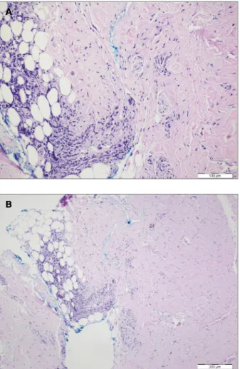
FIGure 2.Fibrocollagenous inflammatory cells consisting of lymphocytes in connective tissue and fat tissue. (H-F, x200)
differential diagnosis of EF and myositis. In addition, MRI is a useful imaging technique in the selection of biopsy site and monitorization of the treatment
pro-cess24,25. In consistency with the literature data, in our
case MRI of the upper extremity showed increased hete rogeneous T2 signal indicating edema and inflam-mation in the fascial planes involving extensor and par-tially flexor carpi ulnaris muscle and tendon, reduced adipose tissue, and signal loss in T1-weighted series. In these adjacent regions, increased subcutaneous adipose tissue was observed. However, there is a limited data on the sensitivity and specificity of MRI in the diagnosis of EF.
The definitive diagnosis is based on the full-thick-ness skin/fascia/muscle biopsy. The biopsy findings often show normal but rarely atrophic epidermis. Al-though half of the patients had sclerosis in the dermis, there are cases without sclerosis. Sclerosis is accompa-nied by lymphocyte, eosinophilia, plasma cells and his-tiocytic infiltration. The fascia beyond dermis is thicken ed by 2-15 folds. Eosinophilic infiltration which is disease-specific in fascia is rarely seen. Mus-cular bio psy results usually show lymphocyte, plasma
cell and histiocytic infiltration22. The pathological
find-ings of skin/fascia/muscle biopsy in consistent with all these findings suggested definitive diagnosis of EF in our case.
The initial treatment in EF is high dose corticos-teroids with a success rate of 70% among patients.
Other beneficial options include MTX26,27,
hydroxy-chloroquine28, cyclosporine29,30, D-penicillamine31and
photochemotherapy32. In recent years, tumor necrosis
factor antagonists, such as infliximab, have been tested in steroidrefractory EF patients and revealtested be
-neficial results33. Although our case was diagnosed two
years after the onset of the symptoms, she responded well to high dose steroids in combination with MTX. Her pain and swelling were substantially recovered. No recurrent disease occurred during the follow-up visits. Bischoff et al. reported that the mean time to dia -gnosis of EF was 8.8 months. The authors observed that patients with poor outcome had a longer time gap between initiation of first symptoms and diagnosis,
when compared to patients with good outcome28. We
have screened a total of 60 reports on EF which has full texts published in English in Pubmed-MEDLINE from 1974 to the present. Among these, in seven cases the mean time of diagnosis was >1 year (Table I). Only five of these cases had eosinophilia in peripheral blood sam-ples, while five had increased ESR and only two stu dies tic anemia, leukemia, and lymphoma, other
malignan-cies and autoimmune diseases including Hashimoto’s thyroiditis, systemic sclerosis and Sjogren’s
syn-drome5,6,13-15. The onset of the disease is triggered by
severe phy sical activity in more than half of the patients and there are also some case reports showing EF asso-ciated with trauma, hemodialysis, insect bite,
pheny-toin use,
exposure to trichloroethylene, and Borrelia
burgdor-feri3,7-9,16,17. A study investigating the cytokine profile in
EF demonstrated increased cytokine abnormalities and transforming growth factor b-1 levels similar to patients with atopic diseases, suggesting that autoimmune mechanisms may also play a role in the pathogenesis of
EF18. The major symptoms include usually
symmetrical acute or subacute pain and edema in the limbs, limi -ted range of joint motion and thickened skin. Review of literature revealed case reports with unilateral
pre-sentation similar to our case19-21. The disease is
characteri zed by “peau d’orange” appearance and
woody induration, as well as edema and erythema2.
Face and hands are rarely involved. A groove sign due to skin indurations on superficial veins is a specific characte ristic of the disease. In our case, symptoms were unilateral and the groove sign was also present. We could not find any triggering factor, as no detailed information was obtained from the patient since the complaints started two years ago. The diagnosis was made in the late stage, as the patient visited our out-patient clinic two years after the onset of swelling.
Laboratory tests reveal increased eosinophilia, in-creased ESR and polyclonal hypergammaglobulinemia
in peripheral blood samples22. The result of ANA, anti
ds-DNA and RF are usually negative. In our case, the patient had no eosinophilia in peripheral blood sam-ples with a slightly increased acute phase reactants. The laboratory parameters, which partially returned to nor-mal levels, may be explained by chronic phase of the disease. This suggests that imaging techniques and biopsy samples are more reliable for the diagnosis of pa-tients with chronic disease, than laboratory tests.
The MRI findings are also highly valuable in the diagnos is of EF which reveals thickening and hyperin-tensity in the superficial muscular fascia in the T1, T2-weighted and STIR sequences without contrast en-hancement, while severe contrast enhancement was observed in the fascia in the T2-weighted and STIR se-quences following the administration of IV contrast agent. The majority of the patients had deep muscular
tA b le I. c lI n Ic A l ch A r A ct er Is tI cs o F cA se s In t h e lI te r A tu r e T im e to M R I B io p sy R es p o n se t o d ia g n o si s L ab o ra to ry r es u lt s fi n d in g s re su lt s T re at m en t th e tr ea tm en t Is la m e t al . 1 2 m o n th s E S R : 2 8 m m /s Ø + 2 0 m g p re d n is o lo n e+ Ye s [3 4 ] W B C : 1 7 .1 x 1 0 9 /l h y d ro x y ch lo ro q u in e (4 0 0 m g /d ay ) E o : 3 6 % S er u m I g : N K h an n a et a l. 1 5 m o n th s E S R : n o t av ai la b le Ø + 4 0 m g p re d n is o lo n e N o [3 3 ] W B C : 7 .9 /µ l In fl ix im ab ( 3 m g /k g /8 h ft ) Ye s E o : 1 8 % M o n o cl o n al g am m o p at h y ( -) D an is e t al . 3 6 m o n th s E S R : 3 m m /h + + 1 m g /k g /d ay m et h y lp re d n is o lo n e Ye s [2 1 ] W B C : 2 2 .8 x 1 0 9 /l E o : 6 0 % S il n y W > 1 2 m o n th s E S R : in cr ea se d Ø + 1 0 0 0 -5 0 0 -2 5 0 m g I V m et h y lp re d n is o lo n e T o x ic it y [3 5 ] E o : 5 % fo ll o w ed b y 3 2 m g P .O . m et h y lp re d n is o lo n e + cy lo sp o ri n e (1 5 0 m g /d ay ) U V A 1 p h o to th er ap y ( 6 0 J/ cm 2 , Ye s th re e ti m es a w ee k , 3 1 i rr ad ia ti o n , to ta l d o se : 1 7 5 0 J /c m 2 ) T ah ar a et a l. 1 2 m o n th s E S R : 6 6 m m /s Ø + C y lo sp o ri n e (1 0 0 m g /d ay ) Ye s [3 6 ] W B C : 5 5 0 0 /µ l E o : 5 0 0 /µ l S er u m I g G : 3 5 5 0 m g /d l K h an n a et a l. 1 2 m o n th s E S R :1 0 0 m m /s Ø + H ig h d o se d ex am et h as o n e an d M il d [5 ] T o ta l b lo o d c o u n t: n o rm o ch ro m ic p am id ro n at e (m u lt ip le m y el o m a im p ro v em en t n o rm o cy ti c an em ia tr ea tm en t) Ig G M o n o cl o n al g am m o p at h y i n th e p ro te in e le ct ro p h o re si s (a ls o d ia g n o se d w it h m u lt ip le m y el o m a) S er u m I g G : 3 6 .0 0 g /l ( N : 6 .8 7 -1 6 .3 0 ) F la m en e t al . 4 8 m o n th s E S R : 4 4 m m /s Ø + G lu co co rt ic o st er o id s + D - p en ic il la m in e N o t av ai la b le [3 7 ] W B C : N (d o se n o t av ai la b le ) M o d er at e eo si n o p h il ia P o ly cl o n al I g G h y p er g am m ag lo b u li n em ia in t h e p ro te in e le ct ro p h o re si s R o se n fe ld e t al . 1 2 m o n th s E S R : 6 1 m m /s Ø + 4 5 m g p re d n is o lo n e Ye s [3 8 ] W B C : 2 5 x 1 0 9 E o : 8 0 % A g re em en t w as v o te d o n a s ca le f ro m 1 t o 1 0 ( fu ll y d is ag re e to f u ll y a g re e) b y 3 6 v o ti n g r h eu m at o lo g is ts . A S A S , A ss es sm en t o f Sp o n d yl o ar th ri ti s in te rn at io n al S o ci et y. A S D A S , A n k yl o si n g Sp o n d yl it is D is ea se A ct iv it y Sc o re . B A S D A I, B at h A n k yl o si n g Sp o n d yl it is D is ea se A ct iv it y In d ex . S D , st an d ar d d ev ia ti o n .
showed increased serum IgG level. These results suggest obscure laboratory findings in late stages of disea -se. The definitive diagnosis was based on full-thickness skin/fascia/muscle biopsy in all these seven cases. The MRI was performed only in one case, suggesting fin dings consistent with EF. The treatment modalities va -ried, however the majority of the patients responded
well to medication5,21,33-38.
In conclusion, EF should be considered in differen-tial diagnosis in case of unknown swelling and pain. Although laboratory parameters may be reduced in ca -ses with late diagnosis, a full-thickness skin/fascia/mus-cle biopsy and MRI may significantly contribute to the diagnosis. Despite late diagnosis, patients may still be -nefit from a high dose steroid therapy.
correspondence to
Elif Aydin
Adnan Menderes University Hospital Department of Physical and Rehabilitation
09100 Aydin, Turkey E-mail: drebulak@yahoo.com
reFerences
1. Shulman LE. Diffuse fasciitis with eosinophilia: a new syndro-me? Trans Assoc Am Physicians.1975; 88: 70-86.
2. Turan Y, Sendur OF, Berkit IK, Arslan H, Cetin ED. Eosinophi-lic Fasciitis: A case report and review of the literature. Turk J Rheumatol. 2010; 25: 208-213.
3. Granter SR, Barnhill RL, Duray PH.Borrelial fasciitis: diffuse fas-ciitis and peripheral eosinophilia associated with Borrelia in-fection. Am J Dermatopathol. 1996; 18: 465-473.
4. Sill P, Pint r D, Ostorh zi E, Maz n M, Wikonk l N, P nyai K, Vo-lokhov DV, Chizhikov VE, Szathmary S, Stipkovits L, K rp ti S. Eosinophilic Fasciitis associated with Mycoplasma arginini in-fection. J Clin Microbiol. 2012; 50: 1113-1117.
5. Khanna D, Verity A, Grossman JM. Eosinophilic fasciitis with multiple myeloma: a new haematological association. Ann Rheum Dis. 2002; 61: 1111-1112.
6. Junc J, Cuxart A, Tural C, Ojanguren I, Flores A. Eosinophilic fasciitis and non-Hodgkin lymphoma. Eur J Haematol. 1994; 52: 304-306.
7. Hayashi N, Igarashi A, Matsuyama T, Harada S.Eosinophilic fas-ciitis following exposure to trichloroethylene: successful treat-ment with cyclosporin. Br J Dermatol. 2000; 142: 830-832. 8. Buchanan RR, Gordon DA, Muckle TJ, McKenna F, Kraag G.The
eosinophilic fasciitis syndrome after phenytoin (dilantin) the-rapy.J Rheumatol. 1980; 7: 733-736.
9. Lakhanpal S, Ginsburg WW, Michet CJ, Doyle JA, Moore SB. Eo-sinophilic fasciitis: clinical spectrum and therapeutic response in 52 cases. Semin Arthritis Rheum. 1988; 17: 221-231. 10. Rodnan G, Di Bartolomeo A, Medsger TA Jr. Proceedings:
Eosi-nophilic fasciitis:report of six cases of a newly recognized skle-roderma-like syndrome. Arthritis Rheum. 1975; 18:525. 11. Ortega-Loayza AG, Merritt BG, Groben PA, Morrell DS.
Eosi-nophilic fasciitis in a female child. J Am Acad Dermatol. 2008; 58: S72-74.
12. Loupasakis K, Derk CT. Eosinophilic fasciitis in a pediatric pa-tient. J Clin Rheumatol. 2010; 16: 129-131.
13. Hur JW, Lee HS, Uhm WS, Jun JB, Bae SC, Park CK, Yoo DH. Eosinophilic fasciitis associated with autoimmune thyroiditis. Korean J Intern Med. 2005; 20: 180-182.
14. Kitamura Y, Hatamochi A, Hamasaki Y, Ikeda H, Yamazaki S. As-sociation between eosinophilic fasciitis and systemic lupus eryt-hematosus. J Dermatol. 2007; 34: 150-152.
15. Jacob SE, Lodha R, Cohen JJ, Romanelli P, Kirsner RS. Para-neoplastic eosinophilic fasciitis: a case report.Rheumatol Int. 2003; 23: 262-264. .
16. Romero AG, Fernandez JG, Calatayud JC. Eosinophilic fasciitis associated with simple traumatism. Acta Dermatovenerol Croat. 2001; 9: 287-290.
17. Mallepalli JR, Quinet RJ, Sus R. Eosinophilic fasciitis induced by fire ant bites. Ochsner J. 2008; 8: 114-118.
18. Dziadzio L, Kelly EA, Panzer SE, Jarjour N, Huttenlocher A. Cy-tokine abnormalities in a patient with eosinophilic fasciitis. Ann Allergy Asthma Immunol. 2003; 90: 452-455.
19. Seror J, Bonnard P, Baudrimont M, Guiard-Schmid JB, Teixeira A, Rozenbaum W, Pialoux G. Febrile pseudotumor lesion of the arm. Atypical presentation of Shulman syndrome. Presse Med. 2003; 32: 498-500.
20. Daniel RS, Lavery S, Maize JC Jr, Brown AN, Bolster MB. Uni-lateral eosinophilic fasciitis: an under-recognized subtype? J Clin Rheumatol. 2009; 15: 247-249.
21. Danis R, Akbulut S, Altintas A, Ozmen S, Ozmen CA. Unusual presentation of eosinophilic fasciitis: two case reports and a re-view of the literature. J Med Case Reports. 2010; 8;4:46. 22. Barnes L, Rodnan GP, Medsger TA, Short D. Eosinophilic
fas-ciitis. A pathologic study of twenty cases. Am J Pathol. 1979; 96: 493-518.
23. Moulton SJ, Kransdorf MJ, Ginsburg WW, Abril A, Persellin S. Eosinophilic fasciitis: spectrum of MRI findings. AJR Am J Roentgenol. 2005; 184: 975-978.
24. Baumann F, Br hlmann P, Andreisek G, Michel BA, Marincek B, Weishaupt D.MRI for diagnosis and monitoring of patients with eosinophilic fasciitis. AJR Am J Roentgenol. 2005;184: 169--174.
25. Desvignes-Engelbert A, Sauli re N, Loeuille D, Blum A, Chary-Valckenaere I.From diagnosis to remission: place of MRI in eo-sinophilic fasciitis. Clin Rheumatol. 2010; 29: 1461-1464. 26. Janzen L, Jeffery JR, Gough J, Chalmers IM. Response to
met-hotrexate in a patient with idiopathic eosinophilic fasciitis, morphea, IgM hypergammaglobulinemia, and renal involve-ment. J Rheumatol. 1995; 22: 1967-1970.
27. Pouplin S, Daragon A, Le Lo t X.Treatment of eosinophilic fas-ciitis with methotrexate. J Rheumatol. 1998; 25: 606-607. 28. Bischoff L, Derk CT.Eosinophilic fasciitis: demographics,
di-sease pattern and response to treatment: report of 12 cases and review of the literature.Int J Dermatol. 2008; 47: 29-35. 29. Valencia IC, Chang A, Kirsner RS, Kerdel FA. Eosinophilic
fas-ciitis responsive to treatment with pulsed steroids and cyclos-porine. Int J Dermatol. 1999; 38: 369-372.
30. Bukiej A, Dropi ski J, Dyduch G, Szczeklik A. Eosinophilic fas-ciitis successfully treated with cyclosporine. Clin Rheumatol. 2005; 24: 634-636.
31. Manzini CU, Sebastiani M, Giuggioli D, Manfredi A, Colaci M, Cesinaro AM, Ferri C. D-penicillamine in the treatment of eo-sinophilic fasciitis: case reports and review of the literature. Clin Rheumatol. 2012; 31: 183-187.
32. Schiener R, Behrens-Williams SC, Gottl ber P, Pillekamp H, Pe-ter RU, Kerscher M. Eosinophilic fasciitis treated with psora-len-ultraviolet A bath photochemotherapy. Br J Dermatol. 2000; 142: 804-807.
33. Khanna D, Agrawal H, Clements PJ.Infliximab may be effecti-ve in the treatment of steroid-resistant eosinophilic fasciitis: re-port of three cases. Rheumatology (Oxford). 2010; 49: 1184--1188.
34. Islam MN, Islam MA, Abdal SJ, Azad MA, Ahmedullah AK, Haq SA. Eosinophilic fasciitis: what matters in management in a de-veloping country - a case report with two and a half-year follow-up. J Health Popul Nutr. 2012; 30: 117-120.
35. Silny W, Osmola-Mankowska A, Czarnecka-Operacz M, Zaba R, Danczak-Pazdrowska A, Marciniak A. Eosinophilic fascitis: a report of two cases treated with ultraviolet A1 phototherapy. Photodermatol Photoimmunol Photomed. 2009; 25: 325-327.
36. Tahara K, Yukawa S, Shoji A, Hayashi H, Tsuboi N.Long-term remission by cyclosporine in a patient with eosinophilic fascii-tis associated with primary biliary cirrhosis. Clin Rheumatol. 2008; 27: 1199-1201.
37. Flamen P, Dierickx L, Everaert H, Reychler R, Bruyland M, Fran-ken PR, Bossuyt A. Fascial Tc-99m MDP uptake in eosinophi-lic fasciitis as demonstrated by SPECT. Clin Nucl Med. 1997; 22: 844-846.
38. Rosenfeld K, Stodell MA. Eosinophilic fasciitis in a father and son. Ann Rheum Dis. 1994; 53: 281.
![tAble I. clInIcAl chArActerIstIcs oF cAses In the lIterAture Time toMRIBiopsyResponse to diagnosisLaboratory resultsfindingsresultsTreatmentthe treatment Islam et al.12 monthsESR: 28 mm/sØ+20 mg prednisolone+Yes [34]WBC: 17.1x10⁹/lhydroxychloroquine (400](https://thumb-eu.123doks.com/thumbv2/123dok_br/18374788.892014/4.880.80.762.131.1069/characteristics-literature-tomribiopsyresponse-diagnosislaboratory-resultsfindingsresultstreatmentthe-treatment-prednisolone-lhydroxychloroquine.webp)
